Bisystole
Historical document, translated for reference. It reflects medical knowledge of the 1920s–30s and is not medical advice.
Summary
Bisystole is a phenomenon of a two-stage contraction of the left ventricle observed by V. P. Obraztsov in aortic valve insufficiency. The article details its clinical manifestations, auscultatory findings, and differentiation from a double apical impulse.
Encyclopedia article (1928–1936)
BISYSTOLE, contraction of the left ventricle in two stages—a phenomenon noted by V. P. Obraztsov in aortic valve insufficiency. The first phase of left ventricle contraction, which is weaker, is associated with the onset of the cardiac apex beat and the tension of the atrioventricular valves, while the second follows after a certain interval following the systolic sound and produces a muscular sound. Consequently, at the apex and especially in the third and fourth intercostal spaces on the left, a doubling of the first sound is heard, the so-called systolic gallop, with the second component of the first sound being strongly accentuated and falling on the middle of systole (mesosystolic sound). Upon a drop in muscle tone, an additional sound (protodiastolic sound) is added to the main second sound at the apex from the rapid stretching of the ventricular wall by the mass of blood flowing into it. Thus, in the presence of bisystole and incipient weakness of the hypertrophied.
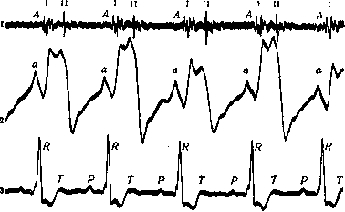
v,0 sec.
Bisystole: 1-sounds; 2-cardiogram; 3- electrocardiogram of the muscle, a four-tone melody is heard. The latter was also noted by Strazhesko, Zimnitsky, and Zelenin (cardiophonographically). Obraztsov sees the main proof of the two-stage contraction of the heart in aortic insufficiency in the appearance of pulsus bisferiens in large vessels and an additional rise on the ascending knee of the cardiogram. In smaller vessels (radial artery), only one rise in the form of pulsus celer is noted at the same time, since (according to the author) the second, larger wave travels at a higher speed, overtakes the first, smaller wave and absorbs it already at the periphery, giving a single rise. However, pulsus bisferiens in the radial artery was sometimes noted by Broadbent, Steel, Lewis, Mackenzie, the latter in cases of aortic stenosis. Broadbent also associates the double pulse wave with a double contraction of the heart. Bisystole should be distinguished from the double impulse, which is also felt at the apex, but has a completely different origin. On the cardiogram, it is expressed by an additional wave either before the systolic rise or after it, separated from it by a greater or lesser distance (presystolic, proto-, mesosystolic additional wave). Obraztsov believes that the presystolic impulse results from the rapid stretching of the ventricular wall—which has already lost its normal tone—by the blood flowing from the atrium (during its contraction). This impulse is felt most clearly when palpating slightly above and medial to the apex beat and is accompanied by an additional sound. The melody of the heart in this case has the character of an amphibrach (---_-). The diastolic additional impulse usually stands closer to the second sound. The mechanism of the diastolic impulse is essentially the same as in the presystolic impulse: the difference is only that the stretching of the ventricular wall is produced here by blood flowing from the atrium due to vis a tergo, without the participation of atrial systole. This type of impulse is also accompanied by an additional sound, giving the heart melody the form of a dactyl (-w--<). It is best heard at the apex and along the left border of the heart. A hypertrophied and degenerated heart particularly favors the occurrence of the double impulse phenomenon. Strazhesko, observing an additional diastolic impulse in aortic valve insufficiency, fully agrees with Obraztsov's opinion on the mechanism of its occurrence; with this defect, the additional impulse is best and most frequently noted (in 62% of cases, Zelenin). The position of the heart, depending on the height of the diaphragm, the shape of the chest, the degree of coverage of the heart by the edges of the lungs—all this significantly affects the appearance of the additional impulse. The clinical significance of the double impulse is quite significant. Its appearance is associated with the onset of decompensation (see figure). Increasing decompensation correspondingly increases the height of the additional impulse. In later stages of decompensation, the additional impulse may disappear, as the conditions for its occurrence change.
V. Zelenin. BISMARCKBRAUN
Related articles
Cite this page
“Bisystole.” Soviet Medical Encyclopedia. English translation of Bolshaya Meditsinskaya Entsiklopediya, 1st ed. (Moscow, 1928–1936), ed. N. A. Semashko. https://sovietmedicalencyclopedia.pages.dev/article/bisystole/