Heart Defects
Historical document, translated for reference. It reflects medical knowledge of the 1920s–30s and is not medical advice.
Summary
This article provides a comprehensive overview of heart defects (valvular heart disease) from a 1930s Soviet medical perspective, covering statistics, individual forms of defects, compensation and decompensation, diagnosis, treatment, and complications. It discusses the etiology, pathophysiology, and clinical manifestations of various heart valve disorders.
Encyclopedia article (1928–1936)
HEART DEFECTS. Contents: I. Statistics ...................430 II. Individual forms of H. D. Insufficiency of the mitral valve . . . 431 Stenosis of the left atrioventricular opening ......"................436 Stenosis of the aortic orifice..............4 44 Insufficiency of the aortic valves.......446 Insufficiency of the tricuspid valve. . . 453 Stenosis of the right atrioventricular opening 454 Combined defects...........45 5 III. Compensation and decompensation in H. D. . . 456 IV. Functional diagnosis in H. D. ... 463 V. Radiodiagnosis of H. D............464 VI. Prevention and treatment of H. D.........468 VII. Compatibility of H. D. and tuberculosis of the lungs . . . 475 VIII. Congenital H. D........... .... 476 Heart defect (vitium cordis), an old term. still in use at the present time, to denote lesions of the heart's valve apparatus. But the concept denoted by this term is broader, since the pathological process leading to damage to the valves never limits itself to them, but to one degree or another also affects the myocardium, and sometimes the pericardium; thus the heart as a whole becomes more or less "defective". Moreover, the symptom complexes accompanying H. D. extend beyond the heart itself and to a large extent depend on various changes in the entire cardiovascular system, on the one hand due to its direct (anatomical) damage, and on the other hand due to impaired blood supply throughout the body, pathological fluctuations in blood pressure, vascular tone disorders, etc. Thus, one must already speak of a "defect" of the entire circulatory apparatus. But the matter does not end there, and in the picture of "heart defect" one must also take into account a number of certain changes in various other organs and systems, which arose in them partly under the influence of the same causes that led to damage to the heart's valve apparatus (infection, arteriosclerosis), partly under the influence of impaired blood supply to these organs, and partly developed independently but had a decisive influence on the entire clinical picture of the defect (sclerosis of the coronary vessels, myocardial damage, severe decline in blood formation, glomerulonephritis, vegetative neuroses, hypertension, etc.). Therefore, in the study of H. D., the researcher's attention in no way should be confined to determining changes in the valve apparatus and the resulting circulatory disorders. One should also keep in mind another important circumstance. If every chronic disease has its own history of development with certain stages, its dynamics, then nowhere is it presented as vividly as in the course of, for example, rheumatic H. D., which, slowly developing from infection (endocarditis), form into certain symptom complexes characteristic of lesions of this or that valve or opening, but acquire completely different significance depending on recurrent outbreaks of infection (endocarditis recurrens), on changing working and living conditions. Therefore, in the classification of H. D., it is completely insufficient to note the anatomical defect, it is also insufficient to indicate the state of the circulatory apparatus at a given time (sufficiency or insufficiency), but it is necessary to note the rate of onset of pathological disorders, strictly relating them to the specific situation, as well as to concomitant diseases. Thus, the diagnosis of H. D., which includes 1) determination of the anatomical defect, 2) sufficiency of circulation, 3) concomitant pathological conditions, and 4) characterization of the dynamics of the entire process in accordance with working and living conditions, will also contain elements of a work prognosis, i.e., will in fact become a predominantly functional diagnosis. I. Statistics. The number of cardiac valve lesions averages about 25% of all cardiovascular diseases; Romberg (according to the latest statistics) gives a higher percentage (76 out of 2218). According to autopsy data, the following percentage falls on the total number of corpses: according to Rosenstein-9.2%, according to Frommel-3.5%, according to Davydovsky (on 53,959 autopsies)--4.1%. However, it should be noted that the statistical evidence from autopsies and statistics of clinic and hospital admissions is not very convincing, as the element of chance is not excluded in them; this is also reflected in the distribution of patients by gender: according to Neustab, H. D. occurs in women 2.7 times more often than in men, while Plesch had 6% more men; according to the statistics of the Med.-biol. institute- 40% men and 60% women. The question of the significance of the hereditary factor is little illuminated, as it encounters great methodological difficulties. In Plesch's statistics, there was a hereditary predisposition (H. D. of father or mother) in only 9.3%, in Neustab-12.1%. Statistics attempting to establish the influence of occupation are very unsatisfactory. If there is no clarity regarding gender, heredity, and profession as etiological factors for H. D., then all authors are unanimous in assessing the role of infections, attributing to them about 3/4 of all H. D., although here too the question is not easily resolved, since the mere presence of one or another infection in the history does not give the right to conclude about etiological dependence. The complaints of patients are also not always conclusive, as a considerable number of H. D. initially runs completely asymptomatic (see below). According to the data of the Med.-biol. institute (500 cases), almost half of all patients (47%) could not note the moment with which they could associate their illness. According to all authors, first place is occupied by "rheumatic" infection (acute articular rheumatism, chorea, etc.): according to Troitsky-45.7%, Guttmann-58.3%, according to Romberg's clinic-47.8%, according to Plesch-49.4%, according to the data of the Med.-biol. institute-42.6%, and patients associated their illness with acute articular rheumatism in only 22.6%, and in 8.8% they blamed angina. All other infections give only 31% according to Plesch, and only 10.6% according to Zelenin. Observations indicate the great importance of polyarthritis. The frequency of anginas should also be noted. Sifilis has predominant importance for aortic defects, and the very frequent combination of aortic defects with kidney disease (cardio-renal syndrome) is also noteworthy. Arteriosclerosis can lead to changes mainly in the aortic valves, although other valves are not protected from the sclerotic process. The role of trauma is small: the aortic valves are more often affected, and the presence of endocarditis apparently facilitates the detachment of the valves. A gunshot wound to the heart with damage to the mitral valve and the His bundle has been described with a completely benign course of the defect. Tumors of the valves (fibromas, myxofibromas) are noted in the literature as rare findings, which can also disrupt their proper functioning. It should be kept in mind that the primary process that led to changes in the heart's valve apparatus leaves a certain imprint on the patho-anatomical picture, which allows one at autopsy without particular difficulty to make a distinction between rheumatic disease, atheromatosis, and syphilis. The clinician, comparing the data of the history, the localization of the lesion, concomitant diseases, the dynamics of H. D., certain peculiarities of the symptomatology, can also with a considerable degree of probability speak in favor of one or another etiology of the suffering, which of course has practical significance. It is also important to form a judgment about the degree of anatomical damage, which, while not the only factor determining the severity of the disease, still often predetermines the nature of the course and outcome of H. D. II. Individual forms of heart defects. Insufficiency of the mitral valve. Among individual forms of acquired defects, according to statistics, the most common is insufficiency of the mitral valve (insuffi-cientia valvulae mitralis, s. bicuspidalis). Although not all data obtained from studying the dynamics of artificial defects in animals can be used in the clinic (in humans, pure forms of defects are rarely encountered, and there are also a number of changes in other organs associated with past diseases, with various working and living conditions), still familiarity with the elementary changes in the work of the cardiovascular system under experimental conditions will facilitate the understanding of the symptomatology of acquired defects in humans. Wiggers thus describes the course of hemodynamic changes in an animal after artificially induced insufficiency of the mitral valve. Following the loss of the valve's ability to completely close the opening, part of the blood during systole is thrown back into the left atrium, and pathological phenomena develop in the following order: 1) due to the backward flow of blood during systole, blood pressure in the aorta falls, maximum intraventricular pressure decreases, systolic and diastolic pressure in the arteries falls, pulse pressure decreases. 2) Pressure in the left atrium is elevated, filling of the left ventricle therefore occurs more completely, diastolic dilatation and initial tension increase, the left ventricle ejects a larger amount of blood into the aorta and back into the atrium.
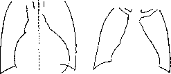
A prolonged state ensues in which, as in normal conditions, the left ventricle in its emptying completely corresponds to the right. 3) The increased amount of blood is taken up by the dilated left atrium, veins, and pulmonary capillaries, which leads to a certain slowing of blood flow in these vessels and a certain restriction of the respiratory surface of the lungs. However, the changes are small, and noticeable fluctuations in pressure in the pulmonary artery cannot be detected. 4) The right ventricle, contrary to widespread opinion, apparently does not participate in the compensation of the defect. In humans, the long existence of relative insufficiency of the mitral valve, for example, in the early stages of aortic defects, gives hypertrophy of the right ventricle. 5) After 5-20 minutes, systolic arterial pressure returns to normal, while diastolic pressure remains somewhat elevated. Thus, the mean arterial pressure falls, the mean pressure in the atria rises, and blood from the arterial system is pumped in larger quantities into the venous system. However, the danger of the defect lies not in the perversions of blood flow, but in those moments that weaken the heart muscle, making it unable to adapt to pathological conditions. Experiments with substances that reduce the contractile force of the myocardium (chloral hydrate, chloroform) showed a rapid increase in congestion in the lesser circulation. Consequently, the key to understanding the phenomena of decompensation lies in the heart muscle. -Etiology. In first place is acute articular rheumatism, in second place is syphilis; a considerable percentage falls on cases with unclear etiology; other infections have little importance, even less is trauma. Pathological anatomy. The mitral valve is disfigured to a greater or lesser degree by scars that pull together its leaflets, which are thickened and often glued to the papillary muscles; the process can also involve the chordae tendineae, which, by shortening, prevent complete closure of the valve; conversely, in case of rupture of the chordae tendineae, the valve during ventricular systole can turn inside out into the atrium. Phenomena of mural endocarditis outside the valve apparatus have been described, even penetrating ulcers of the atria (Plesch). The resulting throwing of blood into the atrium is determined not only by the state of the valve, but also by the direction of the blood wave in the left venticle in connection with the change in its configuration as a result of its expansion and hypertrophy. The left atrium is as a rule dilated and hypertrophied; the pulmonary artery and large venous trunks are stretched and often sclerosed. The right ventricle is hypertrophied. Often the right atrium is also found dilated and hypertrophied. The dynamics of blood circulation differ somewhat from experimental findings; the left ventricle, stretching with a large amount of blood flowing from the overloaded atrium (tonogenic expansion), responds (according to Starling's law) with a more powerful systole and, gradually hypertrophying, performs systole with greater speed and force, as a result of which the resistance of pressure in the aorta is more easily overcome and a smaller amount of blood is thrown back into the atrium. The stretching of the left atrium and the increase in pressure in the lesser circulation are expressed relatively little. Extremely characteristic is also the long asymptomatic nature of this defect in the sense of complete absence of complaints from the patient, which is why heart disease is often discovered only accidentally (during various examinations). The above pathological-anatomical changes in the lesser circulation, as well as in the right atrium, can be attributed to additional disorders associated with the phenomena of already occurring 'decompensation'. -With relative insufficiency, which occurs in approximately 7/3 of all clinically detected defects, an extremely frequent, according to Aschoff almost pathognomonic, post-mortem finding is expressed hypertrophy of the papillary muscles, which occurs along with the general hypertrophy of the left ventricle and depends on the fact that relative insufficiency develops mainly with increased pressure in the greater circulation (hypertension, kidney diseases). In this case, the deviation from experimental findings is even greater. Clinical picture in the stage of absence of disorders of peripheral circulation. It has already been mentioned above that for a number of years after the defect has formed, it may not give any subjective sensations; those suffering from it may be completely unaware of it, carry heavy physical work, and even participate in sports competitions. Objectively, the defect is determined by the following data. The borders of the heart are percussively shifted somewhat to the left (due to the left ventricle) and upward (due to the left atrium or rather its auricle), the right border shows no changes; the impulse has no special deviations. Radiologically [see separate table (p. 463-464), fig. 2] 'smoothing of the waist' is determined, i.e., the disappearance of the angle between the lower left arch (left ventricle) and the vascular bundle, since it is filled by the auricle of the left atrium. The pulmonary artery usually also protrudes more than normal (due to increased pressure in it) (fig. 1 and 2). On auscultation a systolic murmur is heard as a result of the relative insufficiency of the mitral valve. Figure 1. Insufficiency of Figure 2. Insufficiency of the mitral valve ('standing egg').
The increased amount of blood is taken up by the dilated left atrium, veins, and pulmonary capillaries, which leads to a certain slowing of blood flow in these vessels and a certain restriction of the respiratory surface of the lungs. However, the changes are small, and noticeable fluctuations in pressure in the pulmonary artery cannot be detected. The right ventricle, contrary to widespread opinion, apparently does not participate in the compensation of the defect. In humans, the long existence of relative insufficiency of the mitral valve, for example, in the early stages of aortic defects, gives hypertrophy of the right ventricle. After 5-20 minutes, systolic arterial pressure returns to normal, while diastolic pressure remains somewhat elevated. Thus, the mean arterial pressure falls, the mean pressure in the atria rises, and blood from the arterial system is pumped in larger quantities into the venous system. However, the danger of the defect lies not in the perversions of blood flow, but in those moments that weaken the heart muscle, making it unable to adapt to pathological conditions. Experiments with substances that reduce the contractile force of the myocardium (chloral hydrate, chloroform) showed a rapid increase in congestion in the lesser circulation. Consequently, the key to understanding the phenomena of decompensation lies in the heart muscle.
In first place is acute articular rheumatism, in second place is syphilis; a considerable percentage falls on cases with unclear etiology; other infections have little importance, even less is trauma. The mitral valve is disfigured to a greater or lesser degree by scars that pull together its leaflets, which are thickened and often glued to the papillary muscles; the process can also involve the chordae tendineae, which, by shortening, prevent complete closure of the valve; conversely, in case of rupture of the chordae tendineae, the valve during ventricular systole can turn inside out into the atrium. Phenomena of mural endocarditis outside the valve apparatus have been described, even penetrating ulcers of the atria (Plesch). The resulting throwing of blood into the atrium is determined not only by the state of the valve, but also by the direction of the blood wave in the left ventricle in connection with the change in its configuration as a result of its expansion and hypertrophy. The left atrium is as a rule dilated and hypertrophied; the pulmonary artery and large venous trunks are stretched and often sclerosed. The right ventricle is hypertrophied. Often the right atrium is also found dilated and hypertrophied. The dynamics of blood circulation differ somewhat from experimental findings; the left ventricle, stretching with a large amount of blood flowing from the overloaded atrium (tonogenic expansion), responds (according to Starling's law) with a more powerful systole and, gradually hypertrophying, performs systole with greater speed and force, as a result of which the resistance of pressure in the aorta is more easily overcome and a smaller amount of blood is thrown back into the atrium. The stretching of the left atrium and the increase in pressure in the lesser circulation are expressed relatively little. Extremely characteristic is also the long asymptomatic nature of this defect in the sense of complete absence of complaints from the patient, which is why heart disease is often discovered only accidentally (during various examinations). The above pathological-anatomical changes in the lesser circulation, as well as in the right atrium, can be attributed to additional disorders associated with the phenomena of already occurring 'decompensation'.
With relative insufficiency, which occurs in approximately 7/3 of all clinically detected defects, an extremely frequent, according to Aschoff almost pathognomonic, post-mortem finding is expressed hypertrophy of the papillary muscles, which occurs along with the general hypertrophy of the left ventricle and depends on the fact that relative insufficiency develops mainly with increased pressure in the greater circulation (hypertension, kidney diseases). In this case, the deviation from experimental findings is even greater. It has already been mentioned above that for a number of years after the defect has formed, it may not give any subjective sensations; those suffering from it may be completely unaware of it, carry heavy physical work, and even participate in sports competitions. Objectively, the defect is determined by the following data. The borders of the heart are percussively shifted somewhat to the left (due to the left ventricle) and upward (due to the left atrium or rather its auricle), the right border shows no changes; the impulse has no special deviations. Radiologically [see separate table (p. 463-464), fig. 2] 'smoothing of the waist' is determined, i.e., the disappearance of the angle between the lower left arch (left ventricle) and the vascular bundle, since it is filled by the auricle of the left atrium. The pulmonary artery usually also protrudes more than normal (due to increased pressure in it) (fig. 1 and 2). On auscultation a systolic murmur is heard as a result of the relative insufficiency of the mitral valve. Figure 1. Insufficiency of Figure 2. Insufficiency of the mitral valve ('standing egg').
(triangular shape). Regurgitation of blood from the left ventricle into the left atrium due to incomplete closure of the left atrioventricular opening. Barry attributes its appearance to the so-called presystolic phase of systole; according to Frey and Fromm, the murmur is determined at a later period of "emptying." The murmur is as a rule best heard at the apex of the heart, however it is by no means uncommon for its maximum to fall on the point of attachment of the cartilage of the third rib to the sternum, where the left atrial appendage is located (Naunyn's point). Naunyn and Kurchman found it here most often in fresh cases, whereas later (possibly due to an increase in papillary muscles) it moves downward, toward the nipple. Along with the murmur, the first tone is also heard, partly conducted from the tricuspid valve, partly due to tension of the remnants of the mitral valve, but the stronger the mitral insufficiency, the weaker the first tone at the apex. With significant insufficiency, it may be completely absent. With rough musical murmurs, the tone may be drowned out. Auscultation should always be performed both in the vertical and horizontal positions, as well as after moderate physical exertion. Due to some increase in pressure in the small circle, a moderate intensification (accent) of the 2nd tone on the pulmonary artery is as a rule heard. This accent (Soda's symptom) may disappear depending on changes in blood circulation in two opposite directions: 1) with weakening of the right ventricle (and the occurrence of relative tricuspid insufficiency) and 2) with increased work capacity of the left ventricle (increased pressure in the large circle and decreased in the small). When auscultating the lungs, "saccaded" (intermittent) breathing may sometimes be determined due to pulsatory pressure on the respiratory tract from the dilated left atrium. From the side of the pulse and blood pressure, there are no noticeable deviations from normal. The minute volume of blood is within average figures (Plesh). Blood pressure is normal. In view of the absence of circulatory disorders, patients usually do not complain of shortness of breath and do not show noticeable cyanosis. Electrocardiography. The atrial P wave is either somewhat enlarged or, more often, shows no special deviations from normal. The ventricular complex indicates moderate predominance of the right ventricle: in the 1st lead, a reduced R wave and a pronounced S, in the 2nd and 3rd leads, a high R wave, T-relatively low. The distinction between endocarditic murmur in mitral insufficiency from systolic murmurs of other nature is of great practical interest. As has already been noted, in all cases where a systolic murmur was determined in life in the area of the mitral valve, autopsy did not reveal its anatomical damage. What significance does this finding have from the clinician's point of view? It must be borne in mind that systolic murmurs heard at the apex of the heart or in the so-called 5th point (Kurchman-Naunyn) have different origins, and therefore different significance. The murmur in relative (muscular) insufficiency of the valve, arising from weakening of the circular muscle tracts at the base of the left ventricle, indicates changes in the myocardium, which in terms of work capacity of the heart may have a more unfavorable significance than small anatomical changes in the valve with a healthy heart muscle. The systolic murmur, appearing depending on weakening of the papillary muscles, as a result of which the mitral valve everts during systole of the left ventricle toward the atrium, having a somewhat different pathogenesis than in the previous case, should be evaluated the same as in weakening of the muscular ring. On the contrary, eversion of the valve inward into the ventricle with hypertrophy of the papillary muscles (experiments of Savitsky and Mikhailov with the use of adrenaline) can also cause a murmur, which consequently has the opposite significance (vegetative neurosis, overexcitation during mental and physical exertion). It is possible that systolic murmurs encountered in youth are of the same origin (hypertensive). As for the so-called anemic murmurs, undoubtedly with deep nutritional failure and blood formation (cachexia, severe anemia) they may depend on muscular insufficiency of the mitral valve, more probably they are formed not in the atrioventricular opening, but at the mouth of the pulmonary artery in connection with acceleration of the current and decrease in blood viscosity, and are heard better in the 2nd left intercostal space than in the 5th point, and are rarely determined at the apex of the heart. Systolic murmurs in Basedow's disease (see) are apparently of dual origin: those heard on the pulmonary artery depend on the acceleration of blood flow present in this disease, while those determined in the 5th point or at the apex in various stages of thyrotoxicosis are caused by disturbance of the tone of the circular and papillary muscles. What has been said is important to keep in mind practically, since small shifts in temperature with increased function of the thyroid gland in connection with a systolic murmur often serve as a reason for diagnosing endocarditis. The same diagnostic error is caused by incorrect evaluation of the murmur in various infectious diseases: acceleration of cardiac activity (murmur on the pulmonary artery), weakening of the heart muscle (murmur in the 5th point or at the apex of the heart) with significant intoxication and especially with myocarditis. Systolic murmurs have been described that appear in disorders of cardiac rhythm in connection with weakening of muscular strength or change in blood filling of the left ventricle: Jerod (Jerod) found relative insufficiency of mitral valves in blockage (overfilling of the ventricles in a very long diastolic phase); Galli observed with intermittent pulse (pulsus alternans) a systolic murmur corresponding to a weak heart contraction; Pan - with pulsus bigeminus during the extrasystolic wave. Blood filling is also associated with the systolic murmur in insufficiency of the aortic valves (overfilling and dilatation of the left ventricle). For murmurs not associated with anatomical damage to the mitral valves, their considerable variability (depending on body position, mental state, etc.) is generally characteristic. Thus, a systolic murmur determined on auscultation does not yet indicate a heart defect. On the other hand, with anatomical damage to the valve apparatus, murmurs may be absent ("mute" defects); diagnosis in such cases is based on other auscultatory data (various accents, etc.), but mainly on the entire clinical picture (anamnesis, configuration of the heart, electrocardiography, etc.). The course of the defect is extremely varied, and therefore any attempt to give a general characterization is completely futile; in any case, it depends least on the defect itself, i.e., change in the valve, and to a much greater extent on the state of the heart muscle, which in turn is involved in the damage under the influence of infection, intoxication, arteriosclerosis, etc. Patients usually die from superimposed diseases: various infections, especially those associated with exhausting heart cough (the "experiment" of Valsalva and Müller), such as bronchopneumonia and pleurisy (Botkin, Plesh), as well as in the presence of cachexia (Henschen). The prognosis in view of the above is also uncertain. In general, it becomes doubtful after signs of circulatory disorder ("decompensation") begin to appear. However, even here one must take into account that mitral defects, if they generally decompensate more easily and faster than aortic ones, also compensate again better than the latter. Prevention. We have at our disposal no means of preventing the development of valve damage in infections. Proper care during endocarditis, mainly sparing the heart muscle, can ensure a more favorable course of the defect in the future. Treated syphilis gives less chance of valve apparatus damage than untreated syphilis. Prevention of sclerotic insufficiency of the mitral valve consists in the entire complex system of prevention of arteriosclerosis, since valve changes only rather late complicate the picture of widespread arteriosclerosis. The question of the role of physical exertion requires special evaluation. On the basis of physiological data, it should be considered correct to apply certain muscular tensions as a measure strengthening the heart muscle (see below). To the same extent, it is important to maintain the nervous system at a sufficiently high level of tone, "cheerfulness of spirit," by instilling a certain optimism in relation to the defect that has occurred in the heart. Every physician knows the so-called "iatrogenic," i.e., induced by the physician, disorders of cardiac activity (angioneurotic phenomena, interruptions, general depression), which disrupt the work of the cardiovascular system during the period of its pathological reorganization. That is why favorable conditions of work and life will in the future be the decisive factor in preserving "compensation."
The question of whether to immediately place the patient with the described defect under conditions of limited workload should not be decided schematically as a general rule, but should be evaluated in each individual case with full consideration of existing employment opportunities. Professions involving heavy physical labor and requiring significant nervous tension should still be avoided, despite repeatedly observed cases of full work capacity being maintained under such professional loads, since muscular labor is cardiovascular labor. The assessment of the degree of disability will also depend not only on the formal definition of the defect and possible functional testing, but also on the entire set of working and living conditions. Drug therapy in the absence of decompensation phenomena is usually unnecessary; it must be resorted to only in cases of nervous complications (especially in cases of mild cardiac excitability, quinine bromide is readily prescribed in doses of 0.15x3) and in pronounced anemia (Ferrum hydrogenio reductum up to 3.0 pro die). Narrowing of the left atrioventricular opening (stenosis mitralis). Experimental data (according to Wiggers) are less favorable than in the case of mitral valve insufficiency. Moritz's research showed that the work of the left ventricle (calculated according to his formulas) is decreased, while the work of the right ventricle remains unchanged. According to Zvankovskaya's data, in the late stages of the defect, the work of the right ventricle also increases. Blood accumulating in the left atrium due to the abnormal obstruction to outflow places increased demands on it, pressure in it increases (Mc. Dowall): thus the entire task of 'compensation' of this defect falls on the thin-walled and weak-walled section of the heart (left atrium). With slight narrowing, the atrium copes with this task relatively successfully; circulatory disorders are absent. With significant stenosis, the small circle becomes congested, pressure in the pulmonary artery increases. Conversely, the left ventricle is insufficiently filled with blood in diastole, which leads to a decrease in the amount of blood ejected with each systole.
Etiology. If 'rheumatic infection' can still be blamed for the formation of the defect in a significant percentage of cases, it is still encountered less frequently in uncomplicated mitral stenosis than in other defects. Instead, the percentage of cases of unclear etiology significantly increases, which suggests the influence of the body's developmental conditions on the formation of the defect (Duroziez). The same is indicated by the significant predominance of the female sex, accompanying female diseases (endometritis, uterine fibroids), anemia, chlorosis (ovarian hypofunction), as well as general infantilism, underdevelopment of the cardiovascular system, tuberculous intoxication; mitral stenosis is more often determined already in early youth than other defects. Moussoir doubts the congenital nature of mitral stenosis and blames infections for its origin: scarlet fever, lues, tbc. Moreover, the opposite viewpoint is also quite acceptable, according to which the described constitutional anomalies may be a consequence of circulatory disorders in an already formed defect from early childhood. Pathoanatomical and pathophysiological data. Narrowing of the atrioventricular opening is caused by changes in the ring itself, which becomes less pliable due to calcareous deposits, as well as in the valves (adhesions and scarred disfigurement of the leaflets), and also in the chordae tendineae, which acquire cartilaginous consistency and thus impede the passage of blood from the atrium to the ventricle. The degree of stenosis of the atrioventricular opening depends on the combination and depth of all these anatomical disorders (Plesch). The left atrium is always hypertrophied and dilated. The right ventricle, which has to overcome high pressure in the pulmonary artery, becomes hypertrophied. The right atrium is also dilated and hypertrophied, apparently in connection with circulatory disorders in the great circle. The left ventricle in most cases appears atrophied, but sometimes it is found to be completely normal or even hypertrophied (Lenhartz). The latter finding does not correspond at all to the conditions of blood circulation in this defect (relative underloading of the left ventricle) and must be explained by accompanying diseases (nephritis, pericarditis).
Clinical picture. Unlike cases of insufficiency, stenosis cases apparently come under medical observation earlier, which is associated with the greater severity of the disease, which quite soon attracts the attention of both the patient and those around them. Complaints usually come down to general weakness and shortness of breath, especially after physical exertion. Both weakness and shortness of breath depend primarily on the nature of the defect, in which the equalization of blood circulation ('compensation') falls to a large extent on the weak section of the heart (left atrium). Weakness can be attributed in part to the often found general underdevelopment, while shortness of breath may also depend on other factors causing circulatory disorders in the small circle: emphysema, kyphoscoliosis, effusions in the pleural cavities, in the pericardium, etc. In general, shortness of breath is not characteristic of mitral lesions and is, as a rule, associated with impaired left ventricular function (in aortic defects). Plesch believes that in mitral stenosis, due to the small filling of the left ventricle, its accommodative capacity is reduced, which can lead to the development of dyspnoea. In general, shortness of breath is more pronounced only when there is significant congestion of the pulmonary capillaries. Cyanosis in pronounced stenosis is an outstanding symptom: strikingly violet-red cheeks are noticeable, which can be mistaken for a febrile coloration at a superficial assessment, especially in consumptives (errors in the direction of diagnosing tbc of the lungs are generally not uncommon). At the same time, one should keep in mind the sclerosis of the pulmonary arteries (Erpinger and Wagner), as well as toxic enterogenous cyanosis (Ditrich and others). A markedly atheromatous pulmonary artery can be a direct consequence of this defect on the basis of prolonged increase in blood pressure in the small circle. More pronounced cyanosis is found in other, mainly congenital defects (see below). Hemoptysis associated with congestion of the pulmonary vessels is a characteristic symptom of this defect (in mitral valve insufficiency it is encountered much less frequently), and sometimes takes the form of true 'cardiac phthisis' (haemophtisie cardi-aque of French authors): it is either parenchymal hemorrhage or hemorrhagic infarcts due to the great tendency to develop thrombi, which can sometimes rotate freely in the left atrial cavity and, by obstructing the mitral opening, lead to sudden death. Due to the accumulation of blood pigment in the lungs, so-called 'cells of heart defects' are found in the sputum (see Sputum).
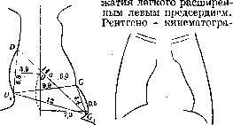
Figure 3. Drying of the left atrioventricular opening. Left atrium-OG. Figure 4. Drying of the left atrioventricular opening. The aortic arch is small and stands high, the middle left arch is noticeably bulged (pulmonary artery and left auricle of the left atrium). The left ventricle is small. The "waist" of the heart is comparatively wide compared to the entire heart. This method gives some idea of the dynamics of the left atrium: during ventricular diastole, the atrium not only does not collapse at first, but in the first second it stretches even more and only after that begins to collapse. This method provides some reference points for differential diagnosis between stenosis of the opening and insufficiency of the valve. Due to overflow of the small circle, there is paralysis of the left recurrent nerve. Notkin collected 52 cases described in the literature in which compression of the nerve occurred either directly by the stretched pulmonary artery or through the left atrium and pulmonary veins; sometimes there was an initial neuritis without special compression of the nerve. X-ray, as well as the success of treatment with digitalis, allows us to exclude an aneurysm of the aorta. Ortner is inclined to explain the paralysis of the recurrent nerve also by the pressure of the dilated right atrium. In view of the fact that blood-filled and dilated atria and large vessels are located in the mediastinum, symptoms of its compression may be detected. Landolfi describes the following mediastinal syndrome in mitral stenosis: anisocoria due to compression of the sympathetic nerve by the dilated left atrium, pulsus differens due to compression of the left subclavian artery, paralysis of the recurrent nerve from pressure of the greatly dilated left atrium, marbling of the skin in the heart area from difficulty in blood flow through the mediastinal veins and painful phenomena. - Cardiac pains. One cannot agree with the opinion of Nothnagel, who believes that chest pains in mitral stenosis do not occur. On the contrary, they occur quite often, have different characters and depend on various causes. The auricular pain point (part of the mediastinal syndrome) consists, according to Vaquez, of independent or pressure-induced pain between the spine and the medial edge of the left scapula. There have even been true attacks of angina pectoris, which according to Hochrein's research can be explained by compression of the place of departure of the left coronary artery.
Auscultation presents a great variety of sound findings: 1) The first tone at the apex due to the small blood filling of the left ventricle is sharply intensified, flapping. 2) The second tone over the pulmonary artery (due to increased pressure in the small circle) is accentuated and often split ("quail rhythm"). Poten explained the appearance of the first flapping tone at the apex by the sudden tension of the changed mitral valve when it opens (claquement de l'ouverture mitral). This auscultatory sign is very important, since isolated intensification of the first tone is not found in any other pathological condition (in arhythmia perpetua along with loud ones, quiet first tones are also found). 3) The second tone over the aorta is usually somewhat weakened, except in cases with arterial hypertension (see below). 4) It is considered the most characteristic to have a diastolic murmur over the apex, which depends on pathological conditions of blood movement from the left atrium to the left ventricle. However, these conditions are so diverse that the murmur is completely absent (according to Henschen's data) in 1/3 of cases of stenosis; according to Plesch, in old age in 2/3 there is "aphonic stenosis". This depends on the fact that in the origin of the murmur both the degree of narrowing and the change in the chordae tendineae (Plesch) and the functional state of the left atrium play a role. The existing murmur gives various variations: sometimes it is heard only at the beginning of diastole, immediately after the second tone ("protodiastolic" murmur of Poten), sometimes, on the contrary, only at the end of diastole, in the presystolic stage, before the first tone ("presystolic"), very rarely it is determined only in the middle of diastole ("mesodiastolic"), sometimes it fills the entire diastole, presenting at the same time characteristic waves of decreasing and increasing intensity (decrease-decrescendo-in protodiastolic murmur and increase-crescendo-in presystolic). The most important of these variations is the presystolic murmur, as it allows one to judge the functional state of the left atrium; with weakening of the atria, it loses its increasing character, and with the cessation of atrial activity (e.g. in arhythmia perpetua) the murmur also disappears.
A direct proof of this dependence was presented by Schellong, who recorded an electrocardiogram and phonogram in heart block with mitral stenosis: the murmur was heard (and recorded) only at the moment of atrial contraction. It is believed that with significant stenosis, the murmur can fill the entire diastole, while with not very sharply expressed narrowing, only the strengthening of the current during atrial contraction (presystole) can cause a murmur (presystolic); this last is most often determined. The sound phenomenon can be reproduced in the form of the following combination of sounds: 1) with presystolic murmur "sh-to-to" ("kh-to-to"); 2) with protodiastolic-"to-to-ssh"; 3) with filling the entire diastole-"sh-to-to-ssh". The most common combination of presystolic murmur and "quail" is reproduced as follows: "sh-t6-to-t6" (Fig. 5). Sometimes it is difficult to say whether there is a presystolic (very short) murmur or splitting of the first tone (depending on the asynchrony in the systole of both ventricles), when reproducing sounds, something like "tra-ta-ta" is obtained. Intensified and split tones and in in in in
in in in in", "in in", "b", "c
Figure 5. Variations of diastolic murmur: a-"sh-tb-to" (presystolic); b-"to-to-shsh" (protodiastolic); c-"sh-to-to-shsh" (protodiastolic and presystolic); d-"sh-to-to-to" (presystolic and "quail"). The various murmurs allowed Botkin to compare the auscultatory findings in mitral stenosis with the sounds of a blacksmith. The noted sound phenomena can be determined by means of tactile sensation: if the palm is applied to the area of the heart, a certain sensation of three impulses is obtained: I a clapping and II a bifid (with "quail"); during presystole or throughout the entire diastole, the palm perceives a tremor resembling the sensation that the hand receives when stroking a purring cat; since the time of Corvisart and Laennec, this symptom has been called "cat's purring" (fremissement catarrhique). This tremor is most distinctly perceived in the extreme left portion of the cardiac impulse (Edens). In regard to differential diagnosis, it must be borne in mind that the most prominent auscultatory symptom, namely the presystolic murmur, as indicated above, may be absent in unquestionable stenosis, and on the other hand (generally, however, very rarely), it is also found in other diseases: the Flint phenomenon in aortic valve insufficiency (see below), in pericardial adhesions (Fischer, Steel), in extremely dilated hearts. Bickel found it together with a systolic murmur in Basedow's disease. Sometimes a diastolic murmur is added, depending on the relative insufficiency of the valves of a greatly dilated pulmonary artery (Graham Steel). The systolic murmur at the apex, often heard in the defect described, depends on the relative insufficiency of the mitral valve, more often of the tricuspid valve, above which it is then heard more distinctly. A constantly determined, sufficiently pronounced systolic murmur at the apex indicates a complex defect of the left atrioventricular orifice, which occurs much more frequently than "pure" (isolated) stenosis and has its own characteristic clinical picture (see below - combined defects). The pulse is always somewhat faster than normal: this is apparently due to a certain decrease in the amount of blood ejected per contraction, so that to maintain circulation at a normal level (to preserve the normal volume of circulation) a greater number of beats per minute is necessary; in this case the accelerating reflex originates either from the pulmonary circulation veins or from the heart itself (Plesh). The indication of Beinbridge should also be taken into account that with rapid venous blood flow, tachycardia more easily ensures the pumping of blood from the venous system into the arterial system, giving a rise in arterial and a fall in venous pressure. The defect often runs in severe forms of disturbed rhythm. Due to the overstrain of the left atrium, it often serves as a source of extrasystoles, which can develop into true attacks of paroxysmal tachycardia. With further deterioration of the functional state of the atrium, individual flashes or a prolonged state of atrial flutter or more often atrial fibrillation may occur, which are reflected in the activity of the ventricles in the form of their disordered systoles (see Heart Arrhythmias, arhythmia perpetua), which to a large extent impairs the nutrition of the myocardium and leads to further deterioration of heart function. In view of the fact that the presystolic murmur is caused by the contraction of the left atrium (see above), it disappears when atrial fibrillation occurs. Indeed, sometimes it is still possible to hear a presystolic murmur with certain heart contractions. This is due to the fact that along with atrial fibrillation, flutter may also occur, i.e., a more or less complete systole of the atrium, finding expression in a clearly defined P wave of the electrocardiogram (see Heart Arrhythmias). The filling of the pulse, due to the small amount of blood entering the left ventricle, and consequently the aorta, is diminished (puls. parvus). Sometimes the filling of the pulse on the left is less pronounced-pulsus differens (Miktedir Effendi). This phenomenon, more noticeable in the recumbent position, Savelyev attributes to pressure on the aorta (according to Landolfi on the left subclavian artery) from the left atrium. Blood pressure as a rule is somewhat lowered. Wiggers, especially with tachycardia, found systolic pressure lowered, while diastolic pressure was elevated. Sahli, on the contrary, considers it the rule to raise arterial pressure and connects this phenomenon with impaired circulation. Indeed, hypertension is found more often in cases of "decompensation". Lewin, as well as Boas and Fineberg, on the basis of extensive statistical material (about 4,000 cases) established a connection between hypertension and age: in patients under 40 years of age, hypertension with mitral stenosis occurred in only 7%, while in patients over 40 years of age-in 55%, and this percentage is higher than usual for that age. Arteriosclerosis was most often the basis, sometimes kidney lesions were found at autopsy. - Electrocardiography is a good aid in making the diagnosis: it provides the easiest way to establish the nature of the disturbance of cardiac rhythm; a pronounced right axis deviation (predominance of the right ventricle) is usually found; the study of the state of the atria is especially valuable: the atrial P wave is significantly increased, sometimes consisting of 2 waves (non-coincidence in the contractions of the left and right atria). Zelenin cites a case where a large P wave was the only supporting point in making the antemortem diagnosis, which was confirmed at autopsy. Course, prognosis. A general characterization is impossible, however there are some more or less characteristic features which can be noted here. It has already been said above that mitral defects, unlike aortic defects, are more often accompanied by general edema, decompensate more easily, but on the other hand are more amenable to therapeutic measures. This position applies more to the insufficiency of the mitral valve than to the narrowing of the mitral orifice. In stenosis, the edemas are less significant (Edens), but there is a greater tendency to form thrombi, hemoptysis, and the appearance of atrial flutter. Wiggers also notes that the overflow of the lesser circulation, being the nearest hemodynamic consequence of stenosis, does not have great prognostic significance in this defect, while in valve insufficiency, disorders of pulmonary circulation are already a sign of beginning decompensation. The prognosis is clouded when, due to the overstretching of the left atrium and the change in its functional state, the heart loses its rhythm (see above): tachycardia (in the frequent form of atrial fibrillation) and the disorderliness of ventricular contractions ("fruitless systoles", beating "in vain"-vide) exhaust the myocardium, the heart dilates, reaches large sizes (cor bovinum); the right ventricle, stretching, causes relative tricuspid insufficiency with the symptomatology characteristic of this defect (see below); circulatory disorders (overflow of the veins of the greater circulation, edema, ascites) reach significant proportions, which in turn impairs the nutrition of the heart itself. Relative insufficiency of the tricuspid valve can occur without atrial fibrillation, and similarly atrial fibrillation (especially the slow form) can exist for a long time (contrary to the opinion of Plesh) without severe circulatory disorders. The fate of the patient is usually decided by the superinfection involving in the lesion the cardiac muscle or pericardium. The fatal outcome, as with other defects, more often occurs from superimposed diseases: endocarditis, pericarditis, nephritis, tbc, diabetes, cancer, etc.
Capacity for work, employment. This defect, due to the mechanism of "compensation" (the main burden falls on the weak left atrium), makes one fear circulatory disorders with significant loads on the cardiovascular system. For women, pregnancy is of very great importance. In choosing professions, in assessing suitability for participation in sports exercises, the presence of mitral stenosis requires extreme caution and prohibits labor processes and physical education techniques requiring great physical exertion. One cannot in this case rely on a simple indication of profession, but must have a precise idea of all the details of the labor process for this patient, since often in professions of so-called light physical labor, local production conditions require lifting heavy weights (carrying some part to a machine, etc.). In labor examination, it is necessary in such cases to specify exactly from which labor processes the patient should be excluded-whether in the order of granting disability (III group) or simple instructions to the factory administration (outside the group). The prohibition of any physical labor with sufficient amplitude of accommodation (absence of circulatory disorders, shortness of breath, cyanosis) is not expedient, since physical labor is a factor in training the cardiovascular system, and consequently a factor in "compensation".
444 ciations» (adaptations). In those cases where circulatory disorders occur, even to a small degree (swelling of the liver, edema in the legs), it is necessary to «cease work associated with physical exertion. The appearance of hemoptysis (especially recurrent), disturbances of cardiac rhythm (primarily atrial fibrillation) forces one to recognize the patient as disabled (Group II disability, which if necessary for bed rest can later be replaced by Group I). Prevention here should primarily be based on preventing various infections (especially rheumatic). In preventive measures for individuals with a diagnosed heart defect, in addition to the aforementioned principles of preventive selection and employment, the degree of anatomical damage to the opening should be taken into account: a coarse (musical) murmur filling the entire diastole with a 'cat's purr', a clear 'quail rhythm', significant stretching of the left atrium. A pronounced hypertrophy of the right ventricle (clear pulsation in the epigastric region) indicates good adaptation to increased pressure in the pulmonary circulation. An important sign of the beginning weakening of the right ventricle is the expansion of the heart to the right and weakening (resp. disappearance) of the accent on the second tone of the pulmonary artery. Methods of functional diagnosis can help clarify the work prognosis. Aortic stenosis (stenosis aortae). Experimental data. When the aorta is clamped, intraventricular pressure increases, emptying of the left ventricle worsens despite lengthening of its systolic phase, blood pressure (especially systolic) falls; due to the slow flow of blood through the narrowed aortic opening, a characteristic pulse wave is obtained with a slow rise and an equally slow fall (pulsus tardus). With a large obstacle to blood flow, the left ventricle stretches, blood stagnates in it; the stagnation is transmitted to the left atrium, which (in Moritz's models) was found to be filled with blood and enlarged; under such conditions, the right heart also participates in overcoming the obstacle. When the experiment is conducted rapidly, the left ventricle responds to the increasing obstacle with extrasystoles. The amount of blood ejected into the aorta in 1 minute is slightly reduced. Pathoanatomical and pathophysiological findings. Since arteriosclerosis plays a major role in the etiology of aortic stenosis, not only the valves, as if stuffed with calcium deposits at their points of attachment to the ring, which leads to narrowing, but the aorta itself and other large arteries, as well as the coronary vessels, show signs of atheromatosis. The left ventricle is markedly hypertrophied, its expansion, unlike experimental data, is rare, occurring only with prolonged decompensation, when stretching of the left atrium and right ventricle is found (Gerhardt). Apparently, with a slow increase in the obstacle in humans, the ventricle adapts to it without noticeable expansion (Edens). Katz and Feil (Katz, Fell) in 9 cases of aortic stenosis found that the tension time is somewhat lengthened, while the ejection time and the duration of the entire ventricular systole are significantly increased.
Etiology. Besides the already mentioned arteriosclerosis, rheumatic, septic, and luetic lesions can also lead to stenosis. In general, however, this is a very rare defect, not exceeding 1.5-5% among other heart valve lesions (Khavkin found 1.5%, Romberg-2.59%, Plesh-5%). Clinical picture. In the vast majority of cases, the onset of the disease is completely asymptomatic and is hardly ever detected. The particular slowness of development of the pathological process and its benign nature occur mainly in arteriosclerotic conditions; in other forms, changes appear relatively early, which come down to the following. On percussion, the heart is enlarged to the left and slightly downward. The impulse is easily determined as a well-defined elastic rise. The X-ray picture shows the same relationships: the heart is enlarged to the left and has the appearance of an egg not lying flat, the left ventricle forms a rounded edge (Gerhardt), in general—a not sharply defined 'aortic heart' (Fig. 6). The aorta is usually not noticeably dilated, unless the process in it itself is far advanced. On auscultation, a coarse systolic murmur is heard, which is easily determined as a 'cat's purr' on the sternum and at the impulse. It should be borne in mind that the murmur also has two points of maximum intensity (at the manubrium of the sternum and under the apex); it can be distinguished from the murmur of mitral insufficiency by the same character of the aortic stenosis murmur at both points. The aortic murmur, due to its coarseness and strength, spreads throughout the chest, is audible on the back, in the vessels, and can even be transmitted to the stool, on which the patient is sitting (Plesh). Its difference from the mitral murmur is its duration and some distance from the first tone; the time required to overcome the resistance in the aorta (tension period) is lengthened (according to Gerhardt, the murmur falls at the beginning of the ejection period). The second tone on the aorta (due to decreased pressure in it) is weakened; the first tone on the mitral valve is also weakened, because (in contrast to stenosis mitralis) the left ventricle is filled with blood and performs systole slowly (atrioventricular valves are tensed sharply). Maximum blood pressure (in the absence of arteriosclerosis) is lowered, while diastolic is slightly elevated (pulse pressure is reduced). The pulse is slow (tardus), small (parvus), and infrequent (rarus); the presence of bradycardia facilitates more complete emptying of the ventricle. The mechanism of this slowing is not clarified. Sometimes extrasystoles occur. On the ascending limb of the pulse, an additional ('anacrotic') wave is often determined. Anacrotic pulse is usually found only with high blood pressure, while with low pressure it is found only in connection with aortic stenosis. In other conditions, this is a bad sign (Wiggers). With very low blood pressure, coronary circulation may be insufficient, which predisposes to the appearance of anginal pains.
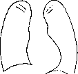
Figure 6. X-ray diagram in stenosis aortae. In differential diagnosis, lues and arteriosclerosis of the aorta must be kept in mind. In syphilis of the aorta, there may also be a long period without any subjective symptoms until sudden death, however anginal pains (aortalgias) appear relatively early and quite readily yield to specific therapy; lues in the anamnesis and a positive RW significantly strengthen the diagnosis. Arteriosclerosis is indicated primarily by the advanced age, a sufficiently full pulse, and a characteristic sphygmogram (arteriosclerotic plateau); the systolic murmur is never as rough as in stenosis. For the latter, the discrepancy between the sharp thrust, strong murmur, and weak pulse (Khavkin) is especially characteristic. It must be kept in mind that a systolic murmur on the aorta can also be detected with compression by tumors, with displacement of the heart (Edens), as well as with developmental anomalies (see below). Kukoverov and Sirotinin made the observation that when raising the hands over the aorta, a soft systolic murmur can be heard, which disappears when lowering the hands. The authors consider this phenomenon a sign of the initial stage of atheromatosis of the aorta (with more pronounced atheromatosis, the murmur is constant). This Kukoverov-Sirotinin symptom is sometimes also found in young subjects, which undermines its diagnostic value. The course, as noted, is very benign. Edema, and even if small, develops only shortly before death, sometimes infarcts in the lungs (Plesh) are also found, as well as attacks of shortness of breath. - Prevention is determined by etiology and pathogenesis. - Treatment (with disruption of 'compensation') is little effective, since the left ventricle, failing due to degenerative changes in the myocardium, cannot increase its contractile ability. If there is a 'reserve', 'compensation' can be temporarily restored. - Assessment of employment due to the rarity of the defect has little practical significance. Based on the foregoing, however, one should not be afraid of significant physical exertion if there are no concomitant diseases. Insufficiency of the aortic valves. This second most frequent defect is very rich in symptoms. Experimental data. With destruction of the valve apparatus, blood in the diastolic phase of the ventricles returns from the aorta back into the heart, producing characteristic changes in the activity of both the left ventricle and the peripheral circulation. Models show a fall in diastolic pressure, while systolic pressure is little changed (Wiggers). Stewart showed that the reverse flow of blood is generally small, and the observed sharp decline in the pulse depends less on the movement of the blood wave toward the heart than on the reflex dilation of arterioles, and consequently facilitated outflow of blood to the periphery. Stewart's curves make it possible to draw the following conclusions: an increase in pulse pressure (amplitude of the pulse) indicates an increase in the amount of blood ejected by the left ventricle, and in turn depends on the degree of diastolic stretching (increase in initial tension); a rapid rise and rapid decline of intraventricular curves indicate that the ventricles empty relatively early. Herzmann, experimenting on 150 dogs, obtained the following data. Hypertrophy of the heart, mainly of the left ventricle, gave moderate figures; it occurs early (from 18 to 110 days), and there is no direct proportionality between the duration of valve damage and the degree of hypertrophy, since hypertrophy depends on other factors besides the hemodynamic overload of the left ventricle (age of the animal, presence of endocarditis, extent of damage). Auscultation corresponds to the data found in humans. Peripheral symptoms appear immediately after the formation of the defect and then remain, while the capillary pulse is detected later. The electrocardiogram gives no indication of left ventricular predominance. As for the general condition of the animals, in Rosenbach's experiments no deviations from the norm were observed; the animals coped with loads as well as healthy ones. Pathological anatomy and pathophysiology in humans. Path.-anat. data on endocarditic, syphilitic, and arteriosclerotic nature of aortic valve damage, according to Plesh, come down to the following changes. There are all possible variants of acute and chronic ulcerative and warty endocarditis. The ulcerative process can affect either individual parts or the entire valve apparatus, papillomatous growths can cover either the entire surface of the valve or only part of it. The process can lead to perforation and even detachment of an individual valve. The process is localized not only on the aortic valves, but mostly also spreads to other parts of the endocardium. In arteriosclerotic insufficiency, calcified hardenings are also found over a large extent of the aorta. A pure defect is rare. Usually it is combined with stenosis, since even a slight change in the valves serves as an obstacle to the systolic flow, and with contraction of the valves, a pronounced narrowing of the opening is formed. Expansion of the bulbus aortae is often found (in syphilis). The left ventricle is dilated and hypertrophied, which fully corresponds to the dynamics of the defect: initial dilation with subsequent hypertrophy (load on the left ventricle). Hypertrophy of the right ventricle is also found, which apparently depends on the overflow of the lesser circulation: stagnation in the left atrium due to difficulty in outflow of blood into the overloaded left ventricle. These data are in full agreement with the studies of Hochrein, who using his technique found in compensated defect only an increase in the left ventricle with normal size of the other chambers; with existing decompensation, an increase in both atria was found. Dynamic observations by Katz and Feil showed that the time of tension is clearly shortened; the authors explain this finding by the greater speed of ventricular contraction and the slight diastolic pressure in the aorta; the time of ejection is lengthened; the duration of the entire systolic phase approaches normal. Plesh suggests that with non-closing aortic valves, blood can already flow from the atrium into the aorta during the diastole of the ventricles. The reverse outflow from the aorta into the ventricle, according to Plesh, can be hindered: 1) by residual blood in the ventricle, 2) at the end of diastole due to an increase in the systole of the left atrium, and 3) by facilitated outflow of blood through the capillaries. In traumatic defect, young subjects in the first days after the formation of a defect in the valve usually do not experience any sensations, later however palpitations, weakness, lack of air, symptoms of myocardial damage join in. Etiology. According to the statistics of Romberg's clinic, syphilis as a cause of valve damage occurred 8 times more often than acute articular rheumatism; other causes were noted only in individual cases. A number of authors described traumatic origins of the defect. Harmer in 476 cases (men) found a positive RW in 37.3%, rheumatic etiology of the defect in 27.7%. Uncomplicated insufficiency of the valves was more often found between the ages of 40 and 50, while in combination with mitral damage - from 30 years and earlier. In cases of pure damage, syphilis accounts for 50%, rheumatism - 7%. Sabatini however emphasizes that rheumatic etiology is generally still more common than is currently accepted. Clinical picture. For many years, if it is not a traumatic defect and if there are no concomitant diseases, insufficiency of the aortic valves does not cause subjective sensations. In more advanced cases, patients complain of dizziness, headaches, unpleasant sensations in the chest and a feeling of generalized pulsation. Objective examination, as indicated, gives an extraordinary wealth of symptoms. On examination, a significant ('aortic') pallor is striking, which does not depend on the composition of the blood, but is a consequence of changes in the capillaries; on the chest, along the line of attachment of the diaphragm - a band of dilated veins (Plesh considers it a very characteristic symptom of this defect). Even with a superficial glance, the sharp pulsation (in the neck) of the carotid arteries is often striking. In developed cases, the play of many arterial trunks (danse des arteres) is noticeable, as well as the diffuse pulsation of the entire patient (homo pulsans). It must be kept in mind that the pulse of peripheral arteries is often visible with severe arteriosclerosis and without damage to the aortic valves. Due to the sharp change in the position of the heart, systolic retraction of the intercostal spaces is observed. The cardiac hump is pronounced even with a relatively late development of the defect. The cardiac impulse is also usually well noticeable in the form of an extensive dome-shaped protrusion (choc en dôme), often shaking the chest and being transmitted to the bed on which the patient lies.
Upon palpation, a double impulse is sometimes detected, which is formed by the systolic displacement of the heart and an additional diastolic wave, explained by Pleshu as follows: because the atrium can already during the diastole of the ventricle push blood through the open valves into the aorta, a presystolic wave can form, which therefore indicates the strength of the contraction of the left atrium. Under such conditions, the sphygmogram may reveal a dicrotic wave, which has a favorable prognostic value: its disappearance will indicate weakening of the left atrium as a symptom of circulatory disturbance. More often, however, a double impulse is observed in the weakening of the hypertrophied and dilated ventricle in cardiosclerosis in the absence of valve damage. The second elevation is connected with the activity of the atria. The pulse sometimes observed in this defect in the form of two equal peaks (pulsus bisferiens) has not found its interpretation. Lew considers it characteristic of the aforementioned myocardial condition. The most characteristic quality of the pulse wave is the so-called rapid (celer) or bounding (saliens) pulse, described by Corrigan in 1832. It is usually explained by the fact that the powerful hypertrophied left ventricle sends a large wave into the aorta (a rapid upward jump), which immediately gives a vertical fall due to the outflow of blood back into the left ventricle. This explanation (in its second part) must be considered incorrect, because the steepest fall of the pulse already occurs during the second half of systole and depends, as indicated above, on the facilitated outflow of blood to the periphery. The wave is quite significant in size (pulsus altus). The number of pulse beats is usually noticeably increased (pulsus frequens). The origin of tachycardia in this defect, which in general favorably influences (by reducing the duration of diastole) the amount of backflow of blood into the ventricle, has not been clarified; it is hypothesized that there is a fall in the tone of the vagus nerve due to reflex from the aorta or from Hering's sinus caroticus (Wigger). Thus, characteristic of this defect are pulsus altus, celer, saliens, frequens, pulsus differens, the difference in filling between the right and left radial artery (usually observed only in syphilitic lesions of the aorta). The bounding pulse is therefore due to large fluctuations in arterial pressure, which is confirmed by direct measurements: the swings (amplitude) of pressure can exceed twice or even four times the normal figures. What is characteristic is not so much the elevation of systolic pressure (in the absence of arteriosclerosis and kidney lesions it can be normal or even below normal) as the sharp fall of diastolic pressure, which may not be detectable at all. Korotkoff tones are heard even at the zero point of the manometer. Relatively high (normal) diastolic pressure indicates complication by accompanying diseases that increase vascular tone, or on very slight expression of the defect. Gausmann observed relatively high diastolic pressure in syphilitic nature of the condition. Hill and his colleagues found that pressure in the femoral artery is significantly higher than in the brachial artery. The authors found this phenomenon in both normal animals and healthy people. According to Hill and Rowland, as well as Lewis and Drury, this difference depends on the increased tone of the femoral artery. Since this increase in tone was not experimentally detected, Frank suggests that the described phenomenon is the result of summation of primary and secondary waves reflected from nearby arterial branches. In any case, this increase in pressure in the femoral artery is characteristic of certain pathological processes: besides aortic valve insufficiency, it was found in arteriovenous fistulas, arteriosclerosis (Hill and Rowland) and thyroid diseases (Taussig). By percussion, as well as palpation of the impulse, displacement of the border to the left and downward is determined. In pronounced cases, the impulse can be determined in the VI and even VII intercostal space on the anterior or mid-axillary line. The left ventricle occupies the entire surface of absolute dullness. Radiographically, the heart has the shape of a lying egg or a sitting duck (expressed aortic heart): a clearly defined angle on the left between the heart and the great vessels, the shadow of which depending on the greater or lesser dilation of the aorta is always increased in transverse diameter [see separate table (pp. 463-464), Figure 3].-Due to the return of blood flow from the aorta into the left ventricle during its diastole, a diastolic murmur is heard, which is distinguished by its duration, soft, blowing character and decreases in intensity (decrescendo). The point of greatest intensity of the murmur is sometimes determined on the manubrium of the sternum (conducted along the entire bone), sometimes to the left, and often to the right of it. Conel and Chillgarth for endocarditic murmurs consider the attachment point of the left third rib cartilage characteristic, for syphilis-the second intercostal space on the right. If the second tone is preserved, the murmur, unlike the diastolic murmur in mitral stenosis, begins at some distance from the second tone, which corresponds to the time required to raise the pressure in the aorta to a certain height. The murmur is better heard in the vertical position, because in it the left ventricle is closer to the chest wall. With a small defect in the valve apparatus, the murmur may be completely absent. The arteriosclerotic process is characterized by a slow (from month to month) increase in the intensity of the murmur. As for the second tone on the aorta, with a small degree of insufficiency of the valves, it can be preserved and then it is usually accentuated. The first tone on the mitral valve is weakened due to the overflow of blood into the left ventricle. A systolic murmur at the apex is heard with the occurrence of relative insufficiency of the bicuspid valve, which can then lead to an increase in the second tone on the pulmonary artery. The presystolic murmur occasionally found in this defect (Flint's phenomenon) is still a subject of dispute among authors. Flint (1860) explained it by the vibration of the mitral valve, resulting from the fact that the backflow from the aorta tends to close the valve, while the contracting atrium, on the contrary, keeps it open. The disappearance of the murmur can therefore indicate weakening of the atrium. Keyt believes that this murmur occurs during the strain stage of the left ventricle, therefore before the first tone, and is formed by the backflow of blood from the aorta. According to Pleshu, it results from vortex movements due to the collision of blood flows from the aorta and atria. Edens believes that there is actually a narrowing of the left atrioventricular opening due to the inward pressing of the aortic cusp of the mitral valve. - Auscultation of peripheral vessels (e.g., femoral, brachial artery) in normal conditions gives no sound phenomena. In this defect, with "careful application of the stethoscope or phonendoscope to the pulsating artery," two, more often one, sharp tones are heard in it due to sharp fluctuations in pressure (Traube's phenomenon). With greater or lesser compression of the vessel, even in normal conditions, a (systolic) murmur is formed; with aortic valve insufficiency, not one but two murmurs are heard-the first, louder and longer, the second-weaker and shorter (Duroziez's phenomenon) (see Duroziez's sign). This sign is by no means common in this defect, and besides it was observed in hypertensives with a large pulse. In addition to the listed symptoms, other phenomena are noted, some rarer, some not fully characteristic of this defect: Musset's phenomenon-systolic nodding of the head (Vinogradov observed it also with insufficiency of the tricuspid valve), F. Müller's-pulsation of the palatine arches, Landolfi-pulsation of the pupils, Snellen-pulsation of the retinal arteries, Gerhardt-pulsatory shaking of the liver and spleen. All these signs depend, on the one hand, on the swings of blood pressure and, on the other, on the fact that the blood-filled powerful left ventricle sends a blood wave, which spreads as a sharp pulsation to distant small arteries. The so-called capillary pulse (Quincke), once considered a pathognomonic symptom of aortic valve insufficiency, is now evaluated differently. According to the widespread view, the capillary pulse (rhythmic change in paleness and redness of the skin or mucous membranes) depends on the fact that the blood wave in the form of a pulsating current reaches the capillaries (in normal conditions, the blood flow ceases to pulsate already in the arterioles) and thereby causes rhythmic redness and paleness.
Even though Claude Bernard established that when the vascular bed is stretched, even veins can pulsate (which was later confirmed by Gibson), capillaroscopy has shown that in the defect described, it is not the capillaries but the small arteries that pulsate (Nesterov, Skulsky, and others); conversely, even precapillaries do not pulsate but are in a state of spasm, which causes the paleness of patients (see above). True capillary pulsation is observed only rarely, namely when the arterial limb of the capillary noticeably expands due to venous congestion. The significance of Quincke's symptom is diminished by the fact that it can be observed in completely healthy people under 40 years of age, depending on the decrease in arteriolar tone, during fever, after various heat procedures, and in Graves' disease. Electrocardiography, in accordance with the marked hypertrophy and dilation of the left ventricle, reveals a pronounced leftogram (sharp discrepancy with the experimental data—see above); the lengthening of ventricular systole (the 'R-T' interval) confirms the data of other methods. The small atrial wave in the stage of full compensation indicates no load on the pulmonary circulation. Extrasystole most often originates from the left ventricle, which fully corresponds to Zelenin's views on the pathogenesis of some extrasystoles: overstrain of a certain part of the heart. As for the pathogenesis of subjective sensations, partly mentioned above among typical complaints, the throbbing in various organs, the sensation of diffuse pulsation, and dizziness are associated with large fluctuations in blood pressure. The occasionally observed semi-fainting state (of the type of minor epileptic seizures—petit mal) during a long (compensatory) pause after a disruption is explained by the fact that due to a significant outflow of blood to the heart, cerebral ischemia occurs (Zelenin). As for the pains behind the sternum and in the heart area (sometimes with typical irradiations), various pathological processes can underlie them: 1) atheromatosis of the coronary arteries, 2) syphilitic lesion of the aorta, 3) mismatch of coronary vessels with the hypertrophied ventricle, 4) ischemia of the heart due to a sharp drop in arterial pressure, 5) reflex from the aorta (Edens).-As for the general assessment of the patient's condition, it is usually overshadowed by these painful sensations, especially when it comes to developed anginal attacks; in most cases, however, the defect itself, not complicated by additional pathological processes, should be classified as benign changes in the cardiovascular system, which fully agrees with the definition of normal (average) figures for the amount of blood ejected by the heart in 1 min.; the absence of oxygen deficiency and cyanosis speaks in favor of undisturbed peripheral circulation; to what extent compensation is due to an increase in the volume of a single contraction (Schlagvolumen) and to what extent to the acceleration of cardiac activity, it is difficult to say.-Is there relative insufficiency of the aortic valves? It is undoubtedly possible to detect it in significant aneurysmal expansions of the syphilitic aorta. The clinical picture in such cases is composed of the symptom complexes characteristic of aneurysm and the defect. Krehl in 1891 described 2 muscle strands in the plane of the interventricular septum from the side of the left ventricle cavity and believed that they could interfere with the closing of the semilunar valves. However, Pollaczek proved the impossibility of such a role for these strands and considers them as a muscular layer protecting the left 'leg' of the His bundle. The course is fully determined by the individual combination of conditions: age, arteriosclerosis (especially of the coronary vessels), lesions of the myocardium (from previous infections and syphilis), etc., as well as the conditions of work and life. In general, it is relatively benign: poorly developed forms often run completely asymptomatic and are brought under medical observation quite late. An early sign of the impending circulatory disorder is a decrease in vital lung capacity even in the absence of accentuation of the second pulmonary artery tone, which joins later; then swelling of the liver is detected, while peripheral edema almost never reaches significant sizes. Edex gives the following interpretation to this circumstance: the sharp pulsation of the arterial trunks facilitates the blood flow through the accompanying veins, while in the liver the role of this 'peripheral heart' (a. hepatica) is negligible: the blood flow in the hollow veins lacks this pulsatory acceleration; venous stagnation and enlargement of the liver result. But this purely mechanical factor in no way resolves the question as a whole. Further circulatory disorder is characterized by attacks of shortness of breath (predominant weakening of the left ventricle). In a number of cases, the general picture of decompensation differs little from that in mitral lesions. Prognosis. All that has been said above in describing the course applies to an even greater extent to the prognosis. In the absence of signs of circulatory disorder, the patient is, as a rule, long-lived and capable of work and life. From sports literature, mention is made of champions in the most severe forms of sports with undoubted insufficiency of the aortic valves. Plesch asserts that arteriosclerotic insufficiency by itself is never a cause of death. The most frequent companion of the defect, leading to a fatal outcome, is kidney damage (especially in syphilitics). Hence the line of conduct of the doctor in resolving the question of employment is clear: the detection of one defect in the cardiac valve apparatus should in no way result in the transfer of the patient to the category of disabled persons. However, when the first clear signs of decompensation appear, patients die quite quickly. The prognosis is especially poor if there is a negative T wave in the electrocardiogram (in lead I). Indeed, the T wave should be given an unfavorable prognostic significance if it is deformed, as this usually indicates myocardial damage. The addition of bundle branch block and especially of block of the terminal branches of the His bundle (see Heart block) aggravates the prognosis. It is also necessary to bear in mind that the aortic valves more often than the mitral valve serve as the site of localization of malignant, septic endocarditis, which makes the prognosis highly uncertain. Defects of the right atrioventricular opening are usually considered to have arisen in the fetal period (congenital). In a large percentage of cases, this does not correspond to reality. Insufficiency of the tricuspid valve (insufficientia valv. tricuspidalis). A pure uncomplicated defect is a great rarity; most often it is combined with mitral lesions. Indeed, a disease that developed in utero usually takes an unfavorable course, and children die from it very early. Genchen in adults, based on his material, did not find a single case of tricuspid insufficiency that could be considered congenital. Etiologically, the defect is associated with endocarditis, which can produce significant anatomical changes in both the valve and the chordae tendineae. Autopsy reveals a huge increase in the right atrium, hypertrophy and dilation of the right ventricle, which fully corresponds to the dynamics of the defect: return of blood during ventricular systole into the right atrium and additional load on the right ventricle (similar to what is observed with the corresponding defect of the left heart). Among the signs of circulatory disorders, the so-called positive, or ventricular venous pulse is described as the main symptom, i.e., a fairly powerful rise of the venous wave coinciding with the contraction of the right ventricle and disappearing when the jugular vein on the neck is compressed (above the point of compression). It should be borne in mind that this phenomenon does not develop immediately but only gradually, when the blood-filled hypertrophied right atrium cannot cope with the load, is additionally stretched, and the stagnation is transmitted to the large veins, which lose the tone of the circular bundle (cuff) described by Keith (Kiss). Only after this is venous pulsation of the liver also detected. In the clinical picture, phenomena of shortness of breath and cyanosis and very easily occurring cardiac insufficiency predominate. The systolic murmur determined at the lower end of the sternum does not in itself solve the diagnosis. Percussory expansion to the right falls mainly on the sharply enlarged right atrium, which can also shift the left border of the heart.-Relative insufficiency of the tricuspid valve joins lesions that give a load to the right ventricle, which gradually degenerates and dilates, involving the atrioventricular ring in the dilation. These include far advanced mitral defects, as well as diseases of the respiratory apparatus, emphysema, kyphoscoliosis, etc.
If relative insufficiency of the tricuspid valve occurs with mitral defects (mainly with stenosis), then in addition to the general symptomatology (enlargement of the heart, systolic murmur at the proc. xiphoid., 'positive' venous pulse, pulsation of the liver, etc.), there is a marked weakening of the accent on the II tone of the pulmonary artery (due to unloading of the lesser circulation). For the same reason (drop in pressure in the left atrium), the diastolic (or presystolic) murmur above the left atrioventricular opening may also weaken or even disappear. In view of the foregoing, the unfavorable prognostic significance of manifested tricuspid insufficiency becomes clear: Kerr and Warren examined 1,000 people and found this defect in 32 patients, of whom 28 died within 1 year. Narrowing of the right atrioventricular opening (stenosis valv. tricuspidalis) according to Chilikin's statistics constitutes 5-6% of all P. s. It usually develops in utero, most often after acute articular rheumatism. Path.-anat. data are reduced to hypertrophy and dilation of the right atrium (less than in the previous defect), to fusion of the valve leaflets, which however never cause such degrees of narrowing as occur in the left ring. Hypertrophy and dilation of the right ventricle depend on the combination with mitral defects. For the same reason, in the clinical picture, auscultatory data (presystolic murmur at the right edge of the sternum) are masked by murmurs of the left heart. Some relief in diagnosis is obtained in the presence of an atrial hepatic pulse and a large A: wave on the phlebogram. Especially characteristic is the variability of the symptomatology and the entire clinical picture and in general the benign course (Chilikin, Dressier). The duration of compensation stands in some contradiction to the hemodynamics of the defect, since the task of equalizing circulation falls mainly on the weak right atrium. In this example, with particular clarity, is revealed that position which should always be kept in mind when analyzing the clinic of defects, namely, that the conditions of hemodynamics by no means cover the entire aggregate of influences on the activity of the damaged cardiovascular apparatus. Combined defects. In the study of combined P. s., the following points must be kept in mind: 1) as already mentioned in the description of individual defects, cases of isolated damage to one valve occur incomparably less frequently than various combinations of changes in both valves and openings. Nevertheless, a detailed description of 'pure' cases is undoubtedly necessary, as it facilitates the understanding of more complex pictures. In fact, from the point of view of narrowly hemodynamic relations, the symptomatology of combined lesions is composed of a combination of partial features inherent in each defect. 2) However, a combined defect is by no means an arithmetic sum of individual pure defects, but represents a new category of pathological condition, which however most often manifests itself in the fact that the clinical picture of combined lesion is formed under the sign of the predominant expression of one or another defect. This circumstance further justifies the necessity of studying typical symptomatology. 3) This definite predominance of one defect and the complexity of symptomatology inherent in the combination of defects make correct diagnosis sometimes extremely difficult, reducing it to a happy chance ('Glucksdiagnose'). 4) Thus, the sporting enthusiasm for diagnostic subtleties is justified neither by objective possibility nor by great necessity. 5) Nevertheless, the attempt to establish a more precise diagnosis has the foundation that combined defects in general give a more severe symptomatology, which, on the one hand, depends on more extensive circulatory disorders, but mainly on the fact that combined defects indicate a greater depth and variety of the anatomical process affecting the entire heart (pancarditis) and the entire organism. Therefore, the indication of some authors on the possibility of mitigation of one defect by another either does not correspond to reality at all or has a purely formal (mechanistic) character. The existing large statistics of Hensen, in addition to some indications on the peculiarities of hemodynamic relations, is also valuable in that it notes the most frequent combinations, in the direction of which diagnostic investigations should be directed. Aortic defects rarely combine with mitral stenosis, more often with insufficiency of the mitral valve. Usually, stenosis is combined with stenosis and insufficiency with insufficiency. Relative insufficiency of the bicuspid valve occurs mainly with arteriosclerotic insufficiency of the aortic valves. It is always necessary to keep in mind that in endocarditic defects, there is often a latent pericarditis, which is revealed at autopsy in 2/3 of cases. Conversely, in defects on the basis of syphilis and arteriosclerosis, pericarditis is rare. In regard to the complication of the dynamics of combined defects and their influence on the condition of various parts of the heart, the following indications exist: in the combination of insufficiency of the mitral valve with stenosis of the corresponding opening, hypertrophy and dilation of the left atrium are especially sharply expressed, because to the incomplete emptying of it during diastole of the ventricles is added an additional amount of blood entering during systole. Systolic overflow of the left atrium also occurs with insufficiency of the bicuspid valve and narrowing of the aortic opening, because due to the difficulty of blood outflow into the aorta, the greatly hypertrophied left ventricle sends a larger wave back into the left atrium through the pathological opening. The greatest load on the left ventricle naturally falls with the simultaneous existence of insufficiency of the mitral and aortic valves, because each of these defects separately leads to dilation and hypertrophy of the left ventricle. As for the double aortic defect, its effect on the left ventricle is as follows: due to the narrowing of the aortic opening, the backflow of blood from the aorta is difficult and thus the overflow of the ventricle and consequently its dilation will not be as pronounced as occurs with insufficiency of the aortic valves, not complicated by stenosis of the opening. Bamberger considers this combination favorable for a pure defect (insufficiency of the valves). If Bamberger's reasoning is correct from the point of view of the mechanics of circulation, they completely ignore the etiology of stenosis and its influence on the entire organism, as well as the condition of the coronary vessels. The opinion about the hemodynamic 'benefit' for mitral defects from the accompanying insufficiency of the tricuspid valve (a safety valve for the overloaded lesser circulation) has been repeatedly put forward in the literature. This assertion does not withstand criticism from any side. If, for example, there is a developed form of mitral stenosis with great overflow of the lesser circulation, then the occurrence of relative insufficiency of v. tricuspidalis indicates only the further development of circulatory disorders (weakening of the right ventricle). III. Compensation and decompensation in P. s. Before proceeding to the presentation of therapeutic and preventive measures in relation to patients with compensated and decompensated P. of the heart, it is necessary to establish the meaning of the terms 'compensation' and 'decompensation' in P. s. By compensation of P. s. is meant the establishment of a certain approximation to the normal functioning of the cardiovascular apparatus, i.e., the absence of noticeable circulatory disorders in the usual working and social environment without limitation of the patient's work capacity. The occurrence of circulatory disorders with some limitation of work capacity is sometimes designated as subcompensation; decompensation, however, implies a state of deep circulatory disorder and loss of work capacity. The conventionality of these designations is self-evident, because, on the one hand, the requirements imposed on the cardiovascular apparatus by life (socio-labor conditions) are completely different in each individual case; on the other hand, there is no sharp boundary between the indicated states, and they represent a process influenced by a large number of constantly changing factors. Familiarity with them is extremely important, as they determine not only the paths of prevention but also the methods of therapeutic intervention. Here, one must first of course keep in mind the pathological changes in the circulatory apparatus itself (mainly in the myocardium), age peculiarities, the degree of general development of the organism, conditions of nutrition, the environment of work and daily life, as well as the influence of emotions. If hemodynamic relations (perversions of blood flow and their consequences) are not the decisive factor in decompensation, then along with the moments noted above, intercurrent infectious diseases (recurrent endocarditis, typhoids, pneumonias, erysipelas, grippe, etc.) acquire special importance, which usually cause circulatory disorders.
In such cases, they depend either on further, more severe damage to the valve apparatus (endocarditis recurrens) or, more often, on changes in the myocardium that undermine the systolic energy of the ventricles. Thanks to electrocardiography, which allows to note barely perceptible by other methods changes in the myocardium, it has been possible to establish a significant frequency of myocardial lesions in various infections, especially in acute rheumatism. At this time, either various degrees of atrioventricular block are detected (see Heart Block) or deformities of the G wave of the electrocardiogram occur (Zelenin and Lias). The also determined friction rub of the pericardium indicates the extensiveness of the process that has affected all the membranes of the heart (pancarditis), which can directly or later affect the ability of the cardiovascular apparatus to maintain circulation. It should also be borne in mind that circulatory disorders are determined not only by the weakening of the contractile ability of the heart muscle (in connection with infectious myocarditis), but also by the state of vascular tone, which is often disrupted in acute infection. Thus, the activity of the main process, the tendency to relapses, leaves its mark on the course of the defect and the occurrence of "decompensation." In subacute or chronically progressing infections, the weakening of the cardiovascular apparatus depends not only on its intoxication, but also on impaired nutrition due to a decline in blood formation (anemia). For the reduction of the accommodating ability of the heart muscle, the local ischemia in connection with impaired coronary circulation (in coronary sclerosis and spasms of the heart vessels) is of no less importance than the general decline in blood formation. As a result, the tendency to circulatory disorders correspondingly increases. In addition to the tendency to coronary spasms with the aforementioned ischemia of the heart muscle, the increased excitability of the autonomic nervous system, causing various forms of cardiac rhythm disturbances (mainly extrasystoles) and myocardial tone, exhausts its functions and leads to a more rapid occurrence of circulatory disorders (see below). In the same direction acts the often present in vegetative neurotics insomnia, nightmares, and obsessive ideas about severe suffering. At this time, disorders arise in the functioning of numerous mechanisms of circulation (see). It is necessary here to note only some peculiar features of the weakness of the left and right heart, as well as to add a few more rough strokes characterizing the state of decompensation and to some extent outlining the paths to the restoration of satisfactory circulation. Insufficiency of the right and left heart. Some authors have attempted to give a more exhaustive clinical picture of the insufficiency of one or the other ventricle. Laubry draws the symptom complex of left ventricular weakness in the following form: early appearing and chronically increasing dyspnea, sometimes passing into attacks of asthma with signs of pulmonary edema; in other cases, angina pectoral attacks occur more often than asthma; signs of myocardial hypotension (expansion of the heart, mainly to the left, transient murmur due to muscular insufficiency of the mitral valve, gallop rhythm); tachycardia (in contrast to right ventricular insufficiency) does not reach great degrees, bradycardia is even encountered; ventricular extrasystoles and pulsus alternans are common; pulse pressure is reduced; stagnation phenomena in the periphery are observed last, comparatively early disturbance of cerebral circulation (insomnia), renal and hepatic is discovered. For right ventricular insufficiency, Laubry considers tachycardia (90-120 beats per minute), expansion of the right ventricle, gallop rhythm as characteristic; however, Laubry does not consider cyanosis pathognomonic. This symptom complex in a clear form in connection with damage to the valve apparatus is encountered only in (congenital) defects of the right heart; more often circulatory disturbances in the small circle are of extracardiac origin (atheromatosis of the pulmonary artery, obliteration of the pericardium, aneurysm of the art. pulmonalis and aorta, etc.). Uhlenbruck calls this combination the "cardio-pulmonary complex" in contrast to the "cardio-renal" one, characteristic of lesions of the large circle. Unlike Laubry, Uhlenbruck did not find noticeable enlargement of the heart in right ventricular insufficiency; on the contrary, with moderate dyspnea there is marked cyanosis: venous pressure is usually elevated, hematuria, bloody vomiting, bloody stool, a sharp increase in the number of red blood cells, peripheral edema are often observed; the pulse is frequent, usually regular; the heart tones are clear, accent of the II tone of the pulmonary artery is as a rule absent. Zelenin considers such a far-reaching differentiation in the symptomatology of right and left heart insufficiency a task of little feasibility, since isolated disturbance rarely exists in reality. At our disposal there is a method, namely electrocardiography, which makes it possible to judge the predominant role of the left or right ventricle (lefto- or rightogram). However, precisely in the period of decline of cardiac activity, we are not yet able to rely on the electrocardiogram to judge which half in this case more loses its working capacity. In the atonal state, it was possible to observe isolated contraction of only one ventricle (hemisystole). By means of electrocardiography, from another side, one can also approach the judgment about the predominant lesion of one or another part of the heart. Zelenin's research established that the place of origin of additional excitation (easily determinable electrocardiographically) can not only indicate partial, strictly localized (mainly near the conduction system) anatomical lesion, but can also be a response to increased (dynamic) load of a certain part of the heart: the occurrence of extrasystoles originating from the right ventricle in emphysema of the lungs, from the left ventricle-in insufficiency of the aortic valves. Eppinger and Rothberger showed that by clamping the pulmonary artery or aorta, one can artificially cause either a righto- or leftogram. In mitral stenosis, due to overstrain of the left atrium, atrial extrasystoles arise, which, with further deterioration of nutrition of the atrial musculature, can be replaced by their flutter or fibrillation (delirium cordis, puls. irregular. perpet., see Heart Arrhythmias). The role of arrhythmias in the phenomena of decompensation is twofold: on the one hand, they indicate the onset of decompensation, and on the other hand, they themselves can cause or deepen decompensation (Kabakov); thus a vicious circle arises which is prognostically especially serious when atrial fibrillation occurs (Plesh), although sometimes patients with heart defect and arrhythmia rec. cope well with heavy physical labor. Symptomatology of circulatory disorders in heart defects. Profuse hemoptysis, especially characteristic of mitral stenosis, often alleviates the patient's condition, reducing pressure in the small circle. With weakening of the right ventricle and stagnation in the veins of the large circle, bleeding (e.g., from hemorrhoidal veins) can restore compensation in the most premortal states. Diarrhea in stagnation in the roots of the portal vein sometimes quite ensures the excretion of fluid, despite the decline in urination (due to a drop in pressure in the renal artery). In general, the water exchange of the body in decompensation is insufficiently studied, and we are therefore not fully oriented in our therapeutic tactics: in particular, there are observations (Kauf-Zak) that with the development of edema, sweating also decreases. The influence of diuretic agents (e.g. salirgan) in restoring compensation is also not quite clear: in addition to ascites, mechanically hindering diaphragmatic breathing, transudates in the lungs, pleura, in the brain, pericardium, impairing circulation, the possibility of edema of the heart muscle itself is not excluded; electrocardiographic analysis in one case of extremely widespread (neurotic) edema proved such a possibility. For characterizing the initial stage of circulatory disorder, the determination of hidden (accumulating in tissues) edemas is important, which can be best detected by systematic weighing of patients and special techniques (intradermal test of McCloy-Oldridge, technique of Kaufmann, etc.). Belova and Goldman, studying patients in Kislovodsk, established the suitability of the first test, and Salishchev - of the second for judging the state of compensation. On methods of direct determination of the volume of circulation - see Circulation. Connection of circulatory disorders in heart defects with the condition of various organs. Respiratory apparatus. An additional force of circulation is, as is known, the negative pressure of Donders, existing in the pleural spaces. On inspiration it increases, on expiration it decreases.
The suction effect of Donders' negative pressure mainly affects thin-walled structures: the large veins, the right atrium, and to a lesser extent, the right ventricle. Thus, the act of respiration promotes circulation. In addition to the aforementioned suction effect of negative pressure, the movement of the diaphragm mechanically squeezes blood from the hollow veins, which enhances the blood flow toward the heart; according to Lewis's observations, only abdominal breathing has a positive effect, while chest breathing has the opposite effect—it impedes the blood flow to the heart. In cardiac patients, when the pulmonary capillaries are filled with blood, the vital capacity of the lungs is also reduced. Peabody and Wentworth therefore recommend the systematic determination (in the same patient) of the vital capacity of the lungs for prognostic purposes. Primary lesions of the cardiovascular apparatus cause a number of phenomena in the respiratory organs: impairment of peripheral circulation leads to congestive pharyngo-laryngitis and bronchitis with distressing cough, extremely burdensome for the cardiac patient (fluctuations in intrathoracic pressure that impede blood flow to the heart). Weakening of respiratory excursions due to the high position of the diaphragm and the tendency for transudation into the alveoli causes the observed, especially in the morning, band of atelectatic rales in the lower lobes of the lungs. Accumulation of fluid (transudates) in the pleural sacs (mainly the right) indicates further circulatory disturbance and worsens it by limiting the ventilated surface of the lungs. With significant enlargement of the heart, mainly the left atrium, atelectatic (crepitant) rales are heard in the area of the III-IV ribs along the left parasternal line (Botkin's symptom). Due to the filling of the pulmonary vessels with blood, sputum in congestive bronchitis and pulmonary edema is colored pink or has a more noticeable admixture of blood. Hemoptysis is observed in pulmonary infarctions due to embolism (accumulation of thrombi in the right heart). With prolonged congestion, brown induration of the lungs occurs, about which the cells of cardiac defects found in the sputum speak. Constant congestion in the lungs predisposes to pneumonia, which takes a torpid (sluggish) course with low temperature. Abdominal organs. The liver, being a large reservoir for blood, enlarges in volume very early, pushing the diaphragm upward and often reaching down to the navel. Patients experience a feeling of heaviness in the right hypochondrium, which is stronger the faster the organ swells (stretching of Glisson's capsule). Objectively, the round soft edge is easily palpable. To what extent the functions of the liver are affected in this condition is not sufficiently clarified. It is possible that the hypoglycemia of cardiac patients, which unfavorably affects the nutrition of the heart muscle, is due to venous congestion of the liver. With prolonged (many months) circulatory disturbance, the liver becomes dense due to the development of connective tissue, which can eventually lead to venous, cardiac cirrhosis of the liver with impairment of portal circulation (ascites, enlargement of the spleen, which also undergoes induration from congestion). The spleen enlarges in size, however not enough to be easily palpable. Attacks of severe pain in the area of the spleen usually depend on infarction in it. The pancreas is also often found in a state of induration; little is known about the disorder of its functions in this case (cf. the hypoglycemia noted above). Congestion in the gastrointestinal tract manifests with dyspeptic phenomena (loss of appetite, nausea, feeling of heaviness in the stomach) and a tendency to constipation or (more rarely) to diarrhea, as well as significant flatulence (upward displacement of the diaphragm and the lying position of the heart). Profuse diarrhea sometimes dehydrates the body (reduces edema) better than other diuretics. It is also necessary to take into account that reflexes originating from the stomach and intestines distended with gas can cause spasm of the coronary vessels and disturbance of cardiac rhythm (Lass's experiments with inflation of the stomach in cardiac patients). The discrepancy between the size and persistence of ascites with the degree of circulatory disturbance in cardiac patients should suggest either the beginning of cirrhosis of the liver or significant induration of the peritoneum, hindering the reabsorption of fluid. Clinic of ascites—see Ascites. Kidneys. Depending on the slowing of blood flow and the fall in blood pressure in the renal artery, the water-excreting ability of the kidneys decreases: diuresis decreases, urine becomes concentrated with a brick-colored sediment of urates. The excretion of table salt is delayed. Due to impaired liver function, urobilin (urobilinogen) is often detected. Constant companions of stagnant urine—albuminuria and hyaline cylinders. The amount of protein rarely exceeds 1-2%. With severe congestion, a certain amount of red blood cells is always present. The presence of renal epithelium and epithelial and granular cylinders indicates the addition of degenerative changes in the kidneys. Quincke noted that the excretion of urine is slowed down and occurs mainly at night (see Nocturia). The water-secreting function can be controlled by comparing the amount of fluid introduced and excreted. However, errors are very common here. For the accurate determination of water movement, it is best to use systematic weighing of patients, as well as methods for determining hidden (intratissue) edemas. Genital organs. Edema of the scrotum and prepuce often serves as an obstacle to voluntary urination and catheterization. Sexual potency in the stage of significant circulatory disturbance falls. Preservation of libido with limited accommodative capacity of the heart can cause sudden death during coitus. On the contrary, in other cases, regulated sexual life, acting soothingly on the nervous system, has a beneficial effect on the cardiovascular apparatus. Menstruations are weakened or even disappear in a number of cases; sometimes, on the contrary, there are menorrhagias and metrorrhagias. Subjective complaints, however, as a rule, predominate over objective findings. On the relationship between pregnancy and P. s.—see Pregnancy. Blood. With overflow of the venous system of the greater circle (especially in connection with congenital defects), secondary (symptomatic) polycythemia and increased blood viscosity are often observed simultaneously with peripheral edemas and cyanosis. Skin, subcutaneous tissue, muscles, bones. The skin capillaries are elongated, the branches of their loops are altered: in cyanosis, the venous limb is dilated, in pallor, the arterial limb is narrowed. With significant congestion, the subpapillary plexus is filled with blood. The bluish discoloration first appears on the extremities (acrocyanosis): on the fingers of the hands and feet, the wings of the nose, the auricles, the lips, then on the extensor surfaces of the shins and forearms. Small skin veins are visibly dilated. The jugular veins are engorged and undulate. The hands and feet are cold to the touch. It should be borne in mind that in neurasthenics, such cooling (of vaso-motor nature) is usually accompanied by increased sweating—cold sweat (secretory neurosis). Edemas (hydrops anasarca, oedema pedum). The accumulation of fluid in the subcutaneous tissue (anasarca) depends on many, not yet clarified causes (see Edema). In some cases, edemas appear relatively early and reach large sizes, in others—complete circulatory collapse occurs with relatively slight edema of the skin and subcutaneous tissue. According to hemodynamic conditions, aortic lesions lead to edemas only in the final stage. Hydrostatic reasons cause edemas to first appear in the lowest places: on the ankles (oedema pedum) in ambulatory patients, on the waist, and then on the chest, arms, and face—in bedridden patients. At first, edemas, appearing during movement, disappear at rest, and then not only do not disappear, but rise higher: the legs turn into clumsy logs, on which pressing with the finger leaves a deep pit. Edemas on the legs reach enormous sizes (the skin shines and may burst), because due to the accumulation of fluid in the cavities (abdominal, in the pleural sacs, pericardial sac) and the high position of the diaphragm, patients due to respiratory disturbance (orthopnea) cannot lie down, but spend all their time in a chair or in bed with legs down. In this position, circulation is facilitated, as part of the blood is excluded from the circulation and in addition the diaphragm is lower. Due to impaired nutrition of the skin, eczematous changes develop on it, and in the presence of cracks, erysipelas. Muscles with circulatory disturbance become flaccid, tire very easily, which is reflected in the general state of lethargy and breakdown of dropsical patients.—Sometimes, especially with congenital defects, drumstick fingers are observed (see).
The hemodynamic consequences of widespread edema are extremely great: the interstitial spaces, filled with transudate, compress the vascular bed; the almost complete absence of movement in the patient eliminates the training effect of the contraction of skeletal muscles on the cardiovascular system; flaccid muscles, having lost their tone, cease to be an additional factor in peripheral circulation; transudates in the cavities restrict breathing and increase the load on the right ventricle; a significant accumulation of transudate in the pericardium (hydropericardium), by impeding the diastolic phase, also reduces the systolic. That is why an increase in diuresis through diuretic agents in cases of cardiac insufficiency makes the cardiovascular system, after the reduction of edema, capable of responding to cardiaca. It must be kept in mind that when the body is freed of water by increasing diuresis, this entire amount of fluid, sometimes measured at ten to twenty liters, is pumped with the active participation of the heart. Therefore, before using diuretics, it is more advisable to remove part of the fluid mechanically (paracentesis, incisions, or Kursmann cannulas on the legs). Nervous system. The influence of the nervous system on the circulatory apparatus is extremely great (changes in vascular tone, cardiac rhythm, etc.). Circulatory disorders, in turn, disrupt the function of the brain: insomnia, heaviness in the head, headaches, dizziness, general irritability, fatigue during mental work are quite early symptoms of impaired circulation in the brain. Patients often complain of a feeling of fear and impending catastrophe. Actual psychoses (of a depressive nature) have been described in the final stages of circulatory disorders (especially with aortic valve insufficiency). Edema of the brain can sometimes cause a comatose state; as a rule, however, cardiac patients die with preserved consciousness. Organs of sense. Vision. Conjunctivitis, hemorrhages into the conjunctiva and retina are encountered. Flashes before the eyes bother patients. With significant edema of the optic nerve, blindness (amaurosis) may occur, which passes with improved circulation. Hearing. Together with congestive pharyngitis, eustachitis is also observed, causing ringing in the ears and hearing loss. Metabolism. The basal metabolism in impaired circulation with symptoms of shortness of breath is increased. In compensated heart defects, the basal metabolism is normal. IV. Functional diagnosis in H.D. There are numerous methods of so-called functional diagnosis of the cardiovascular system, serving to evaluate the degree of compensation or the width of accommodation of the cardiovascular system in H.D., i.e., the body's ability to maintain sufficient circulation under certain "loads" (walking, jumping, breath-holding, Valsalva's test, etc.). Undoubtedly, each of these methods of functional diagnosis adds some new data for judging the degree of sufficiency of the circulatory apparatus, but none of them alone provides a firm basis for judging the stability of compensation. According to observations by Zelenin, positive breath-holding tests (Shange-Gens) with a relatively slow pulse and a developed T wave on the electrocardiogram show the greatest correlation with the state of work capacity. In general, however, the decisive "functional test" is dynamic (repeated) observation of the state of the cardiovascular system under conditions of work and daily life (frequent and prolonged withdrawal from work, decreased labor productivity, etc.). For a correct assessment of the professional suitability of a patient with H.D., in addition to the formal (anatomical) diagnosis and certain functional tests, along with the socio-clinical history, the work-oriented (or conversely, "anti-work") attitude of the given patient becomes very important, since the role of emotions in the rationalization of the work of the circulatory apparatus is very great (Bainbridge), although still insufficiently evaluated to this day. As an example of the influence of the autonomic nervous system on peripheral circulation, the following observation by Lange can be cited: in decompensation, most patients lose the ability of capillaries to react to temperature irritations, while vegetative neurotics and hypertensives retain this reaction. The influence of sympathetic innervation on the heart itself (positive dromotropic and inotropic influence) is well known. In accordance with the above, there are observations (of the medical-insurance department of the Institute of Labor Protection) on disabled persons of group II who successfully cope with moderately heavy work processes with a certain work attitude, and conversely, on completely incapacitated mentally traumatized (or socially unstable) patients with relatively minor defects in the heart valve apparatus without any noticeable circulatory disorders.
V. Zelenin. X-ray Diagnosis of Heart Defects. X-ray diagnosis of heart defects is based on determining changes in the configuration of the contours and size of the heart shadow. Depending on the nature of the defect, the outlines of the heart shadow also change. Hypertrophy of the ventricles without dilation can give a change in the configuration of the heart shadow in the corresponding parts (rounding of the contours), but the increase in the shadow is relatively insignificant (not more than 1-1/2 cm). Dilation of the heart cavities gives a significant increase in the corresponding parts of the shadow on the screen. Dilation can be of two kinds: tonogenic and myogenic. The first occurs compensatorily under the influence of increased systolic volume and systolic residue, i.e., the amount of blood remaining in one or another part of the heart after its contraction; the heart muscle is unchanged in this case, and according to Kirch, the dilation of the heart ventricles occurs due to the lengthening of their cavity without expansion in their transverse diameter. Myogenic dilation, which is a consequence of weakening of the heart muscle as a result of one or another lesion of it, can reach significantly greater degrees, and according to Kirch, the dilation of the cavities occurs in all directions and predominantly in the transverse direction. Therefore, the configuration of the heart shadow in tonogenic dilation, due to the elongation of its parts, mainly the ventricles, changes in the sense of predominant elongation of its shadow, and in myogenic dilation, in the sense of predominant expansion of it. Under the influence of valve defects, not only the configuration and size of the shadow of the ventricles and atria change, but also of the large vessels depending on their dilation or elongation under the influence of changes in their blood filling or changes in pressure in them. An increase in the left ventricle increases the mid-left size of the sagittal heart shadow, as well as its length. In the presence of tonogenic dilation and usually accompanying hypertrophy, the ventricle increases in length, the contours of the apex (IV arch on the left) are rounded, the heart lies more transversely on the left dome of the diaphragm, its waist becomes more pronounced, and the heart shadow takes on the so-called aortic configuration. Myogenic dilation, causing significant expansion of the ventricle in all directions, if it occurs primarily, does not give a typical aortic configuration of the shadow. An increase in the left ventricle is clearly revealed in the oblique II direction by the bulging of the lower left arch. An enlarged left atrium extends upward, to the right, backward, and to the left, as a result of which the auricle gives a more convex shadow of the III arch of the left contour of the sagittal heart shadow, and the waist of the heart is smoothed out. In oblique directions, when the left atrium is enlarged, the parts of the contours formed by it become more convex, the depth size of the heart shadow increases at the level of the left atrium, as a result of which the retrocardiac field narrows in its upper part in the oblique I direction. In some cases of significant enlargement, the edge of the left atrium becomes visible in the sagittal direction to the right above the contour of the right atrium. An enlarged (hypertrophy and dilation) right ventricle increases the heart downward, to the right, and to the left, the heart turns somewhat around the body's vertical axis, so that its apex is displaced backward. Then the left ventricle in the sagittal direction participates to a lesser degree in the formation of the IV left arch. The heart's long line becomes more vertical due to the right ventricle pushing the right atrium upward and to the right, the angle of inclination formed by the heart's long line and a horizontal line drawn along the lower edge of the heart shadow becomes larger. At the same time, the left atrial auricle is displaced somewhat backward and the entire left atrium backward and to the right. The mid-right size of the heart increases. In some cases, the edge of the enlarged right ventricle becomes visible to the right below the II arch (right atrium), forming an additional III arch on the right. Sometimes the edge of the right ventricle (in the region of the pulmonary cone) bulges to the left between the II and III left arches, replacing the contour of the left auricle, and with particularly large enlargement, it forms the IV left arch instead of the displaced left ventricle. Its enlargement is also manifested in the oblique II and right frontal directions by an increase in the convexity of those parts of the heart shadow contour formed by the right ventricle. Enlargement of the right atrium gives a more convex contour of the II arch on the right of the sagittal heart shadow, an increase in the mid-right size of the latter, and darkening of the lower part of the retrocardiac space in the oblique I direction below the shadow formed by the left atrium. In the oblique II direction, the enlarged right atrium can form the entire section of the lower left contour of the shadow, covering the contour of the right ventricle. Stenosis of the left venous opening primarily causes dilation (and hypertrophy) of the left atrium, which causes bulging of the III left arch of the sagittal heart shadow due to the elevation of the left auricle and an increase in the convexity of the left atrial shadow in oblique projections (narrowing of the corresponding part of the retrocardiac space). Enlargement of the right ventricle (hypertrophy) and subsequent enlargement of the right atrium gives expansion of the mid-right size of the heart, rounding of the II arch on the right and its displacement upward. Due to the rotation of the heart to the left by the enlarged right ventricle, the outlines of the heart apex become less convex, the heart's long line takes a more vertical direction. The sharpening of the heart apex shadow apparently also occurs due to some atrophy of the left ventricle, often present in mitral stenosis. Stagnation (increased blood pressure) in the small circle, causing dilation of the pulmonary artery, gives a distinct bulging of the II left arch of the sagittal heart shadow; this bulging is sometimes stronger than the bulging of the III left arch. Enlargement of these parts of the heart shadow, smoothing and thickening the waist of the heart, gives it the characteristic so-called mitral configuration (see separate table, fig. 1). The shadow of the aortic arch on the left is less pronounced, because as a result of the rotation of the heart, the aortic arch also turns to a more sagittal position. The vascular shadows of the pulmonary hili are significantly intensified. Mitral valve insufficiency, primarily causing tonogenic dilation and hypertrophy of the left ventricle, gives an increase in the mid-left size of the heart with a distinctly rounded contour of the apex and some increase in the heart's length, but an aortic configuration of the heart is not obtained, because with more or less radiologically determinable dilation of the left ventricle, there is also dilation of the left atrium with stagnation in the small circle; this causes bulging of the II and III arches on the left and thus thickening of the heart's waist, and consequently partially its mitral configuration. Enlargement of the left atrium is well determined in both oblique I and II directions. Subsequently, hypertrophy of the right ventricle and enlargement of the right atrium develop. The contours of the right part of the heart shadow become rounded, the mid-right size of the heart also increases. The vascular shadows of the pulmonary hili are significantly intensified (see separate table, fig. 2). Combinations of mitral valve insufficiency with stenosis. The heart shadow has the outlines characteristic of the shadow in mitral insufficiency, but the bulging of the II and III arches on the left is much more pronounced, and the enlargement of the shadow of both atria in oblique directions is also more clearly expressed (see separate table, fig. 4) [see also separate table (Vol. XVIII, pp. 79-80), fig. 6]. Insufficiency of the tricuspid valve. The change in the heart shadow is characterized by a significant increase to the right and distinct rounding of the right contour. An increase to the left is determined in those cases when the enlarged right ventricle forms the left contour of the heart shadow instead of the left one. Often with particularly large enlargements of both atria, the shape of the heart approaches a spherical one.
Overflow of blood into the superior vena cava causes dilation of its shadow to the right, sometimes (as Dietlen indicates) its systolic pulsation can be seen. Stenosis of the tricuspid valve is a very rare defect and manifests as an increase in the cardiac shadow contour to the right and significant dilation of the superior vena cava shadow. Stenosis of the aortic valve is characterized primarily by tonogenic dilation and hypertrophy of the left ventricle. The left ventricle elongates and lies on the left dome of the diaphragm, the contours of the apex become more rounded. The mid-left size of the cardiac shadow increases. The waist of the heart becomes more pronounced, the cardiac silhouette acquires an aortic configuration, the pulsation of the left ventricle is strong with slow waves (pulsus tardus). The addition of myogenic dilation of the left ventricle causes it to dilate in all directions, and the waist of the heart becomes less pronounced. The shadow of the ascending part of the aorta, best visible in the first and second oblique positions, appears dilated because the blood stream under high pressure is ejected by the left ventricle through the narrowed opening into the aorta above its narrowing and then, expanding fan-like, stretches the walls of the aorta. Insufficiency of the aortic valves. Initially, there is no tonogenic dilation of the left ventricle, as there is no systolic residue, and only compensatory hypertrophy develops and the filling of the left ventricle in diastole increases. The apex of the heart becomes rounded, and the mid-left size of the heart increases slightly. The heart takes on an aortic configuration (see separate table, fig. 3), which becomes more pronounced when tonogenic dilation is added and less pronounced when, due to heart muscle damage, the left ventricle dilates in all directions. The most characteristic feature is the strong, rapidly increasing and decreasing contractions of the left ventricle, transmitted to the aorta to a marked degree (pulsus celer). The shadow of the aorta is enhanced and elongated, according to Asmanna (Assman), as a result of an increase in the amount of blood ejected into it by the systole of the left ventricle. Combination of mitral and aortic valve defects. The cardiac silhouette acquires characteristic features of both defects. Depending on the predominance of mitral or aortic valve damage, the corresponding change in the configuration of the cardiac silhouette becomes more pronounced (see separate table, fig. 5). Congenital heart defects. Radiodiagnosis is very complex, in most cases only by comparing all clinical symptoms with radiological data is it possible to arrive at the correct conclusion. Combinations of defects and degrees of valve anomalies can be very different. Patent ductus arteriosus. The cardiac silhouette is characterized mainly by bulging of the second left arc due to increased pressure in the pulmonary artery and an increase in the cardiac shadow to the right due to enlargement of the right ventricle and right atrium. The vascular shadows of the hili are clearly dilated. Defect of the interatrial septum (patent foramen ovale) is also characterized by bulging of the pulmonary artery arc to the left on the cardiac silhouette, hypertrophy of the right ventricle, sometimes significant, dense vascular shadows in the hili, sometimes with their pulsation clearly visible. Pulmonary artery stenosis. In cases where there is no clear dilation of the right ventricle, the cardiac silhouette does not differ significantly from normal. With significant degrees of hypertrophy and dilation of the right ventricle, the latter can form the entire anterior surface of the cardiac shadow (determined in the second oblique and right frontal projections), as well as the left contour of the sagittal silhouette. In some cases, the second arc bulges to the left due to dilation of the pulmonary artery above the site of narrowing; according to Asmanna, this bulging may be formed not so much by the pulmonary artery as by the pulmonary artery cone with significant dilation of the right ventricle. Defect of the interventricular septum. In many cases, the cardiac silhouette may remain unchanged; in more severe cases with significant enlargement of the right ventricle and right atrium, the mid-right size of the cardiac silhouette increases, and the left ventricle may also become hypertrophied or dilated; then the cardiac silhouette increases in transverse diameter, the apex of the heart becomes rounded. Systolic strong pulsation of the right contour of the heart is visible only with significant degrees of the defect. Stenosis of the aortic isthmus is characterized by dilation of the ascending part of the aorta, visible in both sagittal and oblique positions, best in the second oblique direction. In some cases, dilation of the right subclavian and innominate arteries is also noted, determined on the screen by darkening of the medial part of the pulmonary apex. This defect is often combined with a patent ductus arteriosus, and then bulging of the second arc to the left is visible on the cardiac silhouette.
n. Potte. VI. Prevention and treatment of heart defects. Prevention of heart defects consists mainly in the prevention of infections, especially those entering through the oral cavity (oral sepsis, angina), prevention of acute articular rheumatism, syphilis, arteriosclerosis, endocarditis. When these diseases occur, persistent treatment can prevent damage to the valve apparatus. Prevention of decompensation. Physical labor, strengthening the heart muscle and facilitating peripheral circulation, is a factor of compensation. However, one should transition to it only after a period of gradually increasing difficulty of gymnastic exercises according to the type of passive, then passive-active, and finally active gymnastics. This also includes dosed walks on level ground or ascents (Terrain-Kur after Oertel), which are especially indicated in subjects prone to obesity. Even though many patients with heart defects cope very successfully with even very heavy work, when choosing professions, one should avoid heavy loads that could exhaust the heart muscle. It must be kept in mind that if the defect was well compensated from the very beginning, it extremely rarely decompensates under the influence of only (pathological) hemodynamic relations: infections (endocarditis recurrens and others), then intoxications and violations of regimen play the decisive role here. Instructions should be given in this direction. Nutrition should be sufficient but not burdensome, i.e., taken at definite times, in relatively small portions, to avoid reflexes from the stomach (Lass) and high position of the diaphragm. The much debated question about the permissibility of meat food should not be decided by a template; with developed sclerosis and changes in the kidneys, meat is forbidden; conversely, with weakness of the myocardium and decline of blood formation, meat is absolutely necessary (Pevzier and others), and its exclusion often clearly reduces working capacity. The special milk diet of Karell is suitable only for obese subjects and then only for a relatively short period. Its benefit is based on low caloric content, absence of table salt and perhaps on the presence of K salts with their diuretic effect. In this respect, the potato diet of Frey is more effective. It is necessary to monitor the proper functioning of the intestine. The total amount of fluid should also not deviate from average figures (1,000-1,500 cm³); single abundant drinking burdens the heart and should be avoided. Alcohol in small quantities somewhat stimulates cardiac activity and is readily accepted by cardiac patients with subjective sensations of weakness, coldness of extremities and feeling of compression behind the sternum. Larger doses of alcohol, lowering blood pressure, unfavorably affect circulation. - Tobacco smoking meets with different evaluations. German authors usually permit moderate smoking (Plesh), considering that the depression of the psyche upon prohibition has a more harmful effect than tobacco intoxication. However, since nicotine is a specific autonomic poison (Langley), smoking in neurotics prone to vascular spasms is absolutely contraindicated. Experiments of Salishev and Chernogorov revealed the influence of nicotine not only on intracardiac nerve apparatuses but also on the heart muscle. In connection with the enormous influence of the psyche on the circulatory apparatus, psychogenic hygiene under working and living conditions acquires great importance (work attitude, etc.). Sleep, affecting the tone of the nervous system, must be carefully protected. Sexual life, which also exerts a certain effect through the nervous system on the circulatory apparatus, must be regulated; its prohibition applies only to states of significant circulatory disorders, especially in combination with attacks of angina pectoris. (Pregnancy with heart defects. Pregnancy.)-Bathing in rivers and lakes as well as other hydrotherapeutic procedures are not only not prohibited but are recommended as measures supporting the tone of the neuromuscular apparatus. Sea bathing in the south, possibly in connection with the hot climate, is tolerated worse, while bathing in northern seas, on the contrary, can be fully recommended. Swimming, as well as other types of light sports (without competitions) are quite permissible under medical supervision. Although cases of sports champions with damage to the valve apparatus (Ackerman) are described in the literature, this does not eliminate the need for a cautious approach to the question of allowing physical exertion in patients with heart defects. Stay at resorts is evaluated from the point of view of a whole system of factors affecting the cardiovascular apparatus (climato-hydrological data, switching of the psyche, proper regimen, moderate sport, Terrain-Kur, hydrotherapeutic procedures). As for balneotherapy in the narrow sense, the mechanism of action of carbonic baths (Narzan of Kislovodsk) and hydrogen sulfide ones (Matsesta) still cannot be considered clarified. Despite many old and newest theories, the position remains in full force that the success of treatment entirely depends on the physician's ability to evaluate the individual reaction of a given patient to the entire complex of resort factors and flexibly guide his behavior at the resort (Groedel). Among the features of Soviet cardiac resorts, one must keep in mind the mountainous nature and high location of Kislovodsk and the subtropical climate of Matsesta (Sochi). In general, the indication for direction to Kislovodsk is the need to strengthen the heart muscle, especially in patients prone to obesity; thus, patients with heart defects in the stage of decompensation and with an unresolved acute endocardial process should not be sent to Kislovodsk and Matsesta; patients with moderate circulatory disorders can achieve definite improvement with careful guidance. Additional indications for sending to Matsesta are residual joint manifestations. An absolute contraindication to resort treatment is the presence of accompanying the defect attacks of angina pectoris. Stay in well-organized sanatoriums with their general regimen, proper nutrition, hydrotherapeutic procedures (artificial carbonic baths, etc.) can bring definite benefit even for more severe patients who cannot be sent to a resort. General massage deserves special mention, which, promoting peripheral circulation, can be applied even in cases when gymnastics and Terrain-Kur are not indicated. Pharmacotherapy. The use of cardiac agents is absolutely not indicated in the stage of complete compensation, and even with initial signs of circulatory disorder, it is better to try to achieve restoration of sufficient circulation first with rest, appropriate diet, followed by the use of therapeutic physical education and perhaps with preventive measures (easing of profession, improvement of living conditions). Only upon hospitalization of a patient with clear signs of decompensation is pharmacotherapy appropriate with the entire arsenal of cardiac agents, with digitalis at the head (see Digitalis). Classic indications for its application are decompensated heart defects with edemas, frequent and irregular pulse, disturbance of urination, enlargement of the liver, stagnation in the lungs, etc. In the absence of deep degenerative changes in the myocardium, the effect from the use of digitalis is in reality so striking that Eden rightly divides the history of pharmacotherapy of heart diseases into two periods: the period before digitalis and after digitalis. Since the time of Mackenzie, it has been known that the insufficiency of cardiac activity in heart defects with atrial fibrillation ('nodal rhythm' according to this author) especially well respond to treatment with digitalis, mainly in connection with its vagotropic action (difficulty in conducting impulses through the His bundle); however, the direct influence on the myocardium cannot be excluded, since often even with a relatively slow form of fibrillation, signs of decompensation under the influence of digitalis can completely disappear. It must be kept in mind that in the further period the action of digitalis can go in two ways: either along the path of blockade or, conversely, along the path of excitation of intracardiac apparatuses of the heart (mainly the left ventricle), causing extrasystoles (originating as a rule from the left ventricle) and even long-lasting bigeminy (see Heart arrhythmias). Mackenzie considered the onset of bigeminy a sign of the optimum effect of digitalis; this cannot be agreed with at all, since any disturbances, especially ventricular ones, exhausting the strength of the myocardium, do not facilitate circulation. By mainly strengthening the contractile force of the left ventricle, digitalis is little suitable for aortic defects, in which the left ventricle anyway works with maximum load; the negative effect of digitalis in insufficiency of aortic valves also depends on the fact that with slowed rhythm a larger amount of blood returns from the aorta to the left ventricle, which deepens the pathological hemodynamics.
The action of other agents of the Digitalis group is generally similar, however, there are indications from authors that strophanthus and sea onion supposedly act better on the right heart (Mendel). It is necessary to take into account that the therapeutic and toxic doses of sea onion are very close and that, according to observations by Zelenin, it comparatively easily blocks the His bundle. Strophanthus is especially suitable for intravenous administration (1/4-1/2 mg of strophanthin or 1-2 drops of T-rae Strophanti in glucose, according to Melikh) in severe degrees of cardiac insufficiency. Pletnev conducted long-term use of intravenous infusions of strophanthus with good results. It should be kept in mind that intravenous infusions of strophanthin, as well as T-ra Strophanti, in individual cases led to sudden cardiac arrest, for which reason careful dosage is required. Mors subita was observed most frequently in cases where prolonged use of foxglove had been conducted beforehand. Due to the cumulative property of this drug, it is necessary to wait at least 1 week before administering an intravenous infusion of strophanthin. Combined pharmacotherapy (Hanp.Digalen and T-ra Strophanti) sometimes gives very good results, especially when the drug is administered rectally in microclysters, which is shown mainly in violation of portal circulation: the enema, being absorbed in the rectum, enters the system of the vena cava through vv. haemorrhoidales, bypassing the portal vein. Subcutaneous (resp. intramuscular) administration of Digitalis preparations (Diginorm, Gitalen) is indicated in acute cardiac failure, when faster and precisely dosed administration of the drug is desirable. Preparations of Adonis (Infusum, Adonilen) are very common with us; the opinion about their low effectiveness is incorrect: even the works of the school of S.P. Botkin showed their definite therapeutic effect; in the treatment of decompensated heart defects, Zelenin sometimes saw from them an even more pronounced effect than from foxglove, with rapid appearance of left ventricular bigeminy. Glycosides of lily of the valley, despite their strong action under experimental conditions when taken internally (T-ra Convall. majalis), prove to be little effective and are used only as an auxiliary remedy for nervous palpitations. Preparations of the foxglove group, according to Wollheim, supposedly are effective only in those forms of decompensation that occur with an increased amount of circulating blood (see Circulation - insufficiency of cardiac circulation), while caffeine, strychnine, adrenaline and camphor are applicable in vascular (peripheral) insufficiency with a reduced amount of circulating blood. A number of authors (Keshni; Rotberger and others) do not recognize any value for camphor as a cardiovascular agent, which does not correspond to daily hospital experience. In view of the fact that in chronic circulatory insufficiency both the heart and blood vessels are involved in the process, the usually used combined therapy with both groups turns out to be the most effective in decompensation of heart defects. As for the general pharmacotherapeutic tactics, in far advanced circulatory disorders, it is advisable to start with large doses of preparations of the foxglove group (digipuratum, diginorm, Pulv. fol. Digitalis) - 0.1 three to four times a day for 2-3 days in combination with diuretin (1.0 to 3.0-4.0 pro die). When the pulse rate decreases and diuresis increases, the dose of one or the other drug can be reduced. As for the total amount of foxglove permissible for use without interruption (taking into account the cumulative action of this substance), it, averaging 3.0-4.0 g, varies extremely depending on both the quality of the preparation and especially on the individual properties of the patient: it is not uncommon for cases to occur where after just a few doses of Infus. fol. Digit. (0.6:200.0) a sharp slowing of the pulse or extrasystole (resp. bigeminy) occurs. In other cases, on the contrary, the patient does not react at all to foxglove treatment, and sometimes one has to try various of the above-mentioned means. The practically important question of how long, in the interests of maintaining 'compensation', to continue the use of preparations of the Digitalis group and which ones exactly, is decided differently by authors. Some keep the patient on small doses of foxglove for a long time (weeks and even months) (0.03 2-3 times a day), while others, making breaks of 1-2 weeks, resort to other agents that do not cumulate: T-ra Strophanti, Adonis vernalis (Adonilen) and others. It should also be taken into account the antagonistic influence of some cardiac agents on various functions of the heart. For example, foxglove, due to its vagotropic action, lowers the automatism of the sinus node and causes bradycardia, and also, by suppressing conductivity along the His bundle, can cause the appearance of blockades of various degrees, while caffeine and strychnine, with their sympathicotropic properties, have the opposite effect on the rhythm. Thus, in cases of strong blocking action of foxglove in a given patient, it can be weakened by the administration of caffeine, strychnine, as well as atropine (belladonna), which is expedient. On the contrary, the blocking properties of foxglove are especially useful in the presence of atrial fibrillation (reduction in the number of ventricular contractions), which is why simultaneous administration of caffeine is undesirable in such cases. In cases where edemas are large, it is risky to pump all the fluid accumulated in the body through the heart; it is much more expedient to perform mechanical removal of fluid before starting drug therapy (trocar in ascites, Kurshmanov's cannulas in edema of the subcutaneous tissue of the legs). The latter procedure, unfortunately, often leads to erysipelas, extremely dangerous for the patient. In myocardial weakness, preparations of the foxglove group are unable to cause sufficient diuresis, and then one has to resort to diuretics that act either on the renal vessels (preparations of the theobromine group, Diuretin, etc.) or on the colloid-osmotic state of the tissues (novasurol, salyrgan). Salyrgan acts especially powerfully after preliminary (two-day) use of ammonium chloride (20.0-30.0 pro die). Sometimes calcium chloride also gives good diuresis. Calcium enhances the action of both diuretin and digitalis, which is why it is recommended in combination with the latter. The diuretic action of potassium salts is well-known (Kalium aceticum, Kalium nitricum). The methods that dehydrate the body should include laxatives (mainly salts) and diaphoretic procedures. But these methods require great caution in their application, especially diaphoretic procedures. In severe weakness of the right heart (cyanosis, dyspnea), especially on the basis of mitral stenosis, one can sometimes achieve remarkable success by large bloodletting (300-400 g). The mechanism of action of bloodletting is by no means limited to only a temporary decrease in the amount of circulating blood, reduction of venous pressure and load on the right heart: one must also take into account the decrease in blood viscosity, changes in colloid-osmotic pressure and the diuretic effect of bloodletting (Vovsi, Vagon and Itsikson). Similar results can be achieved by excluding a certain amount of blood from the general circulation by applying rubber tourniquets to all extremities according to Volgard-after a certain period of time (71/2 hours-1 hour) the heart can emerge from a state of acute weakness, and then when the blood of the extremities is slowly included, the heart is able to maintain circulation. The application of 'cupping' is a favorite procedure in stagnation in the lungs. The undoubted positive effect (relief of dyspnea and improvement of general well-being) is little explained. Therapeutic tactics in relation to arrhythmias in heart defects deserve special mention. The speech is mainly about the use of quinidine in atrial fibrillation (see Heart Arrhythmias). It seems highly expedient to restore the heart to a normal rhythm, but these attempts, which lead to positive results in only a relatively small percentage of cases (25-50%), often lead to the most severe exhaustion of the myocardium, which is why it is necessary to refrain from quinidine even in a state of moderate decompensation (subcompensation). The second contraindication to its use is the tendency to embolisms (infarcts), because when the atria transition from fibrillation to normal systole, conditions are created for the detachment of clots from thrombi (e.g., the atrial appendage). As for extrasystole, it, as was clarified above, can result from excessive digitalization; however, Digitalis, on the contrary, can eliminate extrasystole as a symptom of muscular weakness of the heart, by improving the general circulation, and consequently the nutrition of the myocardium. In the late stages of the course of heart defects, one often has to fight with distressing respiratory disorders for the patients. In severe dyspnea, preparations of morphine and pantopon are used (see.
Cardiac asthma), with Cheyne-Stokes respiration, on the contrary, agents that increase the excitability of the respiratory center are applicable, mainly lobelia (intravenously) or T-ra Lobe Pae (15 drops 3-4 times), as well as ol. Camphorae, oxaphor, hexeton and other camphor preparations. Thus, the therapeutic and preventive tactics in relation to patients with heart defects cover the following 5 groups of measures: 1) with full compensation, no intervention is required; 2) with temporary circulatory disorders in connection with exceptional external circumstances (infection, overexertion, etc.) - treatment mainly with rest, subsequent leave or work in a sanatorium and return to work; 3) frequent illness with limitation of working capacity requires transfer to lighter work; 4) significant and prolonged circulatory disorders indicate transfer to disability with the right to special employment and 5) deep circulatory disorders with complete loss of working capacity serve as the basis for the application of the entire therapeutic arsenal described above. Care for patients in the stage of severe circulatory disorders has its own peculiarities. With agonizing shortness of breath, the cleanliness and temperature of the air in the room (not higher than 18-19°) acquire special importance; inhaling oxygen (from a pillow) alleviates the agonizing feeling of lack of air. With a tendency to some chilliness and especially to cooling of the extremities (hands and feet), hot water bottles are applied. In a very overheated room, patients feel unwell: sleep poorly, experience palpitations, etc. It must be remembered that during attacks of cardiac asthma, the patient is forced to sit, sometimes lower the legs, which must be kept warm. In those cases where the patient due to dyspnea cannot lie down at all (orthopnea), care should be taken to create such a (semi-sitting) position in which one can fall asleep without the danger of falling off the bed (placement in a chair or in bed with the headboard raised). Due to anxious sleep, the maintenance of quietness in the room is especially important. In view of the fact that most patients are fully aware of the danger of their condition, attentiveness and kindness of address become an important therapeutic factor in recoverable cases and alleviate the difficult period in hopeless cases. There is as yet no generally accepted tactic during this pretanatological period: some comparatively early resort to pantopon and morphine for the narcotization of the dying, others manipulate the entire diverse arsenal of means that for a short time raise the activity of the cardiovascular system and sharpen the neuropsychic perceptions. If one takes into account that among severe cardiac patients there are attempts at suicide, the first method must be recognized as more humane, of course only on the indispensable condition when the hopelessness of the patient's condition is quite obvious. Surgical intervention in heart defects. Is it possible to treat heart defects, i.e., to eliminate the anatomical changes that have occurred in the valve apparatus? After the proposal of Brunton (1902) to operate on mitral stenosis with the aim of converting it into a more benign insufficiency of the mitral valve, individual attempts at surgical intervention on people were made (Duff, Allen, Kutter). Kutter (1924) performed 4 operations of incision of the left atrioventricular opening in its narrowing, one of which ended successfully, while 3 patients died (at autopsy, damage to the chords and ventricular septum was found). Prib-ram operated on one patient who underwent the operation but died from pneumonia. Yarotsky proposed to create communication between the atria in cases requiring unloading of the pulmonary circulation. The lack of technique is the main obstacle to progress in this field: recently, instead of the simple, so-called 'blind' valvulotomy, cardioscopes with lighting apparatus have been proposed. Haecker operates with temporary exclusion of the azygos and hollow veins; his technique was improved by Terebinsky, who (on animals) performed a series of successful operations. VII. Compatibility of heart defects and tuberculosis of the lungs. The practically important question of the relationship between tuberculosis of the lungs and heart defects (mainly mitral) is currently resolved as follows. The old opinion of Rokitansky (1845) about the absolute incompatibility of phthisis with heart defects in such a formulation must be recognized as unacceptable. According to the impeccable (sectional) statistics provided by Strazhesko, in 13.6% there is coexistence of both diseases, and findings of pure mitral stenosis are not uncommon. Potent in this defect finds in 16% the presence of a tuberculous process, which allowed this author (together with Tissier) to consider tuberculous intoxication as one of the etiological moments for the development of mitral stenosis. As for the sequence in which both diseases develop, as a rule the presence of the defect precedes the appearance of tuberculosis of the lungs. The assertion of authors that in this case we are talking exclusively about congenital defects (narrowing of the pulmonary artery or right atrioventricular opening) is disputed by Strazhesko, who saw such a combination more often in acquired defects. In general, the opinion of Rokitansky, rejected in its absolute formulation, is now accepted in the sense that in the presence of heart defects, especially those proceeding with stagnation in the lungs, tuberculosis has a very favorable course ('Tripier's rule'). This can be explained by the fact that venous stagnation promotes the development of connective tissue and causes the development of a benign (fibrous) form of tuberculosis of the lungs. On the contrary, those defects that lead to anemia of the lungs (narrowing of the pulmonary artery and left atrioventricular opening) cause degenerative and necrotic processes (caseous). The question of antagonism between tuberculosis and heart defects, according to Strazhesko, is more complex than follows from 'Tripier's rule'. v. Zelenin. VIII. Congenital heart defects. Etiology. Congenital heart defects can be divided into two groups. The main group, covering the vast majority of congenital defects, is caused by a violation of heart morphogenesis and represents defects of the ontogenetic development of the heart. The other group of defects, significantly smaller, is caused by endocardial diseases contracted during intrauterine life. It is sometimes very difficult to strictly distinguish between these two groups. Congenital heart defects may be the result of the combined effect of developmental defects and intrauterine endocarditis. The relationship between these two factors is unclear, since on the one hand, a developmental defect is a predisposing factor for intrauterine endocarditis, on the other hand, the influence of intrauterine endocarditis on the morphogenesis of the fetus cannot be completely excluded. Therefore, it is difficult to establish the etiology of congenital heart defects not only during life, but sometimes even at autopsy. However, it is necessary to strive to distinguish between these two groups of congenital heart defects. With a certain degree of probability, one can still determine whether a given defect is a consequence of a violation of the ontogenetic development of the heart or intrauterine endocarditis. The causes of violations in the ontogenetic development of the heart cannot be considered sufficiently studied. The genetic factor plays an unconditional role. There are observations that congenital heart defects manifested as a family disease. The presence of congenital heart defects was repeatedly observed in brothers and sisters. The same congenital heart defects are found in twins. In addition, in a number of cases of congenital heart defects, blood relationship of parents was established. If to this is added that in many cases heart development defects are combined with developmental defects of other organs in the family, then the role of the genetic factor in the etiology of congenital heart defects becomes even more prominent. Unfortunately, all these genealogical observations are not systematic and therefore do not allow for a final statement about the significance of the genetic factor and the nature of inheritance in congenital heart defects. The connection between the presence of heart development defects and syphilis, tuberculosis and alcoholism in parents is completely unclear. A number of authors, noting in parents of persons with heart defects syphilis, tuberculosis, rickets and alcoholism, considered these diseases, especially syphilis, as an etiological factor in the appearance of heart defects. However, upon a detailed study of the genealogy of congenital heart defects, one cannot come to the conclusion that there are sufficiently convincing data that could indicate a connection between diseases suffered by parents and the presence of heart development defects in children. This does not, of course, apply to those cases when the mother was ill during pregnancy with syphilis or endocarditis. Then we can speak of intrauterine infection. In these cases, the question arises about the influence of endocarditis and other diseases of the fetus on the processes of development of its heart. However, apparently the significance of this factor is not very great. Endocarditis is observed in the last months of intrauterine life, when in fact the formation of the heart is completed. One cannot exclude the possibility of the influence of endocrine organs, especially the thyroid and goiter glands and the pituitary gland of the mother, on the embryonic development of the fetus and in particular, of course, on the heart.
Some confirmation of this influence can be provided by the frequency of congenital H.D. in regions with a high incidence of goiter. The attempt to link the occurrence of H.D. of the heart with mechanical pressure on the pregnant uterus is completely unfounded. Establishing the number of people with congenital H.D. is extremely difficult. Statistical data from various authors vary within very wide limits. This is explained by the fact that some congenital H.D. are not diagnosed at all during life and are only established at autopsy. Furthermore, such H.D. as an open foramen ovale or an open ductus arteriosus may be present at birth and disappear with age. Finally, some congenital H.D. may manifest at a later age. Thus, the method of collecting material is of essential importance. In particular, the question of the frequency of congenital H.D. in men and women is characteristic. Most authors believe that congenital H.D. occur more frequently in men than in women. However, there are also opposite observations, both regarding congenital H.D. in general and regarding individual forms. In view of this, statistical data obtained from pediatric material become significant. Thus, Zhukovsky found 28 cases of congenital heart anomalies among 1,000 autopsies of newborn children, which constitutes 2.8%. Some of these children die shortly after birth, so it is understandable that data from other authors who studied children at an older age are lower. According to Gubobin and Cassel, congenital H.D. account for about 0.5% of all diseases; Maslov believes that congenital H.D. occur in children in 0.2% of cases. Congenital defects caused by violations of the ontogenetic development of the heart. To understand and differentiate various types of abnormal heart development, one must be familiar with the process of ontogenetic development of the heart (see Heart - embryology). When considering violations of ontogenetic development of the heart, it must be taken into account that some forms of abnormal heart development lead to death at the moment of birth. These forms have no clinical significance. All clinical forms can be divided into the following groups: 1) change in the position of the heart, 2) anomalies of the atrial septum, 3) anomalies of the ventricular septum, 4) anomalies of the arterial end of the primary cardiac tube, 5) anomalies of the ductus arteriosus, 6) anomalies of the right and left venous openings. Establishing the form of congenital H.D. often presents considerable difficulties. This is explained to a large extent by the fact that developmental anomalies of the heart in the vast majority (up to 70-80%) are combined. Therefore, some authors considered it completely impossible to differentiate individual forms of congenital H.D., believing it sufficient to establish the fact of a congenital H.D. This point of view is unacceptable. There is no specific picture of congenital H.D. All symptoms described as specific for congenital H.D. (severe cyanosis, erythrocytosis, drumstick fingers, rough murmurs, etc.) are encountered not only in congenital H.D., but, most importantly, are absent in a large number of defects. The precise determination of the form of congenital H.D. is very important, because the prognosis and assessment of work capacity for individual types differ sharply. A characteristic feature of congenital defects caused by heart development anomalies is the frequent (10%) combination with developmental anomalies of other organs (cleft lip, cleft palate, etc.). The most common forms of congenital H.D. are: open ductus arteriosus, defects of the atrial and ventricular septum, and narrowing of the pulmonary artery. These forms cover 95% of all congenital H.D., and according to Spiller's data, the most common is the open ductus arteriosus (in 37%), while according to Russian data (Ostrovsky), the most common is the combined or pure defect of the ventricular septum (in 72%) and stenosis of the pulmonary artery, more often in combination with a defect of the ventricular septum (in 47%). There is a combination of septal defect, narrowing of the pulmonary artery, and displacement of the aorta, originating from both ventricles and sitting astride the ventricular septum with simultaneous hypertrophy of the right ventricle (tetralogy of Fallot). According to Abbot's statistics, among 400 autopsies of children with congenital H.D., the following were found: ventricular septal defect - 36 times uncomplicated and 149 times combined with other anomalies; open foramen ovale - 134; open ductus arteriosus - 106; transposition of great vessels - 46; stenosis of the pulmonary artery - 17 times pure and 73 times combined with a defect of the interventricular septum. According to other data, stenosis of the pulmonary artery is most common; thus, Peacock found it in 119 cases among 181 cases of congenital H.D., i.e., in 66%. The question of the causes of certain combinations of developmental anomalies arouses many disputes. Previously, the so-called theory of stasis predominated, which explains, for example, the combination of pulmonary artery stenosis and defects of the atrial and ventricular septum by the fact that blood, encountering obstacles in the pulmonary artery, seeks other paths to penetrate the left ventricle. This theory, attempting to explain the combination of defects by their usefulness for blood circulation, is methodologically incorrect and is contradicted by facts of different, obviously non-purposeful combinations of anomalies. An attempt was also made to use Roux's law of developmental mechanics to explain this, which, according to Beneke, acts not only in physiology but also in pathology. It must be said that we have not yet approached the solution of this problem. In children with pulmonary artery stenosis, the most prominent symptom - cyanosis - may be observed from the day of birth or develops later; sometimes attacks of cyanosis appear only when the child cries, coughs, or has diseases affecting circulation (e.g., pneumonia). Once appearing, cyanosis may become permanent, only changing its intensity. Instead of cyanosis, sometimes marked pallor may be present. Typical drumstick fingers develop in children quite rapidly even with relatively small degrees of stasis. As for hyperglobulinemia and polycythemia, they are observed only in older children; in the very early age, this phenomenon is often absent. In severe defects, dyspnea is usually clearly expressed. Sometimes there is no dyspnea or it occurs only during breastfeeding, crying, movements, etc. The appearance of pinpoint hemorrhages in cases of cyanosis, bleeding from the nasal mucosa and other mucous membranes are often noted. Children with pulmonary artery stenosis usually lag somewhat in development compared to their peers; their growth and weight are below normal; children grow weak and fragile; their nutrition is usually impaired, and in early age, even on breastfeeding under good conditions, it is often difficult to achieve good weight gains. Changes in the position of the heart - see Heart. Anomalies of the atrial septum may be the result of violations of various stages of ontogenetic development (see Heart). The most significant anomaly is the complete underdevelopment of the atrial septum and the formation of a three-chambered heart (cor triloculare biventriculosum) with the presence of divided ventricles. The anomaly may also consist in the underdevelopment of the first atrial fold and the non-closure of the first interatrial opening (foramen primum). Finally, the underdevelopment of the second atrial fold can lead to the non-closure of the oval opening at the end of intrauterine life (foramen ovale apertum). The most common of these anomalies is the open foramen ovale. In 30-50%, openings of varying sizes can be noted in the membrane covering the oval opening. Even older authors considered that an open foramen ovale is not an anomaly. An open foramen ovale, especially if it is small and if there is no combination with other developmental anomalies, often does not manifest clinically and can be an accidental finding at autopsy. Indeed, Herzheimer found partial non-closure of the oval opening in almost 50% of children under 1 year and in 33% of adults at autopsy; Theremin found an open foramen ovale in 55.8% of all children from 1 to 12 years. Sometimes at the base of the heart, a systolic or diastolic murmur of varying intensity is heard. On the basis of an open foramen ovale in children immediately after birth, sometimes very intense cyanosis occurs, which lasts for several days or even weeks, gradually weakening. The configuration of the heart does not change. The electrocardiogram is normal. No signs of impaired circulation are noted. The same is observed with non-closure of the first opening of the atrial septum, if the communication between the atria is small. When the septal defect is very significant or there is a three-chambered heart, significant impairment of circulation occurs. The clinical picture in defects of the atrial septum is extremely diverse, which depends on the size of the defect and on the simultaneous combination of this defect with other very diverse heart anomalies. With significant septal defects, cyanosis is always observed.
It was previously assumed that cyanosis is observed in all cases of congenital heart defects where there is mixing of venous and arterial blood. At present, it has been established that the main factor in cyanosis is the oxygen depletion - anoxemia - of venous blood. In large defects of the atrial septum, the larger part of blood bypasses the pulmonary circulation. The left atrium expands and hypertrophies due to the increased blood flow to it. Due to increased pressure in the left atrium and pulmonary circulation, the right ventricle hypertrophies. The left ventricle expands and hypertrophies in response to the increased blood inflow. The heart takes on a mitral configuration. Murmurs are heard at the base, described as both systolic and diastolic murmurs. The electrocardiogram remains unchanged due to hypertrophy of both ventricles. Sometimes so-called paradoxical embolism is observed, when an embolus from the venous system, due to a defect in the atrial septum, passes directly into the branches of the arterial system, bypassing the pulmonary artery. The antemortem diagnosis of septal defect is often very difficult. Relatively characteristic are the mitral configuration of the heart, the absence of a sharp accentuation of the second sound, and a normal electrocardiogram. When the heart muscle weakens, circulatory disturbance occurs with corresponding subjective and objective symptoms. Assessment of work capacity and prognosis depend on the size of the defect, but are generally favorable, as circulatory disturbance occurs relatively late. In disturbed circulation, the prognosis is determined by the condition of the myocardium.

Anomalies of the ventricular septum, like those of the atrial septum, occur in various degrees. Cases have been described where the ventricular septa were almost completely absent and the heart was three-chambered with divided atria (cor triloculare biatriatum). Partial defects of the ventricular septum are much more frequently observed. The most common is the non-closure of the interventricular foramen, which is open for a certain period of intrauterine life. In the closure of this foramen, the septum of the lower part of the arterial trunk plays the main role (see Heart). This explains the frequent combination of ventricular septal defects with anomalies of the arterial trunk septum, particularly with narrowing of the pulmonary artery. The first detailed description of the clinical picture of ventricular septal defects was made by Roger, which is why this condition bears his name (morbus Roger). In Russia, this anomaly was found as early as 1872 by Tolochinov in a patient who died from anthrax. The clinical picture of this P. is more characteristic and defined than that of atrial septal defect. Subjective sensations, especially in pure cases, are small and may even be completely absent. Cyanosis is either completely absent or weakly expressed. With a ventricular septal defect, part of the blood during systole enters the right ventricle, which expands and hypertrophies; during diastole, blood enters the left ventricle both by the normal route and through the existing defect. The left ventricle thus also expands and hypertrophies. Data on the degree of expansion and hypertrophy of both ventricles are quite contradictory, with most noting predominant expansion of the right ventricle. In Vogel's cases, there was predominant expansion of the left ventricle. This is explained by the fact that blood enters the right ventricle during systole by a path not characteristic of circulation, and, as experiments with artificial insufficiency of the mitral valve have shown, should not be particularly large; during diastole, however, blood enters the left ventricle from the left atrium and from the unforamen in the septum. The left ventricle expands and hypertrophies. On X-ray examination, the heart is uniformly expanded on both sides, more to the left than to the right (Fig. 7), and often has a typical mitral configuration. For a time, the Deneke symptom was considered a characteristic radiological sign of ventricular septal defect, which consists in the synchronous pulsation of the right and left borders of the heart. Deneke believed that the right border is formed by the hypertrophied right ventricle, which has displaced the right atrium upward and backward. Further observations showed that the Deneke symptom is not observed in most cases of septal defect. It can be noted only in cases of large gram defects of the interventricular septum when both ventricles form ONE CAVITY. BESIDES, the Deneke symptom is also observed in cases where there is no septal defect at all. In childhood, this symptom occurs more frequently.
On auscultation over the sternum, a rough murmur is heard, which begins in systole and ends with the second sound. The point of maximum intensity of the murmur is the lower half of the sternum, and in children often to the left of it, approximately at the level of the 3rd intercostal space. Here, in individual cases, a purring sensation can be noted on palpation. In very rare cases, when the defect is very large, the murmur may be completely absent. For the murmur, its uniform intensity is characteristic, in contrast to murmurs in lesions of the mitral valve, in which a rise and fall of sound phenomena can always be noted. This uniformity clearly appears in the recording of murmurs, when uniform oscillations filling the entire space between the first and second tone are visible on the cardiophonogram (Fig. 4). The murmur is sometimes so harsh that it covers both tones; usually, however, the tones are heard. No accentuation of the tones has been noted in most cases, although some speak of a slight strengthening of the second tone of the pulmonary artery. The electrocardiogram in ventricular septal defects sometimes remains unchanged, but for the most part shows a predominance of the left ventricle with a large R wave in the first lead and a small R and deep S wave in the third lead. The change in the electrocardiogram toward predominance of the left ventricle can serve as an important differential diagnostic sign in pure forms of ventricular septal defect.
The diagnosis of septal defect can be made on the basis of the characteristic clinical picture described above: a rough murmur in the
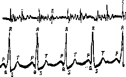
/ Fig. 8. Electrocardiogram and phonogram in interventricular septal defect.
of the lower part of the sternum, enlarged heart, predominantly to the left, predominance of the left ventricle on the electrocardiogram in the absence of cyanosis and accentuation of tones,-II r og-n o z i assessment of working capacity with this defect is comparatively favorable. The prognosis of this defect for childhood is somewhat worse than for adults, because such children comparatively often die from accompanying pneumonias.. (Maslov) and other infections; a comparatively frequent occurrence of endocarditis has also been noted. Blood circulation in persons with a septal defect is not disturbed for a very long time, even with heavy physical labor. Patients reach advanced age (in one case up to 67 years) without disturbance of blood circulation. The cause of death in individual cases was paradoxical embolism, tuberculosis, pneumonia, and only in rare cases with large defects did death occur due to disturbed blood circulation. Anomalies of the arterial trunk mainly concern its dividing septum. The septum may either be completely absent and in these cases a common trunk of the aorta and pulmonary artery is formed, or it may have partial defects - then there will be a constant connection between the aorta and pulmonary artery. The relationship between the septum of the arterial trunk and the ventricular septum may also change, and then displacement of the great vessels and change in their connection with the ventricles will occur. Finally, the septum may unequally divide the arterial trunk, and in this case narrowing or insufficiency of the pulmonary artery or aorta will occur-. In very rare cases, a change in the number of valves of the pulmonary artery and aorta can also cause narrowing and insufficiency of these valves. Narrowing of the pulmonary artery orifice (stenosis art. pulmonalis). Of all anomalies of the arterial trunk, the most common is narrowing of the pulmonary artery. In children, this is clinically the most important of congenital defects, since no less than 4/sBCex children with P. s, who reached 12 years of age, and no less than half of all congenital II. s. falls on this very developmental anomaly. According to Hecht, it apparently occurs more often in boys than in girls. A clinical picture similar to pulmonary artery narrowing and caused by septal anomaly is also given by congenital narrowing of the so-called arterial cone of the right ventricle (conus arteriosus dexter) and intrauterine damage to the pulmonary artery valves. Narrowing of the arterial cone, i.e., that part of the right ventricle through which blood outflow occurs, is essentially not an anomaly of the pulmonary artery, but an anomaly of the heart, particularly the right ventricle. Narrowing occurs either throughout the entire cone or, most often, only its apex is narrowed. In all these cases, an obstacle arises in the path of blood flow from the right ventricle into the pulmonary artery. During life, it is impossible to determine the nature of this obstacle, which is established only at autopsy. The most important and characteristic sign of pulmonary artery narrowing is sharply expressed cyanosis, which is observed in the patient even at complete rest and increases even more with any physical exertion. The absence of cyanosis in pulmonary artery narrowing is extremely rare and apparently occurs only in cases where the degree of narrowing is small. Pulmonary artery narrowing causes reduced blood flow into the pulmonary capillaries. Venous blood contains significantly less oxygen than normal (16 volume percent), which causes the bluish discoloration. The appearance of cyanosis is contributed to by hyperplasia of the capillary network, neocapillary formation, and dilation of the venous limb of the capillaries. Cyanosis is often combined with sharp pallor, resulting in the so-called pale cyanosis (cyanose blanche of French authors). Along with cyanosis, varying degrees of dyspnea can be noted, which sometimes takes the form of attacks accompanied by loss of consciousness and epileptiform convulsions. The right ventricle, which has to overcome the increased resistance of the narrowed pulmonary artery, undergoes hypertrophy. The area of the heart sometimes bulges; epigastric pulsation is noted. On percussion and X-ray examination, the heart is usually enlarged to the right. Often, however, no enlargement of the right border of the heart can be determined, on the contrary, there is some enlargement of the left border of the heart. This is explained by the fact that early hypertrophy of the right ventricle causes the heart to rotate around its axis. The right ventricle during hypertrophy cannot spread to the right, because it must push aside the right atrium, fixed by the vena cava; it can only rotate around its axis, while the displacement of the right ventricle to the left toward the pulmonary artery is completely free. In this case, the right ventricle is located in front, and the left behind. Thus, the left edge of the orthodiagram is formed not by the left ventricle, but by the right or both together. The shape of the pulmonary artery arch on X-ray fluoroscopy is not the same in all cases of pulmonary artery narrowing. The arch may be completely unchanged, but sometimes there is a depression

of the arch. But in the vast majority of cases, bulging and pulsation of the pulmonary artery arch have been noted (fig. 9). This is explained by the stretching of the pulmonary artery by the increased force of the pressure of the blood stream ejected by the hypertrophied right ventricle (Vaquez et Bordet). But this explanation is not entirely convincing.
To the left of the sternum in the second and third intercostal spaces, a scraping systolic murmur is heard, which spreads in all directions, predominantly to the right ventricle. The murmur is not transmitted to the neck vessels. Often this murmur is well heard on the back to the left of Div. Murmur Figure 9. ort-usually almost covers the first tone of the orthodiagram-pulmonary artery, while the second tone is well heard. Accentuation of the second tone of the pulmonary artery is not noted in the vast majority of cases of uncomplicated pulmonary artery narrowing, and if it is observed, it is weakly expressed, in contrast to the sharply expressed accentuation in an open ductus arteriosus. In the area of maximum murmur audibility, a cat's purr can be felt. The murmur, as with ventricular septal defect, has a uniform character, which clearly appears on the cardiophonogram.-Electrocardio- I /~ Figure 10. Electrocardiogram (b and c-I and II leads), phonogram (a) and phlebogram (d) in pulmonary artery narrowing. gram gives a picture of sharp predominance of the right ventricle with small R and large S in the first lead and large R and small S in the third (fig. 10 and 11). Previously, such an electrocardiographic picture was considered characteristic of all congenital P. s. At present, it has been clarified that it is a very important differential diagnostic symptom specifically of pulmonary artery narrowing. On the phlebogram, a sharply increased atrial wave 'a' can be seen, caused by enhanced contraction of the hypertrophied right atrium, and a decrease or complete absence of diastolic collapse due to stagnation in the area of the right heart. The pulse is usually small, sometimes quite significantly accelerated. Blood pressure is lowered. Very often with pulmonary artery narrowing, deformation of the fingers in the form of drumsticks appears. The amount of Hb and red blood cells increases.
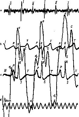
Figure 11. Electrocardiogram (b and c-I and III leads), phonogram (a) and phlebogram (d) in pulmonary artery narrowing.
The appearance of erythrocytosis is caused by enhanced blood formation due to anoxemia and is one of the factors contributing to the maintenance of normal oxygen supply to tissues. Narrowing of the pulmonary artery is often combined with general infantilism and developmental anomalies in other systems of the body. In the course of this congenital defect, the tuberculous lesion of the lungs that accompanies it plays a huge role. Thus, the main symptoms in the diagnosis of narrowing of the pulmonary artery are cyanosis, sometimes pale, then a sharp systolic murmur to the left of the sternum in the II-III intercostal space with the absence of an accentuated second tone, hypertrophy of the right ventricle, rightogram of the electrocardiogram, and an increased 'a' wave of the phlebogram. It is necessary to take into account the extremely frequent combination of narrowing of the pulmonary artery with other defects, most often with a defect of the ventricular septum. This connection becomes understandable when considering the importance of the septum of the arterial trunk in the formation of the ventricular septum. Assessment of working capacity and prognosis in narrowing of the pulmonary artery are unfavorable. The average life expectancy is about 10 years, and by this age half of those suffering from this defect die. Reaching the age of 20 is an exception. In the vast majority of cases, patients are completely unable to perform any work. Death most often occurs due to tuberculosis or accidental infectious diseases. If this does not happen, the patient dies from signs of impaired circulation. Insufficiency of the pulmonary artery valves (insufficientia valvulae semilunaris art. pulmonalis). Insufficiency of the pulmonary artery valves is a rare congenital defect. The clinical picture is not very characteristic, all the more so since this defect in the described cases was always combined with narrowing of the pulmonary artery and often with other defects. Cyanosis is weakly expressed. The area of the heart is protruding. The impulse is weakened. The heart on percussion and X-ray is enlarged to the right due to hypertrophy and dilation of the right ventricle. A diastolic murmur is heard to the left in the second and third intercostal spaces. Sometimes splitting of the second tone was observed. The pulse is small, often rapid. Arterial pressure 120-80. The small amplitude of arterial pressure, unlike the insufficiency of the aortic valves, is an important differential diagnostic symptom. Assessment of working capacity is favorable. In some cases, patients lived to a very old age while remaining fully capable of work. When circulation is impaired, signs of stagnation in the lungs appear first. The stenosis of the aortic outlet, as well as narrowing of the pulmonary artery, can be the result of uneven division of the arterial trunk. Narrowing of the arterial cone of the left ventricle (conus arteriosus sinister) and the extremely rarely observed intrauterine lesion of the aortic valves give the same clinical picture. Cases of aortic narrowing are rare and are of a casuistic nature. However, according to Zhukovsky, who studied the patho-anatomical material of newborns, this defect occurs even more often than pulmonary artery stenosis; apparently most children die in early childhood. Subjective sensations in aortic narrowing are either very weak or completely absent. Cyanosis is usually not observed. A characteristic feature is the sharp hypertrophy of the left ventricle. In the second intercostal space on the right, a systolic cat-like murmur is felt. Here a rough systolic murmur covering the first tone is heard. The second tone is clearly heard, sometimes accentuated. The impulse is sharply expressed. The pulse is small and weak. There are no data on the electrocardiogram in the described cases, but a priori one should expect predominance of the left ventricle. Assessment of working capacity and prognosis is apparently favorable. In the described cases, patients reached 40-50 years of age, were able to work, and in most cases died from accidental causes. Narrowing of the descending segment of the aortic arch. In addition to narrowing at the place of transition from the left ventricle to the aorta, there is also narrowing of the descending segment of the aortic arch (isthmus aortae), between the place of departure of the left subclavian artery and the place of entry into the aorta of the ductus arteriosus. Most authors consider the cause of this narrowing to be the constriction of the aorta by a scarred ductus arteriosus. Others believe that the narrowing is caused by an anomaly in the development of the gill arches. The degree of narrowing varies within very wide limits—from slight narrowing to complete overgrowth and transformation of the aortic arch into a blind sac. The clinical picture depends to a large extent on the size of the narrowing. Subjective sensations, especially with slight narrowing, are almost absent. Cyanosis is usually not observed. The most characteristic feature in the picture of this defect is the size of the vessels branching off from the aorta below and above the place of narrowing, and the formation of collateral circulation between them through powerful anastomoses connecting mainly the branches of the subclavian artery with the branches of the external iliac arteries. With the help of anastomoses, blood is delivered to the abdominal cavity organs and the lower part of the body. The branches of the subclavian artery (aa. mammariae internae, transv. scapulae, cervic. profundae) and the branches of the external iliac artery (art. epi-gastr. and circumfl. ilium prof.) reach special development. The aa. intercostales and aa. lumbales also expand greatly. All these collaterals look like superficially located, tortuous, pulsating vessels as thick as a pencil or even a finger. On palpation, these vessels give a characteristic murmur. On auscultation, characteristic murmurs are heard. The carotid and subclavian arteries pulsate strongly, while the arteries of the lower extremities are barely palpable or not palpable at all. The impulse is sharply expressed, lowered, and is often felt in the VII intercostal space. The left ventricle is dilated and hypertrophied. On X-ray, in addition to the hypertrophied left ventricle, a dilated pulsating ascending aorta can be seen. Sometimes dilation and pulsation of the pulmonary artery are noted. A systolic murmur is heard on the upper part of the sternum. Here a murmur is also felt. The second tone of the aorta is clearly heard, usually accentuated. The pulse in the carotid arteries and in the upper extremities is rising (pulsus celer), in the lower extremities—weak (pulsus tardus). Pressure in the upper extremities is elevated (up to 300 mm), in the lower—decreased. Edemas are often observed, unlike other congenital defects. In most cases (in 37%), a combination with developmental anomalies in other systems of the body was noted, especially often with hypospadias. In the diagnosis of aortic arch narrowing, the main feature is the different development of the arteries of the upper and lower halves of the body, which distinguishes this disease from narrowing at the place of aortic exit. It is difficult in some cases to differentiate from aortic aneurysm. However, the history, RW and other signs make it possible to finally resolve the question. Assessment of working capacity and prognosis is favorable even with significant degrees of narrowing. Patients live to a very old age, maintaining full working capacity even with respect to heavy physical labor. Death occurs from accidental diseases and only rarely from signs of impaired circulation. Displacement of large vessels. A change in the position of the septum of the arterial trunk and its relation to the ventricular septum can lead to various types of displacement of large vessels. Three forms of displacement of large vessels can be distinguished: 1) the position of the septum of the arterial trunk changes, and the aorta and pulmonary artery are placed in such a way that the aorta originates from the right ventricle and the pulmonary artery from the left (true displacement, transpositio vera); 2) the position of the septum of the arterial trunk and the ventricular septum changes in such a way that although there is displacement of large vessels, they still originate from the same ventricles as in the normal state due to the changed position of the ventricular septum; 3) finally, a change in the position of the septum of the arterial trunk can lead to the fact that both large vessels will originate from one ventricle, right or left. In all these cases, various variations in the position of large vessels will be created. Of all these anomalies, only true displacement of vessels has clinical significance, and only when it is combined with an anomaly that allows for a wide connection of arterial and venous blood: with a defect of the atrial or ventricular septum, with an open ductus arteriosus. Otherwise, death occurs as soon as the foramen ovale closes and the ductus arteriosus obliterates, and thus the connection between the small and large circles of blood circulation ceases. Displacement of vessels is an extremely rare congenital defect. The clinical picture depends partly on the nature of the defect with which the displacement of vessels is combined. The main clinical symptom is cyanosis, which when combined with an open ductus arteriosus is especially sharply expressed in the upper half of the body and significantly weaker in the lower.
This is explained by the fact that when blood from the superior vena cava enters the right ventricle and from there into the aorta, the carotid and subclavian arteries, it is completely arterialized, since these vessels branch off from the aorta before the entry of the ductus arteriosus, which carries oxygenated blood. The lower part of the body, receiving blood from the branches of the aorta after the entry of the ductus arteriosus, is supplied with blood that is partially arterialized. Unlike in the case of cyanosis with pulmonary artery stenosis, physical fatigue in children is not observed and there are no attacks of suffocation (Maelyov). The heart has a rounded shape and is located in the midline. On X-ray examination, a displaced pulmonary artery arch is noted on the right top. In most cases, despite the combination of transposition of vessels with other developmental anomalies, the tones on auscultation are clear. This is explained by the fact that life is preserved only in cases of transposition of vessels where the connection between the small and large circles is wide and therefore the defects of the septum are large or the ductus arteriosus is wide. It is precisely in such cases that murmurs may be absent. In addition to cases of true transposition, cases of displacement are described, where the aorta originates from both ventricles. This defect is combined with a ventricular septal defect. On X-ray examination in this case, the aortic arch on the left is absent, and the aorta seems to be riding on the septum (reitende Aorta according to Mohr). The prognosis for this defect is unfavorable. The vast majority (up to 75%) die within the first year of life. There are only individual observations where patients reached mature age. A case of death at the age of 39 is even described. In all these cases, there was a significant circulatory disturbance that made these patients unable to work. Common trunk of the aorta and pulmonary artery. This anomaly is observed in cases where the arterial septum does not develop and therefore the aorta and pulmonary artery form a common trunk. Cases of this defect are extremely rare, are almost always combined with other congenital defects, and it is very difficult to speak of a characteristic clinical picture. In most cases, cyanosis is noted. The heart is usually enlarged to the right. Murmurs are sometimes absent, sometimes pronounced, and their localization depends to a large extent on the associated defects. In all cases, the diagnosis was made after autopsy. The prognosis is unfavorable. In the vast majority of cases, patients die within the first year. Only one case is described where the patient reached the age of 19. Minor defects in the walls of the arterial septum, causing only a partial connection between the aorta and pulmonary artery, have little effect on the circulation and are not recognized during life. Persistence of the ductus arteriosus. The ductus arteriosus connects the pulmonary artery and the aorta during fetal life. Normally, the ductus arteriosus obliterates within the first week of extrauterine life. Obliteration may be delayed and complete by the end of the first year. Sometimes the ductus arteriosus does not completely obliterate, but the connection at this time is so insignificant that it has no effect on the circulation. An anomaly of development is spoken of when the ductus does not obliterate at all and remains completely open. In combination with other congenital defects, an open ductus arteriosus is observed quite often. As an isolated lesion, it occurs much less frequently, but it is still the most common of the congenital defects. The clinical picture depends to a large extent on the diameter of the ductus arteriosus. Subjective sensations are usually very small or even completely absent. Cyanosis is not observed in pure cases. The presence of cyanosis suggests a combination with other congenital defects, mainly with narrowing of the pulmonary artery. However, individual rare cases are described where cyanosis was observed with an uncomplicated open ductus arteriosus. A characteristic clinical symptom is the area of dullness (Gerhardt's zone) to the left of the sternum, a finger's width, above the cardiac dullness, reaching to the II rib. The right ventricle is hypertrophied due to increased pressure in the pulmonary artery. At the same time, the left ventricle also dilates and hypertrophies due to increased blood flow to it. On X-ray examination, bulging and marked pulsation of the pulmonary artery arch can also be established (Fig. 12). During the Valsalva maneuver, pressure in the pulmonary artery increases, blood flow from the aorta through the ductus arteriosus decreases significantly, and the pulmonary artery arch flattens (Bittorf). To the left of the sternum in the II intercostal space, a loud systolic murmur is heard; sometimes the murmur is systolic-diastolic-

Figure 12. Orthodiagram of an open ductus arteriosus.
systolic character, in rare cases purely diastolic. This variability of sound phenomena is apparently explained by the fact that blood through the ductus arteriosus flows during systole in the direction from the aorta to the pulmonary artery, and during diastole, conversely, moves from the pulmonary artery to the aorta. The murmur in patent ductus arteriosus decreases during the Valsalva maneuver and may even completely disappear. When pressure is applied to the abdominal aorta due to the resulting increase in arterial pressure, the murmur intensifies. The murmur is well conducted into the neck vessels. Sometimes the murmur is well audible on the back. Due to the increased pressure, the second tone of the pulmonary artery is accentuated. The accentuation of the second tone is an important differential diagnostic sign. At the site of murmur auscultation, a pronounced purring sensation is felt. In some cases, an unequal pulse was noted on both hands, with the left being stronger. In some cases, a decrease or even disappearance of pulse fluctuations was observed during deep inspiration - pulsus paradoxus. The electrocardiogram is usually unchanged. Cases have been described, however not confirmed by autopsy, where there was predominance of the right ventricle on the electrocardiogram. With a large diameter of the ductus arteriosus, paralysis of the left recurrent nerve is sometimes noted, which in these cases is compressed between the dilated ductus and the aorta. The diagnosis of patent ductus arteriosus is usually relatively simple. It is based on Gerhardt's zone of dullness, on the bulging and pulsation of the pulmonary artery arch, which decreases during the Valsalva maneuver, on the systolic murmur conducted into the vessels, and on the sharply accentuated second tone of the pulmonary artery.--Assessment of work capacity and prognosis are generally favorable. With a small diameter of patent ductus arteriosus, this does not affect the circulation at all. Patients with this condition can even engage in heavy physical labor and reach old age (in one case up to 66 years). With a wide ductus arteriosus, especially when it is combined with other anomalies, circulatory disturbance occurs at a younger age. The cause of death is often endocarditis, accidental infectious diseases, and relatively rarely circulatory disturbance. Anomalies of the right and left atrioventricular openings. Anomalies in the formation of atrioventricular openings lead to their narrowing or insufficiency. Narrowing and insufficiency are caused either by uneven division of the primary atrioventricular opening, with the right or left atrioventricular opening narrowing, or by an anomaly in the development of the valves. In this case, the number of valve leaflets may increase or decrease, which will lead to insufficiency or narrowing, or the free edges of the valves may fuse, causing funnel-shaped narrowing of the openings.-Of all these lesions, the most common and of clinical interest is narrowing of the left atrioventricular opening. In patho-anatomical examination of these cases, either funnel-shaped narrowing of the left atrioventricular opening due to fusion of the valve edges, or narrowing of the left atrioventricular ring itself, or a change in the number of valve leaflets is found; the valves themselves are unchanged, shiny, without any deposits. With very severe narrowing or even atrophy, death occurs in early childhood with signs of severe cyanosis, shortness of breath, and sometimes edema. The heart is dilated on both sides. A presystolic murmur is heard, and when combined with insufficiency, a systolic murmur at the apex of the heart or in the middle of the sternum. Such cases are very rare. Much more often cases are observed where the manifestations of congenital pure narrowing of the left atrioventricular opening appear during puberty (Duroziez's disease). This is usually observed in infantile, asthenic-type women suffering from anemia, hysteroneurosis, poor appetite, and a tendency to hemorrhoidal and nasal bleeding. In these cases, there are no indications in the history of joint rheumatism or any other etiological factor for the defect. A case of such a disease was noted in a family where two sisters (dizygotic twins) and their brother were ill (Sachs). That in these cases we are dealing with a developmental anomaly is indicated, besides the above-described patho-anatomical picture, by the combination of this disease with other developmental anomalies, especially with underdevelopment of the heart and hypoplasia of vessels. Indications of the role of tuberculosis and syphilis of parents in the occurrence of this defect are unproven (Teissier, Huchard). Also unclear is the connection with status thymico-lymphaticus noted by Neisser. For the clinical picture of this defect, the absence of subjective sensations except for increased fatigue is characteristic. On percussion and auscultation, the usual picture of narrowing of the left atrioventricular opening is found. At the apex of the heart, a purring sensation is felt. The heart is slightly dilated to the right. The left atrium is enlarged. At the apex, a crescendo presystolic murmur is heard, ending with an accentuated first tone. The second tone of the pulmonary artery is accentuated and usually split (pheasant rhythm). The X-ray shows the characteristic mitral form of the heart. The electrocardiogram gives a picture of right ventricular predominance.-Assessment of work capacity and prognosis in these cases is much more favorable than in endocarditic narrowing. The course of the disease is favorable and circulatory disturbance occurs very late. Cases of insufficiency of the mitral valves due to congenital developmental anomaly are extremely rare and of a casuistic nature. Usually they are combined with narrowing, and death occurs in early childhood. The clinical picture varies sharply depending on the combination with various other developmental defects/Each described case has its own clinical picture, based on similarity with the clinical picture of mitral valve insufficiency of endocarditic origin. Uneven division of the primary atrioventricular opening can lead to narrowing of the right atrioventricular opening. These cases are significantly rarer than narrowing of the left opening. The clinical picture is characterized by severe cyanosis, shortness of breath, drumstick fingers, and hyperglobulinemia. A pronounced edema is often observed. In the lower part of the sternum, a pronounced systolic and diastolic murmur is heard. Here too a purring sensation is felt. The right ventricle is sharply reduced, and at autopsy it was sometimes found as a small appendage to the left ventricle. The prognosis is extremely unfavorable. Death occurs at an early age, no later than 5 years. Insufficiency of the tricuspid valve is also an extremely rare developmental anomaly. Only about 10 such cases have been described (Blumenfeld), and this defect was combined with other developmental anomalies of the heart. The clinical picture varies sharply. Usually cyanosis and a markedly enlarged right ventricle were found. Systolic murmur in the lower part of the sternum; there too the purring sensation. Interestingly, the positive venous pulse, characteristic of acquired tricuspid valve insufficiency, is often absent. The prognosis is usually unfavorable. Patients die at a young age with signs of impaired circulation. Only one case has been described where the patient reached old age and died from an accidental cause. Congenital defects caused by intrauterine endocarditis. Intrauterine endocarditis can cause damage to the valves: mitral, tricuspid, aortic, and pulmonary artery, and cause their narrowing and insufficiency. Due to the specific conditions of fetal circulation, when the main work falls on the right heart, most often the tricuspid and pulmonary artery valves are affected, much less often the mitral valve, and as the greatest rarity, the aortic valve. The valve damage in its clinical picture resembles the picture of narrowing and insufficiency caused by developmental anomalies. Sometimes it is possible to solve the question of the etiology of the congenital defect on the basis of indirect data. The main criterion in solving the question is the frequent combination of valve anomalies with other heart anomalies and partly other organs, which is characteristic of anomalies, unlike valve damage caused by intrauterine endocarditis. Some assistance in differential diagnosis can be provided by detailed questioning of the mother's condition during pregnancy. Infectious diseases, especially in the last months of pregnancy, can be a source of intrauterine endocarditis. Often during life the question of the etiology of the defect remains unclear. But even in patho-anatomical examination, the question of etiology is not so easily resolved. The presence of verrucous deposits on the valves does not resolve the issue, as, as already noted, valve anomalies predispose to endocarditis. Decisive is the microscopic examination of the myocardium. In intrauterine endocarditis, remnants of inflammatory phenomena can always be found in the myocardium: scars, necroses, accumulations of leukocytes, dilated vessels, giant nuclei in fibers, etc.
Such a lesion of the myocardium is never observed even with severe heart anomalies, even if they are subsequently complicated by an inflammatory process (Mönckeberg).-It is important to note the significant difference in the course of the disease in both groups of valve lesions. Narrowing and insufficiency of the valves caused by intrauterine endocarditis proceed much more severely. Recurrent endocarditis accompanied by elevated temperature is observed much more frequently. Capacity for work is impaired much more severely than with defects caused by developmental anomalies. Circulatory disturbance and death occur much earlier. Features of preventive therapy for congenital heart defects. Precise diagnosis of congenital heart defects is extremely important in establishing the general regimen for the patient and especially in professional selection. In adolescents with narrowing of the pulmonary artery and a large defect of the ventricular septum, one must be extremely cautious in choosing a profession. They can only work in those industries where physical exertion is not required. Some congenital heart defects (true dextrocardia, patent foramen ovale) can be completely disregarded in professional selection. Of course, each individual case must be individualized, and a general assessment given based on the state of circulation. Proper nutrition at definite intervals and suitable food are mandatory from birth. It is necessary to avoid nicotine, alcohol, and any psychic excitations. A breast-fed infant should be breast-fed as much as possible; if there are difficulties with sucking, they are given expressed breast milk from a bottle or with a spoon. Taking into account that children with congenital heart defects are easily susceptible to cooling and that cyanosis easily develops in them, they should be protected from cold. For this purpose, it is desirable to allow such children to spend the winter in a warm climate. However, one should not go to extremes and accustom children too much to warmth. One should cautiously try to harden the body with cool rubdowns, douches, and other similar procedures. Keeping such children in bed for a long time is not advisable, as this can make them greater invalids than they actually are; on the contrary, one should cautiously, taking into account all conditions in each individual case, accustom the heart to exercise. Children should be protected by all measures from infections, knowing that a congenital heart defect predisposes to the development of endocarditis. General schools with a regimen adapted for healthy children are of course not entirely suitable for children with severe heart defects; moreover, crowding increases the danger of infection. In all cases, general strengthening treatment should be carried out using all possibilities to provide such children with more light and air, a full but small-volume diet. All that has been said about the peculiarities of the regimen for children with congenital heart defects refers to severe defects; it is self-evident that in cases where the defect is established only by physical examination, and where, as often happens, there are no subjective phenomena or they are insignificant, there is no need to subject the child to a special regimen, and one should leave them in their usual conditions. The question of childbirth is resolved in each individual case depending on the state of circulation. Therapy for circulatory disturbance is the same as for acquired heart defects. Due to the predominant lesion of the right heart with cyanosis and dyspnea, it is more often necessary to resort to bloodletting and oxygen therapy. One must also remember that patients with narrowing of the pulmonary artery are threatened with tuberculosis of the lungs and take this into account when arranging their living conditions and work.
l. Fogelson.
Related articles
Mentioned in
Cite this page
“Heart Defects.” Soviet Medical Encyclopedia. English translation of Bolshaya Meditsinskaya Entsiklopediya, 1st ed. (Moscow, 1928–1936), ed. N. A. Semashko. https://sovietmedicalencyclopedia.pages.dev/article/heart-defects/