Heart
Historical document, translated for reference. It reflects medical knowledge of the 1920s–30s and is not medical advice.
Summary
This article provides a detailed comparative anatomical study of the heart across vertebrate species, tracing its embryological development from fish to mammals. It examines the structural evolution of cardiac chambers, valves, and the separation of arterial and venous blood flow in different animal classes.
Encyclopedia article (1928–1936)
207 VI. Physiology, pathological anatomy, pathophysiology and clinic of coronary circulation...
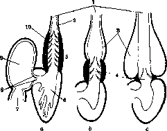
299 I. Comparative anatomy. The heart of vertebrates represents, by its origin, a particularly highly differentiated part of the abdominal (i.e., subintestinal) vascular trunk of chordate animals with a strongly developed muscular wall and a system of valves, playing the role of a pump, pumping blood from the venous system into the arterial system. In fish and amphibians, the heart develops in the region of the posterior part of the head directly under the pharynx. Between the mesodermal sheets (splanchnopleura) of the right and left sides, i.e., in the ventral mesentery, a collection of mesenchymal cells forms, directly adjacent to the entodermal wall of the pharynx. These cells then arrange themselves in a single layer, lining a certain cavity. The rudiment acquires the appearance of a longitudinal tube, which quickly dilates and bends approximately in the sagittal plane in the shape of the letter S, then forming at the site of the middle bend a sharp constriction, marking the division of the heart into two main departments - the atrium on the dorsal side and the ventricle on the ventral. The original rudiment of the heart represents only its inner lining - the endocardium. The walls of the body cavity located on the sides of the rudiment thicken, the cavities themselves increasingly surround the heart rudiment, become isolated from the rest of the body cavity, connect with each other, and thus give rise to the pericardial cavity. The mesenchyme developing from its walls forms the myocardium, while the remaining part of the wall is preserved in the form of the pericardium of the adult animal. Already in some fish, and especially in higher vertebrates (in amniotes), all embryonic sheets, including the entoderm, develop spread out on the surface of the yolk (or yolk sac). In this case, the heart develops from two rudiments, symmetrically located on the right and left between the entoderm and the visceral sheet of mesoderm. The latter deeply surrounds the developing endothelial tube. As the intestinal groove forms and closes from below into the intestinal tube, the right and left rudiments approach each other under the intestine and unite into a single rudiment. The tubular rudiment of the heart bends, as mentioned, in the shape of the letter S and differentiates into a series of departments. This differentiation is simplest in fish, which have a single circulatory system and the heart carries only venous blood. Besides the constriction separating the atrium from the ventricle, a similar constriction forms in the dorsal part of the cardiac tube, separating the atrium from the venous sinus, and in the ventral part a less noticeable constriction separating the arterial cone from the ventricle. At the boundaries between all the mentioned departments, as well as in the arterial cone itself, individual bulges of the endocardium form, which then turn into cardiac valves closing the passages between the departments of the heart. The arterial cone connects with the developing ventral aorta, while the venous sinus continues on the sides into the Cuvierian ducts, and behind into the hepatic vein. The muscular wall develops particularly significantly in both ventral departments - the ventricle and the cone, with the ventricle developing a complex network of muscular trabeculae, more or less protruding into the internal cavity. - The heart of fish thus consists of four successive departments - a thin-walled venous sinus (sinus venosus) into which the largest venous trunks flow; the atrium, separated from the venous sinus by a pair of valves (valvulae sinu-atriales); the ventricle with a powerful muscular wall, separated by a pair of similar valves (valvulae atrio-ventriculares) from the atrium, and finally the arterial cone, sometimes provided with many transverse rows of pocket-like valves (valvulae semilunares). The base of the ventral aorta, departing from the arterial cone, is often dilated and has a thickened muscular wall. Unlike the departments of the heart, which have striated musculature, this 'bulb' of the aorta (bulbus arteriosus) (fig. 1) has smooth musculature and thus already belongs to the arterial system. The arterial cone is well developed only in lower fish. Here it contains several transverse rows of pocket-like valves, the edges of which are suspended by tendon cords to the wall of the cone. In higher fish its size decreases and in bony fish only a rudiment with one row of valves at the boundary between the ventricle and the arterial trunk remains. In terrestrial vertebrates, with the establishment of pulmonary circulation, the heart is fundamentally transformed. Already in dipnoan fish, the pulmonary vein runs along the wall of the venous sinus and independently flows into the atrium; in the latter an incomplete septum develops, and in the arterial cone a special longitudinal valve forms, which also serves as a septum. In amphibians, a similar process of separation of arterial and venous blood flow goes still further. The heart is also 'three-chambered' here, as there is a septum (usually with openings) dividing the atrium into left, arterial, and right, venous, halves; the latter is connected to the still well-developed venous sinus. There is no septum in the ventricle, but its walls have an extremely developed network of muscular trabeculae, deeply incising into the internal cavity and undoubtedly preventing free mixing of arterial and venous blood. The arterial cone is provided with a longitudinal, spirally curved valve, and the base of the arterial trunk is divided by a horizontal septum attached to its end into a ventral department, from which the anterior pairs of arches (carotid arteries and aortic arches) depart, and a dorsal (venous) department, from which the posterior pair of arches (pulmonary-cutaneous artery) departs. - The heart of reptiles is somewhat more perfect. Here there is still no complete isolation of venous blood from arterial blood, but there is still a septum in the ventricle, though incomplete - it departs from the ventral wall and does not reach the dorsal. However, during systole the complete separation of both cavities of the ventricle is obviously achieved. Only in crocodiles is there a complete septum and the heart becomes 'four-chambered'. The venous sinus is already reduced in reptiles and becomes part of the right atrium. The arterial cone is also reduced, from which only a series of semilunar valves remains. The arterial trunk divides to its base into the pulmonary artery, departing from the right ventricle, and two aortic arches, which in reptiles depart - the right from the left ventricle, the left from the right, near the free edge of the septum. Due to its position, the left aortic arch receives mixed blood. - In birds the left arch is completely reduced, and the aorta receives purely arterial blood. The atrial septum also divides the atrio-ventricular opening into two, with each opening receiving half of the original valves (ventral and dorsal), which fuse together along the septum. Thus each atrio-ventricular opening receives one membranous valve on its inner (medial) side. In crocodiles and especially in birds, a muscular fold also develops on the outer wall of the right atrio-ventricular opening, which also plays the role of a valve (in birds this muscular valve becomes the only valve here). - In mammals, as in birds, the heart is four-chambered and arterial blood is completely isolated from venous blood not only in the heart but also in the arterial trunks. However, in mammals the right aortic arch has reduced, not the left. The aorta originates in the left ventricle, and the common trunk of the pulmonary arteries in the right. At the base of both there are three
Figure 1. Diagram of fish hearts (a-selachians, b-Amia, c-bony fish): 1-ventral aorta; 2-arterial trunk; 3-bulbus arteriosus; 4 and 10-arterial cone; 5-ventral part; 6-ventricle; 7-venous sinus; 8-valv. atrio-ventric; 9-atrium.
Figure 2.
Figure 3. Human embryo heart 8.15 mm in length: 1-ventricle; 2-venous end of the S-shaped curved cardiac tube; 3-arterial trunk. Figure 3. Human embryo heart of the 5th week according to His: 1 and 3-right and left auricles; 2-arterial trunk; 4-interventricular groove; 5 and 6-left and right ventricles.
Figure 4. Embryonic heart, starting from the 2nd month (view from behind): 1-anonymous artery; 2-pulmonary artery with both branches; 3-anterior cardinal vein (later superior vena cava); 4-right subclavian vein; 5-posterior cardinal vein (later azygos vein); b-right horn of the sinus; 7-inferior vena cava; S-left horn of the sinus; 9-left subclavian artery; 10-left carotid artery.
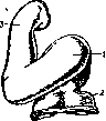
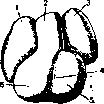
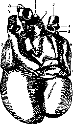
semilunar valves. In the right atrioventricular opening, one medial and two lateral valves develop (valvula tricuspidalis), while in the left, in monotremes there are also three valves, but in all other mammals, the two lateral valves fuse into one, resulting in a bicuspid valve (valvula bicuspidalis, s. mitralis). The edges of these valves are connected by tendon threads (chordae tendineae) to muscular projections (musculi papillares) inside the wall of the corresponding ventricle. In mammals and humans, the cardiac tube forms in the neck region, creating a significant thin-walled bulge that protrudes forward between the head and abdomen of the embryo. The dorsal part of the tube separates from the ventral part by a deep constriction, which forms a narrow 'auricular' canal between the atrium and ventricle. On the sides of the atrium, the auricular appendages of the Heart (auriculae cordis) develop, growing forward and wrapping around the base of the arterial trunk. The auricular canal takes the form of a transverse slit. The yolk and umbilical veins empty into the right auricle. From the upper (cranial) wall of the atrial cavity, a septum develops that gradually descends downward toward the auricular canal. Thus, the right and left atria are separated from each other, and simultaneously the auricular canal divides into right and left atrioventricular openings (fig. 2-5). In the developing septum, a temporary foramen ovale forms, ensuring normal fetal circulation and closing only at birth. The developing pulmonary veins initially open with a common trunk into the left half of the atrium, but later this trunk expands and gradually becomes part of the wall of the atrium itself. As a result, all four pulmonary veins open independently into the atrium. A similar process occurs in the right atrium, whose wall gradually absorbs the venous sinus, so that the main venous trunks (superior and inferior vena cava and coronary vein) empty independently of each other directly into the atrium. The muscular wall of the ventricle thickens significantly, and unlike the atrial wall, numerous muscular trabeculae develop on it. These trabeculae connect with each other to form a characteristic spongy tissue that significantly narrows the ventricular cavity. On the lower wall, the thickening of the musculature forms a projecting inward semilunar fold, which grows upward from below and behind toward the atrioventricular opening and the base of the arterial trunk. Finally, the arterial trunk is divided by a longitudinal septum into the aorta and pulmonary artery in such
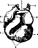
Figure 5. Embryonic heart, starting from the 2nd month (front view): 1- area between the atria; 2-v. cardinalis ant. sin.; 3-left atrium; 4-left ventricle; 5-base; 6-right ventricle; 7-truncus arteriosus (bulbus); 8-right atrium; 9-v. cardinalis ant. dextra.
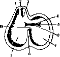
Figure 6. Diagram explaining the position of os-
a way that the pulmonary artery is connected to the cavity of the right ventricle, and the aorta to the cavity of the left (fig. 6). This aortic septum grows into the Heart, fuses with the ventricular septum, and remains as a relatively thin plate forming its pars membranacea. Four endocardial cushions at the entrance to the arterial trunk are divided by this septum; the anterior and posterior are divided into each right and left atrioventricular
rt
tppDrt kas- with respect to ostium BU10> it-°- lumen interventricularis: 1-a. DOGO trunk (aorta and le-puimonaiis; 2-truncus GOCHny artery) is limited by three endocardial cushions, which subsequently transform into semilunar valves. Similar cushions also develop from the myocardium of the ventricles, which differentiate into papillary muscles and chordae tendineae connected with these valves.
I. Shmalgauzen. II. Anatomy and Histology. Anatomy of the Heart. In the human Heart, which has a conical shape, we distinguish the base (basis), directed backward, to the right, and upward, surrounded by large vessels, and the apex (apex), hanging downward, to the left, and forward into the pericardial sac (fig. 7). In the Heart, we distinguish the anterior surface, convex (facies sterno-costalis), formed by the anterior wall of the right ventricle and a small part of the left; the inferior, flattened (facies diaphragmatica), formed by the inferior surface of the ventricles; the posterior (facies vertebralis), corresponding to the posterior wall of the atria; and finally the lateral surfaces (facies pulmonales), directed toward both lungs (fig. 8-9). On the outside, the boundary between the atria and ventricles is the deep coronary groove (sulcus coronarius cordis), interrupted in front by the exit of the pulmonary artery and aorta. In the thickness of the walls of the Heart, between the ventricles and atria, there are strong fibrous rings (annuli fibrosi), which, with the exception of one place (His bundle), completely isolate the atrial musculature from the ventricular musculature and serve as the attachment point for the atrioventricular valves. The left fibrous ring, fusing on the right and left with the root of the aorta, forms thickenings called fibrous triangles (trigonum fibrosum dextrum et sinistrum). Both fibrous rings of the Heart are connected by connective tissue bundles penetrating the walls of the Heart and form, as it were, the skeleton of the Heart. The boundary between the ventricles on the outside are two longitudinal grooves on the anterior and posterior surfaces (sulcus longitudinalis dexter et sinister). Below, to the right of the apex of the Heart, these grooves merge, forming here an indentation, the notch of the Heart (incisura cordis), belonging to the right ventricle. Inside, between the ventricles, the septum (septum ventriculorum) completely separates them. In its greater part, the septum is muscular, and only at the top it has a connective tissue character (septum membranaceum ventriculorum). The atria have a slightly convex posterior surface and an anterior concave surface (in the shape of a horseshoe). Each atrium sends forward one auricular appendage (auricula cordis dextra et sinistra), which with their notched edges surround the roots of the aorta and pulmonary artery. The inner surface of the walls of the atrium is smooth, while the surface of each auricle is covered with the so-called pectinate muscles (mm. pectinati). Into the right atrium (atrium dextrum) empty the superior and inferior vena cava and the coronary sinus of the heart itself. V. cava sup. perforates the upper wall of the atrium, while v. cava inf. perforates its posterior wall. Between the mouths of both vena cava, on the inner surface of the atrium, there is a small elevation (tuberculum intervenosum Lower!). Initially, the place where both vena cava merge was a separate cavity (sinus venarum cavarum). Subsequently, the venous sinus forms a common cavity with the atrium, and only on the inner surface of the atrium, an oblique muscular ridge,
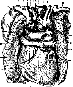
Figure 7. Position of the heart and large vessels: 1-v. subclavia dex.; 2-a. subclavia; 3-v. jugularis int; 4-zamus dex. a. pulmonalis; 5-v. azygos; 6-v. thyreoidea ima; 7-v. anonymasin.; 8- trachea; 9-arcus aortae; 10-v. subclavia sin.; 11- yv. pulmonales sin.; 12-a. pulmonalis; 13-aorta ascendens; 14-atrium sin.; 15-v. cava sup.; 16- pulmo dexter; 17-vv. pulmonales dex.; 18-costal. bordering crest (crista terminalis), on the outer surface of which a shallow groove (sulcus terminalis atrii dextri) is noticeable, is the boundary between the true atrium and the venous sinus. On the septum from the side of the right atrium (septum atriorum), the oval fossa (fossa ovalis) is noticeable, surrounded by a thickened ridge [limbus fossae ovalis (Vieussenii)]. In the fetus, at this place in the atrial septum, there is an oval foramen (foramen ovale), which plays an important role in fetal circulation. This opening is closed by a valve (valvula foraminis ovalis). After birth, the edges of this valve grow to the edges of the oval foramen, but sometimes in an adult a small slit remains connecting both atria. From the anterior part of the limbus fossae ovalis, a fold extends, limiting in front the mouth of the inferior vena cava [valvula venae cavae inf. (Eustachii)]. In the fetus, this fold is well developed and serves to deflect the blood flow coming from the inferior vena cava
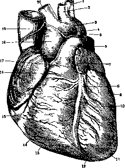
Figure 8. Heart from above and in front: 1-a. subclavia sin.; 2-a. carotis sin.; 3-arcus aortae; 4-place of the turn of the heart sac; 5-lig. arteriosum; 6- a. pulmonalis; 7-auricula sin.; 8-sulcus longitudinalis ant.; 9 and 10-ventriculus sin.; 11-apex cordis; 12-incisura (apicis) cordis; 13 and 14-ventriculus dexter; 15-sulcus coronarius; 16-conus arteriosus; 17 - auricula dex.; 18-aorta ascendens; 19-v. cava sup.; 20-a. anonyma.
to the oval opening. In an adulttit is weakly expressed or may be completely absent. Between the opening of the inferior vena cava and the atrioventricular opening on the posterior wall of the atrium, the opening of the coronary sinus (sinus coronarius cordis) opens, equipped with a semilunar valve [valvula sinus coronarii (Thebesii)]. In addition to the listed openings, a variable number of small veins [foramina venarum minimarum (Thebesii)] open into the right atrium (some of them represent depressions blindly ending in the myocardium). The left atrium (atrium sinistrum) lies more posteriorly compared to other parts of the Heart and only one tip of the auricle is visible from the front when opening the cardiac sac. Four pulmonary veins (vv. pulmonales)-two from each side-flow into the left atrium. Sometimes two veins of one side merge into one trunk before opening, and conversely-on the right side, three venous trunks sometimes flow in. The right ventricle (ventriculus dexter) has a crescent shape on cross section. Its entire inner surface is covered with numerous fleshy ridges (trabeculae carneae) (fig. 10 and 11). The opening connecting the right ventricle with the corresponding atrium (ostium atrio-ventricula-re dex., s. ostium venosum dex.) has an elongated-round shape, along its edges the atrioventricular valve with three cusps (valvula tricuspidalis) is attached. Its three cusps- anterior, posterior, and medial (cuspis ant., post, et med.)-are of different sizes (fig. 12), of which the anterior is larger and the medial is smaller. Sometimes the cusps merge, and sometimes there are more of them. Tendon cords (chordae tendineae) approach the valve cusps, extending from the papillary muscles; chordae tendineae extending from one papillary muscle are attached to two adjacent cusps. The wall of the right ventricle continues upward and to the left in a conical process (conus arteriosus ventri-culi dextri), which is separated from the ostium venosum by a muscular ridge (crista supraventricu-laris). At the site of the pulmonary artery exit (ostium arteriosum dextrum), three semilunar ^valves (val-vulae semilunares a. pulmonalis) are located in front, to the left and right, each bearing on the middle of the free edge a nodule the size of a millet seed (nodulus Arantii). On both sides of the nodule along the free edge of the cla- 18.19^^ ,
;
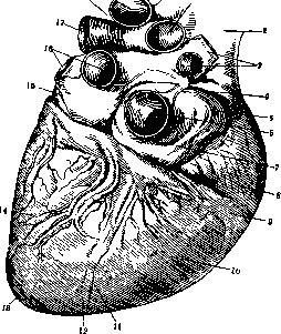
Figure 9. Heart from below: 1-ramus dex. a. pulmonalis; 2-v. cava sup.; 3-vv. pulmonales dex.; 4-place of the cardiac sac turn; 5-sinus venarum cavarum; 6-v. cava inf.; 7- appendix auricularis post.; 8-atrium dextrum; 9-sulcus coronarius; 10-ventriculus dex.; 11-sulcus longitudinalis post.; 12-incisura (apicis) cordis; 13-apex cordis,-14-ventriculus sin.; 15-lig. v. cavae sin.; 16-vv. pulmonales sin,; 17-ramus sin. a. pulmonalis; 18- basis cordis; 19-arcus aortae.
pan go fibrous strips (valvulae semilunares)-the place of closest contact of the valves with each other.-The wall of the left ventricle (ventriculus sinister) is three times thicker than the right, the ventricle itself is longer than the right (fig. 13 and 14), on cross section it has an almost round shape. The venous opening (ostium venosum sinistrum) is equipped with a bicuspid valve [valvula bicuspidalis (mitralis)], which consists of two large cusps (fig. 15), sometimes auxiliary cusps are noticeable between them. The anterior cusp is larger and lies in front and to the right between the venous and arterial openings, the posterior cusp is significantly smaller and lies behind and to the left. Toward the aortic opening, the ventricle forms a slightly twisted cone (conus arteriosus ventriculi sinistri). The aortic opening (ostium aorticum, s. ostium arteriosum si-nistrum) is round in shape, along its edges the semilunar valves (valvulae semi-lunares aortae dextra, sinistra,posterior) are located (fig. 16). Structure of the walls of the Heart. The main mass of the walls of the Heart is formed by a powerfully developed myocardium. The atrial muscle is relatively thin and consists of two layers: a superficial one formed by transverse bundles covering both atria, and a deep one, separate for each atrium, consisting of ring-shaped bundles around the venous openings and around the bases of the cardiac auricles. The ventricular muscle forms three layers: 1) superficial, starting from the annuli fibrosi, going from top to bottom and to the left. At the apex of the heart, the bundles converge in the form of a spiral (vortex cordis), and then they bend inward and, rising upward, pass into 2) inner
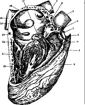
Figure 10. Opened right heart: 1-v. cavasup.; 2 and 17-septum membranaceum ventriculorum; 3- aorta; 4-atrio-ventricular bundle; 5-a. pul-monalis; 6-conus arteriosus; 7- right leg of the atrioventricular bundle; 8-m. papillaris ant. med.; 9- ventriculus sin.; 10-m. papillaris magnus; 11-ventriculus dex.; 12-cuspis medialis valvulae tricuspidalis; 13-foramen sinus corona-rius; 14-fossa ovalis; 15-v. cava inf.; 16-atrium dex.
(deep) layer, which includes trabeculae carneae and mm. papillares; 3) the third layer, the most developed, separate for each ventricle, is formed by circular bundles inserted between the superficial layer and the deep one (fig. 17). In fine structure, the cardiac muscle is a syncytial multinuclear formation consisting of cross-striated muscle fibers, everywhere merging with each other and located like a mesh plexus.-The muscle of the atria and ventricles is isolated and only at one place it is united by means of the atrioventricular bundle of His. Under the endocardium there is a network of muscle fibers with barely noticeable cross striation-Purkinje fibers (Purkinje).
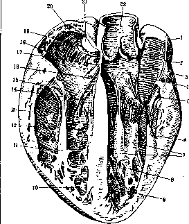
Figure 11. Section of an enlarged heart: 1-atrium sin.; 2- valvula semilunaris sin. aortae; 3-valvula semilunaris post, aortae; 4-cuspis ant. valvulae bicuspidalis; 5-cuspis post, valvulae bicuspidalis (mitralis); 6-m. papillaris ant.; 7-m. papillaris post.; S-ventriculus sin.; 9 and 12- trabeculae carneae; 10-septum musculare ventriculorum; 11- m. papillaris magnus; 13- ventriculus dex.; 14 and 16- cuspis medialis valvulae tricuspidalis; 15-cuspis post, valvulae tricuspidalis; 17-septum membranaceum ventriculorum; IS-foramen v. minimae (Thebesii); 19- musculi pectinati; 20- valvula y. cavae (inferioris, Eustacbii); 21-v. cava inf.; 22- aorta ascendens.
The indicated fibers establish the connection between the atria and ventricles and form a special excitation conduction system (Reizleitungssystem), thanks to which coordinated contractions of individual parts of the heart occur. In the excitation conduction system, one should distinguish: 1) The node of Keith-Flack (see); 2) the atrioventricular node (see Atrioventricular bundle, nodes) and the so-called atrioventricular bundle of His (fasciculus atrio-vent-ricularis His) (see Atrioventricular bundle, nodes). Reizleitungssystem is well expressed in large mammals (horse, sheep, elephant), in humans it is weakly expressed.-The inner surface of the Heart is lined with endocardium (endocardium). The endocardium always contains a significant amount of elastic fibers. At the openings of the vessels, the endocardium passes into tunica intima. The atrioventricular and semilunar valves are folds of the endocardium; in their base there is a small amount of muscle bundles. -The outer surface of the Heart is covered by the visceral leaf of the cardiac sac (epicardium) (see Pericardium). \ Blood supply to the heart-see below. The lymphatic vessels of the Heart are very abundant. They can be divided into superficial ones, lying under the epicardium, and deep ones, located in the thickness of the myocardium. The large lymph vessels of the Heart are located along the course of the coronary vessels. On each side, from the merging trunks, one large one is formed. The right trunk, passing near the aorta lgl. cardiacae, goes to the trachea and flows into the ductus lym-phaticus dexter. The left trunk rises along a. pulmonalis, passes through the lymph glands at its division and flows into the ductus thoracicus. The Heart receives nerves from two systems-from the n. vagus and n. sympathicus, which, exchanging branches with each other, form the cardiac plexus (plexus cardiacus). In it, a superficial and deep layer are distinguished: plexus cardiacus superficialis lies near the aortic arch and at the division of the pulmonary artery, extending more to the left; plexus cardiacus pro-fundus extends more to the right and lies above the superficial one, directly behind the aortic arch at the bifurcation of the trachea. The branches of both plexuses, descending downward, form: 1) plexus coronarius cordis ant., located at the root of the aorta and reaching a. coronaria dex.; its branches supply the right ventricle and right atrium; 2) plexus coronarius cordis post., going to a. coronaria
JO
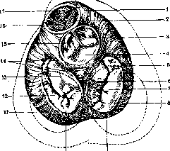
Figure 12. Base of the heart cavities: 1-conus arteriosus; 2-valvula semilunaris dex. a. pulmonalis; 3-valvula semilunaris dex. aortae; 4-valvula semilunaris post, aortae; 5-cuspis ant.; 6-cuspis med.; 7-cuspis post.; 8-annulus fibro-sus dex.; 9-ventriculus dex.; 10-annulus fibro-sus sin.; 11-ventriculus sin.; 12-cuspis post, val-vulae bicuspidalis (mitralis); 13-cuspis ant. val-vulae bicuspidalis (mitralis); 14-trigona fibrosa; 15-valvula semilunaris sin. aortae; 16-valvula semilunaris sin. a. pulmonalis; 17-valvula semilunaris ant. a. pulmonalis. sin.; it supplies branches to the left atrium and left ventricle. Vorobyev distinguishes 6 plexuses (see Vegetative nervous system). The coronary plexuses are accompanied by a mass of microscopic nodes-node fields (Vorobyev). More significant accumulations of nodes are located between the large vessels and along the atrio-ventricular border. The cardiac muscle is richly supplied with nerves, which run in the form of thin non-myelinated fibers along the muscle bundles, surrounding them and dividing dichotomically in places. Such pericellular plexuses end in nodal thickenings on the muscle elements.--In addition to the motor apparatus described in the Heart, there are subendocardial and epicardial plexuses. Plexus endocardicus has three layers; directly under the myocardium, a wide-meshed subendocardial plexus is formed, from which thin branches intertwine in the thickness of the endocardium to form the proper endocardial plexus; from here, thin fibers go to the subendothelial plexus. Similarly, under the epicardium, nerve plexuses are located, from which nerve
fig. 13. Cavity of the right ventricle: 1-auricula dex.; 2-a. pulmonalis; 3-valvulae semilunares; 4-conus arteriosus; 5-m. papillaris med.; 6-cuspis ant. valvulae tricuspidalis; 7-chordae tendineae; 8-trabeculasepto-marginalis; 9-m. papillaris ant.; 10-sulcus longitudinalis ant.; 11-ventriculus sin.; 12-apex cordis; 13-trabeculae carneae; 14-cuspis post, valvulae tricuspidalis; 15-valvula Eustachii; 16-v. cava inf.; 17-fossa ovalis; 18-limbus fossae ovalis; 19-tuherculum intervenosum (Löweni); 20-foramina venarum minimarum(Thebesii); 21-v. cava sup.; 22-aorta.
trunks go under the endothelial cells. The terminal receptor apparatus of the Heart is extremely diverse. Here we find encapsulated apparatus in the form of glomeruli and Golgi-Mazzoni bodies, as well as free endings, both tree-like and glomerular. In the frog, in the wall of the venous sinus, at the confluence of the large veins, there is a large Remak node. From it, two nerves of the septum go to the Bidd node, located along the atrio-ventricular ring. The size of the Heart approximately corresponds to the fist of the subject to whom it belongs. In an adult, the Heart has a length of 12-15 cm, a width of 9-11 cm, and a thickness of 5-8 cm. With age, the size of the Heart increases. The thickness of the left ventricle wall is 10-15 mm, the right is 8 mm. The weight of the Heart is from 250 to 360 g. The capacity of the right atrium is 110-185 cm3, the left is 100-130 cm3, the right ventricle is 160-230 cm3, the left is 143-212 cm3. The capacity of the atria is 1/2-2/3 less than the capacity of the ventricles. Geigel and Brugsch proposed formulas for determining the dimensions of a normal Heart. According to Geigel, the volume of the Heart is calculated based on the surface of the orthodiagram (see Orthography) using the formula V = .34
.33, = t -- x -- or t - x -, where t = surface of the orthodiagram2 %Yn2 4. When the resulting value is divided by the number of kilograms of body weight, a cardiac index is obtained, which normally ranges from 15 to 23. If the cardiac index is below 15, the Heart should be considered small, and if it is above 23, the Heart should be considered enlarged. Brugsch proposed another formula for heart volume: V-r3 x -, where 17a Mr + Ml i.e. half the sum of both distances from the midline of the teleroentgenographic image of the ST. o. the ratio of the absolute volume of the Heart (V=900 cm3) to the volume of the trunk is an indicator of Heart development. The volume of the trunk is calculated by taking the trunk as a cylinder, the height of which is equal to the distance between the jugular fossa and the pubic bone, and the width is the diameter of the chest circumference. The resulting index ranges from 1/3 to 1/3. A size below 1/3 indicates a small Heart, and above 1/3 indicates an enlarged Heart. Both formulas, those of Teitel and Brugsch, are very relative. The position of the Heart changes depending on age, body position, and the phase of cardiac
Figure 14. Opened left heart: 1-valvula semilunaris dex.; 2-valvula semilunaris post.; 3-septum membranaceum ventriculorum; 4-atrium sin.; 5-posterior branch of the atrio-ventricular bundle; 6-ventriculus sin.; 7-m. papillaris post.; 8-m. papillaris ant.; 9-anterior branch; 10-main trunk of the atrio-ventricular bundle; 11-auricula sin.; 12-a. pulmonalis; 13-aorta.
activity and respiration. On inspiration, the Heart descends by 1 cm, when lying on the left side, the Heart shifts to the left, on the right side-to the right. In childhood, the Heart is located high, with age it lowers, and by old age the difference reaches a whole intercostal space. The Heart, enclosed in the pericardium, is located in the anterior mediastinum, resting on the dome of the diaphragm, where it forms an indentation transmitted to the liver (impressio cardiaca). Behind, the Heart is adjacent to the organs of the posterior mediastinum: aorta des-fig. 15. Bicuspid valve: 1-cuspis ant.; 2-cuspis cendens, oesopha- post.; 3-annulus fibrosus; 4-gUS, Ш1. vagi, duc-endocardium; 5-myocardium; tUS thoracicus, VV. a7Wn4 pt Vipmiarv-6-epicardium; 7-m. papillaris post.; 8-chordae tendineae. gOS. In front, the Heart is separated from the sternum by a layer of fatty tissue, and on the sides it is covered by the edges of the lungs with their pleural sacs.
About two-thirds of the entire Heart lies to the left of the midline. A displacement of the Heart to the right (situs inversus) is rarely encountered.- Axis 1 2 3
Figure 16. Aortic valves: 1-a. coronaria (cordis) dex.; 2-septum membranaceum ventriculorum; 3-lunula valvulae semilunaris; 4-nodulus valvulae semilunaris (Arantii); 5-valvula semilunaris post.; 6-a. coronaria (cordis) sin.; 7-valvula semilunaris sin.; 8-cuspis ant. valvulae bicuspidalis (mitralis); 9-myocardium; 10-septum musculare ventriculorum; 11-valvula semilunaris dextra.
The Heart passes from right to left from top to bottom, * starting from behind at the level of Dvi and descending forward to the costal angle, into the space between V and VI ribs on the left. In the latter point, the apex of the Heart is located. Along the vertical line, the Heart is located from the upper edge of the III rib cartilage
Figure 17. Arrangement of fibers of the left ventricle: 1-fibers going from the annulus fibrosus sin. to the right ventricle; 2-fibers to the right ventricle; 3-inner, long fibers; 4-horizontal fibers; 5-superficial and muscle fibers.
to the base of the xiphoid process.--The Heart projects onto the chest wall on the right with a convex line, starting at the II right intercostal space and descending to the lower edge of the V rib; this line is located 1.5-2 cm from the right edge of the sternum. Further, the border turns to the left, crossing the sternum in the area of attachment of the xiphoid process and heading to the V intercostal space to the point of cardiac impulse, located 1-2 transverse fingers medial to the nipple line. From here, the border sharply turns upward and with a convex line to the left reaches the middle of the II rib cartilage. The right venous opening lies on the line connecting the V right and III left costosternal joints, and the left venous opening lies deeply at the level of the III left costosternal joint. The opening of the pulmonary artery is at the level of the III left intercostal space, and the opening of the aorta is closer to the midline at the same level.
n- Bushmakin. Histology of the heart. The wall of the Heart is built from three membranes: the inner-endocardium, the middle or muscular-myocardium, and the outer-epicardium. The endocardium is a relatively thin plate, the thickness of which varies in different parts of the Heart: it is thickest in the left atrium (up to 0.6 mm), thinnest in the ventricles, especially on the papillary muscles and chordae tendineae (fig. 18 and
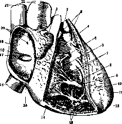
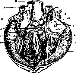
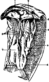
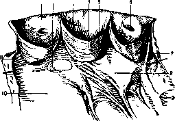
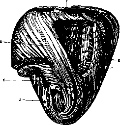
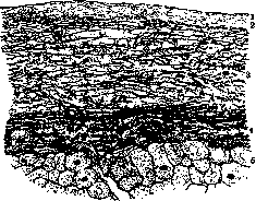
Figure 18. Endocardium of the right atrium: 1-endothelium; 2-superficial connective tissue layer; 3-middle layer with abundant networks of elastic fibers; 4-deep layer with thick elastic fibers; 5-innermost layer of myocardium with transversely cut muscle fibers and elastic fibers in the internal perimysium. Figure 19. Endocardium of the left ventricle: 1-connective tissue layer under the endothelium; 2-middle networks of elastic fibers (dark); 3-deep layer of endocardium with sparse elastic fibers; 4-innermost layer of myocardium, with muscle cells resembling Purkinje cells. The inner surface is lined with endothelium, which is a direct continuation of the endothelium of the vessels and differs from it in that the cells are less elongated. In the tissue underlying the endothelium, 3 or 4 layers can be distinguished: 1) a thin subendothelial layer, formed by a dense plexus of connective tissue fibers immersed in an amorphous ground substance; 2) a relatively thick connective tissue layer with a high content of elastic fibers; 3) an elastic-muscular layer containing networks of thick elastic fibers and in some places smooth muscle fibers; some authors also include as the 4th layer of the endocardium the layer of loose connective tissue at the border with the myocardium, which others consider perimysium internum myocardii. The previously widespread view that the endocardium corresponds to the inner membrane (intima) of the vessels is now disputed; it is equated with the vessel wall as a whole, considering the outer membranes as a special superimposition. The myocardium, forming the thickness of the heart wall, consists of cross-striped fibers of special structure and arrangement (fig. 20). Their cross-striation is of the same type as in skeletal muscles, but they are thinner than them
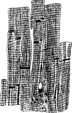
Figure 20. Myocardium. (After M. Heidenhain.)
approximately by 1/3 (9-22 μ), branch at acute angles and anastomose with neighboring fibers, forming continuously extending networks, the point of attachment of which is located on the fibro-cartilaginous rings and the mouths of the large vessels. Features of their structure include: 1) a very thin sarcolemma, the existence of which was long denied; 2) the location of nuclei along the axis of the fibers, with their ends, among accumulations of sarcoplasm, often containing pigment grains; 3) a special arrangement of fibrillar columns having the shape of ribbons: on cross-section, the fields of Cohnheim show a picture of strips running radially from the nucleus, like the spokes of a wheel, separated by layers of sarcoplasm; 4) but the main feature lies in the presence of special cross-striations - intercalated, differing from the ordinary striations in their luster and thickness; these striations often have a stepped shape; they are located at a considerable distance from each other and divide the fiber along its length into special segments. When treated with AgNO3, the intercalated striations blacken, dissolve in 30% KOH, as a result of which the fibers break down into segments (segments of Weismann) containing one or more nuclei; these segments were previously considered muscle cells, the units of which the fiber is composed and into which it breaks down in pathological cases (myocarditis segmentaria Renaut).-Subsequent works (Ebner, Heidenhain, Marceau) refuted this view and established that the muscle fibrils pass through the intercalated striations without interruption; but the significance of the striations has not yet been precisely established - they were at one time considered contraction bands (Exner, Ebner), then areas where fiber growth occurs (M. Heidenhain), and recently elementary tendons (Marceau), which, by dividing the fiber into sections, facilitate its function. Between the muscle fibers there is loose connective tissue, in places with the character of reticular tissue (perimysium), through which blood vessels, nerves pass, and lymph circulates.-The conducting system of the heart muscle or atrioventricular bundle (nodes, bundle of His and Purkinje fibers) consists of muscle fibers modified in a special way: in humans they differ from the usual ones in their thickness, are rich in sarcoplasm, vacuolated, and contain glycogen; in some animals (ungulates) the conducting system consists of large elongated cells resembling plant cells, with light contents and a nucleus in the middle; along their periphery are located cross-striped fibrils, which pass from one cell to another and partly surround the boundaries between cells. There is no reason to consider the conducting system a remnant of embryonic development, since these formations are clearly expressed already in the embryo's heart (see Atrioventricular bundle, nodes). The epicardium is a serous membrane, i.e., a plate of connective tissue covered on the free surface with endothelium. It is firmly attached to the surface of the heart muscle; in the grooves where vessels and nerves pass and where loose connective tissue is located under the epicardium, deposits of fat cells and lobules are often observed.-The valves of the heart are formed by folds of endocardium, at the base of which a dense connective tissue plate from the fibro-cartilaginous rings extends. The latter consist of dense connective tissue, in which sometimes islands of cartilage tissue are found (Tretyakov).
V. Karpov. Anatomy of the coronary vessels. The nutrition of the H. is carried out by means of the coronary vessels. For its work, the H. requires a large supply of nutrient material. Therefore, the muscles of the ventricles are supplied with a capillary network twice as abundant as skeletal muscle. The coronary arteries absorb approximately one-tenth of the total amount of blood entering the aorta from the left ventricle. Both coronary vessels depart from the root of the aorta in the area of the sinus of Valsalva. The arteries of the H. itself are the only large branches departing from the aorta ascendens. A. coronaria cordis dex. (fig. 21) begins in the right sinus of the aorta (sinus Valsalvae), first passes between the right auricle and the a. pulmonalis, then turns right along the coronary groove and, reaching the posterior longitudinal groove, passes into ramus descendens posterior. A. coronaria cordis sinistra emerges from the left sinus of the aorta, immediately divides into two branches. The first, the anterior descending branch (ramus descendens ant.), first goes behind the pulmonary artery, then along the longitudinal groove to the notch of the H. The second, the circumflex branch (ramus circumflexus), lies in the coronary groove, first under the left auricle, then passes to the posterior surface, but does not reach the posterior longitudinal groove. The position of the mouths of the coronary arteries is essential for understanding their filling. The left coronary artery passes through itself approximately 75% of all coronary blood. The main trunks of both coronary arteries run along the surface of the heart and only the second and third order branches penetrate into the depth of the cardiac muscle. The main trunks are covered by the epicardium and surroun
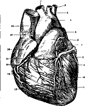
Figure 21. Vessels of the heart from above and in front: 1 - a. anonyma; 2-a. subclavia sin.; 3-a. carotis communis sin.; 4-arcus aortae; 5-ramus sin. a. pulmonalis; 6-a. pulmonalis; 7-atrium sin.; 8- v. cordis magna; 9-ramus descendens ant. a. coronariae (cordis) sin.; 10- sulcus longitudinalis anterior; 11- ventriculus sin.; 12-ventriculus dex.; 13- v. cordis ant.; 14-coronaria cordis dex.; 15-v. cordis anterior; 16-atrium dex.; 17-aorta ascendens; 18-v. cava sup.
covered by epicardial fat. In rare cases, individual muscle fibers are thrown across the main trunks. The winding of the arteries contributes to better filling of the coronary vessels with blood 'under widely varying conditions of heart work. The topography of the coronary arteries varies so much in individual cases that it is difficult to find two hearts equally vascularized. This applies to both humans and dogs, animals on which the main research in the field of cardiology has been performed. Similarly, the distribution of both coronary arteries in terms of supplying different parts of the heart is subject to great variations. In general, however, the right coronary artery supplies most of the right heart, the posterior part of the septum, part of the posterior wall of the left ventricle, and the medial (posterior) papillary muscle of the left ventricle. The left coronary artery supplies the remaining part of the left ventricle, the anterior part of the septum, and a small area of the anterior surface of the right ventricle along the septum. The large papillary muscle of the right ventricle is partially supplied by the left coronary artery. The distribution of subepicardial branches is largely independent of the direction of the superficially lying muscle fibers. The branches penetrating into the depth of the coronary arteries spread mainly perpendicular to the surface of the heart, regardless of the direction of the muscle fibers. These branches penetrate into the papillary muscles and also spread under the endocardium. The peripheral branches anastomose abundantly with each other, which makes it possible to equalize the circulation of the heart in various pathological processes affecting its vessels.-Based on the latest research on the blood supply to the conduction system of the heart, Mahaim speaks of anterior and posterior vascularization of the septa. Anterior vascularization occurs through branches of the descending branch of the left coronary artery, posterior vascularization occurs through branches of the descending posterior artery. In 90% of all cases, the descending posterior artery is a continuation of the right circumflex artery (a. circumflexa dextra); in 10% of cases it is a continuation of the left circumflex artery. The Tawara node is supplied by branches belonging to the posterior vascularization. The main trunk of the His bundle, the right leg, and the initial part of the left leg are supplied by the artery feeding the fibrous part of the septum (a. septi fibrosi), which in 92% of all cases originates from the left coronary artery. The left leg of the His bundle in its anterior ramifications is supplied from the descending anterior branch, the posterior ramifications by branches belonging to the posterior vascularization. However, the distribution of these vessels is subject to great variations. According to the latest research by Tandler, the veins are divided into the following sections: 1. The coronary sinus (Fig. 22), ending proximally at the Thebesian valve. The veins flowing into the coronary sinus are subject to great variations. The main vessels of this section should be considered: the great vein of the heart, the oblique vein described by Marshall, and the common trunk of the right vein of the heart and the interventricular vein. Mostly the sinus is located in the coronary groove (sulcus coronarius). 2. The branches of the coronary sinus: the great vein of the heart (vena cordis magna) begins as the anterior interventricular vein at the apex of the heart, where it anastomoses with the posterior interventricular vein, extending from the apex along the anterior interventricular groove together with the anterior coronary artery towards the base of the heart, passes under the left atrium, circling it, and flows into the coronary sinus. Numerous veins of the walls of the left ventricle and single veins of the right ventricle flow into it. The left marginal (marginal) vein and the posterior vein of the left ventricle on the posterior surface also flow into it. The small oblique vein of the left atrium of Marshall, starting from the pulmonary veins, descends along the left atrium and reaches the coronary sinus along its posterior side. Essentially, the coronary sinus is a direct continuation of this vein. The posterior interventricular vein begins at the apex, extends together with the descending posterior branch of the right coronary artery along the posterior ventricular groove upward, and flows into the coronary sinus either isolated or as a branch common with the right coronary vein. This vein is a collector for the diaphragmatic part of the heart. The right coronary vein is mostly a small vessel passing along the posterior part of the right groove and flowing into the posterior interventricular vein or directly into the sinus. 3. The small veins of the heart. Under this name are combined all veins flowing directly into the right atrium. The largest among them is the vein of Galen, crossing the groove of the heart and flowing directly into the right atrium. Parallel to it are 3-4 smaller veins. In addition to
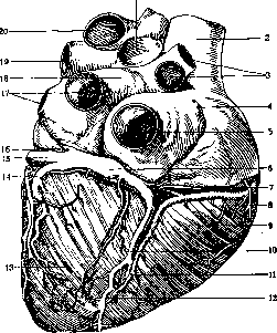
Fig. 22. Vessels of the heart from behind: 1-ramus dex. a. pulmonalis; 2 - v. cava sup.; 3 - vv.pulmonales dex.; 4-atrium dextrum; 5-v. cava inf.; 6-sinus coronarius; 7-v. cordis parva; 8-v. cordis ant.; 9-ventriculus dex.; 10-v. cordis media; 11-ramus descendens post. a. coronariae (cordis) dex.; 12-sulcus longitudinalis post.; 13-ventriculus sin.; 14-v. post, ventriculi sin.; 15-v. cordis magna; 16-v. obliqua atrii sin. (Marshalli); 17- vv.tpulmonales sin.; 18-atrium sin.; 19- ramus sin. a. pulmonalis; 20-arcus aortae.
those described between the cone and the right atrium flows a vein originating in the area of the cone, described by Cruveilhier, as well as a vein collecting blood from the initial part of the pulmonary artery and the adjacent part of the right atrium, described by Zukerkandl. 4. The minimal veins of Thebesius (venae cordis minimae) are either independent ducts originating from small capillary areas, or anastomoses with superficially lying veins. In the first case they are extremely small, their diameter does not exceed several fractions of a millimeter, in the second case they are significantly larger. These veins open mainly directly into the atrium. They are particularly numerous in the right atrium. In the left atrium their number is significantly smaller. Almost constant is a vein in the atrial septum near the place of departure of the aorta. Through it blood flows not only from the atrial septum but also from the ventricular septum. In the area of the ventricles, Thebesian openings are subject to significant variations, they are found mainly at the base of the papillary muscles. All Thebesian veins located in the ventricles are independent centers-collectors of small venous capillaries of the cardiac muscle-and nowhere do they anastomose with large veins, as is observed in the atria. The walls of the cardiac veins, like those of the brain, are thin and small. Medium-sized veins contain, in addition to the endothelium, only connective tissue and elastic fibers. The smallest veins are endothelial formations with a thin connective tissue shell around them. Only in large veins are there strands of smooth muscle, arranged circularly or obliquely in relation to the vascular axis. The veins are fixed by means of the adventitia in the intermuscular connective tissue. In the veins of the heart, senile changes are observed early. Valves in the veins are found only at the place of their flow into the coronary sinus. According to the data of Starling, Evans, and Markwalder, 60% of the blood supplying the heart flows away through the coronary sinus, 40% of the blood-through the Thebesian veins.
D. Pletnev. III. Comparative Physiology. The basic mechanism for the movement of body fluids is: in coelenterates - the movement of cilia and flagella lining the walls of the gastrovascular system; in flatworms and nematodes - contractions of the body and intestinal wall musculature; in annelids, mollusks, arthropods, and chordates - the automatic rhythmic motor activity of the vessel walls. In the latter case, the Heart (or hearts) differentiates as a specialized motor section (or sections) of the vascular system with a number of specific physiological and morphological properties, while at the same time the other vessels lose to a greater or lesser extent their active motor role. As a result of secondary-progressive changes in the latter, new pulsating areas may additionally appear, such as: pre-branchial Hearts of cephalopod mollusks, pulsating sacs at the base of the wings of ephemeropterans and orthopterans, rhythmically contracting veins of the wings of bats, etc. - Along with motors that move intravascular fluid, a number of auxiliary mechanisms arise, both extra- and intravascular. The former include: the rhythmically contracting musculature of the gills of Amblystoma, which propels blood through the gill capillaries, the musculi alares of insects, which stretch the Heart during diastole, the intercostal and diaphragmatic musculature of mammals, which promotes the flow of blood to the atrium during inspiration, etc. The latter include the Heart and vessel valves, present in all animals with a constant direction of intravascular fluid flow. In tunicates, which have a changeable direction of blood flow, the role of the valve is performed by the tonic stricture of the musculature at one end of the Heart, which allows blood to pass in only one direction and is replaced by a similar stricture at the other end of the Heart when the direction of blood flow changes (a morphologically unfixed or "functional" valve). - Morphologically fixed valves are either passive (tendinous) or active (muscular). Examples of the latter are the sphincters of the ostia of the Heart of Dytiscus, the atrioventricular funnel and the aortic valve of the Heart of pulmonates, as well as the bi- and tricuspidal valves of the Heart of mammals (including here the associated musculi papillares as the physiological part of these valves). Active valves are that part of a given section of the Heart that contracts first, thereby creating an obstacle to the backflow of blood before the beginning of systole of this section. Functionally, the Heart, regardless of the number of its anatomical divisions, consists of two parts: the motor part and the preceding reservoir part. In vertebrates, the motor part is represented by the ventricle, and the reservoir part by the atria. The walls of the motor part of the Heart have powerful musculature; the walls of the reservoir part are thin and easily stretchable. In Crustaceae and Nematoda, where the Heart is tubular (single-chambered in the former, multi-chambered in the latter), the entire Heart is the motor part; the role of the reservoir is performed by the pericardial sinus surrounding the Heart, from which blood enters the Heart through the ostia during diastole. In some gastropods, the atrium is porous, and when it is overfilled, excess hemolymph enters the pericardial cavity, which thus plays the role of an additional reservoir. In insects, the entire body cavity performs the reservoir function. - The physiological properties of cardiac muscle differ sharply in vertebrates and invertebrates, which is connected not only with the difference in its origin, but also with the different conditions of its functioning in the typical for vertebrates closed and typical for invertebrates open circulation. The "all or nothing" law is not applicable to the Heart of invertebrates: successive impulses give a different functional effect due to the unequal coverage of the Heart wall by excitation. Unlike the vertebrate Heart, the Heart of invertebrates is capable of tetanus. It lacks an absolute refractory period; the relative refractory period begins with the beginning of systole and lasts almost until its end. In terms of refractoriness, tunicates occupy an intermediate position between vertebrates and invertebrates - the primary-chordate animals, whose Heart, like that of invertebrates, lacks absolute refractoriness but has a more pronounced relative refractoriness than the Heart of invertebrates. According to Carlson, the absolute refractory period is also absent in the cyclostome Bdellostoma. In invertebrates, an extrasystole leads, as in vertebrates, to a compensatory pause if the extra stimulation is applied to the non-leading part of the Heart (e.g., when stimulating the heart muscle of Limulus, which contracts under the influence of impulses originating from the epicardial node). In invertebrates possessing autochthonous automatism in all parts of the Heart (e.g., in Helix), an extrasystole, wherever the stimulation is applied, displaces Engelmann's "physiological period," and a true compensatory pause is absent. - The action currents of the Heart in mollusks represent a simple two-phase oscillation (Arvanitaki and Cardot; Luisada). In Limulus, the Heart contracts tetanically, summing a series of impulses originating from the epicardial ganglion; in connection with this, the cardiogram of Limulus is characterized by great complexity (Rylant's research). In recent years, research has appeared on the development of the electrocardiogram in embryogenesis, showing the correspondence of certain stages of the anatomical differentiation of the Heart to certain waves of the electrocardiogram. Automatism already at the pre-cardiac stage of the existence of vascular motoricity (e.g., in polychaete worms) is true automatism, i.e., the ability of the vascular wall to contract rhythmically in an isolated state in the absence of external influences (Stiibel, Bethe). In the course of evolution, "true" automatism is lost by the non-motor parts of the vascular system, and then also by the non-leading parts of the Heart, which retain only the ability to respond rhythmically to non-rhythmic stimulation (stretching, intracavitary pressure). The place of origin of automatism is, at early stages of phylo- and ontogenesis, the muscle cells (myogenic automatism). In the adult state, myogenic automatism is preserved in the snail. In the larva of Limulus, myogenic automatism was established before the development of the epicardial node. Already at the myogenic stage of Heart development, the ability for automatism is distributed unevenly in it: thus, in the ventricle of the snail's Heart, the leading part is the aortic end, i.e., that end where in other pulmonates possessing neurogenic automatism lies the leading epicardial ganglion. The establishment of a constant leading part may be preceded by a period of alternation of leading parts; this was shown by Child on the larva of Colias, in which the Heart at the beginning of development beats first in one direction then in the other, but with age the periods of Heart beats in the oral direction become longer and longer, until this direction is fixed as the only one. As a constant, non-embryonic phenomenon, the alternation of leading parts is observed in tunicates (Schultze). - With the appearance in invertebrates of epi- or intracardiac nerve nodes, and in vertebrates of a specific intracardiac tissue that produces and conducts automatic impulses, the autochthonous automatism of cardiac muscle regresses; at the same time, the peristaltic type of Heart contractions is replaced by the sequential contraction of entire sections. The same change from peristalsis to sequential contractions of sections takes place in embryogenesis. The problem of the microlocalization of automatism in the Heart of vertebrates remains technically unsolved for now; however, the large number of ganglion nerve cells in the cardiac nodes of vertebrates, with the high ability of these cells to automatism, suggests that it is precisely the nerve cells that must be the site of the flash of automatic impulses of the Heart. A special solution to this problem is attempted by Demoor (whose data are partly confirmed by Haberlandt), who believes that the nerve elements of the leading part play less of a nervous and more of an internal secretory role, secreting a hormone that diffuses into the surrounding muscle tissue and stimulates the muscle elements to rhythmic contractions. For vertebrates, Haskell's rule applies, according to which the gradient of automatism of the Heart decreases from the venous to the arterial end of the Heart. The normal heterotopia established by Mac William, as well as Koehnlein, in some fish (e.g., in the loach) does not violate this rule, because the direct path from the sinus to the arterial cone in them leads through the auricular canal and ventricle, while the atrium is only a lateral outgrowth of the cardiac tube, corresponding to its reservoir role. Haskell's observation that after the Heart of a turtle stops, the sinus resumes beating first, the ventricle second, and only the atria third, allows one to assume here the presence of a direct conducting path from the sinus to the ventricle. - Regarding the location of intracardiac nodes, Biering and Scramlik divide fish into three types: a) with three nodes (leading in the sinus and subordinate in the auricular canal and in the atrioventricular funnel), b) with two nodes (leading in the sinus and subordinate in the funnel), c) with two nodes (leading in the auricular canal and subordinate in the funnel). The arterial cone does not have a separate node.
At higher levels of the vertebrate series, the leading node is the sinus node (understanding this term to also include the node homologous to the sinus node at the site of entry of the vena cavae into the right atrium of mammals), and the subordinate node is the atrioventricular node. Coordination of the rhythms of the Heart's departments is accomplished most primitively through their mechanical interaction, where the contraction of one department stretches the other department or increases its intracavitary pressure, thereby stimulating its contraction (observations of Willems and Eichler on snails, Fredericq on Octopus). The prevalence of this coordination mechanism in mollusks is associated with the free mobility of their atrioventricular boundary; in other types, mechanical coordination plays a minor role, although it is preserved as an additional factor of coordination even in vertebrates. Another coordination mechanism—reflex coordination of the Heart's departments through the central nervous system—exists in snails along with mechanical coordination (Zubkov). Central reflex coordination is the only way to coordinate the rhythms of motor parts of the vascular system that are distant from each other, for example, the branchial hearts of cephalopods, the lymphatic hearts of amphibians. The third coordination mechanism is the conduction of excitation along special epi- or intracardiac pathways. Epicardial conduction is characteristic of the dorsal, segmented, tubular Heart of invertebrates (observations of Carlson on Limulus, Alexandrowitz on Isopoda). This coordination mechanism can reach a high degree of differentiation: thus, histological and oscillographic observations by Rijlant established that in addition to nerve cells producing rhythmic impulses, the epicardial ganglion of Limulus also includes nerve cells conducting these impulses from department to department, and nerve cells ensuring the simultaneous contraction of all muscular elements of one department. Intracardiac conduction is characteristic of the vertebrate Heart. From the atria to the ventricles, excitation is conducted along the specific conducting tissue of the funnel. The higher in the vertebrate series, the more limited the conducting area of the funnel, so that in mammals conduction occurs through one or a few bundles (His, Kolm and Trendelenburg). Intracardiac pressure is low in invertebrates (with the exception of cephalopods), which, according to Scramlik, is related to the incompleteness of their circulatory system. In vertebrate animals, pressure gradually increases with the transition from aquatic to terrestrial life, and with the appearance of homeothermy, it gives a sudden sharp increase, especially in birds. This increase is related not only to the intense metabolism of homeothermic animals and the resulting increase in the capillary network, but also (according to Redfield) to the fact that the osmotic pressure of the blood is high, and normal osmosis through the capillary wall from the blood to the lymph is possible only if the intravascular hydraulic pressure increases simultaneously with the increase in osmotic pressure. In ontogenesis, the blood pressure of mammals is low until the moment of birth, not exceeding 25-30 mm Hg in dogs (Clark). Coronary blood pressure, the exclusion of which in fish and amphibians does not noticeably affect cardiac activity, already in reptiles has great importance (as seen from Haskell's experiments with the restoration of contractions of an isolated turtle's Heart when liquid vaseline is pumped into the coronary vessels), and in vertebrate animals it is a necessary condition for the automatism of the Heart. Central innervation of the vascular system exists even at the pre-cardiac stage. In ontogenesis, central innervation of the Heart appears earlier than intracardiac innervation (research on rat embryos). An example of the most primitive form of central innervation of the Heart is the innervation of the grape snail's Heart, undifferentiated into special inhibitory and excitatory nerves and having terminal apparatuses in the Heart that simultaneously perform both receptor and effector functions (Zubkov). Apparently, in most other mollusks, there is only one type of Heart nerve, and the difference in the effect obtained from irritation of these nerves depends on the state of the Heart itself. According to the data of Carlson, Ransom, and others, in Nudibranchiata and Protobranchiata, irritation of the Heart nerves gives mainly stimulation of the Heart, whereas in Lamellibranchiata and Pulmonata, inhibitory effects predominate. Only in higher mollusks (cephalopods) did Fredericq find a definitely dual innervation of the Heart, and in them the presence of vasoconstrictor and vasodilator nerves has also been established. In arthropods, there is apparently a dual innervation of the Heart. Thus, separate inhibitory and excitatory Heart nerves have been found in decapods; there is also an indication of dual innervation of the Heart in Limulus. Among vertebrates, some fish (Teleostei) have only vagal innervation and no sympathetic innervation; this corresponds to the fact that in embryos of those vertebrates that have dual innervation of the Heart, vagal innervation appears before sympathetic innervation (research by van Kampenhout on cats). A more or less pronounced tone of the centers innervating the Heart, maintained by both humoral and reflex (in particular from reflexogenic vascular zones) effects on these centers, probably exists at all levels of the zoological series, including in vertebrates. For mollusks, it has been experimentally proven in snails (Zubkov) and cephalopods (Fredericq). However, it differs in invertebrates by its greater variability due to the non-isolation of the autonomic nervous system from the animal nervous system and is easily changed under the influence of external irritations. It has been established (Lutz and Wyman) that the area of vascular reflexogenic zones in vertebrates is reduced in the course of evolution: in fish (Squalus), reflex slowing of the heartbeat occurs when pressure increases in the gill vessels and in the abdominal aorta; in amphibians (Necturus mac.)—only when pressure increases in the gill vessels; the reflexogenic area is most limited in the vascular system of mammals (in the dog—the aortic arch and sinus caroticus).-In poikilothermic vertebrates, as well as in hibernating homeotherms, the tone of the centers of the cardiac nerves is subject to seasonal fluctuations. In mammals (at least in those that are helpless for some time after birth), the tone of the vagus center reaches its maximum only during postembryonic development (experiments by Smirnov and others on puppies). The same fact has been established for the development of the tone of sympathetic innervation of the heart.
a. Zubkov.
IV. Physiology. Basic functions of cardiac muscle. In analyzing the activity of the heart and the influence on this activity of extracardiac nerves, Engelmann distinguishes the basic properties or functions of cardiac muscle: the function of automatism, excitability, conductivity, and contractility, while Gaskell also identifies a function of tonicity. This division into separate functions is to some extent schematic: it is impossible to sharply differentiate the individual functions of the heart, since it is not sufficiently known at the present time what underlies these functions. Moreover, the functions of the heart, both under physiological and pathological conditions, are so intertwined that it is often extremely difficult to delineate the role and significance of individual functions. The function of automatism is understood as the ability of the heart to give rhythmically successive contractions without any influence of external irritating factors. An excised heart of cold-blooded animals continues its activity for a long time without change. The same thing happens with the heart of warm-blooded animals if its nutrition is maintained. An excised human heart also possesses this ability, which was first demonstrated by an experiment by Kulyabko. At the end of the 19th century, the question of which parts of the heart possess the automatic function—muscle or nerve tissue (myogenic or neurogenic theory of heart contraction)—was debated for a long time and passionately. Supporters of the neurogenic theory pointed to the heart of Limulus, where contractions originate from a nerve trunk, the removal of which leads to the cessation of the heart. Supporters of the myogenic theory pointed out that automatism is inherent in such formations as, for example, vacuoles, where there are no nerve elements. Attention was drawn to the heart of a chick embryo, where automatic contractions begin long before the appearance of nerve elements. It was not possible to resolve this question by anatomical separation of tissues, since muscle and nerve fibers are closely intertwined. This question has now lost its acuteness in connection with the study of the anatomy and physiology of the conduction system. According to modern data, the automatic function of the heart, both under physiological and pathological conditions in humans and presumably in all warm-blooded animals, is carried out by the conduction system. It cannot be considered finally established whether the contractile myocardium possesses the function of automatism. Apparently it does not possess this ability. It is not entirely clear how the function of automatism is realized. It can be assumed that a constant irritation of a nervous or chemical nature acts on the myocardium. The cardiac muscle, during its contraction, loses the ability to perceive irritation. Only at the end of diastole does the ability to perceive irritation return to such an extent that the strength of the existing constant irritation exceeds the threshold of excitation of the myocardium and the next systole occurs. But it is also possible that the very occurrence of irritation has a rhythmic character. Having caused the appearance of excitation and subsequent contraction, the automatic ability of the heart is exhausted, and a strictly defined time is required for the accumulation of energy for subsequent irritation to occur. According to Engelmann, the metabolism of automatically functioning myocardial cells forms substances that irritate the heart. When the accumulation of these substances reaches a certain level, contraction occurs. Contracting, the cardiac muscle simultaneously destroys the substance causing irritation and loses the ability to perceive irritation. Then again occurs the accumulation of the irritating substance and the restoration of the ability to perceive irritation. According to Hering, the accumulation of irritating substances and the restoration of excitability are not identical processes and do not proceed in parallel. The formation of irritation has a rhythmic character. According to Rothberger, the gradually accumulating potential energy, upon reaching a certain height depending on the excitability of the heart, automatically discharges. Having caused excitation and subsequent contraction of the myocardium, the automatic ability of the heart is exhausted, and a strictly defined time is required for the accumulation of energy to cause subsequent contraction. This conception is in agreement with the latest views identifying the automatic irritant with the potential difference arising in the cells, the height and speed of formation of which depend on the concentration of hydrogen ions. In recent years, works have appeared (De-moor, Haberlandt and others) linking the automatic ability of the heart to the presence of a hormone. Haberlandt considers this cardiac hormone as the main force of normal heart contraction. However, experiments with the removal of the sinus in cold-blooded animals and the sinoatrial node in warm-blooded animals, these main sites of formation of the cardiac hormone, did not confirm the importance of the cardiac hormone as a factor determining the automatic activity of the heart (Averyanov, Fogelson and Fedorov). For the physiology and pathology of heart automatism, it is extremely important to determine which parts of the heart and to what extent possess the automatic ability. Since in the normal heart all parts are interconnected, the part of the heart with the greatest automatic ability will direct the rhythm. If a ligature is placed around the sinus of the frog's heart [Stannius ligature], the part of the heart lying below the ligature stops contracting. After some time, contractions begin again in this segment, and the rhythm of contractions will be slower than before the removal of the sinus; the atria and ventricle contract simultaneously. It is clear that excitation originates from another segment than before, namely from the segment lying at the border of the atria and ventricle. If then a ligature is placed between the atria and ventricle (II Stannius ligature), the atria stop contracting, while the ventricle continues its contractions at a slow pace. Finally, if the ventricle is tied at the border of the middle and lower third (III Stannius ligature), the upper part continues to contract automatically, while the lower part does not contract. From Stannius' experiments it can be concluded that the sinus possesses the greatest automatic ability and is the usual pacemaker. The formation at the border of the atria and ventricle has a lesser automatic ability and takes over the direction of the rhythm when the sinus is excluded. Gaskell proved that the sinus is the pacemaker of the heart by the fact that by heating it he obtained an acceleration of contractions of the entire heart, and by cooling it—a slowing of contractions. Heating and cooling other parts of the heart did not give such an effect. Engelmann approached the resolution of this question somewhat differently. He caused irritation of various parts of a normally contracting frog's heart by an inductive shock. When the ventricle or atria were irritated, their premature contraction occurred, followed by the so-called compensatory pause, exceeding the normal pause between two contractions. But if the sinus of the frog was irritated, the subsequent pause after the premature contraction did not exceed the duration of the usual pause of the normal heart contraction of the frog's heart. This can only be explained by the fact that the impulse for excitation is born in the sinus and, upon reaching sufficient strength, at strictly defined intervals, is directed to the atria and ventricle. With premature irritation of the sinus, the accumulated irritating impulse will discharge, and its increase will occur through the usual interval. With premature irritation of the ventricle, the sinus continues its normal rhythmic activity, but the excitation emanating from it will find the ventricle in an unexcitable (refractory) state. Only the next impulse, originating from the sinus, will cause a normal heart contraction. The duration of the compensatory pause and the preceding premature contraction together with the cycle of the preceding shortened normal contraction will equal the duration of two normal cardiac cycles. When the atria are irritated, the excitation will be directed retrogradely to the sinus. Upon reaching it before the moment of exit of the normal impulse, it will discharge it and cause a disturbance of its rhythm. For the restoration of the automatic ability, the sinus needs the usual period. Then the duration of the compensatory pause will depend on the moment when the premature contraction discharged the impulse emanating from the sinus. The compensatory pause will be shortened in comparison with the pause after premature contractions of the ventricles and will not together with the premature contractions and the preceding shortened cycle equal two normal cardiac cycles. All the above experimental data confirm the role of the sinoatrial node as the pacemaker of the heart. In the heart of warm-blooded animals there is no isolated sinus. The sinus has entered into the composition of the right atrium, forming its so-called sinus part. The role of the sinus is performed by the sinoatrial node. Heating the sinoatrial node in warm-blooded animals leads to an acceleration of the rhythm of the entire heart, while cooling leads to a slowing down.
In analyzing the course of gradual involvement in excitation with local recording of action currents, it was established (Wybauw) that the first point of appearance of the electro-negative wave (Negativitat) and consequently the first point of excitation is the sinoatrial node. All this forces us to recognize that under physiological conditions the pacemaker is the sinoatrial node. The sinoatrial node is the first-order automatic center, while the atrioventricular node is a center with less pronounced automatic ability - a second-order center. In the rest of the conduction system, the automatic ability to generate excitation is even weaker, and this part is a third-order center. Under physiological conditions, the rhythm is controlled by the center with the greatest automatic ability - the sinoatrial node; the atrioventricular node and the rest of the conduction system act only as conductors of excitation. With organic or functional damage to the sinoatrial node, or when it is separated from the atrioventricular node, the latter manifests its automatic ability and takes over the control of the rhythm, and if the connection between the atria and ventricles remains, it controls the rhythm of the entire heart; if it is disrupted, this control is limited to the ventricles. Since the automatic ability of the atrioventricular node is lower, the heart rate drops to 30-40 instead of the normal 70-80. With destruction of the atrioventricular node, control of the rhythm passes to the rest of the conduction system (third-order centers).- Mobitz believes that the atrioventricular node, even under physiological conditions, plays a role not only as a conductor of excitation. In his opinion, the function of the node is to sum up the irritating impulses coming to it from the sinoatrial node. The irritation of the atrioventricular node caused by these impulses is the source of further excitation of the heart. With an original view on the genesis of cardiac rhythm, the French school, represented by Vaquez, Donzelot, and Geraudel, came forward. These authors rejected the generally accepted unitary theory of rhythm, which assumes that the single pacemaker is the sinoatrial node. In their opinion, there are two formations to which the heart rhythm is subject. In the sinoatrial node (Donzelot), excitation is generated only for the atria, while the excitation of the ventricles, the main motors of blood circulation, is born in the atrioventricular node and spreads along the bundle of His and its branches. The bundle of His acts as a conductor between the atrioventricular node and the ventricles, and not as a connecting link between the cavities of the heart. The connection between the atria and ventricles is only apparent, and there is no connection between them. The same factors act on two centers with the same properties under the same conditions. Since these factors act on the atria and ventricles with a constant interval of 0.12-0.18 seconds, the illusion of a close connection between them is created. The main factors causing the alternation of atrial and ventricular contractions are either the intracavitary pressure in them (Vaquez and Donzelot) or the nature of the blood supply to the sinoatrial and atrioventricular nodes (Geraudel). However, this dualistic, as the authors call it, theory of cardiac automatism does not provide a satisfactory explanation of all available clinical and experimental data. In recent years, experimental data have appeared that refutes this theory. To maintain the automatic ability of an isolated heart, a number of conditions are necessary, in the absence of which cardiac automatism cannot manifest. It is necessary to have a certain temperature, different for different species of animals, a certain composition of the nutrient solution with the mandatory participation of NaCl, Ca, K, sugar, the presence of oxygen, and a certain concentration of hydrogen ions. All these factors also affect the normal heart of an animal. The automatic ability of the heart, resp. the number of heart contractions, is different in different species of animals. In the vast majority of cases, the number of heart contractions is inversely proportional to the weight of the animal: in amphibians, in frogs and turtles, about 40-50 per minute, in elephants the number of heart contractions is 25-40, in horses 34-50, in lions 40, in dogs 100-200, in cats 120-140, in rabbits 120-150, in guinea pigs 132-288, in mice 520-780, in bats 600-900. In humans, the number of heart contractions varies depending on age, sex, and body size. The intensity of metabolism and profession are of great importance. The number of heart contractions in a newborn is 120-140 (Filatov), by the year it reaches 110, by 12 years it reaches 82, and by 20 years it gradually decreases to 72 per minute. In old age, a slight increase in the number of heart contractions is noted. According to Volkmann, an increase in body size at the same age reduces the number of heart contractions. The number of heart contractions in women with the same body size as men is greater. In individuals with an increased basal metabolism, the number of heart contractions is increased. The state of basal metabolism is probably also connected with the fluctuation in the number of heart contractions during the day. The smallest number of beats (on average 50-60) is observed at 3 PM and then gradually increases. The function of excitability. The cardiac muscle, like skeletal striated muscles, possesses excitability. The excitation of muscle fibers is accompanied by physicochemical changes in them, of which the most noticeable is the appearance of a negative electrical charge in the excited muscle. The fact that the excited surface becomes galvanometrically negative is explained (Nernst) by a change in the concentration of ions in the muscle cell, with positively charged ions accumulating inside the cell and thus attracting negative ions to its outer surface. It was suggested (Hober) that the membrane surrounding the muscle cell and in the resting state not allowing certain ions, especially anions, to pass through, loosens in the stage of excitation. The permeability of the shell changes, and anions appear on the surface, causing a negative charge of the cell. The question of whether there is an interval between irritation and the excitation caused by it (the time of hidden irritation-Latenzzeit) cannot be considered finally clarified. Apparently, the time of hidden irritation is either very short or, which is most likely, absent altogether. Experimental data indicating the presence of time of hidden irritation are explained by the fact that the result of irritation is recorded at the place of application of irritation. In addition, with the inertia of the apparatus recording the result of irritation, a delay dependent on the methodology may occur. Behind the wave of excitation in the heart follows a wave of contraction, and the wave of excitation precedes the wave of contraction by 0.02 seconds. Einthoven believed that the wave of excitation and the wave of contraction coincide. The observed delay he explained by the lower sensitivity and greater inertia of the apparatus recording contraction. During contraction, the ability of the myocardium to perceive irritation disappears, a period of non-excitability - the refractory period - occurs. Then the ability to perceive irritation gradually returns to the myocardium. In the refractory period, two stages are distinguished. The first - when the heart does not respond even to the strongest irritation - the absolute refractory period, and the second - when the heart can be made to contract by increased irritation - the relative refractory period. In a normally contracting heart, the absolute refractory period coincides or rather almost coincides with the duration of systole. The duration of the absolute refractory phase of the ventricles, like the duration of systole, thus varies depending on the duration of the cardiac cycle from 0.3 to 0.4 sec. (see below - systole). The stage of non-excitability of the atria varies from 0.06 to 0.12 sec. The refractory phase of the sinoatrial node is significantly (by 30%) longer and can reach 0.3 sec. The refractory phase of the rest of the conduction system is also 30% longer than the phase of atrial muscle. The duration of the refractory phase of individual muscle fibers of the ventricles is the same. And since the muscle fibers are excited not simultaneously but with a certain sequence (see below), the excitation also ceases with the same sequence. This process of onset and cessation of excitation causes currents of different directions in the heart, which can be registered by a special sensitive galvanometer - an electrocardiograph. Keith, Lucas, and Adrian in nerves, and then Adrian in the heart of a frog, and more recently Lewis and Master in the human heart, noted immediately after systole a short period of increased ability to perceive and conduct irritation (supernormal recovery phase). Lewis and Master attach great importance to this phenomenon in the pathogenesis of some cardiac rhythm disorders. The more excitable the tissue, the shorter will be the duration of the current necessary to cause changes that result in excitation. The changes showed that the chronaxia (see) of the atria and ventricles is 0.002 sec.
The chronaxia of the His bundle is three times greater. Thus the bundle possesses significantly less excitability. Recently, debates have arisen regarding the strength of normal automatic irritation and its relationship to the magnitude of myocardial excitability, in connection with attempts to explain certain rhythm disturbances by changes in the strength of automatic irritation and its ratio with the excitability of the Heart. According to Gaskell and Engelmann, the strength of automatic irritation is small and is at the level of the irritation threshold of the Heart (Schwellenreiz). According to the data of Schellong, the strength of normal irritation exceeds the excitability threshold of the heart several times. And this is understandable, since irritation in its passage must overcome a certain resistance. Function of conductivity. The question of whether the function of conductivity is inherent in muscle fibers or whether only nerve fibers possess this ability, for a long time, similar to the question of the localization of automatic irritation, caused great disputes. However, recent works have most convincingly proven the presence of conducting ability in the myocardium. Since Engelmann's time, the function of conductivity has been separated from the function of contractility on the basis of an experiment proving that non-contracting atria can conduct excitation from the sinus to the ventricle. However, between the function of conductivity and the function of contractility there exists a close relationship and both functions are intertwined. Thus, Skramlik believes that muscle fibers, only by contracting, transmit excitation. According to the data of Junkmann, fluctuations in conductivity and contractility during the cardiac cycle proceed in parallel. Even closer relationships exist between the function of conductivity and the function of excitability. According to Ashman, the process of recovery of conductivity proceeds in parallel with the process of recovery of excitability. According to Schellong, conductivity is not a special property of cardiac muscle: there is only the transmission of excitation from one muscle cell to the next. A change in the permeability of the membrane and the concentration of hydrogen ions of the excited cell causes a change in the permeability of the membrane and the concentration of hydrogen ions of the neighboring cell, causing its excitation and the appearance of a negative electrical charge. The conducting ability is not the same in different parts of the Heart. The atrial muscle conducts irritation twice as fast as the ventricular muscle. While the atria conduct at a speed of 800-1,000 mm per sec., the speed of passage in the ventricles is 300-500 mm per sec. The greatest conducting ability is possessed by the branches of the conducting system. The speed reaches 3,000-4,000 mm per sec. when excitation passes along direct paths, and falls to 1,500-2,000 mm per sec. when passing along a winding path. The passage of excitation is significantly slowed in the atrioventricular node. Lewis and Rothschild explain the different speed of excitation passage by the difference in the amount of glycogen contained in the fibers of one or another part of the Heart. The maximum amount of glycogen is contained in the branches of the conducting system, and then in order of glycogen content comes the atrial muscle, ventricular muscle, nodes. In the same sequence proceeds the conducting ability of the Heart. Depending on views on the essence of the function of conductivity, the different speed of conduction is also explained by the difference in the refractory phase of different parts of the Heart and unequal excitability (different chronaxia). An increase in temperature causes acceleration of conductivity, while cold slows down conductivity. The concentration of hydrogen ions also affects conductivity, and in an alkaline medium (pH = 7.8) conductivity is accelerated, while in an acidic one (pH = 7.0) it is slowed. Lack of oxygen sharply reduces the function of conductivity. The passage of excitation in a normal Heart proceeds as follows. The wave of excitation originates in the sinus node and spreads at a speed of 800-1,000 mm per sec. along the muscle bundles radiating from this node. The wave passes through all the muscle tissue of the atria; spreading to the hollow veins at the same speed, the wave is directed downward along the septum, reaches the atrioventricular node, and from there is transmitted to the ventricles. The spread of the excitation wave can be compared to the spread of a liquid poured on a smooth surface; the edge of the liquid gradually expands, forming circles of ever-increasing diameter until it covers the entire surface. Upon reaching the atrioventricular node, the course of excitation slows down during passage through the node, then excitation is directed along the His bundle, along its legs and branches, to quickly (at a speed of 1,500-4,000 mm per sec.) spread through the terminal branches and reach the myocardium. Then begins the passage of excitation through the contractile myocardium and the Heart muscle gradually, in a normal Heart with a certain sequence, begins to come into excitation. Since the peripheral branches of the conducting system are scattered under the endocardium, it is clear that the inner surface of the cardiac muscle is excited first, then excitation with significantly reduced speed (400-500 mm per 1 sec.) spreads through both ventricles. Thus, the excitation of the muscle bundles of the ventricles lying on the surface will depend on two reasons: first, on the time when the underlying part of the conducting system comes into excitation, and second, on the magnitude of the muscle layer separating the superficial muscle layers from the peripheral branches of the conducting system. The course of excitation is completely unrelated to the anatomical course of the muscle fibers, but is directed from the inner surface to the outer. The closer the muscle fibers of the Heart are to the peripheral branches of the conducting system and the earlier these branches come into excitation, the earlier the corresponding muscle fiber on the surface of the Heart is excited. The central part of the right ventricle is excited first, because the muscle layer here is in close contact with the branches of the right leg and the separating muscular wall is very thin. In the ventricle, the apex is excited first due to the thin wall separating it from the peripheral branches of the left leg. Different points on the surface of the right and left ventricles come into the stage of excitation at a strictly defined time, with a series of points coming into excitation earlier in the right ventricle, while a series comes into the left, depending on their relationship to the conducting system. The process of cessation of excitation of the muscle fibers of the Heart proceeds in the same sequence as the process of occurrence, because the duration of excitation and contraction of the muscle fibers of the Heart is the same. This process of occurrence and cessation of excitation in the Heart causes currents of action in different directions, the recording of which—the electrocardiogram (see)—makes it possible to judge the course of excitation passage in the heart. Excitation is conducted in all directions and, arising in the ventricles, can be directed retrogradely to the atria. However, retrograde passage is sharply slowed down. Function of contractility. Cardiac muscle, like skeletal muscles, reacts to irritation with excitation and subsequent contraction (see Muscles, physiology). As soon as the excitation threshold is crossed, the Heart responds with a contraction of a certain strength. Further strengthening of irritation does not increase the strength of contraction. This property of the Heart to respond to irritation of sufficient strength with a contraction of a certain strength, not strengthening it with an increase in irritation, was formulated as the law of the Heart "all or nothing" (see) (Bowditch). Essentially speaking, the formulation "all or nothing" is incorrect mainly in its first part "all". With irritation that caused contraction of the Heart, this contraction is not maximal. The strength of contraction of the Heart is subject to completely different laws. It is only important that the strength of heart contractions does not depend on the strength of irritation. And the law "all or nothing" is more correctly formulated as the law of independence of contraction strength from the magnitude of irritation; In this understanding, the law "all or nothing" can be applied to skeletal muscle fibers as well. The strengthening of contraction with an increase in irritation in skeletal musculature is explained by the fact that a gradual increase in irritation captures an increasing number of fibers. For each individual fiber, the given law remains in force. The overall result of contraction is determined by the number of fibers captured by contraction. In the Heart, the speed of spread of excitation compared to skeletal muscle is so great that it is not the strength of irritation that determines the number of contracting fibers. At a given strength of irritation, the fibers of the myocardium either all contract or none at all. Unlike skeletal musculature, the Heart cannot be brought into a state of tetanus. This is explained by the extremely short refractory phase of skeletal muscle, and the subsequent contraction finds the muscle in a state of excitability. The refractory phase of the Heart is significantly longer and therefore summation of contractions does not occur.
To continuous irritation, the Heart responds with separate rhythmic contractions, since continuous irritations, due to the refractory phase, are transformed into single irritations. The force of contraction of the Heart does not depend on the magnitude of the irritation. The energy developed during contraction of cardiac muscle, like that of skeletal muscle, and consequently the force of contraction of cardiac muscle fiber, is directly proportional to its initial length, i.e., the length of the fiber before the beginning of contraction (Patterson, Piper, Starling). For muscle fibers of the Heart as a hollow organ, the initial length of the fibers is their length during diastole, and the length of the fibers during diastole depends on the volume of the cavities of the Heart during diastole. Starling defines this dependence of the force of contraction of the Heart on the diastolic volume of its cavities as the law of the Heart. This law determines the ability of the Heart to increase the amount of blood ejected when the inflow of blood increases. The increased inflow causes increased stretching and consequently intensified contraction, thanks to which the Heart completely empties. This ability, developed in the process of evolution, of the Heart to vary the force of its contractions, adapting them to the amount of blood flowing to it, characterizes what is usually called the reserve force of the Heart. The greater the range of reaction of the Heart, the greater its reserve force. The degree of necessary expansion of the Heart for a given amount of blood flowing to it is determined by the thickness of the myocardium and its condition. The force of contraction of the Heart as a whole is composed of the contraction of individual fibers. It is clear that a thick-walled ventricle with a large number of fibers will have to stretch considerably less than a thin-walled one with a small number of fibers to obtain an equal total force of contraction. And indeed, a healthy hypertrophied ventricle increases the amount of blood ejected with much less expansion than a thin-walled one. Nutrition of the cardiac muscle is also of great importance. The better the cardiac muscle is nourished, the greater its ability to develop energy and the less it needs to stretch to cause a contraction of corresponding force. The chemical processes accompanying the contraction of the myocardium are basically analogous to the processes occurring during the contraction of skeletal muscles (see Muscles and Metabolism, carbohydrate metabolism). Function of tonicity. The question of cardiac tonicity is the most confusing and least studied. This is explained to a large extent by the fact that the function of tonicity does not lend itself to investigation by our modern registration methods. The method proposed by Ohm for determining the state of tonicity based on the systolic collapse of the venous curve (see Pulse) cannot be considered sufficiently well-founded. Since the time of Haskell, most clinicians as well as some physiologists have believed that the Heart possesses tonicity, i.e., the walls of the atria and ventricles during diastole do not relax completely but are in a state of partial contraction. The doctrine of tonicity was based on the experiments of Haskell, in which washing the ventricle of a frog with an acid saline solution caused increased relaxation during diastole, i.e., a decrease in tonicity. Conversely, washing with an alkaline saline solution leads to less complete relaxation, i.e., an increase in tonicity. Henderson, based on his observations of the influence of pulmonary ventilation on the size of the Heart, believes that during diastole complete relaxation of the Heart does not occur. In his opinion, the tonicity of the Heart is variable and the magnitude of the tonicity determines, at a given venous pressure, the stretching of the ventricles and thus the filling of the Heart. As proof of the existence of tonicity, some authors cited intensified contraction with a sudden increase in intracavitary pressure, and when the atria were inflated, the ventricles contracted. The expansion of the ventricles after removal of the sinus was also interpreted (Szent-Gyorgy, Haberlandt) as a result of the loss of the tonicity of the Heart, guided by the sinus, and this expansion was not associated with a change in the contractile function. According to Wichels, cardiac tonicity represents a reflex process associated with nerve fibers, not associated at all with the functions of the myocardium. When checking the experimental data justifying the existence of tonicity of the Heart, it turned out that their explanation does not require the mandatory recognition of the existence of tonicity of the Heart. Thus, the main experiments of Haskell can be explained by the fact that alkalis, lengthening systole and shortening diastole, cause a decrease in the inflow of blood to the Heart and consequently a decrease in its size. Acids act in the opposite way, shortening systole and lengthening diastole and thereby causing a corresponding increase in the amount of blood flowing to the Heart and consequently an increase in the size of the Heart. Starling, based on data obtained by him and his students with the heart-lung preparation, denies the existence of diastolic tonicity. The cardiac muscle in norm during diastole relaxes completely. The only factor determining the degree of stretching of the Heart during diastole under physiological conditions is the amount of blood flowing in during the diastolic period. Consequently, the change in the size and shape of the Heart during diastole is explained not at all by the fact that it is in a state of partial contraction and that the degree of this partial contraction changes. The shape and size of the Heart during diastole are determined by the amount of blood that entered it during the diastolic period. The existence of diastolic tonicity would be a factor hindering rather than promoting blood circulation, since incomplete relaxation of the Heart would impede the entry of blood during diastole. The tonicity of the Heart according to Patterson, Piper and Starling is the physiological power of its fibers, which is the result of the conditions of its nutrition and manifests itself in the ability to develop energy. And since a well-nourished Heart with more powerful fibers will expand less with the same blood filling than a poorly nourished one with weak fibers, the tonicity of the Heart (according to Starling) means the ability of the Heart to perform a given amount of work with the shortest possible length of fibers.-Summarizing all that we know about the function of tonicity, it can be said that there are no data that definitely confirm the existence of this function of the Heart as understood by Haskell. However, the question cannot be considered finally resolved in the negative. The final decision is also complicated by the fact that the Heart contracts rhythmically and has no long period of rest. Under physiological conditions, the duration of diastole is so short that complete relaxation of the Heart may not occur. On the other hand, the presence of tonicity in the Heart of humans is evidenced by the clearly expressed tonicity in the vascular system, which is phylogenetically and ontogenetically close to the Heart. The influence on the functions of the Heart of extracardiac nerves is see Vegetative nervous system, centrifugal nerves of the Heart. The Heart as a whole. Dynamics of the Heart. The cycle of activity of the Heart consists of the following acts: 1) systole of the atria, 2) systole of the ventricles and 3) diastole. Systole of the atria. Excitation and subsequent contraction of the atria begins with the sinus node, first covering the sinus area and spreading wave-like through the atria. The pressure in the atria sharply increases with the beginning of systole. The inflow of blood from the veins into the atria ceases. A slight increase in pressure is noted in the veins. This increase in pressure is caused mainly by stagnation due to the cessation of emptying while the inflow of blood from the periphery continues, and not by the reverse flow of blood from the atria into the veins. The almost complete absence under physiological conditions of reverse blood flow from the atria into the veins, despite the absence in mammals of valves between the atria and veins, which atrophied in the process of evolution, is explained mainly by two reasons. On the one hand, the mouths of the pulmonary veins, coronary sinus, and great cardiac vein possess circular musculature, the contraction of which during systole of the atria causes narrowing of the mouths of these veins and prevents the entry of blood from the atria into them. Similarly, the contraction of muscle fibers surrounding the hollow veins acts. On the other hand, to direct blood from the atria into the veins, the contraction of the atria must overcome the kinetic energy of the blood flowing from the veins, and not only to stop the blood flow and thus overcome its living force, but also to give the blood flow a reverse direction. The weak contraction of the atria is insufficient for this. The increased pressure in the veins causes stretching of their walls and the appearance of a wave on the phlebogram (see Pulse). During systole of the atria, the rate of blood flow through the atrioventricular openings increases. The blood filling of the ventricles intensifies. The contraction of the atria, however, does not play a major role in the blood filling of the ventricles, since the additional amount of blood delivered by these contractions is insignificant. The curves of volume and pressure of the ventricles rise slightly. The pressure of the blood that entered from the atria into the ventricles presses on the semilunar valves and causes a slight increase in pressure in the aorta and pulmonary artery.
By the end of atrial systole, resp. end of ventricular diastole, a series of processes begins to manifest that contribute to the closure of the atrioventricular valves. The blood accumulated in the ventricles slightly raises the atrioventricular valves. The filling of the ventricles with blood causes the displacement of the papillary muscles from the walls of the ventricles, which facilitates a more favorable position for the closure of the valve flaps, which are connected by tendon threads to the papillary muscles. In addition, the valve flaps, due to their elastic properties, tend to assume a position close to the position they take when closed. The manifestation of the elastic properties of the valve flaps is facilitated by the contraction of the flap musculature, which is connected to the musculature of the atria. The narrowing of the atrioventricular openings at the end of atrial systole, caused by the contraction of the muscles surrounding them, is of great importance in the closure of the atrioventricular valves. The sharp drop in pressure in the atria at the end of their systole, with an increase in pressure in the ventricles, creates a significant difference in atrioventricular pressure, which, even before the beginning of ventricular systole, causes a sudden cessation of blood flow through the atrioventricular openings and the closure of the atrioventricular valves. That the cessation of blood flow alone is sufficient for the complete closure of the atrioventricular valves was long ago (in 1848) proven by an experiment of Baumgarten. Baumgarten passed a stream of water through the atrioventricular valves with the aortic and pulmonary artery openings closed with wax. The cessation of flow caused such a sharp closure of the valves that water from the inverted Heart did not pour out. At the end of atrial systole, blood begins to flow again from the veins into the atria. The pressure in the veins drops and their volume decreases. The duration of atrial systole ranges from 0.1 to 0.12 sec. Ventricular systole. The excitation of the atria reaches the atrioventricular node and then is directed along the bundle of His, along its legs, branches and branches, and reaches the contractile myocardium. Then a gradual coverage of the myocardium with excitation and its subsequent contraction begins. The interval between the contraction of the atria and the contraction of the ventricles, equal to the time required for the excitation to pass through the conduction system, ranges from 0.06 to 0.1 sec. Ventricular systole consists of two phases: the tension phase and the emptying - ejection phase. The tension phase corresponds to the isometric contraction of a skeletal muscle, i.e., the type of contraction in which its tension increases without a change in its length. With the beginning of the tension phase, the pressure in the ventricles begins to rise. In the left ventricle, a slow rise in pressure is initially observed, which quickly turns into a steep one. In the right ventricle, the rise in pressure gives several small fluctuations, and then, as in the left ventricle, a steep rise in pressure occurs. The rise in pressure in both ventricles begins simultaneously. The increase in pressure causes the sharp snapping shut of the flaps of the atrioventricular valves, which had already closed before ventricular systole. Thus, the mechanism of closure of the atrioventricular valves consists of two phases: closure and snapping shut. Despite the sharp increase in pressure in the ventricles, bulging of the flaps of the atrioventricular valves does not occur, because this is prevented by the simultaneous contraction of the papillary muscles, which are connected by tendon threads to the edges and inner surface of the flaps. The snapping shut of the atrioventricular valves, caused by the sharp rise in intraventricular pressure, causes a wave of pressure rise in the atria, which quickly falls. In the tension phase, before the opening of the semilunar valves, a small rise in pressure in the large vessels can also be noted. The fluctuations in the volume of the ventricles in the tension phase depend on the course of the closure of the atrioventricular valves. When the intraventricular pressure exceeds the pressure in the large vessels, the semilunar valves open. The opening of the semilunar valves causes a break in the rise of intraventricular pressure, determined by a small wave of decline occurring simultaneously in both ventricles; this shows that the opening of the semilunar valves occurs simultaneously in both ventricles. With the opening of the semilunar valves, the tension phase ends. The reduction in resistance caused by the opening of the semilunar valves allows the isometric contraction of the ventricles to transition to isotonic, associated with the shortening of muscle fibers without changing their tension, which leads to the emptying of the contents of the Heart. The isotonic contraction of the Heart causes an increase in the sagittal and a decrease in the transverse dimensions of the Heart, so that the base of the Heart, instead of an elliptical shape, takes on a circular shape. The length of the heart decreases mainly due to the right ventricle. On the anterior surface of the Heart, between the ventricles, a deep groove is revealed, caused by the fact that each ventricle curves along a special radius. On the anterior surface of the left ventricle, between the lower edge of the Heart and the lower edge of the anterior groove, in the region of the apex of the Heart, a bulge is formed. Due to the characteristic arrangement of the muscle fibers of the Heart, a spiral rotation of the Heart from left to right occurs simultaneously with a slight lowering of the base of the ventricles - the atrioventricular septum and a slight raising of the apex of the Heart. In the inflow pathways - the venous parts of the ventricles - the protruding into the cavity muscle bundles almost completely close the lumen of these parts. The outflow pathways - the arterial cones - of both ventricles, by the contraction of the surrounding musculature, are transformed into narrow slits: in the left ventricle - star-shaped, and in the right - round. Complete closure of the lumen of the Heart does not occur during systole, and therefore complete emptying of the Heart does not occur either. Even with strong contraction, a certain amount of residual blood remains in the Heart. The amount of residual (residual) blood has great physiological significance, regulating blood filling and consequently contraction under changing conditions of resistance (see below). The opening of the semilunar valves does not occur abruptly, but gradually. The pressure in the ventricles, after a small drop caused by the opening of the semilunar valves, begins to rise again and then at the beginning of emptying gives a second small drop. From this moment, a gradual rise in pressure in the large vessels begins. The flow of blood into the aorta and pulmonary artery begins slowly at first, and then gradually intensifies. With the beginning of emptying, the pressure in the ventricles, after the aforementioned small drop, rises sharply again to reach its maximum, which occurs simultaneously in both ventricles. The volume of the ventricles first decreases slowly, then very rapidly. The pressure in the large vessels reaches its maximum somewhat later than in the ventricles and large vessels, and it occurs much more slowly in the large vessels. The rapidly increasing flow of blood through the semilunar valves reaches its maximum before the maximum pressure in the ventricles is reached, and then, with the drop in pressure in the ventricles, it begins to fall rapidly. By the end of systole, along with the axial flow going from the ventricles into the large vessels, a wall-rotational (Wirbelbewegung) flow of the opposite direction appears at the mouths of the large vessels. This retrograde flow, pressing down on the flaps of the semilunar valves, brings them closer and sets them in a position close to closure. The contraction of the muscles surrounding the mouths of the large vessels and connected to the muscles of the arterial cones of the ventricles causes a narrowing of the openings of the semilunar valves and an increase in the retrograde wall flow. With the cessation of blood flow through the semilunar openings, ventricular systole ends. The axial flow, sucking blood from under the valve flaps, lasts for a very short time. The cessation of the axial flow with the continuing retrograde wall flow causes the closure of the flaps of the semilunar valves. The sharp drop in pressure that occurs in both ventricles creates a significant pressure difference between the ventricles and the large vessels. This pressure difference causes a reverse flow of blood and the sharp snapping shut of the semilunar valves, and they remain closed throughout the entire diastole. Thus, the closure of the semilunar valve openings, like the atrioventricular valve openings, occurs in two stages: first closure, then sharp snapping shut, with the valve openings previously narrowing. The closure of the semilunar valves and the sharp drop in pressure occur simultaneously in both ventricles, and therefore the end of ventricular systole occurs simultaneously. The moment of snapping shut of the semilunar valves causes a small drop in pressure in the large vessels (incisura of the aortic sphygmogram). During ventricular systole, the pressure in the atria gradually increases, since the blood coming from the veins cannot enter the ventricles due to the closure of the atrioventricular valves. The ejection of blood from the chest cavity leads to an increased flow of blood from the large vessels into the atria.
The duration of ventricular systole and the temporal relationship between systole and diastole—the systolic index—depend in a healthy person on the duration of the cardiac cycle, i.e., the number of heart contractions. When measuring the duration of systole in healthy people by various methods, different formulas were obtained. Fridericia's formula states that the duration of systole = K3p, where K is a constant equal to 8.22, and p is the duration of the cardiac cycle in hundredths of a second. According to Bazett, the duration of systole = Kp, where K is a constant equal to for men 0.37, and for women 0.4; p is the duration of the cardiac cycle expressed in whole seconds. Observational checks on healthy people showed that the formula proposed by Bazett is more correct, and that the duration of systole in women is somewhat greater than in men (Fogel'son and Chernogorov). As for the temporal relationship between the individual phases of systole—the tension phase and the emptying phase—it depends on the amount of blood flowing to the ventricles and the magnitude of the resistance they have to overcome during their contraction. In normal conditions, the duration of the tension phase ranges from 0.08 to 0.09 sec. Diastole. At the beginning of diastole, all valves of the Heart are closed. The pressure in the atria increases, and their volume increases. In the ventricles, the pressure continues to fall. The tension of the atrioventricular valves gradually ceases, their openings widen due to the relaxation of the muscles surrounding them, and the slow flow of blood through the atrioventricular openings begins while the valves are still closed. The volume of the ventricles begins to increase. When the pressure in them becomes significantly lower than the pressure in the atria, the atrioventricular valves open. The flow of blood through the atrioventricular openings intensifies, which causes a sharp drop in pressure in the atria. The pressure in the ventricles continues to fall. With the continued relaxation of the myocardium, the rate of blood flow into the ventricles reaches its maximum. The volume of the ventricles increases. With the increase in blood filling of the ventricles, the tension of their musculature begins, and the resistance caused by this tension slows down the inflow of blood. The systole of the atria then begins a new cardiac cycle. That period of diastole when all valves are closed, Lewis calls active diastole, in contrast to the following period, which he defines as passive diastasis (diastasis). The duration of diastole varies greatly and depends on the duration of the cardiac cycle. The initial period of diastole lasts 0.10-0.12 seconds (according to Lewis). A number of authors (Goltz and Gaule, Magendie) have suggested that the relaxation of the atria and ventricles during diastole is an active process and causes the suction of blood from the hollow and pulmonary veins, similar to how an expanding rubber balloon after compression sucks in air. Thus, the heart would represent not only a pumping pump, but also a suction pump, as in insects (see comparative physiology). In the Heart of vertebrates and humans, such a role of diastole has not been proven (Andreev). The thrust of the Heart, fluctuations in pressure within the cavities of the Heart, and changes in the shape of the Heart during the cardiac cycle cause in animals and humans in the area of the chest where the Heart is not covered by lungs the appearance of a thrust—the cardiac thrust. The cause of the appearance of the cardiac thrust is the systolic hardening of the apex of the Heart pressing against the chest. This is due to the spiral rotation of the Heart around its sagittal axis and the movement of the apex of the Heart upward and forward. Under physiological conditions, the left ventricle is predominantly involved in the formation of the thrust. The thrust causes vibrations in the corresponding areas of the chest wall. Palpation makes it possible to judge the degree of spread of the thrust and its force. The location of the thrust corresponds quite accurately to the apex of the Heart and in an adult is located in the 5th intercostal space, slightly medial to the left mammary line. The area of the thrust varies, but is usually approximately 2 cm2. The force of the thrust, even under physiological conditions, varies greatly. It depends on the thickness of the lung layer covering the Heart, the musculature of the chest wall, and the thickness of the covering layer. Therefore, even in individuals with a healthy Heart, the thrust may not be pronounced. The absence of a thrust may also be due to the fact that the apex of the Heart during systole presses not on the intercostal space, but on the rib. On the curve of the cardiac thrust (see Cardiography), especially if it is recorded by a mirror sphygmograph, individual phases of the cardiac cycle can be noted. Tones of the Heart. The physiological processes occurring during the cardiac cycle cause the appearance of sound phenomena called tones. The name tones is incorrect, since the audible sound phenomena from a physical point of view are not tones, and the French are quite correct in speaking of these acoustic phenomena as normal noises (bruits normaux), in contrast to noises (souffles), pathological sound phenomena audible when the conditions of blood flow through the openings of the Heart change. The normal melody of the Heart consists of two tones. A number of authors (Gibson, Thayer, Einthoven, Gubergritz) still believe that the Heart in normal conditions during its contraction gives three tones. The first tone of the Heart is very complex and owes its origin to a number of physiological processes occurring in the Heart. The first tone can be divided into two parts: atrial and ventricular. The atrial part of the first tone consists of two phases. The first phase is caused by the muscle tone of atrial contraction. The second phase is due to sound phenomena occurring due to the straightening of the flaps of the atrioventricular valves and their closure at the moment blood flow through the atrioventricular openings ceases. The ventricular part is more complex than the atrial one. Its components include sound phenomena caused by the snapping shut of the atrioventricular valves at the beginning of the tension stage of the ventricles, when the pressure in them begins to rise sharply, and the muscle tone of ventricular contraction. It is possible that the opening of the semilunar valves, the flow of blood from the ventricles into the large vessels, and the tension of the aortic wall also cause sound phenomena that are part of the first tone. Under physiological conditions, all these sound phenomena overlap each other and give us ear the overall first tone. The origin of the second heart tone is much simpler; it appears during the fall of intraventricular pressure and is caused by the closure and subsequent snapping shut of the semilunar valves. In an experiment, when the semilunar valves are removed, the second tone disappears. Talma's attempt to prove that it is not the valves at all, but the vibrations of blood flowing into the aorta, proved untenable. Interesting is the suggestion (Kasatkin) that the tones of the Heart are not caused by vibrations of valve flaps, but by hydraulic shock. Gibson and Thayer auscultated, and Einthoven in a number of cases recorded the third tone of the Heart, considering it a normal part of the cardiac melody. Gibson and Thayer believe that the appearance of the third tone is caused by vibrations of the tricuspid valve when blood enters the right ventricle. Wyss believes that the third tone is caused by the opening of the atrioventricular valves. According to Einthoven, the appearance of the third tone is due to the vibration of the aortic valves. The vast majority of physiologists and clinicians believe that the third tone is not audible under normal conditions and that the normal melody of the Heart consists of two tones. When recording sound phenomena of the heart, it is very rarely possible to record the third tone. Since the sensitivity of recording devices, especially the latest ones, is much greater than our hearing, if the third tone were present, it could always be recorded. The absence of the third tone on a normal phonogram indicates that the normal heart melody consists of two tones. During auscultation of the Heart, the first tone is heard as louder at the apex of the Heart in the 5th intercostal space and at the lower edge of the sternum, while the second tone is heard in the 2nd intercostal space to the right and left of the sternum. The tones of the Heart can be recorded mechanically and electrically. Of the mechanical methods, the most common at present is the method of Om. In Om's method, the sound phenomena of the Heart are captured by a stethoscope and transmitted through a rubber tube to the thinnest gelatin membrane with a mirror attached to it. Receiving a light beam from a light source, the mirror reflects it, and the vibrations of the mirror are recorded on photosensitive paper. In electrical recording, sound phenomena are converted into electrical signals and recorded by an electrocardiograph or oscilloscope. The recording of the first tone shows that it consists of initial vibrations caused by the muscle tone of atrial contraction, giving 2-3 small waves. Then follow the main vibrations with 3-5 high waves and inconsistent final vibrations consisting of small, slowly rising and uncharacteristic waves. The duration of the first tone, recorded by Om's method, is 0.22-0.25 sec. The second tone consists of 2-3 waves. Its duration is 0.06-0.1 sec. The third tone, according to Einthoven, appears 0.13 sec. after the beginning of the second tone and 0.32 sec. before the beginning of the subsequent first tone.
Its force is 200 times weaker than the first tone. The third tone is registered as a small tooth. The duration of the third tone is 0.02-0.03 sec. (Einthoven). The physiology of coronary blood circulation-see below. The significance of the Heart in blood circulation-see Blood Circulation. The work of the Heart is determined by the amount of blood flowing to it and the magnitude of the resistance which it must overcome. With an increase in inflow, the Heart gradually expands and, due to intensified contraction, ejects the amount of blood that has come to it. According to experimental data of Wiggers and Katz, an increase in ejection with increased filling of the Heart is achieved by lengthening the phase of ejection of systole and an increase in the speed of ejection itself. The duration of the entire systole hardly changes, only the tension phase shortens. Thus, the duration of isotonic contraction increases at the expense of the isometric. The speed of ejection also increases at this time. But there is a limit to the expansion of the heart. As soon as the diastolic volume of the Heart becomes equal to the volume of the pericardial sac, further expansion of the Heart becomes impossible. The size of the pericardial sac sets the limit to the action of the law of the heart, i.e., the possibility of strengthening its contractions through increased stretching. Then, due to an increase in venous pressure (Benbridge reflex) and a decrease in arterial pressure, the number of heart contractions reflexively increases. Thus, the heart ejects the amount of blood that has come to it by both strengthening its contractions and increasing their number.-Along with fluctuations in the amount of blood flowing to the heart, the height of diastolic pressure also varies under physiological conditions. Movement, rest, work, state of the autonomic system, psyche-all this affects the height of diastolic pressure. With an increase in diastolic pressure, the Heart has to eject its systolic volume against increased resistance. For this, it needs to strengthen its contractions, but strengthening of contractions is possible only with increased stretching. Therefore, with an increase in pressure, the systolic volume initially decreases and the amount of residual blood increases. Then, since the inflow to the left ventricle remains unchanged, due to the increased amount of residual blood, there is an increase in the filling of the left ventricle, its stretching, intensified contraction and complete emptying. Thus, a healthy left ventricle, with increased pressure, ejects the amount of blood flowing to it due to stretching. The amount of oxygen required by the body changes sharply depending on physical load, environmental conditions, etc. Consequently, the amount of blood flowing to the heart and flowing out per unit of time also varies sharply (see Blood Circulation, minute volume of blood). With each contraction, the heart, overcoming arterial pressure, ejects a certain amount of blood, giving it a certain speed. The mechanical work of a single contraction, expressed in gram-centimeters, is equal to: W = QR + ?£, where Q is the volume of displaced blood in cubic centimeters, R is arterial pressure, expressed in centimeters of blood, P is the weight of the ejected blood in grams, v is the speed at which the blood is ejected, in cubic centimeters per second, d is the acceleration of gravity (980 cm/sec.). When evaluating the work of the Heart, its static part, expressed by the formula QR, is of primary importance, while the kinetic part, which consists of giving the ejected blood a certain speed and is expressed by the formula --, is usually negligible compared to the static. According to Evans' data, if we assume that the pressure in the right ventricle is six times less than the pressure in the left, the work of the Heart can be determined by the following formula If the duration of the ejection period approaches normal, this formula can be represented as where W is the work produced, Q is the volume of blood ejected per unit of time, R is arterial resistance, P is the weight of the ejected blood, v is the average speed of blood in the aorta, s is the duration of the cardiac cycle, and E is the duration of the ejection period.-Taking into account the amount of oxygen absorbed by the Heart during its contraction, one can determine (Evans) by calorimetric method the amount of energy expended per unit of time, based on the fact that each liter of oxygen used gives five large calories. When comparing the amount of energy expended and the magnitude of the mechanical work of the Heart, it turns out that under the most favorable conditions, only about 30% of the expended energy is converted into mechanical work, while the remaining 70% is converted into heat. The efficiency of the Heart is very close to the efficiency of skeletal muscle. With an increase in the work of the Heart, the cause of this increase is of great importance for its efficiency. If the increase in work is caused by an increase in pressure, then the efficiency sharply decreases, but if it is caused by an increase in blood inflow, then the efficiency reaches the maximum possible for the Heart, l. Vogelson. U. Pathophysiology. A violation of the function of automatism can go in two directions: the starting point of normal heart contraction remains the sinus node-sinus nodal rhythm-and only the quantity and sequence of impulses emanating from the node change; or the starting point of the impulse itself changes and some other point of the conduction system becomes the driver of the heart rhythm-heterotopic rhythm, and usually the number of heart contractions also changes. With sinus rhythm, we can have an increase in the number of impulses emanating from the sinus-sinus tachycardia, a decrease in the number of impulses-sinus bradycardia, and irregularity in the alternation of impulses emanating from the node-sinus arrhythmia.-The quantity and sequence of impulses emanating from the node depend, on the one hand, on the state of the sinus node, and on the other, on the effect of extracardiac nerves on the node. Thus, the cause of sinus tachycardia can be an increase in the number of impulses due to the state of the node, or increased excitability (tone) of the sympathetic nerve enhancing the automatism of the node, or decreased excitability of the antagonist of the sympathetic nerve-the vagus nerve. Individual individuals are characterized by an accelerated sinus rhythm, apparently hereditary, due to the state of the node (constitutional tachycardia), and sometimes this acceleration is most pronounced in the standing position (orthostatism). Sinus tachycardia can occur under the influence of various factors affecting both the node itself and extracardiac nerves, and it is not always easy to determine the mechanism of tachycardia in each case. An increase in temperature and a number of toxins cause sinus tachycardia by acting directly on the node. In tachycardia with physical and mental stress, with nervous and mental excitement, with a number of diseases (thyrotoxicosis, anemia, etc.), the main role is played by extracardiac nerves, mainly the sympathetic nerve. An increase in the number of contractions with a weakening of the contractile function of the heart, compensating for the reduced systolic volume, occurs reflexively (see above-physiology of the heart). There are certain observations (Wenckebach, Vogelson, etc.) confirming that sinus tachycardia can be observed in the form of paroxysms-(see Paroxysmal tachycardia). Sinus bradycardia can be caused either by a decrease in the automatic function of the sinus node or by excitation of extracardiac nerves. The action of extracardiac nerves consists in increasing the tone of the vagus nerve or, which is observed much less frequently, in decreasing the tone of the sympathetic nerve. There are a number of people with sinus bradycardia caused by hereditary decrease in the automatism of the sinus node. In most cases, the cause of bradycardia is increased tone of the vagus nerve. This increase in tone can be caused either by direct irritation of the center, trunk, or peripheral endings of the vagus nerve, or by reflex effect on the vagus nerve. In brain tumors, in meningitis, sinus bradycardia is observed, caused by mechanical pressure on the center of the vagus nerve. Sinus bradycardia in mediastinal tumors is explained by irritation of the trunk of the vagus nerve. Irritation of the vagus nerve by vagotropic toxins (digitoxin, pilocarpine, etc.) also causes bradycardia. Pressure on the eyeballs (Aschner) or sinus caroticus (Hering) causes bradycardia reflexively. Bradycardia in some diseases of the stomach, liver, and possibly in hypertension can be explained by reflex effect. Bradycardia in jaundice and a number of toxins (quinine, nicotine, etc.) is due to the direct effect on the sinus node (in jaundice mainly by bile pigments). Bradycardia in sclerotic lesions of the Heart is probably explained by direct damage to the node. Sinus bradycardia caused by the vagus nerve can be distinguished from bradycardia caused by direct damage to the sinus node, because vagal bradycardia is accompanied simultaneously by respiratory arrhythmia (see below) and disappears under the influence of small doses of atropine.
Sinus bradycardia in myxedema is caused by decreased tone of the sympathetic nerve. The same is apparently the cause of bradycardia during prolonged fasting. The cause of bradycardia in convalescents from infectious diseases and in parturient women is not entirely clear. It is possible that in these cases there is damage to the sinus node, since this bradycardia is little affected by atropine. Sinus arrhythmia, consisting of the irregular alternation of sinus impulses, in the vast majority of cases is associated with the act of breathing—respiratory arrhythmia. With increased excitability of the vagus nerve, the phases of the respiratory act—inhalation and exhalation—reflexively affect the vagus nerve, causing a changing sequence in the occurrence of sinus impulses. Cessation of breathing, on the one hand, and shutdown of the vagus nerve (with atropine or by section in an experiment), on the other, cause cessation of arrhythmia. The role of the sympathetic nerve in respiratory arrhythmia is not clear. Cases of sinus arrhythmias (Wenckebach, Vogelson) not associated with the act of breathing have been described. The cause of these arrhythmias is not clear. It is possible that fluctuations in the excitability of the Heart or changing tone of the sympathetic nerve are of importance. In very rare cases, the starting point for impulse generation in the heart becomes the atrioventricular node instead of the sinus node—atrioventricular nodal rhythm (Mackenzie mistakenly explained the appearance of atrial fibrillation by the presence of nodal rhythm; see Atrial fibrillation). The starting point for the origin of excitations can be either the upper part of the atrioventricular node, located in the atria, or the middle part, located at the border of the atria and ventricles, or the lower—ventricular part of the node. Excitation, having arisen in the atrioventricular node, will simultaneously be directed to the atria and ventricles. Depending on the localization of the starting point of excitation, it can reach either the atria or the ventricles earlier, causing one or another sequence in their contractions. For atrioventricular rhythm, however, it is characteristic, regardless of the point of impulse origin in the atrioventricular node, that the atria receive their excitation in a retrograde manner. This gives a clear reflection in the electrocardiogram, in which the atrial P wave is directed downward—negative. Experimentally, atrioventricular rhythm can be caused by cooling, destruction and removal of the sinus node or by ligation of its arteries. A short-term transition to atrioventricular rhythm was achieved by acting on extracardiac nerves. The exact cause of clinical cases of atrioventricular rhythm could not be established. Apparently the main role is played by damage to the sinus node. The condition of the extracardiac nerves is also of importance. In experiments and in individual clinical cases, stimulation of the vagus nerves and the right sympathetic nerve caused a short-term transition from atrioventricular rhythm to sinus rhythm. In atrioventricular rhythm, the number of heart contractions depends on the starting point of excitation and ranges from 80 with localization in the atrial part of the node to 40 with localization in the ventricular part. In addition to persistent forms of atrioventricular rhythm, a transition to atrioventricular rhythm is observed relatively frequently in the form of paroxysms, with the number of impulses emanating from the node sharply increased (see Paroxysmal tachycardia). However, this atrioventricular form of paroxysmal tachycardia, like paroxysmal tachycardia in general, is apparently caused mainly by a violation of the function of excitability, not the function of automatism (see below). Experimentally, by turning off the sinus and atrioventricular nodes, it is possible to make the bundle of His or its branches become the starting point of excitation. In individual cases, the new source of impulse is followed by the rhythm of the entire Heart—idioventricular rhythm. Such clinical cases represent extreme rarity. Cases of ventricular form of paroxysmal tachycardia are significantly more frequent, in which the guidance of the rhythm also originates from a point located in the branches of the conduction system, but this form of disturbed rhythm is mainly caused by a violation of excitability (see Paroxysmal tachycardia). With impaired conductivity between the atria and ventricles—atrioventricular block, when the interruption is localized below the atrioventricular node, the starting point of excitation of the ventricles becomes the bundle of His or its branches. However, this idioventricular rhythm is only followed by the ventricles, while the atria follow the sinus node. Violation of the function of excitability. Under physiological conditions, there is a certain relationship between the strength of normal automatic irritation and the degree of excitability of the Heart, which determines the normal sequence of heart contractions. A focus of irritation that arises under pathological conditions in any point of the Heart can, if the strength of the irritation emanating from it is sufficiently great and the Heart is not in the refractory phase, cause premature contraction of the entire Heart or its individual parts—extrasystole. The starting point of irritation and therefore of extrasystole can be the sinus node, atria, atrioventricular node, and ventricles. Accordingly to this localization, sinus, atrial, atrioventricular, and ventricular extrasystoles are distinguished. Each type of extrasystole corresponds to a certain phlebogram and electrocardiogram (see Heart arrhythmias Extrasystole). The starting point of premature contraction can also be determined by the magnitude of the subsequent—compensatory—pause (see above—physiology of the Heart). When the starting point of the extrasystole is the ventricles, the impulse following the extrasystole usually finds the ventricles in the refractory phase. Only the second impulse will cause a normal contraction. Therefore, in ventricular extrasystole, the duration of the compensatory pause together with the extrasystole and the shortened cycle of the normal contraction preceding the extrasystole will be equal to the duration of two normal contractions. In atrial and atrioventricular extrasystoles, excitation in a retrograde manner will be directed to the sinus node. Reaching it before the moment of exit of the normal impulse, it will discharge it and disrupt its rhythm. For the restoration of automatic ability, the sinus node needs a normal period. Therefore, the duration of the compensatory pause will be shortened compared to the compensatory pause of ventricular extrasystole. In sinus extrasystole, the sinus node will need a normal period to restore automatic ability. Thus, in sinus extrasystole, the pause will be equal to the normal pause of heart contraction and even shorter if there is a delay in impulse conduction between the sinus node and the atria. When an extrasystole occurs very early after a normal contraction and the diastole is sufficiently large, the impulse following the extrasystole, passing through the atria, may find the ventricles having emerged from the refractory phase with restored ability to excite, and the ventricles will contract. Then between two normal contractions there will be an interpolated extrasystole. A form of extrasystole with a constant extrasystolic interval (Kupplung)—the distance of the extrasystole from the preceding normal contraction—and a form with a changing extrasystolic interval are distinguished. The mechanism of occurrence of extrasystole and its relation to the violation of excitability of the Heart cannot be considered fully clarified. Under physiological conditions, there is a relationship between the strength of normal automatic irritation and the degree of excitability of the Heart, which determines the normal sequence of heart contractions. A focus of irritation that arises under pathological conditions in any point of the Heart can, if the strength of this irritation is sufficiently great and the Heart is not in the refractory phase, cause contraction of the Heart. To clarify the pathogenesis of extrasystole, it is first necessary to decide whether the increased excitability of individual areas of the Heart and their ability to give premature contractions are a special extrasystolic quality in contrast to the automatic function of the sinus and atrioventricular nodes. Lewis spoke of a special extrasystolic quality causing premature contractions in contrast to passive jumping contractions emanating from the atrioventricular node with slowed automatic activity of the sinus node. At present, it can be considered established that the appearance of pathological irritation causing extrasystole is merely an enhancement in any point of the inherent automatic ability of the entire conduction system and therefore the starting point of extrasystole is a certain point in the conduction system. The appearance of extrasystole can be caused either by a temporary predominance of the strength of the pathological focus of irritation over the strength of normal sinus automatism or by an increase in the excitability of the Heart in the interval between two sinus impulses.
In connection with this, a whole series of questions arises, the clarification of which is important for understanding the pathogenesis of both extrasystole and paroxysmal tachycardia (see below). It is necessary to determine the strength of the pathological irritation, what factors influence the strength of the pathological irritation, and what is the relationship between the strength of the normal impulse and the pathological irritation to the excitability of the myocardium. For the appearance of a premature contraction, it is necessary that the strength of the irritation originating from the pathological focus be sufficient to cause contraction of the Heart during the relative refractory pause in which the Heart is. The strength of the pathological focus of irritation is small, usually less than the strength of the normal automatic impulse and approaches the magnitude of the threshold of irritation. Irritation of the sympathetic nerve increases the strength of the pathological focus of irritation. Thus, the pathological focus, the strength of which is less than the strength of the normal impulse and therefore insufficient to cause an extrasystole, when the sympathetic nerve is irritated, causes an extrasystole. It is very characteristic that in the experiment, irritation of the left sympathetic nerve causes the predominant appearance of extrasystoles originating from the left ventricle, and irritation of the right one causes extrasystoles originating from the right ventricle. In the presence of a pathological focus, the appearance of an extrasystole can also be caused by irritation of the vagus nerve, which lowers the strength of the automatic impulse and thereby changes the ratio between the strength of the automatic and pathological irritation. Thus, for the appearance of an extrasystole, it is necessary to have a localized point in a certain point of the conduction system as a focus of irritation. The appearance of the focus may be a consequence of inflammatory and degenerative phenomena in the Heart; the focus of irritation may appear due to toxic influences and sharp fluctuations in the concentration of hydrogen ions in the cell. The size of this focus can be very small, but its presence is necessary for the appearance of an extrasystole. The pathological focus may remain hidden and not manifest itself, because its strength is insufficient to cause extrasystoles. And in the detection of extrasystole, the extracardiac nerves play a colossal role. A shift in the autonomic nervous system, leading to the predominance of the sympathetic or vagus nerve, can reveal extrasystole. The role of extracardiac nerves in the genesis of extrasystole is very great, but no shift in the autonomic nervous system by itself can cause extrasystole without the presence of a pathological focus. At the same time, the excitability of the heart muscle is also important - if the excitability increases, the pathological focus, the strength of which was below the threshold of excitation of the myocardium, reaches this threshold and causes a premature contraction before the appearance of the sinus impulse. According to Kaufmann and Rothberger, in a number of cases of extrasystole and paroxysmal tachycardia, the basis is the simultaneous existence of two foci of irritation: normal and pathological with a constant strength of irritation - parasystole. When there are two sources of impulse generation of different frequencies in the Heart, the impulse with the higher frequency should become the pacemaker. The other will passively follow it. For the possibility of the simultaneous existence of two automatic centers, it is necessary that one of them be isolated from the influence of the other, blocked (Schutz-blockade). Such conditions of isolation are created around the heterotopic center. With accelerated activity of the nomotopic center, the appearance of automatism of the heterotopic center is possible only when it is protected from the influence of the nomotopic center. For this, the entrance to the heterotopic center must be blocked (Eintrittsblockade). With accelerated work of the heterotopic center, the detection of automatism of the sinus node is possible only when the exit from the heterotopic center is blocked (Austritsblockade). Alternation of the blockade of entrance and exit determines the sequence and amount of extrasystoles. With reduced automatic ability of the sinus node and at the same time increased automatic ability of the atrioventricular node, the appearance of contractions 'escaping' from the atrioventricular node (escaped beats) can be observed. Cases of simultaneous functioning of the sinus and atrioventricular nodes are also observed, with the atrioventricular node working at a higher frequency than the sinus node. If at this time the sinus impulse finds the ventricles not in the refractory phase, it causes their premature contraction (interfering dissociation by Moebitz). All these observations give the right to raise the question in general about the relationship of extrasystole to the function of excitability. If we consider that the starting point of extrasystole is the conduction system with its branches, possessing to one degree or another the ability for automatism, then the cause of extrasystole can be an increase in the automatic ability of any point of the conduction system. The function of excitability of the Heart may remain undisturbed in this case. Apparently, however, in most cases, an increase in the excitability of the myocardium is an essential factor in the appearance of extrasystole. The occurrence of an extrasystole directly after a normal contraction can be explained only by the increased excitability of the Heart at this moment, noted above (see physiology of the Heart). As for extrasystoles with a constant extrasystolic interval, they are explained by a violation of conductivity (see below). A violation of conductivity explains the appearance of return contractions (see below), incorrectly called return extrasystoles (Umkehr-extrasystolen). In the pathogenesis of some disturbances of cardiac rhythm, a change in the refractory phase plays a role. Shortening of the refractory phase of the atria is characteristic of atrial fibrillation and, along with a violation of conductivity in the atria, is the cause of the occurrence of this disturbance of rhythm (see Atrial fibrillation). In pathological cases with accelerated activity of the Heart, excitability is not restored in all muscle fibers of the ventricles simultaneously. After a short diastole, only a part of the fibers becomes fully excitable, the remaining part may remain in the refractory phase (partial refractoriness). Lewis explains by this the mechanism of the occurrence of an alternating pulse (see below). Violation of the function of conductivity. On the entire path of passage of excitation from the sinus node through the entire conduction system and its smallest branches to the most distant from the endings of the conduction system fibers of the contractile myocardium, an obstacle can arise, which either slows down or interrupts the path of passage of excitation. A distinction is made between interruption-blockade-between the sinus node and the atria (sino-auricular blockade), intra-atrial blockade, atrioventricular (atrioventricular) blockade, blockade of the legs, branches and peripheral endings of the conduction system. All these types of blockade correspond to a certain phlebographic and electrocardiographic picture (see Heart block). Lesion of the myocardium causes a violation of conductivity between individual fibers of the contractile myocardium of the ventricles, however, this cannot always be diagnosed with accuracy during life. It cannot be said with certainty whether sino-auricular blockade is caused by a violation of conductivity. In the absence of special pathways between the sinus node and the atria, the occurrence of blockade can be explained by an irregularity in the formation of the impulse due to a lesion of the sinus node. Intra-atrial blockade is caused by slowed passage of excitation in the atria with significant destruction of their myocardium. Atrioventricular blockade can be incomplete, when the passage of excitation between the atria and ventricles is slowed down or the excitation only occasionally reaches the ventricles, and complete, when there is a complete interruption between the atria and ventricles and their independent contractions. The question of the causes of slowing down the passage of the impulse still causes disputes in the literature. Haskell believed that for rapid passage of excitation, a certain amount of muscle fibers is needed. With a decrease in the number of fibers, the passage of excitation slows down. Rothberger's experiments showed that 70-100 fibers are sufficient for normal passage of the impulse. According to Kries, the excitation coming from the smallest element can spread indefinitely, and the width of the conducting path does not matter (unlimited auxomeria of the heart-unbeschrankte Auxomerie). It is not the thickness of the bridge connecting individual areas of the myocardium that causes slowing of conduction, but damage to this bridge. Retrograde conduction of excitation, as shown by experimental data and clinical observations, goes much slower and through undamaged tissue. With conduction of excitation, the function of conductivity is exhausted in order to be restored again after a certain period of time. In damaged tissue, restoration of conductivity occurs more slowly. This can cause a disturbance of rhythm, consisting in a gradual deterioration of conductivity after each contraction.
This deterioration gradually reaches such a degree that the bundle of His completely loses its conducting ability and the irritation coming from the atria no longer causes contraction of the ventricles (periods of Wenckebach). The loss of conducting ability may occur alternately after each associated contraction or last for two, three, etc. atrial contractions, and during associated contractions the conduction of excitation between the atria and ventricles remains normal. The ratio of atrioventricular contractions will be different and equal to 2:1, 3:1, 4:1, etc. The slowing of conduction can be explained by the fact that damaged cells, when excited, have a weaker electromotive force, as a result of which neighboring cells are excited significantly more slowly. Authors who identify the function of conductivity with the function of contractility explain the slowing of conduction by a decrease in the rate of contraction due to lengthening of the refractory phase. Wenckebach explains the dropping of individual ventricular contractions not by a violation of conductivity, but by a decrease in excitability of the Heart. Complete atrioventricular block can be caused either by the destruction of the connection between the atria and ventricles or by a sharp increase in the tone of the vagus nerve, which can not only slow down but also temporarily completely interrupt the conduction of excitation between the atria and ventricles. In complete atrioventricular block, the atria and ventricles contract at a rhythm independent of each other. The sinus node guides the rhythm of the atria in this case. The starting point of the ventricular rhythm can be any part of the atrioventricular node or the bundle of His and its branches, depending on the nature and localization of the block. The automatic ability of the conducting system decreases from the center to the periphery, thereby determining the rhythm of ventricular contractions. Blockage of the branches and branches of the conducting system causes a violation in the sequence of excitation of different parts of the Heart. Wenckebach explains the occurrence of extrasystole with a constant extrasystolic interval by a violation of conduction. Due to the blocking (functional) of one of the branches of the conducting system, excitation is delayed in the area with this branch. Excitation of this area will occur not through the rapidly conducting conducting system, but through the slowly conducting muscle fibers of adjacent areas. By the time the blocked area is excited, the rest of the ventricular muscle may have come out of the refractory phase. Then the area that was excited last becomes itself a source of excitation and causes premature contraction of the ventricles. A violation of conduction due to the poor metabolic condition of the ventricles at the end of the refractory phase explains extrasystole with a constant extrasystolic interval and de Boer. Great importance is attached to the slowing of the passage of excitation in the atria in the pathogenesis of atrial fibrillation. Regardless of which theory of the origin of atrial fibrillation is considered most reliable (see Atrial fibrillation), slowing of conduction along with shortening of the refractory phase is a mandatory condition for the appearance of atrial fibrillation. Slowing of the passage of excitation in cases where the starting point of the impulse is the atrioventricular node causes a very peculiar violation of rhythm. The impulse, going retrogradely, reaches the atria with a great delay: By this time, the conducting ability of the conducting system and the ability to be excited of the ventricles has been restored and excitation returns back from the atria to the ventricles by the usual path and causes their contraction. Thus, a group of two ventricular contractions and an inserted atrial contraction between them is obtained (return contractions - Umkehrextrasystolen - reciprocal rhythm). Violation of the function of contractility. A sharp violation of the function of contractility is expressed in the appearance of a rare violation of cardiac rhythm - alternating pulse, consisting in the regular alternation of large and small pulse waves. Sometimes alternation on palpation or sphygmography does not appear, and only when compressing the brachial artery with a Riva-Rocchi cuff can an alternating pulse be registered on the radial artery (compressor sphygmography). The mechanism of the origin of the alternating pulse cannot be considered completely clarified. The question is controversial, whether the contraction corresponding to the small pulse wave is caused by a weakening of the force of individual parts of the myocardium, partial hyposystole, or a complete cessation of contraction of these parts - partial asystole. The absence of action currents in individual muscle fibers in experimental alternating pulse (Kisch) speaks in favor of asystole of part of the fibers. As for the contraction corresponding to the large wave, apparently not all fibers of the ventricular myocardium participate in it either, and therefore there is also partial asystole. The height of the pulse wave depends on the number of contracting myocardial fibers: with large waves, a larger part of the myocardial fibers contracts, the remaining smaller part gives a small wave. Haskell explained partial asystole by the reduced excitability of individual parts of the myocardium, while Lewis explained it by a violation of the refractory phase of the ventricles (partial refractoriness). Uneven lengthening of the refractory phase can lead to the fact that after the usual short diastole, a larger part of the fibers will remain non-excitable. Only a small part of the fibers will contract and a weak contraction and a small pulse wave will result. With the next impulse, most of the fibers that were previously in the refractory phase will become excitable and contract, and a smaller part of the fibers will be in the refractory phase and thus a strong contraction of the Heart and a large pulse wave will result. Muskens believed that with alternating pulse the function of conductivity is violated and irritation does not reach certain parts of the myocardium. However, this is apparently not so. In the vast majority of clinical cases of alternating pulse, the ventricular complex of the electrocardiogram is the same for both weak and strong contraction of the Heart. This indicates that the course of gradual coverage of the myocardial fibers of the ventricles by excitation is the same for both weak and strong contraction and therefore a violation of conductivity is not the cause of the alternating pulse. In experimental alternating pulse, the shape of the ventricular complex changes and here a violation of conductivity also occurs. It should be noted that Wenckebach generally denies the violation of any functions of the Heart in alternating pulse, and explains the mechanism of its origin by a change in the blood filling of the ventricles due to the regular alternation of short and long diastole. In the experiment in poisoned or dying Heart, the law of the independence of the force of contraction of the Heart from the force of irritation sometimes ceases to operate. In these cases, the greater the force of irritation, the stronger the contraction. This is explained, as in the contraction of skeletal muscle, by the fact that stronger irritation covers a larger number of myocardial fibers. Under physiological conditions, the speed of spread of excitation is so great that the number of contracting fibers is not determined by the force of irritation (see above - physiology of the Heart). Under pathological conditions, due to sharply slowed conduction, the difference in the speed of coverage of excitation of different parts of the Heart can become such that an increase in irritation will cause an increased wave of contraction. Asphyxia and poisonings cause contractions of increasing force with rhythmic irritation of the same force (Bowditch ladder). With an increase in contractions, their duration decreases. Such ladder-like contractions are explained by the fact that after a long pause the function of excitability is reduced, while contractions gradually increase it again. Violation of the function of tonicity. It is very difficult to speak of a violation of the function of tonicity, which cannot be registered and the existence of which as a separate function is not proven. Clinicians and physiologists (Krehl, McKenzie) also speak of a violation of tonicity when the degree of expansion of the Heart, in their opinion, does not correspond to the degree of violation of the function of contractility. However, even if we agree with the possibility of disproportion in individual cases between the degree of expansion of the heart and the degree of its contractile ability, this cannot be explained by the presence of a weakened diastolic tonicity. A decrease in diastolic tonicity would lead to a significant expansion of the Heart during diastole, while during systole, with preserved contractile ability of the Heart, it would sharply decrease. Consequently, the configuration of the Heart would change sharply during the cardiac cycle.
However, this is precisely not observed in cases that are interpreted as a loss of tonicity function. The configuration of the Heart during the cardiac cycle changes less than in normal conditions. Attempts to experimentally prove a violation of the tonicity function cannot be considered entirely convincing (see above - physiology of the heart). Attempts to explain the varying degrees of relaxation of the cadaveric Heart by a loss of tonus are also not proven. The difference in relaxation can always be explained by the condition of the muscular fibers of the Heart. Thus, the question of the presence of a violation of the tonicity functions remains open. The clinician and pathologist have even less evidence of the presence of the tonicity function and its violation than the experimental physiologist. Summarizing all that has been said about the violation of individual functions of the Heart, it is necessary to emphasize that even in pathological conditions these functions are closely intertwined and it is difficult to speak of a violation of only one function. From the analysis of the pathogenesis of various forms of disturbed rhythm, it is evident that each individual form of disturbed rhythm can be explained by lesions of various functions of the heart in one or another combination. Violation of the activity of the heart as a whole. Change in size and position of the Heart. A change in the size of the Heart is a consequence of a violation of its dynamics. This violation of dynamics can be caused by an anatomical or functional lesion of the heart itself or by a lesion of other organs, the violation of the activity of which causes a change in the blood filling of the Heart or a change in the resistance that the Heart has to overcome. A change in the position of the Heart can be congenital or acquired after birth. In pathology of the Heart, congenital changes in the position of the Heart are of enormous importance. This primarily includes situs viscerum inversus (see). In very rare cases, the change in the direction of the cardiac tube occurs in isolation, without a change in the position of the other organs. This already represents not a variation but an anomaly of development, usually combined with other anomalies of development of the Heart. In addition to such true dextrocardia, caused by a changed direction of curvature of the primary cardiac tube, one can distinguish the so-called dextroversio cordis. In dextroversio, the curvature of the primary tube occurs in the usual way, but the axis of the Heart during embryonic development is displaced and directed to the right. The causes of dextroversio are not always clear. In individual cases, an anomaly of development of the liver plays a role, which under normal conditions, by its pressure on the Heart, causes a rotation of its axis to the left, as well as edema or inflammatory fluid in the pericardium. Dextroversio does not cause the mirror-like arrangement of the Heart and large vessels characteristic of dextrocardia; only the axis of the Heart is changed. A change in the position of the Heart can also be caused by a displacement of the heart as a whole to the right in the last months of intrauterine life (dextropositio cordis). Such displacement can occur either with effusions and tumors in the left half of the chest cavity, pushing the Heart to the right, or with inflammatory processes in the right half of the chest cavity, pulling the Heart to the right during scarring and shrinkage. True dextrocardia, if not accompanied by other developmental anomalies, does not cause any violation of circulation or any subjective sensations. The presence of dextrocardia was established in most cases at autopsy and only occasionally during examination for an incidental disease. Until 1875, out of 99 cases, only 16 were diagnosed during life. In recent years, thanks to new research methods, antemortem diagnosis is made significantly more often. In individuals with dextrocardia, upon inspection and palpation, it can be established that the apex beat is located on the right, on the right mammary line. Cardiac dullness is located on the right, and upon percussion, and especially upon fluoroscopy, it is seen that the silhouette of the Heart is a mirror image of the normal. In this case, the aortic arch located on the right is particularly characteristic. The Heart is auscultated at the corresponding places on the right, and the second tone of the pulmonary artery, now located in the second intercostal space on the right, is sometimes intensified. This is explained by the closer proximity of the pulmonary artery to the chest wall. The electrocardiogram in dextrocardia is extremely characteristic. The direction of the electrical axis of the Heart in relation to the first lead (right hand-left hand) is a mirror image of the normal. Therefore, the electrocardiogram in the first lead will be a mirror image of the normal with downwardly displaced P and T waves. In the second and third leads, the electrocardiogram may have a normal appearance with the only difference that with an unchanged Heart, the largest waves will be of the third, not the second lead. Since in the vast majority of cases dextrocardia is combined with totalis situs viscerum inversus, the liver can be determined on the left, and the stomach and spleen on the right by palpation or X-ray. Such individuals are usually left-handed, and in men, sometimes the right testicle is lower than the left. Rare cases have been described where with totalis situs viscerum inversus, the normal position of the Heart was observed, apparently due to the fact that the axis of the Heart, which should have been directed to the right, turned to the left. A change in the position of the Heart after birth is always caused by its displacement due to pathological processes occurring in the chest cavity. Violation of the dynamics of the Heart. A healthy Heart ejects all the amount of blood that has come to it. When the amount of incoming blood increases, the Heart, by expanding and strengthening its contractions, as well as increasing the number of contractions, empties its contents. With an increase in pressure in the arterial system, the Heart expands due to the accumulation of residual blood and, by strengthening its contractions as a result, also ejects the amount of blood that has come to it. In each specific case, the degree of expansion of the Heart and the number of contractions by which it is emptied varies depending on the condition and quantity of the muscular fibers of the Heart. Violation of dynamics in lesions of the pericardium, myocardium, and endocardium. Violation of emptying of the Heart can be caused by two main reasons: either with a normal state of the myocardium, obstacles arise that prevent the Heart from taking in the amount of blood flowing to it and expanding sufficiently to empty its contents, or, due to changes in the myocardium, the heart, despite maximum expansion, cannot eject all the amount of blood that has come to it. The capacity of the pericardial sac determines the degree of possible expansion of the Heart. In certain diseases localized in the chest cavity (pericarditis, mediastinitis, tumors of the mediastinum, etc.), the capacity of the pericardial sac decreases. As a result, the possibility of stretching the Heart and thereby its ability to strengthen its contractions decreases. The Heart, unable to expand, cannot cope with the amount of blood flowing to it. This causes a violation of circulation with the symptoms characteristic of this type of circulatory disorder (see Pericarditis). Lesion of the myocardium causes a violation of the functional state of the Heart. To eject the same amount of blood, a Heart with a damaged myocardium will have to expand more than a normal one. Such a Heart, with any increase in the amount of blood flowing to it, more quickly than a healthy one, reaches the limit of its stretching, determined by the capacity of the pericardial sac. Then, to empty its contents, the Heart resorts to increasing the number of its contractions. The same thing will be observed with an increase in the resistance that the Heart with a damaged myocardium has to overcome. With the same resistance, the damaged Heart expands more than the normal one. An increase in resistance quickly causes maximum stretching of the Heart. Consequently, with ordinary fluctuations in blood filling and resistance, the cavities of the damaged Heart increase, and a persistent increase in its size occurs. The degree of possible expansion - the range of reaction, or what is defined as the reserve force of the Heart, with a damaged myocardium is significantly less. With increasing damage to the muscular fibers of the Heart, despite maximum increase in size and increase in its contractions, it cannot completely empty its contents. The equilibrium between the amount of blood flowing to the Heart and flowing away from it is violated. A violation of circulation occurs on the basis of a damaged myocardium, characterized by a number of symptoms (see Myocarditis, Cardiosclerosis). The possibility is not excluded that the violation of the function of the cardiac muscle can be caused not only by morphological changes in the muscular fibers but also by fatigue. Heavy or improper work can seriously affect the functional ability of the Heart. This condition can arise suddenly or develop gradually. Overstrain of the myocardium, like any skeletal muscle, can be caused by the severity of the work, an excessively accelerated pace of work, and its duration. Such overstrain is most often observed in individuals leading a sedentary lifestyle or who have had infectious diseases and have begun intensive physical work. This has also been observed in soldiers at the beginning of military service or after long marches - 'soldier's heart.' With a lesion of the Heart, the contractile force of its fibers sharply decreases.
The Heart expands, since the weakened myocardium has to expand more strongly to eject the amount of blood that has entered it. Such expansion becomes permanent, reducing the efficiency of the Heart. It is caused by overstrain and disruption of metabolic processes in the cardiac muscle. Kutschera-Aichbergen insists that the disruption of functional capacity is due to the loss of calcium and lipoids by the cardiac muscle. The possibility of a mechanical rupture of muscle fibers is not excluded. Damage to the valve apparatus causes insufficiency or narrowing of the corresponding openings. Narrowing of the opening leads to an increase in residual blood in the cavity located in front of the damaged valve during systole. The volume of the cavity during diastole expands as a result, and the intracavitary pressure increases. With insufficiency of the opening, blood filling also increases in the cavity located in front of the damaged valve, since blood enters by two paths: the normal path and back through the damaged valve. The volume of the cavity increases and the intracavitary pressure rises. This increase in cavity volume and increase in intracavitary pressure, depending on the location of the damaged valve, can also affect other cavities of the Heart (for details, see Heart Defects). With an unchanged myocardium, the enhanced contraction resulting from increased blood filling ensures the delivery of a sufficient amount of blood to the periphery - normal minute volume. Prolonged stretching and enhanced contraction caused by increased blood filling causes hypertrophy of the heart. Anatomically, in hypertrophy, the diameter of the muscle fiber increases. It has not yet been established which morphological substrates of the fiber increase in this case: sarcoplasm, diameter of fibrils, or their number. Hypertrophy of any muscular tissue, and in particular the myocardium, is caused by irritation resulting from prolonged stretching and enhanced contraction, provided the tissue is well-nourished. Under physiological conditions, this occurs with increased work in which the given muscular tissue is involved. Hypertrophy is a very important adaptive mechanism of the body that developed in the process of evolution. Attempts to represent hypertrophy of the Heart as a pathological process (Albrecht) are clearly untenable. Hypertrophy is divided into concentric, without an increase in cavities, and eccentric - with an increase in cavities. Such a division, based on pathological-anatomical material, should completely disappear. Hypertrophy of the myocardium is impossible without irritation caused by stretching, and therefore without expansion of the cavity whose walls are hypertrophied. With an increase in pressure in the pulmonary artery or aorta, the right or left ventricle has to eject its contents against increased resistance. The systolic volume initially decreases, and the amount of residual blood increases. Then, since the inflow remains unchanged, the increase in residual blood leads to an increase in blood filling of the ventricle, its stretching, enhanced contraction, and complete emptying. Prolonged stretching and enhanced contraction thus cause hypertrophy of the corresponding ventricles. Stretching of the ventricles and their subsequent hypertrophy-
HEART
222:
Processes that cause an increase in the size of the Heart. Such an increase in size is defined (Kirch) as tonogenic, in contrast to miogenic, which occurs when the myocardium is affected. The process of hypertrophy of the Heart, caused by increased work of the Heart or its individual chambers, initially has an adaptive character, contributing to the emptying of the Heart. Subsequently, prolonged stretching of the heart chambers leads to the development of connective tissue in the heart. When hypertrophy of the heart reaches a certain degree, the development of vessels in the hypertrophied Heart lags behind the development of the musculature (Epinger, Knaffl). The Heart begins to suffer from insufficient blood supply, which becomes prominent during work. Additionally, in hypertrophy of the Heart, the conduction system hardly participates. All this causes degeneration of the hypertrophied fibers and disruption of the functional state of the hypertrophied Heart. Thus, a hypertrophied Heart, after a certain period of time, which varies greatly in different cases, can transform into a Heart with a degenerative and severely functionally weakened myocardium with all the symptoms of impaired cardiac function. See also Heart defects, compensation and decompensation in heart defects. Change in position and character of the cardiac impulse. Pathological processes occurring in the Heart can cause a change in position and character of the cardiac impulse. Expansion of the Heart causes displacement of the impulse to the left, especially with expansion of the left ventricle. However, expansion of the right ventricle can also cause displacement of the impulse, since under pathological conditions the enlarged right ventricle also participates in the formation of the apical impulse. Furthermore, an enlarged right ventricle can displace the Heart to the left. A change in the position of the Heart also causes displacement of the impulse. In dextrocardia (see above) the impulse is located on the right side symmetrically to its normal position. With any displacement of the heart to the right or left, caused by pleural effusion, pneumothorax, pleural adhesions, etc., the position of the cardiac impulse changes accordingly. Intensification and weakening of the impulse can be caused by factors not related to the dynamics of the Heart. Extreme thinness, wrinkling of the edges of the lungs covering the Heart can cause intensification of the impulse and its wide spread. Conversely-obesity, emphysema of the lungs, pericardial effusion, left-sided pleurisy, tumors can cause complete disappearance of the impulse. Among factors related to the dynamics of the Heart, the speed of increase in intraventricular pressure during the systolic phase and the force of ventricular contraction influence the impulse. Intensified contraction of a hypertrophied Heart causes intensification of the cardiac impulse. An intensified shaking impulse, characterized by the speed of elevation, is not caused by the force of myocardial contraction, but by the speed of increase in intraventricular pressure, mainly with shifts in the autonomic nervous system due to the effect of the sympathetic nerve. A so-called chronic aneurysm of the Heart located in the area of the apex, caused by the disappearance of part of the myocardium and its replacement with scar tissue, can sometimes simulate an intensified impulse. In these cases, there is a sharp dissociation between the strong impulse and the weak pulse with general circulatory disturbance. When palpating the impulse, the noise occurring in the Heart (see below) can sometimes be felt simultaneously, and sometimes noises that are barely audible or not audible at all are well felt. The presystolic noise is particularly well felt in narrowing of the left atrioventricular opening, giving a special sensation of a purring cat (fremissement catarrhale, according to Laennec). Changes in sound phenomena of the Heart. In pathological processes occurring in the Heart, changes in auscultated sound phenomena are observed. The intensity of the heard tones can be increased or decreased, and this can apply simultaneously to both tones, or there can be weakening or intensification-accentuation-of one of the tones. The intensity of heart tones depends partly on processes outside the Heart, and partly on physiological processes occurring in the Heart itself. Weakening of both tones, as well as weakening of the impulse, occurs with thickening of the outer coverings of the Heart and with displacement of the Heart from the chest wall-emphysematous lungs, pericardial effusion, etc. Conversely, extreme thinness or wrinkling of the edges of the lungs cause intensification of the tones. Infiltration of the edges of the lungs also causes intensification of the tones due to improved conduction of sound phenomena. The intensity of the first tone is mainly determined by the mechanism of closure of the atrioventricular valves, which consists of two phases: the closure phase and the slamming phase. The mechanism of closure of the atrioventricular valves is mainly determined by the speed of increase in intraventricular pressure, regulated by extracardiac nerves, with the sympathetic nerve accelerating and the vagus nerve slowing the increase in intraventricular pressure (chronotropic effect of extracardiac nerves). The force of myocardial contraction has little effect on the mechanism of closure of the atrioventricular valves. Accelerated increase in intraventricular pressure brings both phases of the atrioventricular valve closure process closer together and intensifies the first tone. This is observed with increased tone of the sympathetic nerve and with certain cardiac rhythm disturbances when there is a sharp approximation between atrial and ventricular contractions. Conversely, with increased tone of the vagus nerve, weakening of the first tone is observed. Accentuation of the first tone in mitral stenosis is usually explained by reduced filling of the left ventricle. However, a parallelism between the degree of filling and the intensity of the first tone cannot be established (Fogelson). For accentuation of the first tone in narrowing of the left atrioventricular opening, the approximation of both phases of closure of the atrioventricular valves probably has significance. Accentuation of the second tones is explained by increased pressure in the aorta or pulmonary artery. Increased pressure apparently brings both phases of closure of the semilunar valves closer together. In addition to accentuation, doubling of tones is also very often observed. With sharp doubling of the first tone, a three-membered rhythm is obtained, the so-called gallop rhythm (see). Numerous theories have been proposed to explain gallop, suggesting the appearance of new sound phenomena. However, gallop is merely a pronounced doubling of the first tone. Doubling of the first tone is caused by lengthening of it and an increase in the interval between its atrial and ventricular parts, so that they are perceived as separate sound phenomena. This increase in the interval can be the result of either an increase in the duration of atrioventricular conduction-atrial gallop-or a block in the bundle of His (bundle branch block)-ventricular gallop (Fogelson). The generally accepted explanation for doubling of the second tone is the non-simultaneous closure of the aortic and pulmonary artery valves. This non-simultaneous closure is explained either by an increase in the pressure difference between the aorta and pulmonary artery, with the valve of the vessel with increased pressure closing first, or by a difference in filling of both ventricles. Pot explains the often observed doubling of the second tone in narrowing of the left atrioventricular opening by the fact that the opening of the altered flaps of the atrioventricular valve produces a new sound phenomenon. Joining with the second tone, this sound phenomenon creates the impression of doubling of the second tone. However, doubling of the second tone can also be explained by a change in the mechanism of closure of the semilunar valves, with the interval between the closure phase and the slamming phase lengthening. A change in the timbre of tones is observed also under physiological conditions, much more frequently under pathological conditions. Resonance in air-containing cavities can give the tones a metallic tint. Vibration of the chest wall and even the stomach may possibly have significance. Tones are called impure when they are auscultated not as separate beats but as an unclear sound phenomenon. They are sometimes caused by incomplete splitting of tones, sometimes they represent a weak noise. Under pathological conditions, with changes in the conditions of blood flow through the atrioventricular or ventriculo-vascular openings, characteristic sound phenomena occur, which are defined as noises. From a physiological point of view, as already noted, tones are also noises. Due to the great periodicity of their oscillations, noises even more closely approach tones than Heart tones from a physical standpoint. In addition to noises caused by processes occurring in the Heart, noises of non-cardiac origin-paracardial-are sometimes heard in the area of the heart. Cardiac noises can be caused by two main reasons: either a change in the lumen of the heart openings or a change in the speed of blood flow through these openings. In addition to these main causes, the possibility cannot be excluded that the appearance of noises can be caused by changes in the physicochemical properties of the blood. One of the main causes of change in the lumen of openings is deformation of the valves. Depending on the localization and nature of valve damage, noises occur during systole-systolic noises, or during diastole-diastolic noises (see Heart defects).
Noises can be of an increasing nature - crescendo, decreasing - decrescendo, or their combinations, i.e., first increasing, then decreasing, and vice versa. The change in the lumen can also be congenital (see Heart defects - Congenital heart defects). Incomplete closure of the openings can also occur with unchanged valves. Under pathological conditions, the expansion of any of the cavities of the Heart can cause such an increase in the opening connecting it with the adjacent part of the cardiovascular system that the valve apparatus will be insufficient to close this opening. In these cases, they speak of the relative insufficiency of the given valve, since the insufficiency is caused by the enlarged opening. Incomplete closure can also occur with an unchanged size of the opening and unaffected valves. It must be taken into account that even under physiological conditions, the valves cannot completely close the openings if they have not previously narrowed due to the contraction of the muscles surrounding the opening (see above - physiology of the heart). With damage to these muscles, the preliminary narrowing of the opening will not occur and consequently complete closure will not be achieved. Acceleration of the passage of blood flow with unchanged openings is the second main cause of noises. Such noises, due to their instability and connection with the functional state of the organism, are often called functional. Noises due to anatomical damage to the muscles surrounding the openings often fall into the category of functional noises. This is explained by the fact that there are no differential diagnostic signs that would make it possible to distinguish these noises. The intensity of the noise does not necessarily indicate its valvular origin. And only the timbre of the noise, in particular its metallic and scraping character, indicates in the vast majority of cases the presence of valvular damage. As for attempts to judge the condition of the damaged valves or the degree of the defect based on the timbre, they are undoubtedly untenable, since the timbre of the noise depends not on the size of the valve damage, but on the accidental configuration of the damaged valve. Noises are recorded on the phonogram in the form of waves located between the tones, sometimes partially or completely covering them. The character and magnitude of the vibrations depend on the timbre of the noise. Extracardiac-pericardial-noises are usually caused by friction of the pericardium or friction of the pleural leaves lying near the heart. Violation of nervous regulation of the Heart. Vegetative neuroses (see) cause changes in the activity of the Heart, causing at the same time a number of unpleasant subjective sensations. An increase in the tone of the vagus nerve causes a decrease in the automatism of the heart. The number of heart contractions decreases. Respiratory arrhythmia clearly manifests itself. The atrioventricular interval increases due to the slowing of the passage of the impulse. In very rare cases, a complete interruption between atrial and ventricular contractions is possible, caused only by an increased tone of the vagus nerve. Lengthening of the time of blood inflow-diastole-causes increased stretching of the Heart. The Heart in vagotonia is expanded on both sides. On the electrocardiogram, the P and T waves are reduced, which coincides with the experimental data obtained when the vagus nerve is irritated (see Electrocardiogram). With an increased tone of the sympathetic nerve, the number of heart contractions is increased. The atrioventricular interval usually shortens. The Heart - in the absence of myocardial changes - is of normal size, the impulse is strengthened, the heart tones are intensified. The strengthening of the impulse and tones is caused by a rapid increase in intraventricular pressure, caused by the action of the sympathetic nerve. On the electrocardiogram, the P and T waves are increased, which also coincides with the experimental data when the sympathetic nerves are irritated. The shift in the vegetative system is especially important in the presence of pathological changes in the Heart. In these cases, an increased tone of both sympathetic and parasympathetic nerves can reveal such pathological processes in the Heart that, in the absence of a shift in the vegetative nervous system, do not manifest themselves. A decrease in the automatism of the sinus node with an increased tone of the vagus nerve, if at the same time there is an increase in the automatism of the atrioventricular node, can cause during a long diastole the appearance of contractions emerging from the atrioventricular node. In the presence of a pathological focus of irritation that does not manifest itself, an increased tone of the sympathetic nerve can, either by increasing the strength of the pathological focus of irritation, or by increasing the excitability of the Heart, or acting in both ways, cause the appearance of extrasystoles. Extrasystoles in the presence of a pathological focus of irritation can be caused by an increase in the tone of the vagus nerve due to the resulting decrease in the automatism of the sinus node. An increase in the tone of the sympathetic nerve in the same way as in extrasystoles can cause paroxysmal tachycardia. Reflex action on the vagus nerve can stop an attack of tachycardia (see Paroxysmal tachycardia). In pathological processes in the atria, extracardiac nerves can, by affecting the conductivity in the atria and their refractory phase, cause the appearance of an attack of atrial fibrillation (see Atrial fibrillation). A shift in the vegetative system, mainly an increased tone of the vagus nerve, significantly contributes to spasm of the coronary vessels, especially in the presence of sclerotic processes in them. Sclerosis of the coronary arteries is a predisposing factor to spasm. In the presence of sclerosis, spasm can easily lead to blockage of a branch of the coronary vessels and myocardial infarction. The great instability of the vegetative nervous system is characteristic of the period of puberty, especially in boys (juvenile heart). Therefore, in adolescents, such rhythm disturbances are very often observed, which in more mature age, when the vegetative system becomes more stable, disappear. In all these cases, the change in the function of the vegetative nervous system does not cause, but only reveals pathological changes in the Heart. This became known only in recent decades. Just recently, a whole series of rhythm disturbances, as well as angina pectoris, were classified as neuroses of the Heart. In relation to extrasystoles, such individual statements are still noted now. However, it must be considered established that only in the presence of a focus of irritation can extracardiac nerves reveal extrasystoles. One can speak of neuroses of the Heart only in those cases where, in the absence of damage to the Heart, there is a violation of the function of the vegetative nervous system coordinating and regulating the activity of the heart.
l. Vogel'eon. VI. Physiology, pathological anatomy, pathophysiology and clinic of coronary circulation. Physiology. Experimental studies on the coronary vessels were conducted by the following methods: on isolated vessels, on the isolated Heart by the method of Langendorff and Starling, and finally on the vessels of the living animal as a whole by the method of Morawitz and Zalm and by the method of Hochrein and Keller. Studies on isolated Hearts and vessels show how under non-physiological conditions the Heart and vessels react to various artificial influences. It is therefore natural that these experiments, as well as experiments on isolated vessels, have only an orienting significance. - The circulation in the coronary vessels is related to the height of the blood pressure inside the aorta. However, there is no direct relationship between the pressure in the coronary arteries and the blood pressure in the aorta. Thus, at the same pressure in the aorta, the blood flow in the coronary vessels changes due to a decrease in elasticity and hardening of the aortic walls. Changes in the elasticity of the aortic walls occur in humans with age earlier than morphological changes can be detected in the aortic wall. The blood flow in the coronary artery decreases with increasing flow velocity in the aorta, caused by an increase in systolic volume and a decrease in resistance in the aorta. Anrep and his colleagues explain the disproportion between the blood pressure in the aorta and the blood flow in the coronary vessels by the fact that the blood flow in the coronary vessels is regulated by the diastolic blood pressure in the aorta and the obstacles that arise in the coronary artery under the influence of the Heart's systole. According to Anrep, with strong contractions of the ventricles, the coronary vessels can completely close, as a result of which blood flow in them stops. In individual cases, a reverse wave may even appear. During diastole, the vessels are open, and blood flow in them depends on the level of diastolic pressure in the aorta. With moderate systole, the difficulty for blood flow in the coronary vessels is small, and circulation in them occurs without obstacles. With atrial fibrillation, the coronary vessels are widely open. In addition to intra-aortic pressure, various mechanical, chemical, and nervous factors influence coronary circulation. Since all these factors not only act on coronary circulation in isolation but also influence each other, it is natural that blood flow in the coronary arteries is a complex function of numerous intra- and extracardiac factors. Mechanical factors. In addition to the mentioned increase in blood pressure at the root of the aorta, the speed of blood flow is also important. Moreover, an increase in blood pressure itself is associated with other factors. If aortic pressure increases due to increased peripheral resistance, blood flow in the coronary artery increases to a greater extent than in the case where the increase in aortic pressure is associated with an increase in systolic volume. The innervation of the coronary vessels has been studied in most experimental animals as well as in humans, but not with exhaustive completeness. The available data are often contradictory. For example, some researchers associate the constriction of coronary arteries with the sympathetic nerve, while Morawitz and Zan, Anrep, and others attribute vasodilating functions to the sympathetic nerve. The fact that after removal of the stellate ganglia, upon irritation of the sensory nerves of the Heart, which should cause pain, upon exsanguination, upon asphyxiation of the central nervous system, there is no reflex irritation of the vessels, is taken by the named authors as proof that the expansion of coronary vessels occurs through the sympathetic nerves. Green believes that the fibers dilating the coronary vessels originate in the thoracic ganglia up to the sixth. Vasodilators pass in the same fibers together with accelerators. - Similarly, opinions about the role of the vagus nerve are equally contradictory. Porter, Wiggers, Anrep and Segal, Rein assert that upon irritation of the vagus nerve, blood flow in the coronary vessels decreases. Upon irritation of the constricting branches of the vagus nerve, the coronary vessels constrict for a long time. Upon transection of the vagus nerves, blood flow in the coronary vessels increases. From further works of Rein, it follows that vasoconstricting fibers pass in both vagus nerves. The effect of the latter on the lumen of the coronary vessels can be blocked by atropine. The degree of constriction of the coronary vessels upon irritation of the vagus nerve does not depend on the duration of irritation, whereas the reactive dilation of the vessels following the irritation depends on the strength and duration of irritation. However, it is possible that this consecutive hyperemia is caused by local causes (Hochrein). Leriche and Fontaine, based on simultaneous measurements of pressure in the central and peripheral parts of the left coronary artery in an experimental animal, come to the conclusion that the vagus nerve does not carry vasoconstricting functions. - Along with the vagus and sympathetic nerves, the depressor and sinus nerves apparently participate in the regulation of blood flow (Hering, Hochrein and Keller). Irritation of the depressor in the same animal depending on the frequency and strength of irritation often gives contradictory reactions (Hochrein). Upon irritation of the depressor, there is an increase and decrease in arterial pressure, an increase and decrease in coronary blood flow, apnea and hyperventilation. This must be explained, on the one hand, by the fact that hemodynamic and respiratory symptoms have different thresholds of irritation, on the other hand - the depressor, despite its apparently homogeneous structure, is not a single nerve but represents a sum of nerves, in which individual branches react differently to the same strength and frequency of irritation. The path of sensory nerves goes from the Heart through the inferior cardiac nerve, thoracic ganglia, starting from C to D to the central nervous system, with numerous connecting branches between them. Another path passes through the vagus nerve to the superior cervical ganglion and from there to C and iv, to the spinal cord, through the carotid plexus to Gasser's ganglion. In addition, mediastinal branches D- connect the Heart and aortic plexus with the border column and spinal nerves. By electrical irritation and by pinching on exposed neck nodes, mainly the stellate ganglion, Leriche caused severe anginal attacks in the region of the Heart, shoulder, and left hand, and he even managed to topographically isolate the site and paths of irritation. By irritating the upper pole of the cervical ganglion, he caused pains in the Heart itself, whereas when irritating the lower pole of it, he caused pains in the shoulder and arm. Pletnev and Khesin in their therapeutic experiments obtained typical anginal pains when introducing a needle into paravertebral ganglia D-D. As with Leriche, with Pletnev, the pains stopped immediately upon the introduction of novocaine. The studies of the named authors can be considered to have established that pains within the Heart can arise both in the Heart itself and be transmitted centripetally to the centers where they are perceived, as well as outside the Heart and be directed centrifugally to the Heart. Hormones undoubtedly influence the coronary vessels, but their role in this direction cannot be considered sufficiently clarified. Under the influence of adrenaline, blood flow in the coronary vessels increases, which is explained, on the one hand, by an increase in pressure inside the aorta under the influence of constriction of peripheral vessels, on the other hand, by dilation of the coronary vessels. However, the purely dilating effect of adrenaline and its close relatives ephetonine, sympatol is not recognized by all. Sato observed on the Langendorff heart an initial constriction of vessels, which later turned into dilation. Anrep and Geisler observed when using adrenaline on the heart-lung preparation an enhancement of systole. In connection with their aforementioned viewpoint, the cessation of coronary blood flow they associate with vigorous contraction of the Heart's musculature. Upon cessation of the effect of adrenaline on the cardiac muscle, a clear dilation of the coronary vessels is observed. Based on experiments on a whole animal with undisturbed circulation, Hochrein asserts that the increase in coronary blood flow after adrenaline, ephetonine, and sympatol cannot be attributed only to an increase in intraaortic pressure. It is simultaneously the result of active dilation of the coronary vessels. After vagotomy, the dilating effect of adrenaline becomes even more evident (Rein). Under normal conditions, the dilating effect from adrenaline is small, since it is overcome by the vasoconstricting effect of the vagus nerve. Under natural conditions, it is necessary to take into account the dilating effect on the coronary vessels of metabolic products arising during contraction of the Heart - a factor of great importance for blood flow in a working Heart. When using preparations from the posterior lobe of the pituitary gland (pituitary extract of Park and Davis and tonifen I. G. Farbenindustrie), inconsistent, sometimes opposite results were obtained.
According to Gohrein, the most probable explanation is that the mentioned preparations are not free from other specific constituent parts, primarily from histamine. Among chemical factors, the concentration of hydrogen ions is of significant importance. When the medium is alkalized, contraction of the coronary vessels is observed, while with increased acidity of the medium, their dilation occurs. All these observations were obtained on isolated preparations of the Heart, and naturally, their application to living animals must be made with great caution. With artificial inhalation of a mixture of CO2 and O2, blood pressure slightly decreases with unchanged number of heart contractions in cases of increased CO2-coronary blood flow initially remains unchanged, only to decrease on the 20th second, continuing for approximately 1 minute after cessation of CO2 supply, after which an increase in coronary blood flow occurs and after 1-11/2 minutes, a return to normal. With these fluctuations in coronary blood flow, peripheral arterial pressure remains unchanged. The described nature of the experiment is the rule (Gohrein). In general, with fluctuations in hydrogen ion concentration in the acidic direction in intact animals, an increase in coronary blood flow is observed. With increased alkaline concentration, a decrease in blood flow is observed. However, other influences on the organism must also be taken into account. Lack of oxygen also leads to an increase in coronary blood flow. Among metabolic products, intravenous injections of lactic acid increase coronary blood flow despite a drop in peripheral arterial pressure. The influence of a number of breakdown products—purine, guanosine, adenosine, etc.—is of particular interest due to their introduction into therapy. Histamine, acetylcholine, adenosine, nucleosides, and a number of other not yet fully determined substances are obtained from various organs and used as 'cardiac remedies'. Various 'cardiovascular hormones'—hormocardiol, lacarnol, kallikrein, ephenin, miol, and a number of lysates—are extracts or breakdown products of various organs with varying content of the mentioned chemical substances. With intravenous and subcutaneous injections, all the mentioned preparations influence the circulation. At certain doses, improvement in circulation in the Heart, skeletal muscles, brain, and a drop in arterial pressure are observed. This influence on the cardiovascular system is largely connected with the state of the vagosympathetic tone, since after cutting these nerves in animals, coronary blood flow remains unchanged. According to many authors, the active principle is reduced to breakdown products, in particular breakdown of cell nuclei, the chemical principle of which is adenosine-triphosphoric acid, resp. adenylic acid. With the introduction of pure preparations of adenosine, adenylic acid, and adenosine-triphosphoric acid, an improvement in coronary blood flow is observed. Blood pressure drops with unchanged number of pulse beats. Systolic volume, as well as the amount of circulating blood, decrease. After denervation of the Heart, adenylic acid has less influence. Histamine causes in the dog, while maintaining the physiological conditions of its life, an increase in blood flow of the coronary arteries despite a drop in general arterial pressure. Thus, the improvement in blood flow depends on considerable dilation of the coronary vessels. The vasodilating influence of histamine is also observed after denervation of the Heart. When considering pharmacological means affecting the coronary vessels, doses that have a therapeutic effect will be given primarily. a) Influence on isolated coronary vessels has been the subject of numerous works. In small doses, most 'cardiac' remedies cause contraction of the surviving vessels, others have no effect on them. Such are preparations of strophanthus, digitalis, barium chloride, and various of the above-mentioned hormones and parahormones. The exception is amyl nitrite. Under its influence, prolonged dilation of the vessels occurs. After passing a solution of Tyrode, no reversibility of the dilation is observed. This dilation is not the result of injury to the surviving vessel, but is the result of strong binding of amyl nitrite by the vascular wall, since when passing histamine, the vessels quickly contract. b) The effect when using cardiac remedies on the whole animal can be divided into three groups: 1) increase in blood flow of the coronary arteries may be caused by increased pressure in the aorta or dilation of the coronary vessels. There are known remedies that affect by increasing peripheral pressure and simultaneously dilate the coronary vessels, as well as remedies that increase pressure and cause constriction of the coronary vessels. Dilation and constriction of the coronary vessels may depend on changes in innervation or direct influence on the vascular walls. No direct correlation between coronary blood flow and the magnitude of pressure in the aorta is observed when using pharmacological remedies, as in physiological experimental conditions. Atropine affects the vagus nerve and through it the lumen of the coronary vessels. The vasoconstrictive influence of the vagus nerve is eliminated by atropine (Rein). These pathways are more resistant to the influence of the latter than chronotropic pathways, since doses of atropine that cause increased pulse rate simultaneously do not affect coronary blood flow. Barium salts have a vasoconstrictive influence on the coronary vessels both on the isolated Heart (Kravkov, Sato) and on surviving vessels (O. Mayer), as well as on the whole animal; in the latter also with central vascular regulation eliminated (cutting both vagosympathetics). Under the influence of barium salts, pressure in the aorta increases and simultaneously coronary blood flow increases. From this it is assumed that coronary blood flow with their use increases both due to increased pressure in the aorta and due to the direct influence of barium salts on the coronary vessels themselves (Mayer and Gottlieb). Lobeline also increases blood flow in the coronary vessels both by increasing pressure in the aorta and by direct influence on the coronary vessels. 2) A number of remedies increase blood flow in the coronary arteries independently of pressure in the aorta. These include remedies of the purine group: theobromine, theophylline, caffeine, etc. Both on the isolated Heart and in the whole animal—in the latter under the influence of intravenous injections—blood flow in the coronary arteries increases without substantial change in respiration, arterial pressure, and number of heart contractions. Cardiazol and camphora, such favored remedies in cardiac diseases, have negligible influence on blood pressure and coronary blood flow. Their main influence is on the respiratory excursions of the animal. After intravenous injections of camphora, blood flow in the coronary arteries clearly increases. Calcium, with intravenous use, causes strengthening of coronary blood flow without substantial influence on blood pressure. After cutting the vagosympathetics on both sides, blood flow increases even more. From this it is concluded that calcium has a direct dilating influence on the coronary vessels. The beneficial influence of intravenous introduction of grape sugar is attributed by some authors (Gohrein) to strengthening of coronary blood flow. One of the most strongly vasoconstrictive remedies is nicotine. Under its influence, outflow of blood through the coronary veins strongly decreases. This vasoconstrictive property of nicotine is observed both with a drop in blood pressure and with its rise. 3) Of the remedies that lower blood pressure and dilate the coronary vessels, the nitrites (amyl nitrite, nitroglycerin, erythrol-tetranitrite) should be named first. This dilating action on the coronary vessels is observed both on the isolated Heart and in the whole animal. The influence on the vessels is also observed after cutting both vagosympathetics, and the dilating influence of these remedies remains prolonged, which indicates their binding by the vascular walls. Coronary blood flow and work of the Heart. The dependence of the work of the Heart on coronary blood flow is evident from the data obtained by Starling on the isolated heart. Starling increased obstacles for the work of the left Heart by changing arterial pressure and measured the amount of blood ejected by the left ventricle, fluctuations in venous pressure, and fluctuations in the amount of blood flowing through the coronary vessels. Table 1. Arterial pressure in mm Hg Amount of blood in cc, ejected by the left ventricle in 1 min. Venous pressure in cm H2O Amount of blood in cc, flowing from coronary vessels in 1 min. 851.80 9.6-12.4 40.80 840.40 8.8-12.0 50.30 840.75 8.0-11.2 70.75 867.40 8.0-10.8 117.40 860.30 12.0-22.0 260.30 840.30 8.4-10.4 80.30 While maintaining the same amount of blood flowing from the left ventricle of the Heart with changed pressure, i.e., studying the still sufficient Heart, Starling obtained the following fluctuations in the work performed by the heart, expressed in kilogram-meters and in oxygen absorption by the tissues of the Heart. Table 2.
Indicators of the work of the Heart. 1. Fluctuations in work. The amount of blood ejected by the Heart in 1 hour, expressed as 31 42 234 20 31 130 371 60. Arterial pressure in mm Hg. Oxygen consumption in 1 cm3 in the course of... Perfect Heart work in kg{m. The tables presented illustrate the value of blood flow in the coronary arteries for the useful work of the Heart and at the same time indicate its relationship with the protoplasmodynamics of the heart muscle. The ratio of coronary blood flow and respiration in general can be expressed by the position - respiration improves blood flow. An increase in blood flow begins simultaneously with the phase of inspiration. Respiratory fluctuations after cutting both vagosympathetic nerves are more pronounced than before the cut. Blood pressure does not change significantly at this time. Artificial increase in intrapulmonary pressure in experimental animals in most cases contributes to a decrease in blood flow. Pathological anatomy. According to data, atherosclerotic changes in the coronary vessels are distributed by age as follows: at ages from 15 to 20 years they occur in 10.6%, from 20 to 25 years - 10.8%, from 25 to 30 years - 22.7%, from 30 to 35 years - 27%, from 35 to 40 years - 34.1%, from 40 to 45 years - 31.6%, from 45 to 50 years - 50%. Boas and Donner on the material of workers engaged in physical labor in 615 cases of heart diseases noted in one third as the primary cause changes in the coronary vessels. 71% of these people were under 51 years of age. The data presented allow us to note: 1) early appearance of changes in the coronary arteries, 2) a significant jump in the sense of an increase in changes in the coronary arteries in connection with the sexual maturation of the body, 3) the value of physical stress as a stimulus for the development of these changes. The last group, however, must include a large correction, since in none of the presented statistics can nicotine be excluded as an external etiological moment, which plays a significant role in the occurrence of changes in the coronary arteries. Similarly, a complementary exogenous factor is the abuse of alcohol. The role of the latter in the occurrence of atheromatous changes in the coronary vessels, however, is much less than the role of nicotine. On extensive material, it has been established that sclerosis of the coronary vessels develops earlier than changes in the aorta (Orlean). Path.-anat. changes in the coronary vessels manifest in two forms: 1) atheromatous changes with early developing deposits of lime in them, 2) a more dangerous form without lime deposits. In some cases, lime deposits spread to the smallest branches of the coronary vessels. These changes are clearly visible on X-ray plates. Often people with such sclerotic changes in the coronary vessels during life do not suffer from either decompensation phenomena nor angina pectoris. On the changed coronary vessels, especially in old age, focal aneurysmal bulges are sometimes observed. Opposite to the calcifying process is that form of atherosclerosis which is characterized by the appearance of nests of fat deposits, necrosis and softening with little development of connective tissue. This form is predominantly focal in nature. Clinically, this form of the disease is accompanied by more severe functional disorders. Atheromatous senile sclerosis usually spreads more to the left coronary artery. However, senile sclerosis often affects the right coronary artery as well. With atheromatous changes, the descending branch of the left coronary artery is mainly affected, in second place is the circular branch going under the left auricle. With kyphoscoliosis, isolated sclerosis of the right coronary artery is often observed. Koch distinguishes a special clinical form of the disease of the coronary arteries. These are usually obese people, pniks, aged 30 to 40 years. Both during life and after death, the aorta and peripheral vessels show no major deviations from normal. In people of this type, the right coronary artery is affected as often as the left. Changes are observed mainly in the intima and consist in the appearance of yellowish fat nests. Lime deposits are observed as an exception in the left coronary artery. The fat foci are rich in cholesterol. Along with them, abundant amounts of lipoids are found in the adrenal cortex, gallstones are common, sometimes consisting almost exclusively of cholesterol. Death in people of this group is often the result of thrombosis of one of the coronary arteries. Isolated lesions of the coronary vessels in syphilis are rarely observed. Most often there is a transition of the specific process from the wall of the aorta to the mouth of the coronary vessel. Thickening of the edges of the mouth and scars lead to a significant narrowing of the entrance to the coronary vessels with all the resulting functional consequences. A rare form of vascular lesion is nodular periarteritis (periarteriitis nodosa). The described changes in the coronary vessels more often lead to narrowing of the lumen than to its expansion. Aneurysmal expansions of the coronary vessels more often develop on the basis of arteriosclerosis than on the basis of their infectious lesion. The formation of thrombi in the changed vessels, corresponding to the predominant change in the left coronary artery, is more often observed in the latter than in the right. In connection with the closure of the branches of the coronary vessels by either a thrombus, embolism or by prolonged spasm, ischemic infarcts develop, which are most often located in the depth of the muscle (Ashoff). In individual cases they spread to the endocardium and epicardium. In the latter case, a fibrinous pericarditis (pericarditis epistenocardica) develops over the area of the infarct. Fresh infarcts of the myocardium have a dirty-brown color. In them, hemorrhages of various sizes are not uncommon. Microscopically, necrosis and granular disintegration of muscle fibers are observed. The infarcted areas are subsequently infiltrated by leukocytes. The dying contractile tissue undergoes organization, i.e. is replaced by connective tissue, resulting in a connective tissue scar. Depending on the size and degree of changes in the infarcted area, ruptures of the Heart, aneurysms of the ventricles, formation of mural thrombi in aneurysmal bulges with subsequent spread of emboli of various sizes through the vascular bed are observed. Infarcts of the heart develop less often on the basis of embolic closure of the coronary vessels than on the basis of arteriosclerosis. With the multiple development of connective tissue changes in the Heart, a path.-anat. picture of cardiosclerosis with clinical manifestations of Heart insufficiency develops. Pathophysiology. The basis of the pathophysiology of coronary circulation is the change in the ratio between the need of the muscle cells of the Heart for blood and the possibility of satisfying this demand. The reasons changing these ratios can be mechanical, chemical and nervous. When analyzing phenomena, it is always necessary to consider both cardiac and extracardiac causes. With insufficient blood supply to the Heart, the condition of the heart muscle, the condition of the root of the aorta are important. The modulus of elasticity of the latter changes earlier than visible morphol. changes occur. Apparently, colloido-chemical changes occur in the structure of the elastic tissue. With a decrease in the elasticity of the vascular wall, the blood flow into the coronary vessels decreases. The latter is also observed with a significant acceleration of blood flow in the root of the aorta (paroxysmal tachycardia, Basedow's disease, etc.). Reduced filling of the coronary vessels is observed, in addition to anatomical changes in the vessel itself (syphilis and other causes), in narrowing of the mitral valve. With thickening and scarring of the flap of the mitral valve, the entire mitral valve opening is drawn into the left ventricle, in connection with which there is tension on the root of the aorta and that part of the aorta from which the coronary vessel departs. The entire sinus of Valsalva is stretched in length. At autopsy, the artery opening is found narrowed without an anatomical substrate in it (Gochrein). The sensitivity of the heart muscle to oxygen starvation and to lactic acid, which forms during work, is significantly greater than the sensitivity of striated muscle. These changes have all the greater importance that the Heart reacts to all shortcomings not with its individual parts, but as a single organ. Cessation of blood circulation in the coronary vessels is accompanied by various changes depending on the size of the affected vessel and the degree of development of the process. It has been experimentally proven that the application of a clamp, ligation or the introduction of lycopodium through the carotid artery directly into the coronary vessels immediately leads to death, if the interruption of blood circulation occurs in a large vessel or simultaneously in many branches of small caliber. When blood flow ceases in small caliber vessels, blood circulation quickly recovers. If there is a violation of blood circulation in vessels of medium caliber, in connection with which a decrease in Heart activity, arrhythmia is observed, then these quickly equalize if the clamp is removed in time (Lukyanov). These experiments create the prerequisites for understanding clinical cases of cessation of blood flow in the coronary vessels.
In thrombosis or embolism, either sudden death or the patient recovers after a short or long period of time with or without functional insufficiency of the Heart, depending on the size of the affected vessel. It is interesting to note that in some cases, along with a decrease in heart activity, a severe attack of angina pectoris develops (stenocardia e thrombose), whereas in other cases, the formation of a thrombus inside a coronary vessel is not accompanied by any pain (thrombosis sine stenocardia). Such dissociation indicates that the basis of angina pectoris, as a phenomenon experienced by the patient, is not a mechanical moment, but a disturbance of the entire autonomic nervous system. Instant death or second death (Sekundenherztod) is the result not of an instantaneous stoppage of heart activity, but of the transition of normal heart contractions to ventricular fibrillation (Hering). Clinically, acute obstruction of the coronary arteries can be diagnosed in the vast majority of cases. Closure of the lumen of the left coronary artery leads to a decrease in the activity of the left ventricle while the right continues to work well, as a result of which, on the one hand, there is a decline in the pulse, and on the other, congestion of blood in the pulmonary circulation-pulmonary edema. With obstruction of the right coronary artery, acute swelling of the liver is observed, as well as a small amount of blood entering the pulmonary circulation (Pletnev). If the caliber of the affected artery is not large, then at the first moment the pulse does not drop as sharply as with damage to the left coronary artery. Subsequently, a general decline in heart activity naturally occurs (Pletnev). In addition to the picture described, changes in the electrocardiogram are observed in most cases. However, there are cases where, despite the presence of thrombotic processes and associated infarcts, no changes in the electrocardiogram occur. This is observed both in the clinic and in experiment and is explained by the presence of 'silent' zones, although in reality there can be no truly silent zones in the living Heart. Wolferth and Wood recommend in such cases to take electrocardiograms by placing the electrodes not in the classic locations but on other parts of the body (chest, back). In general, however, with the help of an electrocardiogram, a fine local diagnosis of thrombosis of a coronary vessel and the associated myocardial infarction can be made. In connection with obstruction of the coronary vessels, shock of varying intensity may develop. It was previously assumed that this shock arises reflexly from the coronary vessels. At present, it is thought that this shock is the result of autolysis of the rapidly dying muscular tissue of the Heart. Clinic. The clinical symptoms observed in diseases of the coronary vessels can be divided into two groups. One group includes general symptoms, i.e., those observed in other diseases of the Heart, and in some cases the cause lies in the Heart itself, in others outside the Heart. In diseases of the coronary vessels, as well as in diseases of all other organs, the clinical symptomatology by no means always corresponds to the anatomical changes. For example, people die with severe pain in the area of the Heart with a picture of angina pectoris, whereas on autopsy no serious changes in the coronary vessels are found, and conversely, there are many people with far-advanced changes in the coronary vessels without clinical manifestations of the disease. This is explained by the fact that histological processes are usually the result of far-advanced changes, and the disruption of the normal picture is the result of chemical and nervous causes. Where the most severe anatomical changes are observed, subjective symptoms are often absent. The tissues are so altered that there is no longer life in them and they cannot react with pain, i.e., as a manifestation of the final stage. One of the common general symptoms in disorders of coronary circulation is changes in heart rate-tachycardia, bradycardia, and arrhythmia of various kinds. Tachycardia is observed either in connection with a disturbance of the automatism of the sinus node (regular atrial tachycardia), or in connection with atrial flutter and fibrillation, or in connection with a disturbance of the atrioventricular rhythm, or finally in connection with a disturbance of the ventricular contraction rhythm. In the last two cases, tachycardia is an expression of piling up extrasystoles, reaching in some cases ventricular anarchy. The electrocardiogram is an essential aid in recognizing these types of circulatory disorders. However, a change in the electrocardiogram is not an absolute diagnostic sign of a disease of the coronary vessel; the decisive factor in this regard is the overall picture of the disease. Similarly, severe lesions of the myocardium and changes in the coronary vessels, diagnosable by other methods, may exist in the presence of a normal electrocardiogram (cf. silent zones of Morawitz and Hochrein). Nevertheless, the analysis of the electrocardiogram has significance in the overall picture of the disease. With insufficient blood supply to the sinus node, proper bradycardia is observed. Arrhythmia in the form of extrasystoles can arise as a result of thrombotic processes of the coronary vessels, as well as as a result of the development of acute ischemic foci on the basis of spasms of the coronary vessels. Anomalies of conduction of the His bundle are observed in connection with phenomena of narrowing or thrombosis of the coronary arteries supplying this bundle. Similar changes are also observed in infectious diseases, most often in rheumatic carditis. - Arhythmia retrograde, caused by atrial flutter, is observed in anatomical changes of the coronary vessels. However, since atrial flutter is also observed without any changes in the latter, care should be taken in establishing an etiological connection between the changes in the vessels and atrial flutter. - Thrombotic processes of the coronary vessels in some cases are accompanied by attacks of severe angina pectoris, in others they occur without pain symptoms. It naturally follows that thrombosis as such does not in itself cause the pain syndrome of angina pectoris (thrombosis sine dolore). The latter is based on the most severe disturbance of the entire autonomic nervous system, perceived by the patient as a psychogenic painful experience. Similarly, it is permissible to ask whether the neurovegetative syndrome in the form of a pain attack is not the result of the most severe spasm of the coronary vessels and, as a consequence, the formation of a thrombus. Cases found at autopsy of hemorrhages and infarcts in the vicinity of normal coronary vessels speak in favor of the possibility of such a hypothesis. In connection with this hypothesis, it is unnecessary to isolate vaso-motor angina as a special form, since the pathogenesis of angina pectoris in sclerosis of the coronary vessels remains the same without this. The difference amounts only to the difference in the vascular anatomical substrate on which the attack is played out. This also explains the difference in the prognosis of both forms of angina pectoris. The same functional vaso-motor pathogenesis underlies nicotine angina pectoris. Another symptom complex in sclerosis of the coronary vessels is cardiac asthma, see Heart asthma. A third clinical type in diseases of the coronary vessels is paroxysmal tachycardia. Lewis observed in animals ventricular paroxysmal tachycardia with closure of the lumen of the coronary vessels. Similar observations have been made by Hochrein and Keller and Damir. However, one should not overlook that the most severe lesions of the myocardium occur without attacks of paroxysmal tachycardia, nor that in the study of deceased patients who had manifestations of persistent paroxysmal tachycardia, in some cases the most careful macro- and microscopic examination of the Heart gave negative results (Wenckebach). In connection with this, the assumption arises that the cause of these attacks is not so much organic changes of the Heart as chemical and nervous causes, in connection with which the question arises of an innervational-vascular or neuromuscular origin of paroxysmal tachycardia. In connection with widespread anatomical changes in the coronary vessels, sclerotic changes develop in the myocardium-cardiosclerosis (see). Naturally, in the treatment of diseases of the coronary vessels and the Heart, a significant role belongs to vasodilators, which include the group of purine substances (caffeine, tembromin and its derivatives, etc.) and so-called cardiac hormones (hormocardiol, lacarnol, miol, evtonon, padutin and lysates of various organs, first of all muscle lysates, pancreas, liver, etc.). The means that stop pain symptoms are first of all NITRITES.
D. Pletnev. VII. Pathological Anatomy. Due to the complexity of morphogenesis, heart malformations are extremely diverse. Complete absence, agenesis of the heart, or the presence of a rudimentary heart are found only in cases of severe fetal underdevelopment, in monsters called acardius (see). Underdevelopment, hypoplasia of the heart, may manifest in its small size ('drop heart'), which is often a partial expression of general weak constitution. Incorrect position of the heart is observed in the form of inverted 'mirror' arrangement, where everything that is normally located on the left is found on the right and vice versa (situs inversus). This condition may affect only the heart (or even its ventricles) or all organs of the chest cavity or finally all organs in general. From situs inversus, dextrocardia must be distinguished, in which the heart is developed correctly but is positioned incorrectly, namely, its apex is turned to the right. With congenital splitting of the sternum, the heart may be found protruding outward (ectopia cordis). A common malformation, occurring in 25-30% of all adults, is the persistence of the oval foramen in the septum between the atria; the degree of non-closure of the oval foramen may vary—from a narrow slit at the edge of the foramen, detectable only when probing this edge with a probe, to complete non-closure in the form of a wide opening in the septum between the atria. The basis for the persistence of the open oval foramen is the imperfection or complete absence of the process of closure of this foramen, which functions during intrauterine life and normally closes soon after the birth of the child. A similar origin has the persistence of the ductus Botalli. Insufficient development of those folds in the cavity of the embryonic heart, which subsequently form the septa dividing the atria and ventricles, results in various defects in these septa. In the septum between the atria, an opening is most often observed in its lower part; sometimes there is complete absence of the septum between the atria and the presence of one common atrium with two ventricles (cor triioculare biventriculare). With an incorrect ratio between the forming atrial septum and the openings of the venae cavae or pulmonary veins, a kind of splitting of it may occur and the formation of an extra atrial cavity (cor triatricum). Incomplete development of the septum between the ventricles of the heart most often affects the upper membranous part of the septum, in which there may be a defect connecting the cavities of the left and right ventricles. The main trunk of the His bundle usually passes under the mentioned defect; sometimes with a large defect, the trunk of the His bundle crosses the defect, which gives the impression of the presence of two defects—anterior and posterior. Complete absence of the formation of septa between the ventricles is the basis for the formation of a three-chambered heart with two atria and one ventricle (cor triioculare biatricum). Simultaneous underdevelopment of the septa between the atria and ventricles results in a two-chambered heart (cor biloculare). Much less significance has that irregularity in the formation of the ventricles that leads to the bifurcation of the apex of the heart (apex bifidus). Various irregularities may affect the origin of the aorta and pulmonary artery from the heart. As is known, the aorta and pulmonary artery are formed due to the division of one arterial trunk (truncus arteriosus) into two vessels; in this case, in addition to the process of division, the simultaneous rotation of the dividing trunk around the axis corresponding to its longitudinal axis is important. Irregularities in the process of division of the truncus arteriosus and in the course of the above-mentioned rotation give various irregularities from the side of the orifices of the aorta and pulmonary artery or their incorrect ratio with the cavities of the ventricles. Among the developmental maladies belonging here, stenosis or complete atresia of the outflow of the pulmonary artery are most often encountered, which is the result of too right-sided formation of the septum in the truncus arteriosus; the voluminous aorta in such cases may be located above the septum between the ventricles and receive blood from both ventricles, or (with incomplete atresia, i.e., stenosis of the pulmonary artery) the aorta normally originates from the left ventricle. In the heart with this malady, there is always an opening in the septum between the ventricles, an open oval foramen, and often also a persistent ductus Botalli, through which blood from the aorta enters the upper part of the pulmonary artery and the lungs; if the ductus Botalli is closed, then pulmonary circulation occurs through the bronchial arteries. Much less often, the division of the truncus arteriosus occurs incorrectly in the sense of deviation of the septum to the left, which leads to narrowing or atresia of the aortic orifice; in this case, the oval foramen and ductus Botalli usually remain open, through which blood from the pulmonary artery enters the great circle. From the stenoses and atresias of the orifices of the aorta and pulmonary artery of the above origin, stenoses that are the result of intrauterine endocarditis must be distinguished. A number of faulty ratios of large vessels with the ventricles of the heart are based on irregularities in the above-mentioned rotation of the dividing truncus arteriosus around the longitudinal axis. This physiological rotation may not occur at all or be very weakly expressed; then the pulmonary artery is located behind and originates from the left ventricle, while the aorta originates from the right ventricle ('true transposition' of large vessels). Besides this type of vessel transposition, there is also transposition in inverted (mirror) development of the heart, when the right ventricle is on the left side and the left on the right; in these cases, the aorta as if originates correctly from the left ventricle and the pulmonary artery from the right ventricle, but in reality in this heart, in place of the left ventricle there is the right, and in place of the right there is the left ventricle, which is established by the appearance of the venous valves and by the distribution of the His bundle; this type of transposition is called 'corrected transposition' of large vessels. In recent years, Spitzer, to explain transposition, opposes the theories of violation of the rotation of the dividing truncus arteriosus a new phylogenetic theory. From the side of the valves of the heart, there are cases of decrease and increase in the number of cusps, which usually concerns the valve apparatus of the aorta and pulmonary artery; in the aorta, a decrease in the number of cusps to two is more common, in the pulmonary artery, cases of an increase in the number of cusps to four are more frequent. Sometimes in the valves of the aorta and pulmonary artery, small openings ('fenestrated valves') are encountered. In newborns, 'valve hematomas'—accumulations of blood in the pockets, which are like splits of the valve tissue—are sometimes observed. Delay in the reverse development of the so-called sinus valve in the right atrium results in the presence of net-like, resembling lace or cobwebs, membranes, usually in the area of valv. Thebesii of the right atrium. Irregularities in the development of the His bundle and the formation from it of strands extending from the ventricular wall to the papillary muscles explain the so-called false chordae tendineae of the ventricles, more often of the left. From the side of the coronary vessels of the heart, there are cases of abnormally high origin, multiplication of the number of orifices, origin of one of the coronary arteries from the pulmonary artery, underdevelopment and complete absence of one of the arteries. Similarly, various irregularities may affect the emptying of venous vessels into the cavities of the atria. Among the manifestations of tissue disturbance of metabolism in the heart, brown atrophy is very often observed, manifesting in a decrease in the volume of the heart, where the coronary vessels are arranged in the form of snake-like curved strands, and in a clearly brownish tint of its color from the surface and on the section. Microscopically, the muscle fibers are thinned with brown pigment of lipofuscin at the poles of the nuclei. Brown atrophy is observed as a manifestation of cachexia in old age and with various exhausting diseases. Cloudy swelling, in which the heart muscle acquires a dull, boiled and flabby appearance, and under the microscope, swelling of the muscle fibers and graininess of their protoplasm is noticeable, occurs with various infections and intoxications. Under the same conditions, as well as with severe anemias, there is degenerative fatty degeneration of the heart muscle, in which many small fat droplets are observed in the muscle fibers; depending on whether this change affects the heart muscle completely or only areas along the course of small veins [see separate table (for Syphilis), fig. 1], the heart has a diffusively clay-like dull appearance or appears variegated, resembling the skin of a tiger ('tiger heart'). Progressive fatty degeneration of the heart manifests in the accumulation of fatty tissue under the epicardium and between the bundles of muscle fibers; such fatty degeneration mainly affects the right heart, the wall of which may be completely penetrated by fatty tissue; the separated and compressed by fat muscle fibers undergo atrophy.
In severe obesity, the Heart appears to be completely surrounded by a fatty sheath. -In venous congestion, vacuolization is found in the muscle fibers of the heart. -In certain infections (diphtheria, meningococcal meningitis, streptococcal sepsis, etc.), waxy necrosis of the muscle fibers is observed; subsequently, these fibers may undergo petrification. -Amyloid deposition in the muscular tissue of the Heart and in the walls of its vessels is not often encountered. A distinctive change is the fragmentation of the muscle fibers of the Heart, which consists in the muscle fibers being divided into larger or smaller homogeneous pieces; it has been established that this division of muscle fibers does not correspond to the cementing lines (Kitt-Hnien) of the fibers, but lies outside them (some assume the existence of division along cementing lines, which they call segmentation). To this day, there is no agreement of views as to whether fragmentation develops during life or represents a postmortem phenomenon. Apparently it has an agonal origin, and for its development some special predisposition on the part of the Heart muscle is necessary. -Deposition of lime in the Heart muscle is observed in toxic necrosis of the muscle (e.g., in poisoning with mercuric chloride), as well as without necrosis in metastases of lime. In the valves of the Heart, manifestations of atherosclerosis are often encountered (for its consequences, see Heart defects). Of circulatory disorders in the Heart, the most common are manifestations of anemia, associated with difficulty or cessation of blood flow in one part or another of the coronary artery system: this may be a consequence of arteriosclerosis of the coronary arteries, syphilitic lesions of them, or the result of neurogenic spasm of the arteries. With prolonged narrowing of the arteries of a certain area, atrophy of the muscle fibers subsequently occurs in the latter, followed by proliferation of connective tissue. With rapid occlusion of an artery by a thrombus, embolus, or severe spasm, an ischemic infarct develops in the corresponding area of muscular tissue, and the dead tissue subsequently undergoes organization and replacement by connective tissue with corresponding consequences (for more details, see Angina pectoris, Myomalacia, Cardiosclerosis). -Hemorrhages into the Heart occur under the endocardium, under the epicardium, and in the muscle, usually in the form of petechial spots and small bruises; this is observed in diseases of the blood, in hemorrhagic diatheses, in poisoning with phosphorus, mustard gas, lewisite, in certain infections (diphtheria); the most serious consequences are hemorrhage under the endocardium of the left ventricle in the area of the bundle of His. Significant hemorrhage into the cavity of the cardiac sac most often occurs when the integrity of the Heart or aorta is violated. -Rupture of the Heart is rarely observed; it usually occurs in the wall of the left ventricle in the area of an infarct, myomalacia, or when the wall of an aneurysm of the left ventricle is thinned; less often, rupture occurs in the wall of the right ventricle, for example, with severe obesity of it. The blood that has flowed from the Heart fills the cavity of the cardiac sac, and when the latter is filled to a certain extent, it prevents further outflow of blood (the so-called cardiac tamponade). As in any tissue, so in the tissues that make up the heart, venous congestion and edema are observed. Thrombosis in the Heart occurs in the form of mural thrombi in the cavities of the Heart. The formation of thrombi is facilitated by slowing of blood flow and damage to the endocardium of the inner surface of the cavities. Marantic thrombi form in the decline of cardiac activity in the auricles of the atria, at the edges of the oval window, and between the trabeculae of the ventricles in the form of protruding rounded white formations, often softened in the central parts (the so-called cardiac polyps). With stenosis of the mitral valve, a thrombus formed in the left auricle and usually having an irregular surface can reach considerable size. It may detach from the wall and rotate freely with the blood flow in the atrium, gradually acquiring a spherical shape and a smooth surface (a 'free spherical thrombus'). Thrombi also form in aneurysms of the Heart in the form of layered deposits. In endocarditis, in myomalacia, thrombotic masses are deposited on the surface of the damaged endocardium. Thrombosis in the cavities of the Heart can become a source of embolism in the small and large circles of circulation. -Inflammation in the Heart see Myocarditis, Pericarditis, Endocarditis. Tuberculous lesions of the Heart are comparatively rare. Most often, tuberculous pericarditis is observed (see). Less often, tuberculous lesions of the Heart muscle occur; they manifest in different forms. Sometimes, in general miliary tuberculosis in the muscle, miliary tubercles are visible under the endocardium; in addition, solitary caseous tubercles can develop in the Heart, arising in the wall of the ventricles or atria and sometimes reaching enormous sizes (up to goose egg); subsequently, they undergo petrification. A diffuse fibrous myocarditis of tuberculous origin is also described. Tuberculosis of the endocardium is a great rarity; it is observed both in the area of the valves and in the parietal endocardium (e.g., in the atria) and manifests as proliferation of tuberculous granulation tissue with tubercles; thrombotic masses are deposited on the surface of such proliferation. -S yphilis of the Heart--see below. -Actinomycosis, lymphogranulomatosis usually spread to the Heart from the mediastinum, its lymph glands, from the lungs. -Regeneration is not inherent in the muscle fibers of the Heart, and all injuries: the walls of the Heart heal by scarring; special 'myocytes' observed by some authors in the vicinity of the site of injury have no relation to the regeneration of muscle fibers. Hypertrophy of the Heart is very common: with increased work of any part of the Heart, the muscular wall of this part undergoes hypertrophy in the form of thickening of it to one degree or another. The basis of hypertrophy is an increase in the volume of muscle fibers (proliferation of muscle fibers does not occur). If, with hypertrophy, the cavity is not dilated, they speak of concentric hypertrophy, with dilation of the cavity--of eccentric hypertrophy. Hypertrophy of the left atrium occurs with stenosis of the mitral valve; hypertrophy of the left ventricle--with defects of the aortic valve, with arteriosclerosis, with aneurysm of the ascending aorta, with nephroscleroses, with great muscular work; hypertrophy of the right ventricle--with emphysema of the lungs, with obliteration of the pleura, with chronic bronchitis, with kyphoscoliosis, etc. If no obvious cause of hypertrophy is found, the hypertrophy is called idiopathic; usually its basis is hypertension of unclear origin. Over time, the hypertrophied heart undergoes dilation, which coincides with weakening of its contractile ability, with decompensation. Primary tumors of the Heart are a great rarity. Among them, the more common type are tumors of the atria (more often the left), usually growing from the area of the oval window, hanging on a broad base or on a narrow stalk into the atrium and often narrowing the corresponding venous valve opening with their mass. The surface of the tumor can be smooth, sometimes the tumor has a papillary character; sometimes such tumors, in particular those having a papillary appearance, develop on the valves. Microscopically, these tumors most often reveal the structure of fibromas, often with a strong edema of their tissue, myxomas, hemangiomas and lymphangiomas, more rarely cavernous angiomas, lymphangioendotheliomas, lipomas, leiomyomas, sarcomas. At the present time, it can be considered established that only part of these formations belong to true tumors, while others, mainly having the structure of fibromas, myxomas, are not tumors, but organized polypoid thrombi with a strong edema of the connective tissue that has replaced them. In the muscular wall of the Heart, very rarely special tumors from striated muscle elements are encountered, belonging to rhabdomyomas, usually in the form of multiple white nodes scattered in the muscular walls of the ventricles; more rarely only one tumor node is found. Microscopically, the tumor consists of spindle-shaped or rounded, often branched or spider-like large elements with transverse striations of the protoplasm and with one or more nuclei; glycogen is present in the vacuoles of the protoplasm. As a rule, rhabdomyoma of the Heart is found only in early childhood (up to 3 years), usually as an accidental finding at autopsy. Undoubtedly this tumor is the result of faulty development of the wall of the Heart, and on the basis of the similarity of the elements of the tumor with Purkinje cells and the frequent connection of the nodes of the tumor with subendocardial tissue, it can be thought that the basis of the formation of tumors is the faulty development of the bundle of His. Extremely distinctive and still unexplained is the combination of rhabdomyomas of the heart with a developmental defect of the brain called tuberous sclerosis; sometimes small tumors of the kidneys of mixed structure (myo-, fibro-, lipangiomas), as well as adenoma sebaceum of the facial skin, and sometimes other tissue developmental defects were observed simultaneously. The Heart is secondarily affected by sarcomas and carcinomas of different origins, melanomas, lymphosarcomas, hypernephromas, etc.
Such a lesion of the Heart occurs either metastatically, hematogenously or due to the spread of the tumor to the Heart from adjacent parts. Metastatic nodes of tumors in the Heart are not often encountered; usually they are located in the thickness of the muscle, bulging under the epicardium. Tumors of the mediastinum (e.g., lymphosarcomas), cancers of the esophagus, bronchi, stomach, metastasizing to the glands of the mediastinum, spread to the Heart mainly along the epicardium and subsequently grows into the muscle of the Heart. In addition to these routes, tumors occasionally penetrate directly into the Heart through large veins; for example, sometimes a hypernephroma grows in the form of a strand inside the inferior vena cava upward and reaches the right Heart; here the tumor can be implanted into the tissue of the heart. Similar cases have been described in relation to teratoid tumors of the testicle, chondromas of the femur, and stomach cancers. Among animal parasites, cysticerci are found in the Heart, sometimes multiple; they are usually located in the outer layers of the muscle, bulging under the epicardium. Echinococcus of the Heart is very rare; it belongs to the unilocular echinococcus and is most often found in the septum between the ventricles or in the wall of the right ventricle. Its rupture leads to embolism with the chitinous shell or daughter vesicles; such an embolism of the pulmonary artery can be the cause of Death.
A. Abrikosov. VIII. General Diagnosis. Anamnesis, Heredity, Subjective Sensations. To evaluate the state of circulation, to diagnose this or that lesion of the heart, and to judge the degree of its influence on the entire 'organism, it is necessary to conduct a certain plan of clinical research, which thanks to modern instrumental methods in only a few ways yields to physiological experiment, having before it also significant advantages: the actual physiological nature of the experiment, accounting for subjective sensations and data from the anamnesis. Familiarity with the conditions of labor and life, with past infections and intoxications, makes it possible to more correctly evaluate findings when studying the present state (status praesens). Of previously transferred infections, the decisive importance belongs to: true rheumatism (chorea, acute polyarthritis, primary rheumatic carditis after angina or without it, etc.) and syphilis, and of intoxications—tobacco smoking, alcoholism, and some professional poisonings (lead poisoning, etc.). As for other acute infections (typhus, influenza, scarlet fever, especially diphtheria), their influence is usually manifested within a relatively short period of time adjacent to the disease. The role of heredity due to the lack of exact genetic data is insufficiently illuminated and therefore cannot be used in the diagnosis of diseases of the H-. An important point in the medical and labor anamnesis is to determine the time of onset of the disease of the cardiovascular apparatus, the first manifestations of circulatory disorders (decompensation), the degree and rate of loss (or reduction) of work capacity, and the circumstances aggravating the course of the pathological process. These moments serve as the main criterion for constructing a prognosis, which is in fact a functional diagnosis. Subjective sensations of the sick, this fine criterion of arisen pathological states, must be given greater importance than is sometimes done (Mackenzie). Therefore, the description of various attacks in the past and complaints of patients during the present examination must be given sufficient attention. A meaningful presentation of subjective sensations and accompanying phenomena in a number of cases decides the diagnosis. For example: 1. Pains in the region of the heart or sternum with characteristic irradiations (into the arm, back, neck, etc.), arising under certain circumstances (movement, excitement, stomach distension), indicate angina pectora (see Chest Angina); with the decline of cardiac activity and anxiety (resp. fear of death), accompanying these pains, one can think of the obstruction of a large branch of the coronary artery (elevation of temperature, leukocytosis and data from electrocardiography decide the diagnosis); 2. A sudden flash of palpitation, reaching extreme degrees (200-300 beats), and just as sudden its ending is characteristic of paroxysmal tachycardia (see). The presence of individual interruptions, perceived by the patient as thrusts in the region of the H. (extrasystole) or a standstill, confirms the diagnosis. Individual thrusts, felt with very slow pulse (30-40 beats), correspond to the simultaneous contraction of the atria and ventricles in atrioventricular block (Strazhesko). In atrial fibrillation, which can also occur paroxysmally, patients often complain of trembling of the H., which fully corresponds to the complete disorder of ventricular contractions (delirium cordis; see Atrial Fibrillation). 3. Attacks of cardiac asthma (see Asthma, Shortness of Breath), which have great diagnostic and prognostic significance, are usually described fairly definitely by patients. When complaining of shortness of breath on movement, it is necessary to determine what degree of effort causes it (climbing stairs to the 5th-6th floor, fast running, or conversely, slight physical effort). Unlike the fairly clearly defined syndromes described above, patients sometimes complain of a number of indefinite sensations, associated with a feeling of anxiety, fear (phobias), either in the form of individual pricks in the region of the heart or in the form of catching of the breath, dissatisfaction with inspiration, or the subjective sensation of palpitation with normal rhythm of the H. (see Palpitation). All this—cardiac accompaniments of psychoneuroses. It should generally be kept in mind that with organic lesions of the H., patients only as an exception complain of the H. itself, but usually suffer from the consequences of circulatory disorders: insomnia, heaviness and painful tension in the region of the liver, disturbances of the functions of the gastrointestinal tract, asthma, edema, etc. (see Heart Defects, symptomatology of circulatory disorders). Objective Examination. In objective examination, it should be kept in mind that a lesion of the H. not only in the sense of its functional state, but also in the sense of anatomical defects (defects) cannot be fully characterized without studying the phenomena occurring in the vascular bed; similarly, the pathology of vessels can be fully evaluated only in comparison with the processes taking place in the H. itself. Thus, the larger part of phenomena obtained in the examination of vessels is used to characterize the activity of the heart and only the smaller determines the anatomical and functional state of the vessels themselves. In addition, some modern methods make it possible to evaluate various aspects of the entire dynamics of circulation. A. Simplest Methods. I. Inspection. a) The color of the skin has certain significance, especially in comparison with other findings. Cyanosis as a consequence of circulatory disturbance and anoxemia (see) of tissues is often especially expressed on the fingers of the hands and feet (acrocyanosis), on the tip of the nose, lips, and mucous membrane of the oral cavity. Extremely characteristic for narrowing of the left atrioventricular opening (stenosis mitralis) is the peculiar bluish-pink coloration of the cheeks (mitral cheeks). Extreme degrees of more diffuse cyanosis are observed in congenital heart defects. As a rule, cyanosis increases with physical exertion. If cyanosis is characteristic for mitral defects, then with lesions of the aortic valves, a special (aortic) pallor of the skin is observed. A peculiar coffee-with-milk color is sometimes found in the skin of patients with prolonged septic endocarditis (endocarditis lenta). Yellowish or subicteric coloration of the skin and sclera is encountered with significant swelling of the liver. b) Edema, dropsy (oedema, anasarca) of the subcutaneous tissue can be determined by the eye only with significant development (swelling of the shins, greatly enlarged scrotum), while at the beginning of circulatory disturbance, palpation or even (with hidden edema) the use of special techniques (see below) is required. Being mainly an expression of hemodynamic disturbance (see Edema), cardiac edemas are located according to the law of gravity: in ambulatory patients on the ankles, in bedridden patients—on the surface in contact with the bed. Swelling of the face, so characteristic of kidney diseases and dropsy (see), is encountered in cardiac patients (and then not symmetrically) only with the head in a low position. With prolonged circulatory disturbance, edemas may not obey the law of gravity. With the accumulation of transudate in the abdominal cavity, the abdomen has a peculiar shape (frog belly) and is significantly enlarged in size. To distinguish from meteorism, abdominal tumors, etc., palpation and percussion are required. It should be remembered that in ascites of another origin (for example in cirrhosis of the liver) due to compression of the iliac veins, significant edema of the legs can occur, which is also observed with local circulatory disturbances (varicose veins of the leg, thrombophlebitis, etc.), usually located unevenly on both legs. According to the conditions of hemodynamics, edemas appear quite early in mitral defects and myocardial lesions and occur only in the final stages of aortic defects. c) The finding of drumstick fingers (see) during inspection of the extremities has certain significance, as they are often encountered in congenital defects and prolonged endocarditis. d) Cardiac hump is called a limited protrusion in the region of the H., encountered with significant hypertrophy of the H., developed in childhood (e.g. in congenital defects). It should not be confused with the deformation of the chest in severe rachitics. e) Observations of pulsatory movements. 1) Cardiac impulse. A diffuse cardiac impulse is encountered in emaciated subjects with significant hypertrophy of the H. With significant development of the subcutaneous fat layer, in edema (anasarca), in covering of the H. by emphysematously expanded lung, as well as at the height of deep inspiration, with accumulation of fluid or air in the pericardial cavity, with weakening of cardiac activity, the impulse is not visible and not even palpable (see below). On the contrary, in emaciation, in shrinkage of the left lung, in pressure on the H. from behind by a growing tumor, hypertrophy of the H., mental excitement and physical exertion, the impulse is better visible. The average transverse diameter of the impulse is 1x/a-2 cm.
The cardiac impulse shifts along with the displacement of the entire Heart: when lying on the left side, it is displaced on average by 3-5 cm, and in individuals with a mobile heart (cor mobile - with a small heart and wide chest cavity, with rapid emaciation, with arteriosclerotic elongation of the aorta) it can move as far as the mid-axillary line; when lying on the right side - only by 1-1/5 cm; in dextrocardia, the impulse is detected in the corresponding place on the right. With fluid accumulation in the pleural cavity, the impulse shifts along with the entire heart to the opposite side, while in a fibrosed lung - to the side corresponding to the location of the pathological process. When located on the diaphragm, the Heart, due to its high position, changes the direction of its axis, which is also reflected in the location of the impulse detection (the most common cause of errors when judging the size of the Heart). For this reason, in asthenics the impulse is determined closer to the midline, in hypersthenics, as well as with fluid accumulation in the abdominal cavity, with tumors, pregnancy, and meteorism, the impulse shifts to the nipple line or even beyond it. The same is observed in early childhood (5-8 years) due to the more transverse position of the Heart. With expansion and hypertrophy (eccentric) of the Heart, mainly of the left ventricle (with aortic valve insufficiency), the impulse shifts to the left and downward (into the VI or VII intercostal space, reaching the anterior or mid-axillary line); with hypertrophy (concentric) of the left ventricle (aortic stenosis, arterial hypertension), the impulse shifts to the left without descending below normal. With hypertrophy of the right ventricle, some displacement of the impulse to the left is also observed; along with this, a pulsation in the epigastric region (pulsatio epigastrica), characteristic of a hypertrophied right ventricle (mitral or congenital defects, pulmonary emphysema), is noticeable (especially during inspiration). Displacement of the entire Heart below the costal margin (diffuse pulsation) may occur with massive cancer of the left lung (Zelenin). A negative impulse, i.e., systolic retraction instead of protrusion, occurs with adhesions at the base of the Heart, preventing it from shifting downward during systole, and with shrinkage of the left lung and adhesion of the pleural leaves along the anterior edge of the upper lobe, as a result of which the lung does not fill the space formed between the chest and the Heart during its systolic displacement. More significant retractions of the chest wall are observed with adhesive mediastinopericarditis and with tricuspid valve insufficiency: in the latter case, the retraction occurs due to rapid emptying of the blood-filled and enlarged right ventricle. A double impulse is rarely visible to the eye (more often palpable): the additional wave, as shown by the apex impulse recording (see Bistolia), corresponds to atrial contraction. The phenomenon is observed with marked relaxation of a hypertrophied left ventricle (myodegeneration in chronic glomerulonephritis and aortic valve insufficiency). 2) In the second left intercostal space in emaciated subjects with decreased vascular tone (for example after hemorrhages), pulsation of the pulmonary artery is sometimes noticeable. 3) To the right of the sternum or over the sternum itself (in its destruction), a pulsating (often only palpable) tumor is detected - an aortic aneurysm. 4) Visible pulsation of arteries (carotid, brachial, sometimes radial, femoral, etc.) is more often observed with aortic valve insufficiency (dancing arteries), but also occurs with impaired vascular tone (Basedow's disease, febrile condition), as well as with widespread arteriosclerosis (noticeable pulsatory displacement of a tortuous vessel). In atheromatosis of the aorta, the subclavian artery receives a stronger impact of the blood wave and noticeably pulsates in Morheim's fossa (Trenbach's symptom). 5) When examining the veins, a developed network of them on the anterior surface of the chest indicates impaired blood circulation in the mediastinum (tumor, aortic aneurysm), which together with bluish discoloration and edema of the face and upper extremities constitutes the mediastinal syndrome. Engorgement of the neck veins (especially noticeable during speech and coughing) is a sign of their overfilling. With a further increase in venous pressure, especially with changes in atrial dynamics, pulsation of the veins in the form of two relatively low waves (negative venous pulse) or in the form of one rather high wave resembling carotid artery pulsation (positive ventricular venous pulse with tricuspid valve insufficiency) can be easily determined. 6) For studying the capillary (precapillary) pulse (rhythmic reddening and blanching of the skin), artificially induced hyperemia of the forehead skin is used, or the color of slightly pressed nail beds or lip mucosa is observed. Capillary pulse (Quincke's phenomenon) is observed with aortic valve insufficiency and impaired vascular tone (Basedow's disease, fever). e) General examination of the patient, observation of their behavior allows orientation in the general condition: forced sitting position (orthopnea), dyspnea with participation of the nasal alae and cough when unable to utter a short phrase - typical Cheyne-Stokes breathing (see Dyspnea), the characteristic appearance of a constrained patient during an attack of angina pectoris - all these grave pictures are very different from the noisy behavior of a neurotic patient emphasizing insignificant details of their sensations. II. PALPATION (see Palpation). Palpation reinforces and deepens the data obtained by inspection and is always used together with it. 1) Minor edema on the shins and lower back may be overlooked and easily detected by pressing with a finger (pit); one must press hard, not minding some pain. Conversely, in obesity and endocrinopathies (dysthyroidism), the visible 'edema', 'swelling' gives a peculiar sensation of doughiness (pasty) to the touch, without leaving a pit after pressing. 2) The cardiac impulse, invisible to the eye, can be palpated, and during palpation, the height, width, and strength of the impulse are determined. The rapid increase in pressure within the left ventricle contributes to a higher impulse. With hypertrophy of the right ventricle, the impulse can be palpated almost along its entire extent from the nipple to the sternum, as well as in the epigastric region, behind the xiphoid process. The strength of the impulse is proportional (all other conditions being equal) to the strength of cardiac contraction. On extracardiac factors affecting the impulse, see above under inspection. In aneurysm of the Heart, there is a sharp discrepancy between the strength of the 'impulse' (produced by the aneurysm) and the filling of the arteries (the pulse), since the left ventricle actually contracts weakly. 3) Vibration of the chest wall in the area of the Heart, determined by the applied palm, is perceived either as short impulses, corresponding to the intensified heart tones (see below), or gives a sensation of more prolonged scratching, resembling the sensation obtained when stroking a purring cat: cat's purring (fremissement catarrh of Corvisart). It occurs with valve defects when low pathological sounds (of low frequency) are formed. III. Percussion of the heart see Percussion. It is best to percuss finger to finger, according to Plesch's method, in the intercostal spaces, as this, in addition to determining the quality of the sound (dullness), also provides tactile sensations (resistance). The most characteristic pictures are found with narrowing of the left atrioventricular orifice (upward displacement of dullness due to enlargement of the left atrium) and the triangular shape with fluid accumulation in the pericardial cavity. Percussion findings in aortic defects, mitral insufficiency, and myocardial lesions sometimes give similar pictures, especially in the stage of significant circulatory disturbance (decompensation). IV. Auscultation of the Heart (see Auscultation). Since most of the sound phenomena are associated with the activity of the valve apparatus and the condition of the Heart's orifices, the listening sites are chosen according to the proximity of these structures to the chest wall, see also the table. Firmort's Table. Listening Site | Valve Bicuspidal Site of Cardiac Impulse Edge of closure of the valve against the III left costal cartilage Tricuspidal V-VI costal cartilage and adjacent part of the sternum Sternal end of the III left intercostal space to the V right costal cartilage Pulmonary Artery Valves II left intercostal space near the sternum At a distance of approximately 1 cm from the anterior chest wall, behind the attachment of the III left costal cartilage to the sternum; changes in position often occur Aortic Valves II right intercostal space near the sternum At the height of the III intercostal space, posteriorly, downward, and to the right from the ostium pulmonalis, approximately 4 cm behind the sternum The pulmonary artery and the right atrioventricular orifice (tricuspid) are listened to at their anatomical projection sites (second left intercostal space and lower part of the sternum), while the left atrioventricular orifice (bicuspid) and the aorta are listened to at other points.
Due to the impossibility of distinguishing sound phenomena of adjacent openings due to the proximity of projection points, the aorta is listened to in the second right intercostal space (or directly on the sternum at angulus Ludovici), while the bicuspid valve is listened to at the apex beat, as sound phenomena are well conducted along the blood flow to this area. From this rule it follows that with abnormal blood flow, other locations may be more favorable for auscultation. For example, the noise caused by backflow of blood from the aorta into the left ventricle (in aortic valve insufficiency) will spread downward along the sternum and even "somewhat to the left of it; while the noise of aortic stenosis, on the contrary, will be conducted upward along the sternum and is well heard in the neck (above the carotids) and behind at the spine. To understand the mechanism of noise propagation, it must also be borne in mind that the contact of hypertrophied or dilated parts of the Heart with the chest wall affects sound conduction: with enlargement of the left atrium and its appendage adhering to the attachment point of the third rib to the sternum (in connection with mitral defects), it is precisely here that noises are best heard; this is the so-called 5th point of Naunin, i.e., the anatomical projection point of the mitral valve; Kurshman noticed that in the initial stage of bicuspid insufficiency, the noise is most distinctly heard precisely at the 5th point, while later (with left ventricular hypertrophy) it is clearer at the apex of the Heart: with significant left ventricular hypertrophy, the noise is also well heard toward the axillary line. Normal tones. At each point in the normal state, two tones (1st and 2nd) are heard: between them is the systolic phase of the ventricles; between the 2nd and the following 1st is the diastolic phase. Due to the greater length of diastole and the difference in the character of the tones at average heart rate and normal rhythm, distinguishing the 1st tone from the 2nd presents no difficulties. With arrhythmia and tachycardia, difficulties arise, which are sought to be eliminated by correlating the moment of tone onset with the best palpation of the beat (the 1st tone coincides with the beat) or with the pulsation of the carotid artery (which lags somewhat behind the 1st tone: by 0.1 sec); comparison with the pulse of the radial artery cannot be made (lag of 0.15-0.24 sec). The first tone, due to its complexity (see above-physiology), is more prolonged and allows considerable variations in its character, while the 2nd (exclusively valvular) tone is higher, shorter, and varies only in regard to its intensity. Since the main component of the 1st tone (as well as for the 2nd) is the sound associated with valve closure (valvular), the intensity of the tones will be different at various auscultation points (local and conducted tones). Thus, the first tones are louder at the apex and lower down the sternum: -Lw (trochee); the second tones are louder at the base: w- (iamb). Fierordt measured the intensity of the tones by a special method, finding that in healthy people the tones are arranged in intensity as follows: 1) 1st tone at the apex (the strongest), 2) 2nd at the pulmonary artery, 3) 1st at the tricuspid valve, 4) 2nd at the aorta, 5) 2nd at the apex, 6) 2nd at the tricuspid valve, 7) 1st above the pulmonary artery and 8) 1st above the aorta (the weakest-3 times weaker than the 1st at the apex, sometimes completely absent). The author also points out that the aortic 2nd tone is heard over a larger area (is conducted), while the pulmonary artery 2nd tone is heard over a more limited extent. Age is significant in that in young subjects (due to the closer proximity of the pulmonary artery to the chest wall than the aorta), the 2nd tone at the pulmonary artery is louder than the aortic 2nd tone (see below). In older subjects (with aortic valve sclerosis), the opposite relationships exist. In newborns, measuring by his method, Fierordt found tones sometimes equal in intensity to those in adults, but more often weaker (at rest). In children with thin and flat chests, the tones are louder than in adults. Phases of respiration change the resonance of the tones (covering the Heart with the hand): it is best to listen at the height of expiration. It is always necessary to auscultate in both vertical and horizontal positions, as this changes both normal sounds (tones) and pathological sounds (noises); the latter are sometimes heard only in the horizontal position (presystolic noise) or in the vertical position (diastolic noise at the aorta). In the horizontal position, especially in subjects with a mobile Heart, the tones are heard less distinctly, and sometimes a noise appears in place of a former tone. Weakening or intensification of all tones occurs under the conditions indicated in relation to the cardiac beat (see above). The force of cardiac contraction, resp. the speed of increase in intraventricular and arterial pressure, plays an essential role: intensification of tones during muscular tension or nervous excitement. Fierordt in one epileptic during a seizure found an increase in the intensity of tones by V/2 times. Changes in the intensity of individual tones at different openings occur only under pathological conditions and therefore have great diagnostic significance. Intensification of the second tones ("accent" in Schoda's terminology) indicates increased pressure in the corresponding vessel: accent on the 2nd tone of the pulmonary artery indicates congestion in the lesser circulation of blood (mitral defects, mainly stenosis, acute and chronic diseases of the lungs with exclusion of a significant part of the vascular bed- lobar pneumonia, widespread fibrosis, emphysema); accent of the 2nd tone at the aorta- on increased pressure in the greater circulation (essential arterial hypertension, glomerulonephritis, arteriolosclerosis); it was mentioned above that with sclerosis of the valves or wall of the aorta, accent of the 2nd tone may occur even with relatively low pressure (this is especially characteristic of syphilitic aortitis). Disappearance of pathological accents indicates weakening of the corresponding ventricles or unloading of the vascular bed (for example, disappearance of the 2nd tone at the pulmonary artery with the appearance of tricuspid insufficiency: unloading of the lesser circulation). With aortic stenosis, the 2nd tone is weakened despite left ventricular hypertrophy. Intensification of the 1st tone is practically important only in relation to the mitral valve as one of the signs of narrowing of the left atrioventricular opening. Whether insufficient filling of the left ventricle or other factors play a role in this is not fully established. In any case, in the opposite condition (overfilling of the left ventricle with aortic valve insufficiency), the 1st tone over the apex is always weakened. Weakening, resp. disappearance, of the 1st tone at the bicuspid or tricuspid valve, besides overfilling of the corresponding ventricle, also depends on weakening of the energy or decrease in the speed of its contraction, as well as on the condition of the valve itself, often preceding the appearance of tone impurity and later noise. Noises. Intracardiac noises depend either on blood passing through a narrowed opening (with incomplete destruction of the valve or stenosis of the arterial orifice), or due to increased speed of its movement, causing intensified friction of its particles against the normal vascular bed (in so-called inorganic noises), or from vortex phenomena when normal and pathological blood streams collide (e.g., with aortic valve insufficiency), or finally these noises depend on deformation of the inner lining of the vessel (atheromatosis of the aorta). (For details see above-physiology of the heart, as well as Diastolic noise, Presystolic noise, Systolic noise and Heart defects.) Auscultatorily, a noise is easily distinguished from a tone, sometimes taking on the most varied shades: sometimes a hissing sound of blowing predominates, sometimes a rough scraping, sometimes a whistling or whistling, or other forms of musical timbre at times extremely distressing for the patient. In intensity they are extremely varied: from slight tone impurity to rough musical noise audible at a distance. According to the phase in which they appear, noises are divided into systolic and diastolic. Depending on the defect in the valve or the degree of narrowing of the opening, noises are sometimes short, barely perceptible, and sometimes, on the contrary, occupy the entire phase and give distinct increases and decreases in sound intensity (crescendo and diminuendo; see Heart defects). With incomplete destruction of the valve, the corresponding tone may be heard simultaneously with the noise. The place of origin of the noise is judged by the greatest intensity of the noise at a certain point and by the direction of its radiation. For example, the systolic noise with mitral valve insufficiency is better heard over the apex of the Heart and spreads to the axillary lines, while the systolic noise with aortic stenosis is most intense over the manubrium of the sternum (or to the right of it) and is conducted to the neck vessels. To distinguish by the intensity of the noise whether there is anatomical damage to the valve apparatus is possible only with very resonant, musical noises.
For differentiating murmurs, the following observations are used: with an organic valve defect, the murmur intensifies or appears for the first time after muscular exertion (squatting or body movement); in the absence of an organic defect (especially with muscular insufficiency), murmurs under these conditions (and, according to Mikhailov's observations, after adrenaline injection) sometimes, but not always, disappear instead. With slow beating of the Heart (e.g., after taking digitalis), it is easier to understand the auscultatory findings. Generally, it is meant that so-called "functional" murmurs are as a rule systolic and are heard over the apex, at the fifth point, and especially over the pulmonary artery, and are very variable over a relatively short time. The final conclusion is made based on the totality of all symptoms. The relationship of the murmur to the phase of the cardiac cycle indicates the form of the defect: a systolic murmur over the mitral and tricuspid valves indicates their insufficiency; a systolic murmur over the vessels indicates narrowing of their orifices; a diastolic murmur over the vessels indicates insufficiency of the aortic and pulmonary valve valves; a diastolic (or1 presystolic) murmur over the atrioventricular valves indicates narrowing of the atrioventricular openings. Additional tones are obtained either due to splitting of the components of the first tone (separately captured atrial tone-with delayed conduction through the His bundle; non-coincidence in ventricular contraction-with blockade of one of the bundle branches and with violation of myocardial tone) or from splitting of the second tone (due to non-simultaneous closure of the semilunar valves). With splitting of the first tone, a three-membered rhythm resembles the sounds of a galloping horse (gallop rhythm) in 3/4 time; with splitting of the second tone, there is a rhythm in 2/4 time, resembling the drumming of a quail ("quail rhythm" of Obraztsov). It is observed almost exclusively with mitral stenosis in connection with overflow of the lesser circulation. Potain explained the appearance of the second additional tone over the apex in this defect by the sudden tension of the mitral valve: claquement d'ouverture de la mitrale. B. Phonographic analysis allows for a more precise understanding of the auscultatory findings. C. Instrumental and s s l e -I d o v a n i y a. X-ray fluoroscopy and radiography! Heart-see below. See also Sphygmograph, Cardiography, Electrocardiography, Plethysmography, Capillaroscopy. D. Study of the components of blood circulation. Modern clinical methods allow the complex problem of blood circulation to be broken down into a number of factors, which can thus be separately evaluated and used for diagnosis. 1. Measurement of the phases of cardiac activity is important in that both the duration of ventricular systole, and especially the ratio of systole to the entire period revolutionis cordis-systolic index-in a certain degree characterize the adequacy of the cardiac muscle (Bazett, Fogelson and Chernogorov): the systolic index increases with myocardial insufficiency. Already during auscultation, it is sometimes not difficult to detect a change in the duration of systole, especially in the direction of decrease (closely spaced loud tones with nervous excitement). Precise measurement is performed based on the recording of curves, mainly the electrocardiogram and phonocardiogram. 2. An extremely important indicator of circulatory sufficiency is the amount of blood ejected by the Heart with one beat (stroke volume) or in 1 min. (minute volume)-see Blood Circulation. 3. In various forms and degrees of circulatory disturbance, it is desirable to determine the amount of circulating blood (see Blood Circulation), which is also important in regard to the use of various cardiac agents acting on this factor of blood circulation. 4. The speed of blood flow (see Blood Circulation) increases in connection with circulatory disorders. According to observations in Zelensin's clinic, normal figures for blood flow speed are characteristic of indicating circulatory sufficiency and are found in persons who maintain their work capacity. 5. An increase in venous pressure (determined by the blood method, see Blood Pressure)-an early sign of circulatory disturbance, mainly with mitral defects and myocardial insufficiency; often correlates with violation of work capacity. 6.Determination of tissue use of O2 (see Blood Circulation) allows one to characterize the relationship between impaired circulation and the state of tissue metabolism. E. Determination of hidden, tissue edemas makes it possible to detect the beginning of circulatory disturbance. For this purpose, the following methods are used. Intradermal test of Aldrich and McClure: the time of absorption of a blister from intradermal injection of a salt solution is accelerated in the presence of edema. Kaufman's test: increase in diuresis when the patient is placed with legs elevated. Systematic weighing of the patient. F. For studying the amplitude of accommodation of the cardiovascular system, on which an attempt is made to build a work prognosis, there are a number of methods based mainly on one or another load on the body (resp. the cardiovascular system) (see functional diagnosis of Heart). All these techniques will acquire the necessary value only with parallel and repeated comparison of them with work (production) and social indicators.-Thus, for anat. and fnkts. diagnosis of the cardiovascular system, the entire complex clinical arsenal of methods must be used, starting with the anamnesis and taking into account, along with the subjective sensations of the patient, the data of objective research at the present moment (status praesens), obtained by both simple and complex methods and verified by long-term observations under conditions of work and daily life.
II.
Zelonin. IX. Functional Diagnosis. The functional capacity of the heart, vascular system, and the innervating section of the nervous system must first be considered together as the functional capacity of a single apparatus, which performs a specific function through the coordinated work of its parts—the function of blood supply to the entire organism. A feature of the circulatory apparatus is that it serves all other functions of the organism through its own function. When there are increased demands on the work of a particular system or organ, it must correspondingly increase its work, develop, and improve in proportion to the development of that apparatus, system, or organ that must work intensively. No organ can work intensively without a corresponding increase in blood supply. But there is certainly a limit to the possibility of increasing the work capacity of the circulatory apparatus, and it undoubtedly varies individually. Therefore, it is hardly possible to speak of any general norm of work capacity for the circulatory apparatus. The questions must be posed as follows: 1) does the work capacity of the circulatory apparatus in this case correspond to the requirements imposed on it by the various systems or apparatuses of the organism under the given external conditions of its life, 2) is the circulatory apparatus in this case and under the given external conditions capable of performing more perfect work in the sense just indicated and to what degree. What has just been said applies primarily to determining the functional capacity of the circulatory apparatus under physiological conditions. It is clear what importance determining the functional capacity of the cardiovascular system has, for example, in establishing suitability for various professional activities, various types of military service, various types of physical education, etc., but the boundary between physiological and pathological conditions here is not clear. Thus, with age, the functional capacity of the circulatory apparatus apparently decreases to a greater extent than the functional capacity of the nervous system or skeletal musculature; it wears out especially quickly. Moreover, in old age, determining the degree of work capacity of the circulatory apparatus is particularly important because in this age it is much less capable of increasing its functional capacity. Consequently, for the purpose of possibly better preservation of working capacity, determining the functional capacity of the circulatory apparatus and fixing on the basis of the obtained data the size and type of physical or mental load have particularly important significance here. We understand the functional capacity of the circulatory apparatus in the sense of its ability to supply blood, more precisely to perfuse the various organs, tissues, areas of the body with a certain amount of blood of a specific composition necessary to them per unit of time to perform the work required of them under the given conditions. How can one determine the presence of the proper degree of this capacity of the circulatory apparatus? Understanding the definition of the functional capacity of the circulatory apparatus in the broadest sense, we must include among the methods of determining the functional capacity of the circulatory apparatus those methods that determine, as it were, not functional but anatomical changes, such as, for example, determining the size of the heart. It is quite obvious that we can also form an idea of the change in their functional capacity from anatomical, resp. morphological, changes of the organs. There is no doubt that with our usual methods of clinical investigation, we can in most cases form a fairly accurate idea of the functional capacity of the circulatory apparatus, but unfortunately this applies only to more severe degrees of impairment of this capacity. Even in outpatients, these methods often do not achieve the goal, and in healthy people they in the vast majority of cases give nothing. Here we often have only subjective phenomena. Indeed, regarding subjective indicators of decreased functional capacity of the circulatory apparatus, it should be recognized that they are in general more sensitive than objective ones. As is known, weakness and especially shortness of breath are the most important and earliest manifestations of circulatory insufficiency. Shortness of breath appearing in the subject during certain physical movements represents for the physician the most important indication of developing cardiac weakness, and determining from the patient's words the appearance of shortness of breath when climbing to the 2nd, 3rd, or 4th floor is already a primitive form of functional diagnosis with load. But precisely the subjectivity of these symptoms limits the possibility of their use. In the ordinary therapeutic work of a physician, assessment of the functional capacity of the circulatory apparatus based on analysis of all subjective and objective signs remains to this day the most reliable and sensitive method of functional diagnosis of the cardiovascular system and cannot be replaced by any other methods. But these methods do not always satisfy because they usually cannot be expressed in certain exact values. Here come into play those research methods that determine the impairment of functional capacity of the circulatory apparatus directly, for example, methods for determining minute volume of blood, circulation speed, etc. It is clear that between the usual research methods, which make it possible to judge the functional capacity of the circulatory apparatus not directly but indirectly, and the methods that determine the impairment of function directly, there is no clear boundary. As for all these methods of indirect and direct determination of the functional capacity of the circulatory apparatus, all of them, or most of them, are applicable to determining this capacity either on the basis of the presence of deviation of the corresponding function under conditions of more or less complete rest of the organism or after the so-called load. For example, we can conclude about decreased functional capacity of the circulatory apparatus if the minute volume of blood at complete rest is markedly reduced, but this method of determining the functional capacity of the circulatory apparatus at rest, without load, of course does not detect moderate and milder degrees of impairment of functional capacity of the circulatory apparatus, when this apparatus is able to satisfy relatively small requirements. Here come into play the methods of determining the functional capacity of the circulatory apparatus by means of the so-called load. Thus, from all of the above, it follows that we must include among the methods of determining the functional capacity of the circulatory apparatus all methods, starting from simple inspection and questioning of the subject and ending with special direct methods of determining the functional capacity of the circulatory apparatus with load. It has already been pointed out above how important the application of load is in determining the functional capacity of the circulatory apparatus. Theoretically, the functional capacity of the circulatory apparatus would be best determined by that maximum load which it can withstand without any harm to itself and to the organism as a whole. It is clear that determining this maximum functional capacity is permissible only under physiological conditions and even then under the condition of medical supervision. In more advanced age, such a method is not applicable, not to mention patients with cardiovascular and other diseases. Consequently, the principle of determining the functional capacity of the cardiovascular system by the maximum work it can give is practically applicable only under certain limited conditions. In most cases, it remains to apply a smaller load, and then the question arises, how large this load should be. As has already been indicated, the functional capacity of the circulatory apparatus varies within very wide limits. For example, it is clear that the functional capacity of the cardiovascular system in a porter or record-setting athlete, on the one hand, and in an elderly woman working in an office and not engaged in any physical labor or physical education, on the other hand, will be completely different, yet in both it can be quite normal. Should the load therefore be varied individually? To this question, only a conditional answer can be given: depending on the purpose and conditions of examination of the functional capacity of the cardiovascular system. Depending on the purpose of examination, two principles can be applied: either it is determined at what load a certain reaction from the cardiovascular system appears, or it is determined what reaction from the cardiovascular system occurs at a certain load. As a load, physical movement is used almost exclusively. This is the most common, usual load for the circulatory apparatus; it can be varied as much as desired and, most importantly, can be fairly accurately dosed. The nature of the load should be as identical as possible; moreover, the load should be easily applicable and in particular should consist of movements that would not require any strain of attention, any special training. These requirements are best met by walking or running on level ground and especially climbing stairs.
If a specific load is chosen, then its degree can be varied within any limits depending on the purpose of determining the functional capacity of the cardiovascular system, depending on the profession, on the habit of physical work, on physical culture, on the age, etc. of the subject. Then it remains to establish certain norms of reaction of the cardiovascular system to various degrees of this load for different ages, professions, etc. By what reaction of the cardiovascular system or of the organism as a whole should the functional capacity of the cardiovascular system be determined? A brief definition of the function of the cardiovascular system has already been given above. As for the violation of this function, one can distinguish local circulatory disturbances, e.g., disturbances of blood supply to the heart or brain due to sclerotic changes in the arteries supplying these organs, and disturbance of circulation throughout the body due to disturbance of the function of the circulatory apparatus as a functional unit. Indeed, the examples of local circulatory disturbances just cited show that such local circulatory disturbances very often do not remain local, but cause and are accompanied by general circulatory disturbances. When in practice we speak of determining the functional capacity of the cardiovascular system, we mean primarily the function of this apparatus as a whole and such disturbances of this function which reflect more or less on the blood supply of the entire organism. On the other hand, it is of course always necessary to keep in mind that although the final effect of the work of the circulatory apparatus is one and the same-maintenance of proper circulation throughout the body-this effect is nevertheless achieved by the combined work of the individual parts of this apparatus (see Circulation). We can have disturbance of the function of circulation as a whole when there is disturbance of the function of one of these main parts. However, each of them in itself represents a most complex apparatus, and disturbance of its function can be the result of disturbance of the function of its individual particles; thus, for example, the activity of the Heart can be disturbed due to damage to a small area of the system that generates and conducts impulses. Consequently, when determining the functional capacity of the circulatory apparatus as a whole, we must primarily set ourselves the goal of determining the presence of disturbance of its functional capacity as a functional unit, as a single system; in addition, we must always have in mind the further task-determining the functional capacity of the individual parts of this apparatus, i.e., of the heart, vessels, or their nervous apparatus, and of the individual constituent elements of these parts, for the purpose of elucidating the source of disturbance of the entire function of circulation in the organism. Here it is very important to keep in mind that the character and manifestations of insufficiency of the function of circulation undoubtedly differ depending on which part of the entire apparatus is disturbed, causing insufficiency of the entire circulation in the organism. Thus, there is a substantial difference in the manifestations of circulatory insufficiency due to cardiac insufficiency on the one hand, and due to vascular insufficiency on the other. This difference must always be kept in mind when determining circulatory insufficiency, since individual indicators of this insufficiency can vary depending not so much on the degree of circulatory insufficiency as on which constituent part of the circulatory apparatus is disturbed, causing this insufficiency. For example, in cardiac insufficiency the volume of circulating blood increases, while in vascular insufficiency it decreases. Depending on this, the rate of circulation in vascular insufficiency decreases to a lesser degree than in cardiac insufficiency, with the same minute volume of blood. However, although in cardiac and vascular insufficiency the pathogenesis, the distribution of blood, and the entire clinical picture are different, they still have one main consequence in common-insufficiency of circulation throughout the body, both in the small and in the large circle, and in all tissues and organs. The adequacy of blood supply to all organs is of course best determined by the amount of blood they receive per unit of time. This amount of course varies greatly depending on the need of the organs for blood, determined by their activity at a given moment. For adequate blood supply to working organs, a sufficient amount of blood must first of all enter the aorta per unit of time, i.e., the so-called minute volume of blood must be sufficiently large. We can determine this minute volume with the body at complete rest, and only a clear decrease in the minute volume can be considered a sign of circulatory insufficiency, whereas a normal minute volume at complete rest is of course not yet proof of adequate circulation-the organism must also have the ability to increase this minute volume many times, since the need for blood supply to organs increases many times during their intensified work. In these cases and during muscular work, the minute volume must first of all be increased. This increased minute volume of blood in any case passes completely through the lungs for the corresponding increase in external respiration: 1) for the absorption by the blood of Oa from the alveolar air, which is needed first of all and in increased quantity for the intensification of the work of a given organ, and 2) for the enhanced release of CO2, brought from the working organ in increased quantity. It has already been mentioned above that intensification of external respiration is of course the most important condition for the possibility of increasing muscular work. But this enhanced absorption of O2 by the blood by increasing the minute volume will give a full effect only in case the increased minute volume entering the aorta is distributed properly, i.e., if the blood is directed in proper proportions to the working skeletal and respiratory muscles, to the intensively working cardiac muscle, and to the corresponding intensively working parts of the nervous system. Here the most important role of the complex apparatus that manages the distribution of blood is revealed. With proper function of this apparatus during intensified physical work, dilation and opening of all capillaries occurs both of the working muscles and of the lungs, and between these two capillary networks the largest part of the blood in the circulatory system circulates rapidly. There can be no doubt that a decrease in the minute volume is an inevitable phenomenon both for cardiac and for vascular insufficiency. Consequently, circulatory insufficiency due to disturbance of the function of the main parts of the circulatory apparatus must inevitably manifest itself by a decrease in the minute volume of blood. One should only not forget, when evaluating the suitability of the minute volume of blood for determining the functional capacity of the circulatory apparatus, of another function of this apparatus-the function of thermoregulation. During muscular work, intensification of circulation is necessary not only for intensification of the blood current through the lungs and the working musculature, but also for enhanced release of heat generated during physical work. Consequently, the increase in the minute volume of blood will also be used for the enhanced current of blood through the skin for the purpose of enhanced heat release. This part of the minute volume must be the greater, the higher the ambient temperature and generally the more difficult the heat loss. Thus, the minute volume of blood is that quantity which apparently best determines the function of the circulatory apparatus as a whole, and in particular, the determination of the degree of increase in the minute volume with one or another load should give the most accurate picture of the working capacity of this apparatus. Unfortunately, the technique for determining the minute volume of blood is still comparatively complex, and therefore this method has so far been used only for scientific research purposes. The most perfect and easily applicable method under physiological conditions at present is considered to be Grolman's acetylene method. Under physiological conditions with significant acceleration of blood current (intensified physical movements) and in some pathological conditions (e.g., in hyperthyroidism, anemias), where circulation is also accelerated, the application of the acetylene method meets certain difficulties. When the minute volume increases above 10 liters, this method as a whole seems inapplicable. In these cases, preference should be given to the method of Bock, Dill, and Talbott with CO2 or of Krogh or Lindhard with nitrous oxide. The application of Grolman's method is also questionable in pathological disturbances of the function of the respiratory apparatus, when obtaining uniform mixing of gases in the lung-bag system is difficult or when changes in the lungs disturb their permeability to gases.
It is necessary, however, to note that when applying the most perfect of the methods currently available, values for the minute volume of blood are obtained in healthy people and under conditions of determining basal metabolism, which vary within 30-50% (according to Grollman, for example, 50-75 cm3 per 1 kg of weight or 1.90-2.49 per 1 m2 of body surface) and even more, while during physical exertion individual fluctuations are even greater. Therefore, the determination of the minute volume of blood in practice cannot yet serve as the main method for determining the function, resp. functional capacity of the circulatory apparatus, all the more so because very recently doubts have begun to arise about the possibility of applying Fick's formula (see Circulation) for determining the minute volume of blood, and this formula is the basis of all methods for determining the minute volume of blood. The difficulty of determining the minute volume of blood in humans, which requires the application of gas analysis methods, has forced the search for another indirect path for determining this value. Liljestrand and Zander proposed using the amplitude of the pulse oscillation of arterial pressure (pulse pressure) taking into account the pulse rate. Unfortunately, the value of the systolic volume of blood (Schlagvolumen) represents only one of the factors determining the magnitude of pulse pressure; other factors, mainly the elastic properties of the vascular walls, cannot be taken into account, and yet they differ greatly in variability. Therefore, under certain conditions we can observe a significant parallelism between the minute volume and pulse pressure, resp. pulse pressure multiplied by pulse rate (Amplitudenfrequenz-Product), but we have no guarantee that under other conditions such correspondence will not be grossly violated. Therefore, this method is completely unreliable, and the data obtained by it do not deserve any trust. Broemser proposed a method for determining the minute volume by means of a formula constructed by him on a strictly scientific basis on the basis of our modern hemodynamic knowledge, but the quantities entering this formula, for example, the diameter of the aorta, maximum and minimum arterial pressure, can be determined in humans only very approximately. The determination of other quantities—the speed of propagation of the pulse wave, the length of systole, etc.—requires complex apparatus and the development of special techniques. In any case, this method also seems for the present time very unreliable. In any case, Broemser's method cannot yet be used to check the suitability of other methods for determining the functional capacity of the circulatory apparatus. Since the movement of blood is the basic manifestation of the function of the circulatory apparatus, for determining the functional capacity of this apparatus, first of all, attempts were made to apply the determination of the speed of this movement. Methods for determining the functional capacity of the circulatory apparatus by measuring the speed of movement of blood can be divided in principle into methods for determining the average speed of movement of blood in a certain place of this or that vessel and into methods for determining the time required for a blood particle to pass through this or that section of the circulatory system, i.e., determination of the speed of circulation. The first have less importance for determining the functional capacity of the circulatory apparatus as a whole, since between the speed of blood flow in this or that vessel and the average speed of movement of the entire mass of blood through the circulatory system the relationship is not constant, since the speed of blood flow in individual vascular areas is determined to a large extent not only by the general conditions of circulation but also by local factors, primarily the functional activity of the corresponding organ or part of the body. Of course, it would have very great significance if it were possible to determine the speed of blood flow in the ascending aorta or pulmonary artery, since the determination of the speed of blood flow here would to a large extent bring us closer to the determination of the minute volume, but precisely in humans for such a determination only superficial vessels are accessible, and in them precisely the speed of blood flow depends to a large extent on various local influences. Meanwhile, in recent time very perfect methods for measuring the average speed of blood flow in a certain place of the blood vessel have been proposed, as for example Broemser's differential sphygmograph and especially the so-called Thermostromuhr of Rein. However, these methods have almost no value for practical purposes of determining the functional capacity of the circulatory apparatus. Methods of the second category approach more closely to the solution of the problem of determining the functional capacity of the circulatory apparatus as a whole, namely methods for determining the so-called speed of circulation. The principle of determining the speed of circulation was introduced into the clinic, if not counting Bornstein (Bornstein, 1912), Koch and Blumgart (E. Koch,s 1922; Blumgart, 1927). The principle of this method consists in determining the speed of movement of a substance introduced into the blood along a certain path of the circulatory system. Initially, attempts were made to determine the duration of a complete circulation of blood through the small semicircle and through a certain loop of the large one; thus, this or that substance was introduced into the vein of one arm and its appearance in the corresponding vein of the other arm was determined. The so-called radium method of Blumgart allows to determine the appearance of radium C, introduced into the cubital vein of one arm, in the right heart, in the lungs, and in the arteries of almost any part of the large semicircle. At present, the histamine, decholine, and calcium methods are most in use. With these methods, the duration of the path from the cubital vein to the capillaries of this or that area of the large semicircle is determined. The first question arising with regard to the principle of this method is the question of how far, according to the speed of movement of the introduced substance, one can judge the average speed of movement of blood along this path. With these methods, the appearance of the first most rapidly moving particles of the introduced substance at the end of the path is determined, more precisely the appearance of such a mass of the introduced substance which is capable of giving a noticeable reaction; it is not quite clear what concentration of this substance in the blood at the end of the path is necessary for this. It would be more correct to determine both the appearance of the first particles and the rate of increase in concentration and the appearance of the maximum concentration of this substance in the corresponding section of the circulatory system. These values would make it possible to judge more accurately the average speed of movement of blood along this path, but the determination of such values would considerably complicate the technique. The speed of passage of the fastest particles of blood through a certain loop of the large semicircle and in particular through a loop, the end (capillary network) of which lies on the periphery of the body, depends mainly on the state of the tone or degree of contraction of the vessels of this loop, while this tone is determined by various moments, primarily by the greater or lesser functional activity of the corresponding organ or part of the body, surrounding temperature, nervous influences, etc. Therefore, between the speed of movement of blood through this or that peripheral loop of the large circle, on the one hand, and the average speed of movement of blood through the entire circulatory system, on the other, complete correspondence may not exist. Consequently, the speed of movement of blood through the vessels of the peripheral loop of the large circle is a poor criterion for determining the function of the circulatory apparatus as a whole, and therefore this section of the complete circulation—the loop of the large semicircle with its capillaries—is not advantageous for this purpose. On the contrary, the speed of movement of blood through the small semicircle is a value of very great importance from this point of view, since this value must to a large extent correspond to the average speed of movement of blood through the entire circulatory system. A change in the minute volume must of course be accompanied by a proportional change in the amount of blood flowing per unit of time through the small semicircle, and this latter value changes not only depending on the magnitude of the lumen of the pulmonary capillaries in total, but mainly on the speed of blood flow through the small semicircle. In addition, it is necessary to take into account the circumstance that the difference in the length of the various loops of the blood path in the lungs is relatively small. Therefore, the speed of passage of the small semicircle for different blood particles does not differ so strongly. Thus, the speed of movement of blood through the small semicircle of circulation is a value of very great importance for determining the function of the entire circulatory apparatus. Therefore, if the speed of movement of blood in this or that peripheral vascular loop of the large semicircle of circulation is not advantageous, and the speed of movement of blood through the small semicircle, on the contrary, is a valuable criterion for determining the function of circulation, then undoubtedly preference should be given to such a method for determining the speed of circulation which would determine, as far as possible, only the speed of circulation through the small circle.
To this requirement to the greatest extent are satisfied those methods in which is determined either the appearance of a substance introduced into a vein in a large artery, as in Blumgart's radium method, or in the sinus caroticus, as for example in the method of Robb and Weiss with sodium cyanide, unfortunately presenting other inconveniences. In second place come methods in which is determined the appearance of a substance introduced into a vein in the capillaries of the greater circulation, for example the histamine and decholine methods. The most substantial objection against the method of determining the function of the circulatory apparatus by measuring the speed of circulation consists in that the speed of circulation is not a magnitude which alone directly determines the function of the circulatory apparatus—namely the blood supply of the organism, i.e. that general quantity of blood which in a unit of time all organs and tissues of the organism receive in sum, in other words, the minute volume. Only by comparing the speed of circulation with the quantity of circulating blood is obtained a correct conception of the minute volume. The ratio of these three magnitudes is expressed in the formula of Vierordt: time of circulation (circulation time) = 60 × quantity of (circulating) blood / minute volume of blood or minute volume of blood = 60 × quantity of (circulating) blood / circulation time
From the relationships expressed by this formula, it follows that, for example, the minute volume can increase with unchanged circulation time of the blood if the amount of circulating blood increases, and with a decrease in the amount of circulating blood, the circulation time can be shortened with a reduced minute volume. However, numerous clinical observations lead one to assume that changes in the speed of blood circulation, particularly pulmonary circulation, most often occur in the same direction as changes in minute volume; corresponding changes in the amount of circulating blood usually only increase or decrease the magnitude of the corresponding fluctuations in circulation speed compared to fluctuations in minute volume. Thus, a decrease in the amount of circulating blood, for example due to vascular insufficiency in infectious diseases, can, theoretically speaking, increase the speed of blood circulation compared to normal, but more often, as corresponding determinations show, with vascular insufficiency the speed of blood circulation decreases, but this decrease is not as strong as the decrease in circulation speed with cardiac insufficiency, where the amount of circulating blood is usually increased compared to normal. The question of the suitability of methods for determining circulation speed for evaluating the function of the circulatory apparatus in vascular insufficiency requires further development in any case, but it is already beyond doubt that these methods have great value for determining the functional capacity of the circulatory apparatus in cardiac insufficiency. Of course, combining the determination of circulation speed with the determination of the amount of circulating blood would be especially valuable for determining the functional capacity of the circulatory apparatus, since by this method we would obtain, as the recently cited formula shows, a more or less exact representation of the minute volume. Unfortunately, the determination of the amount of circulating blood is still a relatively complex method, not applicable under conditions of purely practical work. In this respect, the method for determining circulation speed undoubtedly has a great advantage, since it is not complex and can be easily applied under any conditions without complex equipment. Of course, it cannot replace the determination of minute volume, but still, the speed of blood circulation, primarily the speed of circulation in the lesser circulation, is apparently a quantity of great value for determining the function of the circulatory apparatus based on its most important manifestation—the speed of blood flow. From the above formula, it is clear what great importance the amount of circulating blood has for the function of the circulatory apparatus. And since we know that this quantity in the animal organism changes within rather wide limits depending on physiological and pathological influences, the great hemodynamic importance of this quantity has become especially obvious. As we have seen above, the determination of the amount of circulating blood in combination with the determination of circulation speed gives us an idea of the minute volume. The organism's ability to change the amount of circulating blood by emptying or filling as needed the so-called blood depots is the most essential element of the functional capacity of the circulatory apparatus. The above-mentioned increase in minute volume of blood during physical work occurs partly due to an increase in the amount of circulating blood through the transfer of blood from depots to that part of the circulatory system through which blood circulation occurs. Therefore, the determination not only of the amount of circulating blood but also of the degree of possibility of its increase or decrease undoubtedly enters into the tasks of a complete determination of the functional capacity of the circulatory apparatus. The technique for determining the amount of circulating blood at present time is still relatively complex and not entirely reliable (see Blood, total mass of blood). In any case, the determination of the amount of circulating blood is the most important method for determining the hemodynamic factors of the organism, but a method that can only be applied under the conditions of special scientific research institutions. Another important factor of blood circulation—arterial pressure—in combination with pulse rate is widely used for the purpose of determining the functional capacity of the circulatory apparatus. The determination of the reaction of arterial pressure and pulse rate to one or another load constitutes the essence of various methods for determining the functional capacity of the circulatory apparatus, in particular the most widespread method of Martinet in its numerous variations. The degree of increase in pressure—maximum, minimum, and pulse pressure, the degree of acceleration of the pulse under the influence of a certain load, and the speed of return of these values to the original level are determined. Undoubtedly, in choosing this method, one is guided not so much by the value of these quantities for determining the functional capacity of the circulatory apparatus as by the accessibility of methods for measuring them. Of course, one cannot deny the enormous importance of measuring arterial pressure—it is one of the most valuable diagnostic acquisitions of modern clinical medicine, but the value of this method is not in the field of determining the functional capacity of the circulatory apparatus, since it is not arterial pressure but the speed of blood flow that is the fundamental manifestation of the function of blood circulation and determines its sufficiency or insufficiency. As for the reaction of blood pressure, as well as pulse pressure, to physical load, it depends less on the performance capacity of the cardiovascular system than on the excitability of the entire nervous system and in particular on the nervous apparatus regulating the function of blood circulation, as well as on the condition of the endocrine glands. It is impossible to determine to what extent the reaction from the side of blood pressure and pulse rate depends on nervous excitability and to what extent on the change in performance capacity of the cardiovascular system itself, precisely in those cases when it is especially important to differentiate these two states, i.e., in all cases where there are no more gross, obvious signs of cardiac or vascular insufficiency. Nevertheless, when evaluating the functional capacity of the circulatory apparatus from the point of view of determining suitability for one or another profession, sports, military service, etc., this method, with correct interpretation of its results, could give valuable indications when the following requirements are met: 1) as much standardization of the technique as possible, and in particular of the load and its corresponding dosage, 2) when establishing certain norms for different ages, sex, profession on the basis of sufficient material according to the laws of variational statistics, and 3) when establishing precise guiding rules for interpreting various deviations from these norms by studying a sufficient number of healthy and sick individuals with all corresponding precise research methods before and after the load, in parallel with the performance of corresponding tests, i.e., through experimental-clinical analysis of the reaction of the healthy and sick cardiovascular and nervous system to a given load. None of these requirements is currently met to a sufficient degree. Another path to determining the value of these functional tests—the path of their practical verification based on revealing the coincidence of test results with actually detectable work capacity—has also not been used to the corresponding extent. The same that has been said about tests of the Martinet type with one or another physical load also applies to tests with the determination of the effect on the pulse and blood pressure of an increase in intrathoracic pressure during the Valsalva test, cessation of breathing, etc.; such tests are for example the tests of Gonczy, Stange, etc. Here another most complex factor is added—the influence of respiratory function on blood circulation, which further complicates the analysis of results. For judging the functional capacity of the circulatory apparatus, blood pressure and pulse rate are also used in various formulas and indices, as for example in the indices of Kabanov, Nikolaev, etc. Since these formulas aim to give an evaluation of the minute or systolic volume of blood, they have already been mentioned. However, either the incorrectness of their construction (specifically Kabanov's formula) or their complete uselessness (Nikolaev's formula) has been proven (G. Lang). If arterial pressure is a poorly suitable and only indirect criterion for evaluating the functional capacity of the circulatory apparatus, then venous pressure deserves more attention in this direction. There is no doubt that an increase in venous pressure is a regular consequence of cardiac insufficiency and for its evaluation and differentiation from vascular insufficiency it can be used with great advantage. But this is the only regular relationship known to us so far between venous pressure and the functional capacity of the circulatory apparatus. Undoubtedly, venous pressure is determined not only by the conditions of blood outflow into the right atrium, but also by a whole series of other factors, of which the inflow of blood from capillaries into veins and the greater or lesser tonic contraction of the musculature of the veins themselves appear to be the most essential.
Unfortunately, the influence of these factors on venous pressure still cannot be easily analyzed. All the listed methods for determining the functional capacity of the circulatory apparatus use various manifestations of the function of the cardiovascular system itself—minute volume, circulation rate, arterial and venous pressure, pulse rate. These methods constitute the first and main group of methods for determining the functional capacity of the circulatory apparatus. The second group can include hemodynamic methods, which use manifestations of blood stagnation in various organs to determine the presence of circulatory insufficiency. These methods specifically determine the first manifestations of cardiac insufficiency, since only cardiac insufficiency, not vascular insufficiency, manifests as blood stagnation in the veins of the systemic circulation. The method of determining the functional capacity of the heart based on vital lung capacity should also be included in this group, since the reduction in vital capacity in cardiac insufficiency is mainly due to congestive filling of the pulmonary capillaries and small pulmonary veins. Consequently, a reduction in vital capacity is a sign of insufficiency, mainly of the left heart. The determination of the functional capacity of the heart based on vital lung capacity was proposed by American authors and is very widespread in America. Based on the verification work of German and Russian authors (Likachev, Kevdin), the individual fluctuations of this value are so great in normal conditions that this test can only be used for the comparative determination of changes in cardiac activity in the same person. Since the circulatory apparatus is the system that serves all other organs, and the blood supply to them to a greater or lesser extent determines all the corresponding biochemical processes in these organs, a disturbance in blood supply should cause a disturbance in these processes. It is quite natural to use the first manifestations of disturbance of these processes as signs of disturbance of the functional capacity of the circulatory apparatus. But it must be kept in mind that such methods are associated with great difficulties, since there is always the possibility of disturbance of the corresponding biochemical processes due to causes lying outside the circulatory system. For example, when determining disturbance of water excretion after a corresponding load as an indicator of reduced functional capacity of the circulatory apparatus, it is always necessary to consider all other most complex conditions of water metabolism—the influence of renal and hepatic function, blood composition, endocrine factors, lymphatic exchange, etc. The value of these tests for clinical purposes can be acknowledged, but only when they are used in conjunction with other clinical methods for determining the functional capacity of the circulatory apparatus, kidneys, and liver. They are naturally not suitable for outpatient examination and in any case, when used in isolation, cannot be considered reliable. In view of the special role that the liver plays in the circulation as a large reservoir located directly before the right heart, manifestations of disturbance of this organ's function as signs of disturbance of the functional capacity of the heart should attract special attention. Undoubtedly, it is the liver of all the peripheral (in relation to the heart) organs and tissues that is most frequently and strongly involved in suffering during cardiac insufficiency. Of course, its damage in this case is secondary in time, but one can raise the question, 'does not the liver bear the main share of those chemical peripheral influences that maintain the primary pathological state, creating a vicious circle' (Myasnikov and Samarin). D. D. Pletnev proposed for the purpose of determining the functional capacity of the circulatory apparatus to use the function of the liver as the main organ of intermediate carbohydrate metabolism in the sense of determining the nature of the hyperglycemic curve after glucose administration. This method apparently cannot serve as either a sufficiently sensitive or sufficiently specific indicator of the functional capacity of the circulatory apparatus, not to mention its comparative complexity. Gollweyer proposes as the most sensitive method for determining the appearance of cardiac congestion in the liver the determination of the reduced color index of urine using a step photometer before and after a load. This test is apparently very sensitive, but is not applicable in cases of kidney and liver damage. In addition, it requires special expensive apparatus. Much greater interest than the above-mentioned methods for determining cardiac insufficiency are the methods for studying metabolism in circulatory insufficiency and, in particular, the determination of basal metabolism, blood reserve alkalinity, and the content of lactic acid in the blood. As has already been indicated, it was quite natural to look for signs of circulatory insufficiency in the disturbance of biochemical processes in the body in general, since these biochemical processes depend on the circulation. The great merit of Eppinger lies in the fact that through his work and hypotheses he first drew attention to this aspect of the phenomena of circulatory insufficiency and raised the question of this insufficiency from the point of view of metabolic pathology. His work specifically highlighted the importance of an increase in the amount of lactic acid in the blood as a sign of circulatory insufficiency, the question of acidosis in cardiac patients associated with this accumulation of lactic acid in the body, and in part the question of the increase in basal metabolism in cardiac insufficiency. At the present time, it has already become clear that the concepts of biochemical processes, especially the role of lactic acid, which formed the basis of Eppinger's theory, are incorrect, but there is no doubt that an increase in basal metabolism is a regular manifestation of circulatory insufficiency due to cardiac insufficiency and is accompanied by acidosis (compensated), a certain decrease in reserve alkalinity. However, both the increase in basal metabolism and the decrease in reserve alkalinity vary within very wide limits and are not always parallel. Undoubtedly, these phenomena represent a great interest, not only theoretical, but the determination of basal metabolism and reserve alkalinity as practical methods for determining the functional capacity of the circulatory apparatus still cannot be used even in the clinic, since the above-mentioned individual fluctuations of these values require further analysis. Undoubtedly, the so-called oxygen debt should be recognized as a very correct criterion of circulatory insufficiency, i.e., that excess amount of oxygen which is absorbed by the subject after the completion of a certain physical work. Its increase in cardiac insufficiency and circulatory insufficiency in general is apparently very regular and proportional to the degree of insufficiency. Of course, this method is only applicable under the conditions of special scientific research. The hope of finding in the increase of lactic acid in the blood a reliable indicator of circulatory insufficiency has apparently not been justified, since this phenomenon is too inconsistent (John, Gollwitzer-Meier, Brodovich). The pathological changes in metabolism that are characteristic of circulatory insufficiency are undoubtedly much more complex. In any case, the study of metabolism undoubtedly represents great interest, not only theoretical but also practical, and it is quite probable that it is in this direction that a solution to the problem of possibly early detection of disturbances in the functional capacity of the circulatory apparatus as a functional whole will be found.
Lang. X-ray examination. The Heart is one of those organs of the human body which in a clinical setting can be most richly and fully studied by all possible methods of research, in accuracy equal to physiological experiment. And yet the X-ray method among other clinical methods finds ever greater and greater application. This is explained mainly by the fact that X-ray complements the data of percussion, auscultation, electrocardiography, etc., with clear and precise visual representations of the anatomical properties of the Heart and its individual cavities, as well as giving a number of most valuable indications of a functional nature. The examination of the Heart is based mainly on fluoroscopy, i.e., the X-raying of the patient with observation of the image on the screen. The Heart is in conditions of natural contrast with the surrounding pulmonary parenchyma, and therefore its visibility is achieved without the use of artificial media, which is necessary when examining most internal organs. Fluoroscopy should always precede any of the other methods of X-ray examination of the Heart, since thanks to it it is possible to observe the Heart in any position of the patient and with different directions of the rays. The fluoroscopy of the Heart is performed in three basic positions: anterior, first oblique, and second oblique. In the anterior position, the shape of the Heart, its position in the chest cavity, its relationship with other organs, the total size, respiratory mobility, and the nature of contractions of certain parts are determined. In this same position, functional tests are also performed. Since in the anterior position it is not possible to see all parts of the Heart, oblique positions are used for completeness of research with the patient turned 45-60° with the right shoulder toward the examiner (1st, or right oblique position) or with the patient turned 45-60° with the left shoulder toward the examiner (2nd, or left oblique position) (Fig. 23). To obtain spatial representations of the Heart, it is necessary to examine with 'gradual rotation' (fliessende Rotation) of the patient, thanks to which it is possible to see all parts of the Heart from any angles. The examiner, thus summarizing the plane images which appear on the screen with each of the basic projections, thereby creates for himself a spatial, three-dimensional representation. The fluoroscopic data can be recorded radiographically, i.e., by producing the appropriate photograph, and it must be emphasized that for the Heart, as for any other movable organ, the photograph has very relative value. The disadvantages of fluoroscopy (resp. radiography) in the usual examination at a close distance between the tube and the object are the increase in projected dimensions and the deformation of the cardiovascular shadow. These disadvantages can be largely eliminated by telefluoroscopy or telephotography, i.e., examination at a distance from the focus of the tube of not less than 2 m. Telephotography is therefore a more objective method than fluoroscopy at close range and competes in this respect with orthography (see). The basic orthodiagram is made in the anterior position. It is necessary in addition to the Heart and large vessels to also outline on the orthodiagram the contours of the lung fields, which provides a basis for a number of comparisons. Orthography is performed in various phases of respiration, but the most favorable phase, in which it is recommended to perform the orthodiagram, is expiration during quiet breathing. It is precisely during this respiratory phase that possible rotations and displacements of the Heart, which occur under the influence of held inspiration, do not take place. Moreover, during the recommended phase of respiration, the individual arches of the cardiovascular bundle are more clearly differentiated. The contour fixation is done in the diastolic phase, which is longer and better captured. The right and left borders of the Heart, the entire inner edge of the chest, as well as the domes of the diaphragm can be easily recorded orthographically. It is only possible to delimit the Heart above from the vessels and in most cases below at the border with the liver. Nevertheless, it is possible with a high degree of probability to represent the cardiac oval if the contour of the Heart is continued between both cardio-diaphragmatic and vessel-atrial angles. The contours can be outlined most conveniently by using standard points, which are marked in the amount of three for each arch. The correct arch can be restored from three points, harmoniously developing the curve. The orthodiagram obtained in this way serves as a document which can be used to observe changes in the Heart under the influence of various conditions (Fig. 24).
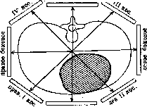
Besides the above-discussed methods of examination, which are routine, more complex methods are also used, which are necessary mainly for scientific research purposes. Such include the construction of plastic Hearts according to the Palmieri method, Sternman's method, and horizontal sections by Sternman's method. To obtain horizontal sections, four orthodiagrams are made in different positions with strict accounting of the angle of rotation of the patient around the body axis. Then, placing each obtained orthodiagram at the angle at which these orthodiagrams were produced, it is possible to transfer to the horizontal plane a 'section' of the organ at any level. Plastic Hearts, i.e., models, are also made orthographically. For this purpose, instead of the needle of the orthodiagraph, a steel string is installed, cutting through a mass of clay or plasticine while the contour of the Heart is being outlined by a point registering the central ray on the screen. Of great interest is the radiocinematography of the Heart. This method objectively records the curves of movement of individual points of the contour of the functioning Heart. Radiocinematography is now used in two forms: 1) according to Gott and Rosenthal and 2) according to Stumpf. In the first method, a lead plate is placed in front of the patient, in which there is a slit 3-4 mm wide. Through this slit X-ray rays fall on the moving film, on which a curve of the pulsating contour of the Heart is obtained. The second method differs in principle in that instead of one slit, multiple slits moving in one direction are used, and on the radiograph new sections of the moving contour of the Heart gradually appear, giving a series of curves by which one can see the nature of the pulsations in graphical representation. The significance of radiocinematography for functional diagnosis is great, and thanks to it it may be possible to resolve some important questions of cardiac pathology.
The analysis of the X-ray image of the cardiovascular mass presents considerable difficulty for the reason that one plane image represents an organ of geometrically irregular shape, with a complex oblique position in the chest cavity. A number of factors creating individual peculiarities and variability in the position of the Heart further aggravate the difficulty of accurate anatomical interpretation of the shadow. However, a number of careful studies of the X-ray anatomy of the Heart in situ by filling its cavities with contrast masses, by outlining the walls of the cavities with metal strips, by inserting pins, etc., have made it possible to sufficiently decipher the anatomical substrate of the shadow in various projections. Precise knowledge of the normal relationships of the cavities allows the examiner to outline these relationships on the orthodiagram, based on the arches of the outer contour, to perform 'animation of the orthodiagram' according to Sternman. In the anterior position (anterior sagittal projection) (Fig. 25) the left contour of the cardiovascular shadow represents 4 arches: aorta,
Fig. 25. Anatomical parts of the Heart in various positions: a-anterior; b-left lateral; c-right oblique; d-left oblique; 1-left ventricle; 2-right ventricle; 3-left atrium; 4-right atrium; 5-aorta; 6-pulmonary artery; 7-superior vena cava.
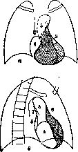
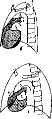
25. Anat. departments of the heart in various positions: a-anterior; b-left lateral; c-right oblique; d-left oblique; 1-left ventricle; 2-right ventricle; 3-left atrium; 4-right atrium; 5-aorta; 6-pulmonary artery; 7-superior vena cava.
pulmonary artery, left atrial appendage, left ventricle. The right contour consists of 2 arches: the superior vena cava (and partly the aorta), right atrium. Thus the boundaries of the proper heart are formed: on the left - by the left ventricle and left atrial appendage, on the right - by the right atrium. The upper and lower edges of the heart merge with the shadows of adjacent organs, i.e., the subdiaphragmatic space and the vascular bundle. To visualize the entire 'cardiac oval', it is necessary to extend the arches of the lateral contours until their intersection. This restoration of the contour according to the principle of 'harmonic development of the curve' by Sternman allows one to outline and measure the area of projection of the entire heart. A significant part of it belongs to the right ventricle, which forms the anterior and lower walls of the heart. The left ventricle, forming only the left edge of the anterior surface of the heart, lies mainly posteriorly, forming the lateral part and part of the posterior-inferior surface of the heart. The left atrium participates in the formation of the left cardiovascular contour only with its appendage and extends onto the anterior surface of the heart, but its main mass is located posteriorly, forming the upper part of the posterior wall of the heart. The right atrium lies on the right, partly forming the anterior and posterior walls of the heart. All the described relationships can be fully visualized on a transparent 'glass' model of the heart (fig. 26). From the different positions of the heart cavities, it is evident that to determine individual cavities, certain projections are optimal. For example, the right ventricle and left atrium are best seen only in oblique projections. The anterior position is therefore not exhaustive, although it reflects changes in the volume of all cavities. The capabilities of the radiological method are not limited to anatomical decoding, which is rightly compared to 'a small biopsy'. X-ray examination also allows one to capture elements of the functional anatomy of the heart ventricles, which clarifies modern diagnosis. As early as Leonardo da Vinci, and after him a number of other researchers, noted the functional isolation and different structure of the two parts of each heart ventricle - the interpapillary and extrapapillary. The first, inlet, part has a trabecular structure of the walls, while the walls of the outlet part of the ventricle are smooth, especially as they approach the arterial orifice. These functional-anatomical divisions correspond to different roles in the hemodynamics of the ventricles (fig. 27). From the venous ring to the apex of the ventricle is the path of blood inflow, while from the apex to the arterial ring is the path of its outflow. The optimal visibility of the functional-anatomical divisions of the ventricle is achieved by using selected projections for each division, when the central ray is perpendicular to it. For the left ventricle: inflow path - anterior sagittal position, outflow path - left oblique position. For the right ventricle: inflow path - left oblique position, outflow path - right oblique position. The other projections and transitional positions, summed up in 'gradual rotation', create a complete spatial orientation in the state of each ventricle. Enlargement of the heart ventricles, according to observations by pathologist Kirch (E. Kirch), proceeds regularly, going retrograde to the blood flow, i.e., starting from the outflow path and subsequently passing to the inflow path (fig. 28). This allows radiologically dividing all ventricular enlargements into 3 types according to their extent. This is presented with radiological symptoms in the table (see art. 273). Enlargement of the atria is observed mainly in oblique and lateral projections. In the right oblique position, the left, resp. posterior, contour of the heart is formed by both atria (fig. 25, c). An enlarged left atrium protrudes into the posterior mediastinum, and with very severe degrees of enlargement, it can reach the right contour. Types of heart ventricular enlargement according to Kudish. Functional-anatomical extent Left ventricle Right ventricle Outflow path Anterior position: elongation and descent of the cardiac apex. Left oblique position: elongation of the cardiac oval. Anterior position: slight increase in the height of the heart, blunting of the cardiac waist by the enlarged pulmonary cone. Right oblique position: protrusion of the pulmonary cone. Lateral position: uniform filling of the retrosternal space with protruding anterior contours of the ventricle and pulmonary cone (narrowing of the retrosternal space). Outflow path + inflow path with predominance of the first Anterior position: elongated wide apex with elevation of the upper border of the left ventricle arch. Left oblique position: protrusion of the left ventricle contour posteriorly, immersed in the diaphragm and forming with it an angle close to a right angle. Anterior position: to the symptoms of enlargement of the 1st type is added the high position of the right atrio-vascular angle, rounding of the contour of the right atrium. Right oblique position: more pronounced protrusion of the pulmonary cone. Left oblique position: protrusion forward of the contour of the right ventricle, immersed in the diaphragm and forming with it a straight or obtuse angle, similar to the left ventricle. Lateral position: protrusion forward of the contour of the ventricle adjacent to the anterior chest wall; shortening of the anterior mediastinum, displacement posteriorly of the also enlarged pulmonary cone. Outflow path + inflow path with predominance of the second Anterior position: same as with the 2nd type, but more pronounced. Left oblique position: sharply protruding ventricle contour approaches close to the spine, forming with the diaphragm a straight or even obtuse angle. The same symptoms as with the 2nd type, but more strongly expressed. Protrusion of the ventricle in the left oblique position predominates over the enlargement of the pulmonary cone in the right oblique position i heart, forming in the anterior picture an additional third arch on the right. The existing concept of the protrusion of an enlarged left atrium in the area of the middle left arch based on the works of Asmann (Assmann) should be considered refuted. The upper part of this arch
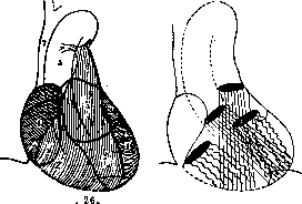
Fig. 26. Spatial relationships of heart cavities ('glass heart model'): 1-left ventricle; 2-right ventricle; 3-left atrium; 4-right atrium; 5-aorta; 6-pulmonary artery; 7-superior vena cava. Fig. 27. Blood flow paths in heart ventricles: straight lines - outflow path; wavy lines - inflow path.
is formed by the pulmonary artery, while the lower part bulges in pathological cases due to the approach and protrusion in the contour of the outlet part of the right ventricle (the so-called pulmonary cone). The right atrium, less frequently involved in the enlargement of heart cavities, in the anterior position mostly protrudes to the right due to displacement by the enlarged right ventricle. At the same time, its arch rounds, and the upper boundary (right atrio-vascular angle) shifts upward. Enlargements of the right atrium itself more often give protrusion of the lower part of the arch, giving the entire heart a somewhat flattened appearance. In the right oblique position, protrusion of the lower part of the cardiac contour is also detected (fig. 25, c). Since the shadows of the lung roots can significantly obscure the posterior mediastinum in the right oblique position, it is sometimes difficult to distinguish the contour of the left atrium and judge its size. Therefore, examination of the left atrium in the left oblique position is more reliable, where, protruding above the arch of the left ventricle, the enlarged atrium can bulge posteriorly or upward (fig. 25, d), partially covering and reducing the so-called 'aortic window'. Thus, the radiological method of heart examination makes it possible to detailed orientation in the anatomical relationships of the cardiovascular complex, which is manifested in the creation of its various configurations. However, a number of constitutional peculiarities in the structure and position of the heart determine the peculiarities of its shape and size. Therefore, it is very important to study the wide variants of normalcy, which
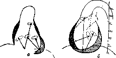
Fig. 28. Enlargement of heart ventricles (a-anterior position; b - left oblique position). Arrows indicate the spread of enlargement along the blood flow paths. Heavy hatching - left ventricle; light hatching - right ventricle.
often
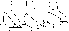
Fig. 29. Three types of heart position: a-vertical; b-diagonal; c-horizontal.
represent borderline states with pathology and complicate the decision regarding the presence of the most important syndromes from a diagnostic standpoint. Depending on the size and shape, and on the ratio of the three main measurements (width, height, and anteroposterior diameter) of the chest, the position of the Heart can vary considerably (Fig. 29). In the long, narrow, and flat chest of an asthenic individual, the Heart usually occupies a vertical position. It is suspended on the vascular bundle and barely touches the diaphragm. Conversely, in the hypersthenic, short, wide, and deep chest, the Heart lies on the diaphragm, often somewhat displacing the vascular bundle upward. The various positions of the Heart can be reduced to 3 types: vertical, diagonal, and horizontal. The angle of inclination of the long axis of the Heart is >45° for the first type, 45-40° for the second, and <40° for the third. The angle formed by the long axis of the Heart with the sagittal and horizontal planes is also different. These constitutional features of the Heart's position create its different projections on the X-ray screen, causing differences in the configuration and size of the Heart. For one and the same area, the three hearts illustrated in Fig. 29 have different transverse diameters, height, etc. Even more important is accounting for the position of the Heart in the same individual with changes of a physiological nature (phases of respiration, pregnancy) or of Latin origin (tumors of the abdominal cavity, ascites, meteorism, etc.). In all cases, changes in the position of the diaphragm strongly affect the position of the Heart, causing its displacements-rotations. Accounting for the rotations of the Heart has been developed by Kienbock and Schick. Displacements of the Heart in three main planes or along three main axes are distinguished. Since the base of the Heart is fixed by the vessels and the mediastinal connective tissue, we are talking about the displacement of the more mobile apex in one plane or another. Displacements in three planes of a Heart of the same size are accompanied by changes in the area of the cardiac projection, the transverse diameter of the Heart, and its height. It should be noted that some displacements of the Heart at first glance simulate the enlargement of individual chambers on the frontal view, and only oblique transilluminations help establish the true nature of the deformation. Displacements of the Heart arising for various reasons usually represent complex combinations of all three types of rotations, which significantly complicates their interpretation. For example, even during inspiration and expiration, a complex combination of rotations creates two completely different pictures of the same Heart. The difference is especially great in the experiments of Müller (see above - functional diagnosis of the heart) and Valsalva (see Valsalva's experiment), reflecting the extreme degrees of inspiration and expiration. And although they sharply change the blood filling of the Heart, a considerable part of the changes in the size and shape of the Heart is due to its rotations. During deep inspiration, the apex of the Heart is significantly displaced downward (in the frontal plane), forward (in the horizontal plane), and the entire Heart is slightly displaced forward (in the sagittal plane). Reverse movements occur during expiration. Special consideration should be given to the displacement of the entire cardiovascular mass together with the mediastinum. Among the causes causing such total displacement, first of all, are the contraction of the lung and pleura, atelectasis of the lung, which draw the mediastinum to the side of the lesion, accumulation of fluid in the pleural cavity, pneumothorax, tumors of the lung and pleura, which displace the mediastinum to the healthy side. Pleuro-pericardial adhesions can cause displacement of the entire mediastinum or predominantly its individual parts, often creating the impression of a pathological deformation of the cardiovascular mass. Finally, it is important to consider the effect of various curvatures of the thoracic spine on the position of the Heart. Large scolioses often displace the entire cardiovascular mass to the side and, by rotating the Heart, create its lateral projection on anterior transillumination. Sharp kyphoses, by elevating the diaphragm, hide the Heart behind it, but most importantly for consideration is the effect of small degrees of kyphoscolioses, which, displacing the Heart only to a small degree, create a picture simulating pathology of the Heart or the vascular bundle. While maintaining a certain regularity of displacement, different types of scolioses often create unexpected deformations of the cardiovascular mass, making it difficult to identify their causes. The mobility of the Heart with changes in body position also has diagnostic significance. Limited displacement of the Heart (resp. mediastinum) is characteristic of adhesive pericarditis. Cases of increased mobility of the Heart (Wandering heart) are also observed. A special chapter in X-ray diagnostics is constituted by congenital anomalies of position (and development) of the Heart. X-rayoscopic examination in these cases allows distinguishing isolated dextrocardias, which are most often inaccessible to diagnosis by other methods except electrocardiography. The study of this rare anomaly (Reinberg and Mandelstam, Rosier) also makes it possible to establish the presence of an inverted position of the heart chambers. Thanks to this, the group of dextrocardias can be classified as follows. Classification of dextrocardia according to Reinberg and Mandelstam: 1) dextrocardia in situs viscerum inversus totalis, 2) congenital isolated dextrocardia with inversion of the heart chambers, 3) congenital isolated dextrocardia with normal relationships of the heart chambers, 4) acquired dextrocardia. Pathology of the Heart is mostly associated with an increase in the size of its chambers, and consequently with an increase in the size of the
N. Figure 30. Measurement of the orthodiagram: a-German method; b-French method. Possessing a number of methods for determining the true size of the projection of the Heart (see Orthography), methods much more accurate and complete than percussion, has long sought by measuring the Heart to find the most objective way to evaluate a healthy and diseased Heart. All existing methods for measuring the projection of the Heart and vessels on the orthodiagram or teleoroentgenogram can be divided into groups: 1) linear measurements, 2) area measurements (planimetry), 3) volumetric measurements (volumometry). The greatest number of methods have been proposed for linear measurement. Of these, we will dwell only on the main ones. 1. The German method (Moritz, Groede 1 and others) (fig. 30, a). From the midline of the body (M) the maximum horizontal distance of the right (Mr) and left (Ml) contours of the Heart is measured, which together make up the transverse diameter of the Heart (Tg). The length of the Heart (L) is drawn from the right atrio-vasal angle to the intersection of the contour of the apex of the Heart with the dome of the diaphragm. The width of the Heart (Wt) is measured by the sum of the perpendiculars dropped onto the length from the points of the right phrenico-cardiac and left atrio-vasal angles. 2. The French method (Lian, Vaquez et Bordet) (fig. 30, b) is based on the principle of chords connecting the ends of the arcs of the cardiac contour; the 4 cardinal points of the cardiac oval B, Dx, G, Gx serve as the basis for constructing measurements. The chords GGx and DGx determine the dimensions of the left and right ventricles; the measurement of the right ventricle also includes DGx, which coincides with the length of the Heart. The perpendicular erected to GGx to the apex of the arc of the left ventricle determines its depth. Finally DDx is the chord of the arc of the right atrium. The line DG is replaced by the measurement from the midline to G of the size of the left atrium, however this measurement has not found application due to the least anatomical substantiation. In addition to the dimensions given, the transverse diameter of the Heart is also measured. It should be noted that a significant defect of the method is obtaining completely different and hardly comparable dimensions of the cavities-GGx = the oblique height of the left ventricle, DGx - the oblique transverse diameter of the right ventricle. The following corrections have been introduced into the method of the French authors: 1) Sternman and his students, and then Arkussky, noted that with a deep breath of the subject, at the apex, one can note the muscle division between the left and right ventricles. The lines GGx, BxGx and BGx are drawn to the point of muscle division a. 2) Arkussky on cadavers established that the edge of the right ventricle in normal condition borders the medial edge of the inferior vena cava, which usually has a diameter of 1 cm. The point Dx is marked by him 1 cm inward from the right phrenico-cardiac angle. In our USSR the question of standardizing the method of linear measurements was discussed by a commission appointed by the I All-Ukrainian Congress of Roentgenologists and Radiologists. The recommended standard method consists of the following measurements (fig. 31). Measurement of the transverse diameter of the Heart (Ts) is made by two measurements of the extreme distances of the right and left contours of the heart (Mr and Ml) from the midline drawn through the center of the body of the II thoracic vertebra. The midline may not coincide with the percussion lin. mediana and this does not allow direct comparison of the right and left sizes obtained by percussion and orthodiagraphically. However, the study of the ratios of the sizes Mr and Ml has to be considered as having lost its significance due to the well-known variability of the influence of individual enlarged cavities of the Heart on these sizes. It is sufficiently known that an enlarged right ventricle can equally increase Ml as well as Mr. In view of this, only the sum of both measurements-Ts is taken into account. The transverse diameter of the lungs (Tr) is measured by a horizontal line drawn between
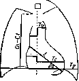
Figure 31. Standard
Method of measuring the anterior orthodiagram of the heart. With two rib arches at the level of the right phrenico-cardiac (cardiohepatic) angle. The height of the chest cavity, presented in reduced measurement diaphragma-clavicula, is measured by the distance between the highest point of the right dome of the diaphragm and a horizontal line passing through the lower edge of the sternal end of the right clavicle. The length of the Heart (L) represents the longitudinal axis of the Heart. It is plotted on the orthodiagram from the right atrio-vasal angle to the most distant point of the apex of the Heart. The angle formed by the length of the Heart (L) with the horizontal (Z.o) determines the position of the Heart in the frontal plane. Measurement of the area of the Heart (after completing its upper and lower boundaries) is done with a planimeter (see Planimetry). In the absence of a planimeter or grid, the following methods are used: 1) cutting out a model of the Heart from millimeter paper, followed by counting the cells or 2) a more accurate weight method: the figure of the Heart cut out of thick paper is balanced on scales with the same paper, cut into perfect squares. Their measurement determines the area of the geometrically irregular figure of the Heart. X-ray measurement of the volume of the Heart represents a more complex combination of linear measurements of the sagittal and frontal orthodiagrams (resp. teleoroentgenograms) and is based on comparing the Heart with regular geometric bodies. A number of methods for measuring the volume of the Heart have been proposed, below we present 2 of them: 1. The method of Rohrer (as well as Kahlstorf a), according to which the Heart is likened to an obliquely lying paraboloid. Its volume V is calculated by the formula: F = 0.63 Pic- Lmax, where Pic is the area of the anterior projection of the Heart, Lmax is the maximum anteroposterior diameter of the heart. The coefficient 0.63 is taken as the average between 0.66 for a sphere and 0.59 for a paraboloid. 2. The method of Sternman a, according to which the Heart is likened to an oblique parallelepiped constructed on the three main diameters of the Heart: V^0,^(Bx-B2-B3), where Bx is the length of the Heart, P2 is the width of the Heart, B3 is the anteroposterior diameter of the heart. Evaluation of the data obtained in roentgenometry presents great difficulties due to the fact that the size of the Heart varies considerably depending on a number of factors. Among the latter are both the constitutional features of each individual and certain physiological influences. Thus, the influence on the normal size of the Heart is exerted by: 1) Sex: in women the Heart is smaller than in men, all other conditions being equal. 2) Height: somewhat larger sizes of the Heart are observed in taller individuals. 3) Age: with age the size of the Heart increases. 4) Weight is a factor with which the size of the Heart is most closely related. A greater weight almost proportionally corresponds to larger sizes of the Heart. However, all 4 of the above factors are only a consequence and part of the general constitution of the organism and of course are not equivalent in individuals of different constitutions. Thus, for example, the tall stature of a hyposthenic, creating prerequisites for the vertical position of the Heart in a long chest cavity, cannot be evaluated the same as the same stature of a hypersthenic. At the same time their weight will also be different. With the same weight of a tall hyposthenic and a small normosthenic with athletic features, the general development of musculature is completely different, which basically corresponds to the development of the muscular organ-the Heart (Dibbelt). V. Müller established that the weight of the cardiac muscle is proportional to the weight of skeletal muscle. Consequently, weight is also not a sufficiently reliable criterion if it is high due to the presence of fat deposits in the body.-Only the sum of these individual characteristics, namely the entire somatic constitution of the object, determines the broad standards within which further correlation can be carried out according to the above-mentioned partial characteristics. Reflection of the general constitution, as already indicated, is the shape of the chest cavity and the corresponding position of the diaphragm. In the presence of certain physiological and pathological influences of neighboring organs, these 2 factors can also change. Thus, deformations of the chest skeleton, already mentioned, high position of the diaphragm in meteorism, pregnancy, ascites, tumors of the abdominal cavity, paralysis of the dome of the diaphragm, etc., significantly influence. There is no need to dwell in detail on such conditions as the position of the body, phase of respiration, phase of cardiac activity, amount of blood in the body, pulse frequency. Finally, it is important to note the significance of professional-social factors, which, having a great influence on the general constitution of the organism, also have essential significance for the partial constitution of the heart. The influence of all the above-mentioned factors is reflected both on the true size of the Heart and to an even greater extent on the roentgenometric sizes, which in turn depend on the change in the position of the Heart. In addition to numerous tables of normal sizes of the Heart (mainly its transverse diameter), correlated with individual of the indicated factors (the main ones of Dietlen, Groedelfl, Otten and others) and the general constitution of the object (Kudish and Lurie), many formulas from the simplest to extremely complex and cumbersome have been proposed. Of the indices having the greatest practical significance, we will mention only a few simple and more or less accurate: the cardio-pulmonary coefficient of Gredel-HLQ - Tr: Tc = 2.00; normal fluctuations by 10% in both directions, i.e. from 1.9 to 2.1. More simply put, the transverse diameter of the Heart in normal conditions is approximately equal to half the transverse diameter of the lungs. The same relationship is expressed in Martin's index, which in percentages determines the ratio Tc:Tr; for normal conditions this index is 45-50%. When fluoroscopy at the usual (about 70 cm) distance, one can use the Kreuzfuchs index, which divides the lower part of the chest into 12 parts, of which in normal conditions 5 belong to the Heart, 4 to the right and 3 to the left lung fields, i.e. the transverse diameter of the Heart is equal to 5/12 of the transverse diameter of the lungs. Unfortunately, both tables and indices of the size of the Heart give for each group significant fluctuations (often up to 50% of their value). This deprives them of diagnostic value precisely in those cases where they would be most important-in cases of initial, slightly expressed pathology. In the presence of sharp changes in size, their obviousness makes fine calculations unnecessary. In conclusion, at present it can be considered that the main value of measurement has been preserved only when accounting for the evolution of the size of the same Heart; this accounting is most accessible when superimposing and comparing repeated orthodiagrams; at the same time, the possibility of detecting displacements of the Heart is more fully achieved. However, neither an increase nor a decrease in the size of the Heart can yet serve as absolute indicators of its functional state, as proven by a number of works and practical observations. In the presence of an increase in the cavities of the Heart, the main question arises, whether it occurred due to an increase in the capacity of the cavities or due to an increase in the mass of the muscle. In other words, is there a passive dilation or hypertrophy of the muscle or both simultaneously. Judgment about this based only on the rounded or elongated shape of the apex is a primitive, unsatisfying the demands of modern clinic. Only the study of the configuration of the Heart and cardiac pulsatory contractions on the screen or preferably on the radiocinematogram provides a certain possibility of functional evaluation of the Heart. Cardiac activity is a product of a number of functions of the cardiac muscle, and their qualification in the roentgenological data makes a significant contribution to the complex of clinical investigation of the cardiovascular system. For the use of roentgenological data, it is necessary to divide the complex of cardiac activity into the active-systolic phase and the passive-diastolic. The characteristic of the observed single contraction of the Heart (systole) consists of its extent in space and time and is included in the following scheme: Amplitude of contraction
Duration of contraction
Z^**^!
(Average speed). , Shallow *---------------------------* Fast Schema of descriptive characteristics of heart contractions according to Kudish; I- tense; II- excited; III- flaccid; IV- small. Four generally accepted names thus acquire quite definite meaning. One can think that each type of contraction corresponds to a certain state of the two main functions of the heart muscle-its contractility and tone. In particular, there are indications that elements of violation of the contractile function of the muscle are reflected in a decrease in the amplitude of contractions, while the tonic function affects the speed of a single contraction, giving in dystonies an accelerated labile contraction. Accounting for the tempo and rhythm of contractions reflects the state of the other known functions of the heart muscle. A separate characteristic of the diastolic phase can note accelerated diastolic filling of the ventricles, which depends on the tone of the muscle, but mainly on violations of hemodynamics with increased blood supply to the ventricles. Not being a direct diagnostic symptom of certain forms of diseases of the cardiovascular system, the character of pulsations can often to a certain extent reveal the nature of hemodynamic disorders, and above all serves as a significant indicator of the state of the heart muscle. These positions undoubtedly gain in reliability with objective radiocinematographic confirmation of the data obtained on the screen during ordinary fluoroscopy. Comparison of the functional-anatomical spread of ventricular enlargement with the character of its contractions makes it possible to judge the hypertrophy or dystrophy of its muscle. Functional tests performed under the screen also come to the rescue. These include-Zehbe's test (symptom of expiratory flattening of the heart on the diaphragm) and Valsalva's experiment (phases of straining and exhalation with compression and then rapid filling of the heart). Thus, the radiodiagnosis of the heart can be broken down into a series of stages of analytical thinking and is based on the following basic principles of diagnostic approach: geometric approach-accounting for planar and spatial features and changes in shape, displacement of axes, violation of absolute values and size relationships; volumetric characteristics of the heart and its fluctuations in connection with the phases of cardiac activity.-Topical orientation-location, displacements and internal dystopias of the cardiovascular mass, changes in relationships with adjacent organs.-Constitutional analysis-accounting for the features of the somatic constitution of the object, the normal partial constitution of the cardiovascular mass and its deviations from constitutional standards and relationships.-Anatomical interpretation-dissection of the shadow mass into its anatomical elements in their spatial relationships.-Functional-anatomical assessment-deciphering of the functional-anatomical sections of the cavities in the anatomical scheme of the heart under study, and qualification of the state of both "blood flow paths; evaluation of existing changes in the sense of their spread along the blood flow paths.-Functional-dynamic analysis--research on the screen or better on the radiocinematogram of the curve of the cardiac contour in the pulsatory phase; evaluation of each phase from the point of view of qualification of the main functions of the heart muscle and violations of hemodynamics; functional tests under the screen.-Diagnostic approach-decision on the norm or presence of pathological changes in the heart and circulatory apparatus; summation of the radiological syndrome of pathology.-Differential-diagnostic analysis-accounting for and revealing external influences, decision on extra- or intracardiac disease.-Nosological interpretation of the syndrome-identification of typical asymmetric enlargements of cavities, characteristic for valve defects, characterization of functional disorders from the side of the heart muscle, fitting into the syndrome of organic or so-called functional disease of the muscle, in the absence or presence of violations of the function of the valve apparatus. The radiological conclusion on the examination of the heart should include an objective-protocol part and diagnostic assumptions or conclusions. The latter is necessary for the reason that the radiologist, specially studying the significance and value of each individual symptom in his field, basing this study on broad clinical and patho-anatomical comparison, not only can but is obliged to make all possible objective conclusions from the data of his study. It is especially important in cardio-radiology to correlate the data of all various methods of clinical research, and although in each case the diagnostic significance of one of the methods may dominate, the diagnosis derived from the sum of all data belongs to the clinic with all its research methods and to the specific patient with all his morphological and functional individual characteristics of his body.
S. Reenberg, V. Kudish and B. Stern. XI. Syphilis of the Heart. It is necessary to differentiate cardiovascular lesions in the so-called secondary syphilis from syphilitic lesions of the late (gummatous) period, which represent a special case of visceral lues. Statistical data. There are no direct indications of the prevalence of syphilitic lesions of the cardiovascular apparatus among the population. An indirect judgment can be made from the following calculations: almost 3Д of all aortic valve insufficiencies are of syphilitic nature; in turn, syphilitic aortic valve insufficiency and aortic aneurysms (according to Romberg's statistics) constitute */4 of all organic cardiovascular diseases (see Syphilitic aortitis). If one takes into account the rarely diagnosed initial forms of syphilitic aortitis, then the lesions of the heart muscle, conduction system, and coronary vessels not included in the above calculation (14% of angina pectoris - on the basis of syphilitic coronaritis), then the share of syphilis in the etiology of cardiovascular lesions must be recognized as very high. According to the statistics of Chiari, Fahr, Frankel and others, cardiovascular phenomena in known syphilitics are found in 40-80%. Wiesner, Rasch in newborn syphilitics found typical mésaortitis in 67.4%. Thus, the cardiovascular apparatus can be affected by syphilis already within the nearest year after infection (Pletnev, Pulay). There are well-documented cases where only 50 years after infection an aortic aneurysm was discovered and developed rapidly (Ktilbs). According to Donath, Weintraud and others, vascular syphilis manifests on average after 20 years (from 5 to 40), usually falling in the age range from 30 to 60 years (Stabler and Fukushi). Men are affected more often than women (according to Pletnev in the ratio of 2.8:1), which according to the author depends on the greater abuse of tobacco and alcohol by men. Among other manifestations of visceral syphilis, lesions of the cardiovascular system occupy a dominant position: according to the statistics of Petersen, Chiari and Stolper, they are equal in frequency to syphilis of the brain and spinal cord combined (Breitman), and the disease of the coronary vessels of the heart ranks first after syphilitic lesions of the cerebral arteries (Breitman, Stolper, Runenberg). In order to establish the syphilitic etiology of heart diseases, it is necessary to know that in persons suffering from progressive paralysis, specific vascular lesions occur in 74-82%, in tabetics - in 24-39%, in neurosyphilitics in general - in 40^5%. In stage II of syphilis, along with skin and mucous membrane eruptions, general phenomena are found indicating involvement of the cardiovascular apparatus. Along with general malaise, patients complain of shortness of breath, pain in the heart area, palpitations. Objectively, various forms of rhythm disturbance are found. Fournier considered sinus tachycardia and respiratory arrhythmia to be constant and early symptoms of secondary syphilis and explained it mainly by lesions of the nervous apparatus. P. Marie and Delpe at this time speak of syphilitic Basedowism (acute thyroiditis). Hallopeau recognized the possibility of lesions of sympathetic fibers. Bradycardia was observed less frequently. Extrasystole, which passes into paroxysmal tachycardia, is also not often encountered. Atrial fibrillation has not been registered even once (Pletnev). As for transient blockades of various parts of the conduction system, their detection requires repeated and systematic electrocardiographic recordings. Arterial hypertension was described as a rare finding. Hypotension with a fall in pulse pressure is much more common, which, in Pletnev's opinion, could be explained by the influence of the syphilitic virus on the autonomic (vasomotor) centers, the hormonal system (especially the adrenal glands), as well as on the Heart. The question of acute myocarditis in stage I of syphilis cannot be considered resolved: moderate heart enlargement, a soft systolic murmur at the apex are more often found in developed anemia, on account of which they could be attributed. However, it is impossible to deny the specific intoxication of the myocardium, similar to that observed in typhoids, for example, the temporary deformation of the T wave of the electrocardiogram (although Turner-White denies this possibility). The presence of acute coronaritis cannot be rejected: T. P. Pavlov found quickly transient changes in the heart vessels. In the so-called tertiary period of syphilis, the entire cardiovascular apparatus is involved in the lesion: all membranes of the Heart, aorta, coronary vessels, peripheral arteries, capillaries and veins. Changes in the aorta are the most common companion of late syphilis (see Syphilitic aortitis and Aortic aneurysm). Lesions of the inner membrane of the Heart. The endocardium is involved in the lesion secondarily: 1) either from the side of the aorta (ascending 'valvular' aortitis) 2) or from the side of the myocardium, which is always affected (gummas, infarction, diffuse changes). The valvular endocardium is affected less often than the parietal. The fibrous form of the lesion of the latter is more often observed in the left ventricle (near myocardial gummas), above the papillary muscles, involving the tendinous cords as well. Gummatous formations are usually located on the septa of the ventricles and atria, more often, as with fibrosis, gummas occupy the left ventricle, serving upon their disintegration as a source of embolism in the systemic circulation. From the valve apparatus, due to the spread of the syphilitic process from the aorta, and sometimes from the myocardium, the aortic valves are mainly affected. Lupa distinguishes 3 degrees of their changes: 1) macroscopically undeterminable, 2) thickening of the base of the valves without insufficiency and 3) thickening with wrinkling of the edges and insufficiency of the valves. Less often, nodules are scattered over the surface of the valves in very large numbers (Voloshin). In general, the wrinkling does not reach such degrees as occurs in rheumatic endocarditis. As for the etiology of aortic valve insufficiency, as early as 1880 Lancer and Dieulafoy pointed to the syphilitic nature of aortitis with involvement of the valves. Subsequently, this etiology was recognized as the most common (Scott-94%, Lambert-75%, Pletnev-315 out of 342 cases). However, other authors (Libmann, Schottmuller) warn against underestimating the rheumatic etiology. Spirochetes, often found in the aorta, are not found on the valves. The mitral valve, although much less frequently, is also affected by the syphilitic process. Neumann describes grayish or yellowish-white fibrous nodules along the line of closure of the valves. Gouche does not at all consider the involvement of the mitral valve rare. Brooks in 74% of cardiac lues found involvement of the endocardium, and in 34% both the aorta and the mitral valve were affected. One must always keep in mind the possibility of relative insufficiency of the mitral valve depending on the lesion of the myocardium and in particular the papillary muscles (Virchow, Mracek). The localization of the process at other openings is rare. The occurrence of endocardial lesions belongs to late syphilis, many years after the moment of infection, although as an exception, valve defects were also found in the early period of lues. The symptomatology of syphilitic insufficiency of the aortic valve has some peculiar features. It must be borne in mind that a diastolic murmur over the aorta occurs also in aortitis without valve insufficiency and in their relative insufficiency due to aneurysm of the ascending part; the same applies to pulsus celer. Gausmann considers the absence of such a significant drop in diastolic pressure as is often observed in rheumatic insufficiency of the aortic valves characteristic. The latter phenomenon could be explained by the relatively limited destruction of the valves (Neumann) and the disturbance of the tone of the peripheral arteries (resp. arterioles)* Some authors (Zelenin) noted in syphilitic insufficiency a drop in diastolic pressure to zero. Differential-diagnostic value is the variability of murmurs, especially in connection with specific treatment, as well as the presence of anginal pains (with frequent combination with supravalvular aortitis) and various forms of blockade (see below). Romberg considers marked tachycardia characteristic. Lesion of the myocardium. The myocardium is as a rule affected secondarily: the process involves the coronary vessels (endarteritis luetica), along which (in the hearts of newborns) foci of infiltration are noticeable; in the muscle, fibrosis develops or gummas form. They are most often located in the left ventricle. The right Heart is affected (according to Breitman) in congenital or acquired syphilis of the pulmonary artery, in syphilis of the lungs, in aneurysm of the aorta compressing the a. pulmonalis, the right atrioventricular opening and the right atrium, in aneurysm of sin. Valsalvae. The atria are also affected in syphilitic mediastinitis. Gummas are most often single, sometimes multiple, varying in size from a millet seed to a billiard ball.
Sometimes they are isolated (surrounded by a ring of dense connective tissue), but sometimes they have a tendency to grow, involving both the endocardium (see above) and the epicardium. Softening and being replaced by scar tissue, gummas give rise to partial aneurysms of the Heart. More significant aneurysmal protrusions occur only after myocardial infarction on the basis of occlusion of a branch of the coronary artery. Here it should be mentioned that as a rule the vessels supplying the left ventricle are occluded. Symptomatology. In addition to the general symptoms characteristic of myocarditis and cardiosclerosis (see), it must be borne in mind that due to the most frequent localization of the process in the left ventricle, phenomena of predominantly left ventricular failure are observed: attacks of cardiac asthma, left ventricular extrasystole in the absence of peripheral circulation disorders. Sinus tachycardia, once it has arisen, usually has a tendency to progress. Of other forms of rhythm disturbance, the most suspicious for syphilitic etiology is atrioventricular block, which in young subjects almost always develops on the basis of gummatous accumulations in the myocardium along the conduction system (see below). Atrial fibrillation and puls. alternans have also been observed. For the semiotics of myocardial infarction and Heart aneurysm, see above, as well as Myocarditis and Aneurysm of the heart. Nicolaï (Ni-colai) observed very low electrocardiogram waves in syphilitic Heart disease. Significant improvement of all symptoms under the influence of specific therapy sometimes turns out to be the only basis for the diagnosis (ex juvantibus), since the Wassermann reaction does not always give reliable indications. Pericardial involvement. In view of the fact that changes in the pericardium usually occur secondarily when the process spreads to the muscular layer, the visceral leaflet is more often affected, and extensive lesions of the pericardium are encountered less frequently, only on limited individual areas (roots of large vessels, anterior surface of the heart, apex). Dry fibrinous pericarditis was observed by Ricord and Virchow. The gummatous form is less common. In fibrous or adhesive pericarditis at the root of large vessels, their compression and generally significant deformation of the Heart can occur. Complete obliteration of the pericardial cavity with miliary gummas in the strands was described by Jurgens. Balzer observed a large number (approx. 30) saccular aneurysms of pericardial vessels along the anterior branch of the coronary artery. Recognition of syphilitic pericardial involvement is not easy, since the friction rub is rarely audible; if it is determined, especially at the base of the Heart or there are systolic intercostal retractions with other signs of syphilitic Heart disease, a specific pericarditis can be assumed. Lesion of the nervous apparatus of the heart and conduction system. The question of changes in the intra- and extracardiac nervous apparatuses of the Heart has not been sufficiently clarified. As for the conduction system, single or multiple gummatous, sclero-gummatous, and sclerotic changes have been observed, both in the area of the interventricular septum (Monckeberg, Aschoff) and in the vessel supplying the septum (a. septi). Depending on the spread of the process, either only the common trunk of the His bundle, or one of its 'legs', or the trunk and leg together are involved. If a gumma or scar involves the conduction system, restoration of conductivity is impossible; but if the bundle is only compressed by a gumma, specific therapy can restore conductivity to normal. Gummas located near the terminal branches of one or the other leg, being a source of extraventricular excitation, can cause persistent extrasystole, more often left ventricular (see above). Lewis considers 25% of blocks to be of syphilitic nature. (For the symptomatology of block, see Heart block.) Block in the lesion of the main trunk (atrioventricular block) can be recognized even without electrocardiographic recordings, which are absolutely necessary for the recognition of leg block, often accompanied by gallop rhythm. It should be noted that neurogenic (transient) blocks with attacks of fainting and complete arrest of the ventricles can occur on the basis of syphilitic lesions of the cerebral vessels (Zelenin). Pancarditis, panaortitis, lesions of peripheral arteries, veins, and capillaries. From what has been stated above, it is clear that syphilitic lesions of the cardiovascular apparatus never limit themselves to any one department, but as a rule involve to one degree or another both the coverings of the Heart, and the aorta, and the coronary vessels, sometimes giving a very complex clinical and patho-anatomical picture. Peripheral arteries are involved in the process (endarteritis obliterans) usually later than cerebral and coronary vessels; gangrenous decay of tissues can stop under the influence of specific therapy. Syphilitic phlebitis and periphlebitis, as well as changes in capillaries (Lukomsky), are rarely described, more often in the early period of syphilis. Syndromes and minor signs. As already mentioned, the presence of cardiovascular disorders in neurosyphilis should always make one think of specific lesions of the Heart. Direct indications for poorly treated syphilis and, in particular, indirect indications for lues ignota (unnoticed infection) should also be evaluated in the same way: scars, changes in the bones of the leg, saddle nose, perforation of the soft palate, etc. In congenital syphilis, the myocardium is most easily involved in the disease. Spirochetes were found both in the Heart and in the vessels. In congenital syphilis of the Heart, all the pathological conditions discussed above in acquired syphilis in adults have been found: single and multiple gummas, sclerosis of the myocardium (cirrhosis), coronaritis, phlebitis and capillaritis with hemorrhages into the cardiac muscle and pericardium, aneurysms of the Heart (Barlow).- Developmental defects. Heart defects of syphilitic origin were described by Virchow, Lancerot, and others: non-closure of the interatrial and interventricular septum together with narrowing of the pulmonary artery orifice. Nobecur encountered syphilitic etiology relatively rarely (in 19 cases of congenital defects, only 2 times). Unlike the findings in acquired syphilis, in congenital syphilis valvular changes of the right heart (tricuspid defect) and a very rare defect in adults, namely narrowing of the left atrioventricular orifice, as well as aortic stenosis, which does not occur in acquired syphilis, have been found. Ambard, on the basis of RW data, is ready to consider most cases of mitral stenosis as having developed on the basis of congenital syphilis. The so-called disease of Duriez (congenital mitral stenosis) Schlesinger tends to associate with intrauterine syphilis. Sometimes the correct diagnosis is made only at autopsy. Myocardial involvement in very small children is often discovered only by significant cyanosis (Breitman). The symptomatology of this condition in adults differs in no way from that discussed above. For confirmation of the diagnosis of congenital disease, the simultaneous existence of general underdevelopment and various endocrinopathies is not without significance. Treatment. To expect benefit from specific therapy is possible only in the presence of a gummatous process and in cardiovascular lesions in the early stage of syphilis. Where a scar has already formed, anti-syphilitic treatment is powerless. As for the choice of drug and persistence in its application, the following rules should be used. In case of significant circulatory insufficiency, it is necessary with cardiac and vascular agents (Digitalis, Adonis, Diu-retin, etc.) to achieve equalization of circulation (compensation). It is best to start, even in the absence of noticeable circulatory disorders, with iodine (Natr. jodatum 10.0-15.0 xx 200.0; 2-3 tablespoons for 3-4 weeks depending on the patient's tolerance to it) or with Biett's mixture (Hydrargyri bijodati 0.1; Kalii jodati 8.0; Aq. destill. 200.0; 2-3 tablespoons, 3 portions). Mercury ointment rubs (frictions) are also quite well tolerated. Among the drugs that little burden the cardiovascular apparatus is bismuth (Bijochinol) (15 injections of 2.0 every other day). There is no unanimity regarding salvarsan (Neo). Ehrlich himself considered it contraindicated in cardiovascular diseases. At present, salvarsan is especially recommended in aortitis and aortic valve insufficiency. In pronounced myocardial cirrhosis, in sclerosis of peripheral arteries and attacks of angina pectoris, extreme caution must be exercised. According to Schlesinger, salvarsan can cause an anginal attack; Schotmüller, on the contrary, did not see bad results. In poorly treated syphilis, the course of treatment must be repeated 3-4 times with intervals of 4-6 months. Caution in the use of particularly energetic treatment is based not only on the fear of causing cardiovascular insufficiency due to intoxication by one or another drug, but also on the fact that rapidly disintegrating gummas on the endocardium can become a source of embolism, while in the muscular layer in the absence of a stable scar, they can lead to acute aneurysm of the Heart. The view has been expressed on the possibility of late complications from the cardiovascular apparatus (aortic aneurysm, etc.) with energetic treatment of syphilis.
Schlesinger blames mainly salvarsan (the salvarsan era) and therefore uses this remedy with particular caution: starting from 0.05 to 0.075, rarely up to 0.3. Stovarsol was tested with a favorable effect (0.25, from 1 to 3 tablets for three days; with intervals of three days, 25-30 tablets). Schlesinger again proposes the almost universally abandoned Decoctum Zittmannii (no. 100.0-200.0 g pro die for many months). Zelenin uses mercury preparations because of their diuretic effect even with moderate circulatory disorders (edema).-Should one aim for the disappearance of RW in specific therapy? According to Krulle, the rapid disappearance of RW indicates the destruction of antibodies and a weakening of the body's reactive capacity. The opposite phenomenon should also be kept in mind: the appearance of RW after the use of mercury and iodine ('provocation'), which can be used in suitable cases to establish etiological diagnosis. I cannot consider Schlesinger's opinion that a positive effect from antisyphilitic therapy does not yet indicate syphilis (denial of the diagnosis ex juvantibus) as justified. Prognosis. If in early syphilis the prognosis is generally favorable, in late syphilis it is generally unfavorable. Although Breitman's data on sudden death (mors subita), occurring in 33-50% of patients with syphilis C, are exaggerated, the danger of a sudden fatal outcome is always very great. Yuchar in 35 cases of sudden death found ruptures of the aorta, rupture of gummas of the heart muscle into the ventricles, embolism of the carotid artery, ruptures of heart aneurysm (after myocardial infarction), etc. Pletnev points to the possibility of a sudden interruption of conduction through the His bundle and disorders of coronary circulation (occlusion or spasm). The overall clinical picture determines the prognosis: frequent attacks of cardiac asthma and severe anginal attacks greatly darken the prognosis, which is aggravated by the inversion of the T wave of the electrocardiogram, especially the electrocardiographic picture characteristic of blockage of the terminal branches of the conduction system (see Heart block).-Work placement is based on the accommodating ability of the cardiovascular system (see functional diagnosis).
V. Zelenin. XII. Surgical diseases of the heart. Injuries to the heart belong to rare observations and therefore it is understandable that individual reports usually concern only isolated cases. There are almost no large series of observations from the same institution. F. Hesse (Friedrich Hesse, Dresden) and Costantini described 6 cases each, Borzymowsky-5 cases, Rychlik-7 cases, Ranzi (Eiselsberg)-13 cases, Janellidze-13 cases, Hesse (Obukhovskaya Hospital, Leningrad)-48 cases. The largest collective statistics belong to Janellidze, who from 1896 to 1921 collected 535 cases of heart suture and analyzed the entire question in an extensive monograph (1927). At present, about 600 cases of heart injuries have been published. The largest number of operated heart injuries falls on the USSR (109 cases, of which Leningrad ranks first-69 cases; of the last 48 cases, 48 were operated on in the Obukhovskaya Hospital and published by Hesse). Second in number of cases is Germany (103 cases), followed by France (83 cases), Austria (50 cases), USA (46 cases), Finland (19 cases), Poland (15 cases), England (11 cases), etc. According to Janellidze, stab-incised wounds were most often observed (72.7%); in 24.8% the injury occurred after gunshot wounds; in 0.5% lacerated wounds were noted. Stab wounds with a 'col' (Borchardt) were observed least often. In 32.2% it was a suicide attempt, in 62.6% the wounds were inflicted by other persons, and finally in 5% they were the result of an accident. Half of the suicides used firearms to inflict the wound, while wounds inflicted by other persons were most often (87.3%) stab-incised wounds. Statistics from peacetime show that gunshot wounds to the heart are observed much less frequently than stab-incised wounds. This is explained by the fact that gunshot wounds to the heart and especially with modern pointed bullets in most cases lead to fatal outcomes at the site of injury. That is why surgery of heart injuries did not become widespread in armies participating in the imperialist war (Fromont, Haecker, Hesse, Janellidze). On the other hand, the number of forensic autopsies after gunshot wounds to the heart is quite significant. The pathological literature on gunshot wounds to the heart under wartime conditions was collected by Giercke (1920). Injury to the heart without injury to the pericardium was observed in 3.03% of all gunshot wounds to the heart (Luxembourg). According to Janellidze, injuries to the atria occurred in 12%, to the ventricles in 88%. The left ventricle is injured more often in gunshot wounds (68 cases of left ventricle injury, in 30 cases of right ventricle injury). In stab-incised wounds, both ventricles are injured equally often (in 167 cases the left and in 153 cases the right). In 92.5% the wound penetrated the cavity of the heart. Wounds perforating the entire thickness of the heart are more often observed in gunshot wounds (27.8%), which explains their more serious prognosis. Much less often perforating wounds in stab-incised injuries (5.6%). Injuries to the posterior wall of the heart were often overlooked, and patients died from bleeding. In one case, several hours after the first operation, it was necessary to resort to a repeated operation because the bleeding continued, and a wound on the posterior surface of the heart was found and sutured. Injuries to the coronary vessels were observed in 7.7%. In 41 cases the coronary arteries were injured, in two cases the veins. In 85.3% the left coronary artery was injured, most often its descending branch. Injury to the valves and septa of the heart quickly leads to death. Of neighboring organs, the pleura was injured in 90%, the lungs in 17.6%, and the diaphragm and abdominal organs in 5.6%. According to the location of the heart injury, Janellidze distributes the material of the collective statistics as follows: Hesse, based on the material of the Obukhovskaya Hospital, gives the following figures: Left ventricle. Right. Right atrium Left
» . 3 » (6,к%) 1 » (2%) Wounding of the left ventricle with its thicker walls and better ability to adhere gives a better prognosis, while the highest mortality is observed with damage to the thin-walled atria. The time elapsed from the moment of wounding to the operation is very important for prognosis. Based on Hesse's material, this is particularly clear: with a period of up to 4 hours, out of 26 cases, 12 died = 46.1%; after 4 hours, out of 7 cases, 6 died = 85.7%. According to the collective statistics of Danelidze, mortality in cases operated on within 1 hour after wounding was 48.8%, and after 1-4 hours - 51.5%. The earlier help is provided, the better the result. But cases have been described where surgical assistance was provided after several days and still successfully. Diagnosis of heart wounds. Wounds of the Heart give an extremely confused picture. In essence, there are no typical signs of a wound of the Heart. Wounds of the lung and even of the medium-sized arteries of the chest cavity can give a picture that makes the surgeon think of a wound of the Heart. The following symptoms should be noted: 1) subjective sensations - a feeling of fear, pain in the area of the Heart, a feeling of pressure, anxiety, weakness, dizziness, shortness of breath, radiating pains in the abdomen. These signs are extremely variable and are often masked by loss of consciousness (50% according to Hesse; 53.6% according to Danelidze) or are dulled by the drunken state of the wounded (25%). Based on Hesse's material, the general condition was severe in 42 cases and satisfactory in 6 cases. Cases were observed where patients walked to the hospital on foot. The gradual deterioration observed in 12.5% is of very great importance. The above-mentioned radiating pains in the abdomen can reach a considerable degree. In 10 cases (out of 21), the pains were so sharply expressed that they forced the surgeon to resort to laparotomy. The pains are explained by damage to the intercostal nerves and the direct muscles of the abdomen. In some cases, the pains are explained by phenomena of congestive liver, caused by cardiac weakness. 2) Phenomena of shock, which can be the result of reflex of the epicardium or the result of bleeding. 3) The most valuable symptom are the signs of increasing internal bleeding and its consequences. Strong external bleeding from a wound of the Heart is observed very rarely (in 22.8% according to Hesse). Wounds of the Heart usually bleed systolically, less often diastolically. External systolic bleeding is observed extremely rarely. In 5.2% of all cases of Heart wounds, there were not even signs of anemia (Danelidze). The pulse is usually rapid (100-120) and of slight filling. Often the pulse is arrhythmic and not palpable. Pulsus differens cannot be interpreted as a symptom of cardiac wounding. From the literature data, it is seen that most often in Heart wounds, signs of severe internal bleeding are observed. Nevertheless, in a number of verified operations for Heart wounds, there were no signs of internal bleeding. This is explained by the fact that sometimes the muscles of the Heart, the fibers of which go in different directions, quickly shift, and there can be cases of Heart wounds in which not a single drop of blood will come out. Blood from the wounded Heart can pour out, with wide communication between the pericardial cavity and the pleura, into the latter cavity. Then phenomena of hemothorax will result. Based on Hesse's material, this was observed in 70.8%. These cases are especially difficult to differentiate from severe wounds of the lung and blood vessels of the chest cavity (aa. mammaria interna and intercostales). If communication between the pericardial cavity and the pleura does not exist or is small, blood pours out into the pericardial cavity and phenomena of hemopericard result. In these cases, the diagnosis of Heart wound is facilitated, since the outflow of blood into the pericardial cavity causes phenomena of cardiac compression or, according to old nomenclature, "tamponade of the Heart" (Rose). It must be borne in mind that only accumulation in the cardiac sheath of more than 150 cm3 gives an increase in cardiac dullness, determined by percussion. Sometimes phenomena of pneumothorax and hemothorax can influence the boundaries of cardiac dullness, for example, increasing left-sided pneumothorax can shift the Heart to the right. When blood flows into the pericardial cavity, the accumulating blood produces pressure on the thin-walled right atrium and the vena cava. At the moment when the intrapericardial pressure exceeds the pressure of the vena cava and atria, which is usually observed with an accumulation of 400 cm3 of fluid, bleeding from the wounded Heart ceases - the Heart wound is "tamponaded", but due to strong compression of the right atrium, the access of blood to it from the vena cava ceases. "Tamponade" of the Heart can be acute with rapid outflow of a large amount of blood. In such cases, patients quickly die. But the same phenomenon can have a subacute character with slower outflow of blood. In such cases, the pericard manages to stretch or the blood partially flows out into the pleural cavity. The clinical signs of "tamponade" of the Heart consist in a feeling of pressure on the Heart, pains in the Heart, left shoulder and epigastrium and a feeling of fear. The pulse becomes arrhythmic and then disappears. Severe shortness of breath sets in. Engorgement of the jugular veins and cyanosis of the face are determined due to stagnation in the veins of the great circulation. For the same reasons, engorgement of the liver is observed. Rapidly appearing "tamponade" of the Heart can kill the wounded person, but slowly increasing due to stopping of bleeding can save life. "Tamponade" of the Heart is the most reliable sign of Heart wound, but it sometimes occurs with isolated wounds of the pericardium, with wounding of pericardial vessels with intrapericardial hemorrhage. Hesse observed "tamponade" in 37.5% of his cases, Danelidze observed the same phenomena more often (69.2%). In cases of cardiac "tamponade", the most urgent intervention is indicated. When freeing the pericardium from blood, cardiac activity can resume. Hesse, in the conditions of the GPMГ where cardiac suture could not be considered, punctured the pericardium and obtained improvement in cardiac activity. 4) Acoustic phenomena on auscultation are considered conclusive for Heart wound and are explained by the presence of blood and air in the pericardial cavity. Thus is explained the "mill wheel" murmur, in which buzzing, friction and noise as if of bursting bubbles are determined. But experience (Lerlche, Baudet, d'Ombredanne, Hesse and others) showed that this sign is not always indicative, since phenomena of pneumothorax and hemothorax create favorable conditions for the occurrence of murmurs. Thus, it is not always possible to distinguish extrapericardial murmur from intrapericardial. The tones of the Heart undergo changes. Usually the tones, due to the appearing layer of blood between the Heart and the ear of the examiner, are weakened (in 72.9%, Hesse), but here also hemothorax and pneumothorax can mask or intensify this phenomenon. 5) The external wound in injuries of the Heart can occupy a considerable area: in stab-incised wounds from II to "VIII rib horizontally and from the left axillary to the right mamillary line vertically. The wound lay outside the projection of the Heart onto the chest according to Simon in 38%, according to Hesse in 43.7% and according to Danelidze in 57.7%. In gunshot wounds, an even wider area is possible. Wounding of the Heart is possible from behind through the posterior mediastinum and from the side of the abdominal cavity (Fuchsig). On the other hand, wounds in the area of the Heart very often do not penetrate into the latter. Probing, proposed by Rehn for determining the direction of the wound canal, is strictly contraindicated. In gunshot injuries with entrance and exit wounds, a conclusion about the course of the wound canal is within the realm of possibility. Wounding of the right ventricle usually occurs from the left half of the chest cavity, less often the external damage lies on the right. The left ventricle is wounded exclusively from the left half of the chest cavity. The right atrium is wounded more often from the left. The left atrium can be wounded exclusively from the left and most often through the III intercostal space. 6) Examination with X-rays sometimes can help, but it is indicated mainly in determining foreign bodies. The symptom of immobility of the cardio-pericardial shadow is important. X-raying in different directions for the freshly wounded is very painful and can lead to collapse. A severely wounded person with suspicion of Heart wound does not belong in the X-rayг cabinet but on the operating table. Thus, there are no absolutely reliable signs of Heart wounding. The diagnosis must be made based on the combination of symptoms present. Almost always some signs are present, while others are absent. They appear in varying degrees and in different combinations.
It is often difficult to determine what portion of the damage is attributable to the Heart and what portion to the opened pleura and the wounded lung. In any case, with increasing experience, the number of incorrect dia- Heart
The diagnosis is made in 48% of confirmed cases of cardiac injury operated on, not made in 24%, cardiac injury suspected in 28%. For 1911-18, the diagnosis was made in 65.2%, not made in 17.4%, cardiac injury suspected in 17.4%. Zeidler (1911) held the view that the diagnosis of a wound of the Heart is impossible and therefore any wound of the chest should be expanded. Modern surgery poses the question differently and at present we operate only for certain indications, carefully monitoring the combination of individual symptoms. In unclear cases, operation is considered indicated when there are signs of increasing internal bleeding. The source of bleeding must be found in such cases regardless of whether the Heart or one of the other organs is wounded. Treatment. Until 1896, treatment of wounds of the Heart was strictly conservative and gave 10% to 15% recovery (Fischer). At present, operative intervention, giving 40% to 50% recovery, is the method of choice. The operation of cardiac suture is a relatively complex intervention and requires composure on the part of the surgeon and impeccable asepsis. This operation can only be performed in a well-equipped hospital institution and by an experienced hand. The pleura and pericardium belong to the most infection-sensitive tissues and not a small part of patients, who have well tolerated the operation itself, die from purulent pleurisy and pericarditis. Although in the literature there are a whole series of cases operated on under the most unfavorable conditions (Wendel, Tedesco), one should still strive to operate on cardiac wounds only under favorable conditions of a modern operating room. Under wartime conditions, cardiac suture is hardly feasible on a field hospital. Cardiac suture in most cases was performed under anesthesia. Local anesthesia is often insufficient. With chloroform anesthesia, recovery was obtained in 44.2%, with ether anesthesia - in 53.3%, therefore the latter is preferable. For access to the wounded heart, an innumerable number of methods have been proposed. For example, Italian surgeons alone have proposed more than 20 methods. All operative methods should be divided into 2 groups: 1) typical methods with the formation of bone-musculo-cutaneous or at least cutano-muscular flaps (Fontan, Delorme, Ninni, Rotter, Kocher and others); 2) methods of individual expansion of the wound along the course of the wound canal with resection of one or several ribs for convenient access to the heart (Zeidler, Grekov). The results speak in favor of the application of simple expansion. More complex flap methods gave 41.6% recovery, the method of layered expansion 46%. The layered method is less traumatic. In 10% it was possible to limit oneself to the resection of one rib, in 24.7% - two ribs. The layered method also has the advantage that it guarantees against overlooking injuries of neighboring organs. Based on Hesse's material, the lung was wounded in 29%. One pericardiotomy without opening the pleura, even in case of injury to the latter, unfortunately cannot be applied because it is impossible to exclude injury to other organs (lung, diaphragm). On the other hand, not quite definite diagnosis of wounds of the Heart forces one to operate by expanding the primary wound, in other words, in the vast majority of cases - through the pleura and this is the greatest danger of this intervention. The Heart cannot be pulled into the wound. Grasping the Heart with instruments is unacceptable. If possible, a cardiac suture should be applied to the Heart in its normal position (in situ). It is necessary to operate as bloodlessly as possible. The most severe bleeding is observed from wounded atria. The left ventricle bleeds the least. For de-vascularization of the Heart, temporary clamping of the hollow veins at the point of entry into the right atrium is recommended (Rehn, Hacker, Lawen, Sievers, Carrel). This procedure is tolerated for 9-10 minutes. Sauerbruch recommends clamping the right atrium. If the operation is performed using a high-pressure apparatus, it is recommended to stop the pneumopressure at the moment of applying the suture. Then the lung collapses and takes in more blood. The application of the cardiac suture should be performed at the most convenient moment for this. A knotted silk suture should be preferred. The auricle of the Heart can be successfully ligated in case of injury. The most unpleasant complication is the cutting through of sutures, especially dangerous in alcoholics with degeneration of the heart muscle. Sutures should be tightened carefully and not too tightly. In case of sutures cutting through, it is necessary to place the sutures further from the edge of the wound and deeper. Sometimes a mattress suture can help. In case of failure, free transplantation of fat, fascia, muscle (Lawen and Juracz) and pericardium should be used. In case of cardiac arrest, cardiac massage is recommended. When using this procedure, a complete effect was observed in 9.5%, partial in 16.6%, temporary in 24% and complete failure in 50% (Dzanelidze). One should not forget about artificial respiration. Some authors used intracardial administration of saline solution, but in 10 published cases this procedure was not successful. Blood transfusion has a great future in cases of Heart wounds. Unfortunately, despite the widespread use of this method in general, it has so far been applied only in exceptional cases. Here it is possible to use reinfusion of blood that has flowed into the pericardium and pleura or transfusion of blood by one of the usual methods. After suturing the wound of the Heart, the pericardium and pleura should be sutured tightly. Smooth course of wounds was observed only in 9.5%. Usually complications were observed. In 54%, complications with infection of the pleura and pericardium occurred. Of 299 fatal cases, death at the operating table was noted in 62 (20.7%). During the first day, 79 patients died (26.4%), during the first week - 92 patients (30.8%), second week - 31 patients (10.4%), third week - 30 patients (10.1%). The cause of death in 299 patients was as follows: blood loss and shock - 150 patients (50.2%), secondary bleeding - 6 patients (2%), purulent pleurisy and pericarditis - 40.5% (Dzanelidze). The prognosis also depends on the age of the patients: up to 20 years - 56% recovery, from 20 to 40 years - 45% and from 40 to 60 years - 32%. Women tolerate cardiac wounds better. Mortality among women is 50.8%, among men 54.7%. The larger the wound of the Heart, the worse the prognosis. Wounds up to 2 cm give 45.6%, wounds larger than 2 cm only 26.6% recovery. In cases where the wound reached a length of 5 cm, there was not a single recovery. In cases of simultaneous injury to organs of the abdominal cavity, only one case of recovery (Yushkova). In blunt injuries and ruptures of the Heart, operative intervention has not yet been used and can hardly count on success. Sometimes pathological ruptures can occur at the site of hemorrhagic softening of the heart muscle after obliteration of a coronary vessel. - Distant results of cardiac suture after injury. This question has been most fully processed by Dzanelidze (1927) and Hesse (1925). The first bases his conclusions on 113 observations collected from the literature, the second reports on 119 cases of distant results, of which 12 he observed himself. The figures and conclusions of Dzanelidze and Hesse are almost identical. According to Dzanelidze's data, good results were obtained in 96.6%. Hesse gives 77.3% excellent, 22.7% conditionally good and only 1.7% poor distant results. In one case, death resulted from a previously sustained wound of the Heart. From these observations it follows that distant results in patients who have successfully survived after cardiac suture are excellent. Taking into account the significant adhesions that nevertheless form between the Heart and the cardiac sac, one must be amazed that the Heart copes so well with its work. Among the cases collected by Hesse and Dzanelidze, there are several cases of very long observation periods: 10 years (Rehn, Burchardt), 11 years (Proust, Bloch and de Coumont, Dzanelidze, Hesse), 12 years 8 months (Hesse), 18 years (Gambini-Botto) and 24 years (Hesse). Based on this material, it was established that only 1% of patients who had sustained a wound of the Heart subsequently became disabled. In 18.4%, partial, and in 80.1% full working capacity was preserved. In a whole series of cases, the wounded and sutured Heart coped with heavy tasks and proved to be up to the task. Some patients without special complications from the Heart tolerated infectious diseases (lobar pneumonia, epidemic influenza, typhus, relapsing fever), childbirth and heavy loads on the Heart (physical labor, participation in military operations, long marches on foot, etc.). Patient Hesse 24 years after cardiac suture well tolerated an operation for perforated gastric ulcer. Some patients chronically drank alcohol and did not feel any difficulties from the Heart. Among those wounded by sharp weapons, 21.3%, among those wounded by firearms 29.6% conditionally good distant results, which is explained by the more destructive effect in firearm injury to the Heart. The same explains the less successful results in double wounds (37.5% conditionally good results) compared to single wounds (22.6%). Injuries to the atria compared to injuries to the ventricles give a slightly worse distant result (25.8% and 21.8% conditionally good results).
Ligation of the peripheral branches of the descending branch of the coronary artery does not affect the worsening of the long-term result, as is evident from 10 cases collected by Hesse. ^The most important question is about the long-term results in wounds of the Heart complicated by purulent pericarditis. Hesse collected 9 cases of recovery after this severe complication. It turns out that even in these cases the long-term result can be satisfactory. The phenomena of adhesive pericarditis and adhesive mediastino-pericarditis are not uncommon (27%) consequences of cardiac suture. The Heart, bound in its movements by the adhesions formed, over time undergoes hypertrophy and its walls thicken. In 2 cases, complete obliteration of the pericardium led to severe phenomena of Heart insufficiency and fatal outcome (Vishnevsky). In such cases, it is necessary to operate (thoracolysis praecardiaca). The possibility cannot be excluded that such patients can be saved by Brauer's operation (thoracolysis praecardiaca). From these considerations, most surgeons (Zeidler, Grekov, Danelidze, Hesse, etc.) believe that to avoid Heart insufficiency on the basis of adhesive mediastino-pericarditis, one should refrain from plastic and flap methods in cardiac suture for accidental wounds. Final resection of ribs and costal cartilage creates the conditions for Brauer's 'thoracic window' and preventive conditions favorable for the function of the Heart. - After cardiac wounds, changes in the myocardium are sometimes observed, which in turn can lead to dilation of the Heart. One can assume that on this basis relative cardiac and valvular insufficiencies may arise. In two cases, aneurysms formed at the site of the scar - the result of anemic infarction and partial necrosis of the wound edges. An unpleasant complication is postoperative perichondritis of the costal cartilages, requiring excision of the affected cartilages within healthy tissues. Based on X-ray examination, it can be stated that in some cases fibrous scars can disrupt the conductivity of heart contractions. In electrocardiography, disorders of the conductivity of the His bundle were observed. These phenomena may disappear later. A special interest is the question of histological changes in the scar tissue of the Heart after injury. This question has long interested researchers who have published a number of experimental works characterizing the process of healing of Heart wounds. As for human material, here only individual observations have been published. The largest material in 7 cases of histological examination of cardiac scars after injury belongs to M. I. Hesse and E. R. Hesse. The data of experimental and histological examination of human material indicate that Heart wounds always heal by the formation of connective tissue scar, which forms relatively slowly and originates from the epicardium and intermuscular connective tissue layers. But since the latter are generally poor in connective tissue, proliferation during scarring is weakly expressed and develops slowly. The later appearance of granulation tissue is apparently also to be connected with this circumstance. Regeneration of muscle tissue in the healing of Heart wounds plays no role and hardly occurs at all. The opposite assertion has not been confirmed by anyone. The appearance of myocytes (Anichkov) could be considered as a hint at regeneration, but the question of these cells remains open for now. M. I. and E. R. Hesse note that myocytes are usually found in those places where muscle fibers are either disintegrating or passing into fibrous tissue. 29» Operations on the Heart. Most often operations are performed on the Heart for foreign bodies. These operations have become especially frequent since the world war. Often in this case the absence of symptoms of foreign bodies in the Heart is surprising. Foreign bodies can be located in the pericardium, cardiac muscle, and in the cavity of the Heart. Ren described 32 cases of foreign bodies in the Heart, of which in 21 cases there were no symptoms. In 6 cases, it was necessary to operate. Foreign bodies of the Heart cavity are usually encapsulated (Girke, Sauerbruch). With localization of foreign bodies in the Heart in the area of automatic nerve centers and cardiac nerves, significant complaints may appear: irregular activity of the Heart, pain during breathing and movements of the chest, stabbing pains in the Heart and shortness of breath. Sometimes disorders of conductivity were observed (Koetzle, Leporsky, Sauerbruch). Patients almost always turn into severe neurasthenics. Foreign bodies of the Heart can be thrown by embolic route (Morestin, Schloffer, Schmidt). After the war, these cases have become more frequent. Boret described 2 cases of finding a bullet in the pulmonary artery and 2 cases in the iliac artery. Kinderlin reports 20, Hirsch 29 cases of displacement of foreign bodies of the Heart along the blood flow. Rubesch described a case of complete obstruction of the femoral artery by a bullet that originally entered the Heart. Cases of retrograde embolism have also been described. Hirsch observed a wound of the jugular vein, in which a bullet was later found in the Heart. According to Hirsch, 11 such observations have been described in total. Infection and thrombosis are also possible if an infected foreign body is retained in the Heart. Indications for removal are given differently by different authors. For fresh wounds with a bullet lodged in the Heart, Sauerbruch advises operating if removal can be done without special technical difficulties. Foderl and Miiller advise to wait for the expectant method of treatment. Removal of a bullet from the cavity of the Heart and septa is most dangerous. Here, an instantaneous reflex cardiac arrest is possible. In old cases, one should operate only with clear indications, as the mortality of these operations is high. Klose reports on 15 such operations, of which 3 cases ended fatally. The technique of operation in removing foreign bodies of the Heart. For fresh wounds, one should use layered expansion of the wound. The approach is usually transpleural. For late intervention, it is necessary to operate extrapleurally. When opening the cavity of the Heart, it is recommended to preliminarily apply two nodal sutures. In this case, the danger of bleeding is not great. In case of suspicion of infection - drainage of the pericardium. For superficial location of a foreign body in the myocardium, it should be excised together with the inflamed area. In the very recent time, the question has arisen about the possibility of performing operations on the Heart in valvular defects. The question arose in 1902, when Brunton expressed the assumption about the possibility of converting the most severe stenoses of atrioventricular valves into insufficiency, more easily tolerated by patients. A number of authors introduced valvulotome through the jugular vein or carotid artery to the corresponding valves. Operations performed on animals did not give an idea of the size and place of damage. Brunton, Tollemer, Cushing, Bernheim, etc. introduced the same instrument through an incision in the wall of the ventricle or atrium. Schepelmann for this purpose proposed a special chordotome. This method of operating blindly was also used on humans. Duayen operated for stenosis of the pulmonary artery valve and lost the patient on the table. Kuttler (1924) reports on 4 operations on humans for stenoses, of which one ended successfully. In the other 3, on autopsy, damage to the chords and ventricular septa was found. Pribram (1926) reported a case of operation for stenosis of the mitral valve by valvulotomy with immediate success, but the patient died on the 6th day from pneumonia. Allen and Braham proposed a cardioscope, which is introduced through a narrow opening in the Heart. A significant step forward was made by Jaeger and then Haecker, who went the way of temporary de-vascularization of the Heart. This made it possible to experimentally cut the wall of the ventricle in experimental animals and work on the valves. Terebinsky developed Haecker's technique, trying not to disturb the circulation of the Heart itself and maintaining arterial pressure after clamping the azygos and venae cavae by introducing a Ringer-Locke solution through the femoral artery. After the Heart collapsed, Terebinsky cut the wall of the ventricle, spread the wound with blunt hooks, and after aspirating blood from the Heart, obtained good access to the valves. Terebinsky's operations lasted from 24 to 4 minutes. In 1930, 10 dogs of Terebinsky with artificially reproduced valvular defects were alive. In 1933, Terebinsky together with Bryukhonenko moved to experiments with artificial circulation using the 'autojector' apparatus of Bryukhonenko with stabilized germanin blood. Terebinsky managed to temporarily turn off the pumping work of the Heart for up to 10-13 minutes and perform open operation on one or another valve, suture the Heart and switch back from artificial to normal circulation. Unfortunately, the animals quickly died from subsequent bleeding. Thus, surgical intervention on the Heart for valvular defects, despite great achievements in experiments on animals, has not yet gone beyond the stage of experiment. Puncture and injection of medications into the Heart.
For injection into the Heart, adrenaline, digalen, strophanthin, coramine and caffeine were used, but this method, as well as massage of the Heart, has narrow limits. In organic lesions of the Heart and central paralysis, injection therapy gives no results. Good results have been obtained only in reflex arrest of the Heart. Time should not be lost, as the increasing overload of blood in the right ventricle soon shuts off the central nervous system. Kussmaul and Tenner proved that the brain retains its viability after complete cessation of circulation for no more than 2 minutes. The best results are obtained from intracardial administration of 0.001 adrenaline, which acts on the end apparatus of the sympathetic nervous system. The arteries of the great circle of circulation constrict, the coronary vessels dilate, and a direct effect on the heart muscle is obtained. The injection should be done slowly and carefully to avoid tetanic contraction of the muscle (Heintz). Small doses are indicated in view of the possibility of too great an increase in blood pressure on the basis of narrowing of the arterial lumen. Injection into the Heart can be done in two ways. Most often, injection into the Heart is performed on the left side of the sternum in the V intercostal space. The heart muscle must be perforated. The thickness of the wall is 7-8 mm. More rarely, the technique of Marfan is used: puncture of the Heart at the attachment of the VII costal cartilage to the sternum. The needle is passed behind the sternum and enters the pericardial cavity from below. Before injection, blood should be aspirated to make sure that the needle has passed into the cavity of the Heart. E. Giese. XIII. Parasites of the Heart. The Heart can be a place of localization of various parasites or a stage during the migration of parasites through the host's body on their way to their final habitat. Transit through the Heart with the blood flow occurs in the larvae of ascarids, Strongyloides stercoralis, hookworms and some others during their migration through the body, with the larvae passing through the right half of the Heart. In cases where individual larvae pass through the capillary network of the lungs into the pulmonary veins, they enter the left half of the Heart with the arterial blood flow and then into the great circle of circulation. Schistosome cercariae (bilharzia) also pass through the Heart after penetrating human skin and passing through the veins. The same path is taken by trichina larvae on their way to the muscles, in whose fibers they become encapsulated. From the simplest organisms, relapsing fever spirochetes and malaria parasites circulate through the Heart with the blood flow, but they have no direct relation to parasitism in the Heart. In cases where the Heart itself is the habitat of parasites, they are localized either in its cavity or in the thickness of the heart wall. In the cavity of the Heart, some filariae live, for example, Dirofilaria magalhaesi (Brazil), in the pericardium-Loa loa. Dirofilaria immitis is found in the right ventricle of the dog's Heart. In the heart wall, parasites develop either in the interstitial connective tissue or in the thickness of the muscular elements. In the connective tissue, cysticerci (Taenia solium) and echinococcus are localized. The Heart is sometimes riddled with cysticerci (see separate table, fig. 1 and 2), which are visible not only on a section but also protrude as bumps on its surface. The Heart is affected by cysticercosis much less frequently than other organs. Echinococcus produces even greater deformations, the cysts of which can grow in various parts of the heart wall. Echinococcus of the Heart can lead to sudden death. It also belongs to the rare parasites of the Heart. In the thickness of the muscle fibers of the Heart, the parasite
Fig. 32. Leishmania-like forms of Trypanosoma Cruzi in the heart muscle (Chagas). Trypanosoma Cruzi-the causative agent of Chagas disease (Brazil). This parasite, in the form of leishmania-like masses, infiltrates the muscle fibers of the Heart, causing their degeneration (fig. 32); the Heart is affected along with the fibers of other muscles and cells of other organs. For comparison, we note that trichina larvae that invade striated muscle fibers never settle in the muscle fibers of the Heart. Other trypanosomes, such as Trypanosoma rhodesiense, Tr. gambiense, Tr. brucei and Tr. equiperdum, cause myo- and pericarditis in various animals, and the trypanosomes themselves accumulate in the foci of inflammation. Trypanosome experimental myocarditis is often observed in monkeys (Hoeppli). In the human heart muscle, sarcosporidia sometimes parasitize.
E. Pavlovsky. Lit:. Anatomy and physiology.-B ii k e v., Die Bewegung der Korpersafte (Hndb. d. vergleichenden Physiologie, hrsg. v. Winterstein, B. I, Jena, 1911); Buddenbrock, Grundriss der Yergleiehenden Physiologie, p. 758-783, B., 1928; Carlson A., Vergleichende Physiologie der Herznerven und Herzganglien bei Wirbellosen, Erg. d. Physiol., B. VIII, 1909 (lit.); Handbuch d. norm. u. pathol. Physiologie, hrsg. v. A. Bethe, Gr. Bergmann u. a., B. VII, T. 1-Herz, B., 1926 (lit.); Tandler j., Anatomie des Herzens, Jena, 1913; Tigerstedt R., Physiologie des Kreislaufes, B. I-IV, B.-Lpz., 1921-23. Pathology and clinic.- Anichkov A., O vospalitel'nykh izmeneniyakh miokarda, diss, SPB, 1912; Vakez G., Bolezni serdtsa, ch. 1-2, L., 1927; Gol'dshteyn V., Ritm serdtse ta yego narusheniya, DVOU, 1933 (bez gor.; na ukr. yaz.); Zelenin V., Klinicheskie lektsii, M., 1916; Zelenin V. and Lyass M., Poroki serdtsa, M., 1932(lit.); Zimnitsky O., Lektsii po serdechnym i pochechnym b-nyam, M., 1927; Kevdin N., Funktsional'naya diagnostika serdtsa, Smolensk, 1928; Klinika detskogo serdtsa, sb. rabot pod red. V. Ivanova i N. Osinovskogo, L., 1933; Kogan-Yasny V. and Pletnev D., Visseral'nyy sifilis, L., 1930; Kurlow M., Perkussiya i auskultatsiya serdtsa, Tomsk, 1928; Lang G., Klassifikatsiya i nomenklatura bolezney serdechno-sosudistoy sistemy, Ter. arkh., t. KhII, vyp. 1, 1934; Lyune, Fiziologiya i patologiya serdtsa, M.-L, 1923;Reyiberg S. and Mandel'shtam M., Dekstrokardiya, Vestn. rent. i radiol., t. V, 1927; Savitsky N., Serdtse-metodika issledovaniya i diagnostika, M.-L, 192S; Fogel'son L., Bolezni serdechnoy myshtsy, M., 1932; Fokht A., Patologiya serdtsa, M., 1921 (lit.); Dondek 3., Lecheniye b-ney serdtsa, M.-L, 1929; Shchabamov D., Serdtse i sport, Kharkov, 1929; Shvartsman, K fiziologicheskomu issledovaniyu serdtsa, Odessa, 1928; Bordet E., La dilatation du coeur, P., 1928; Braun L., Herz und Angst, Wien, 1932; Glerc A. et Deschamps N., Coeur et vaisseaux, Precis de pathologie medicale, t. IV, P., 1931; Coster an R., Le coeur senile, P., 1930; D i e 11 en H., Herz und Gefasse im Rontgenbild, Lpz., 1923; Edens E., Die Krankheiten des Herzens u. der Gefasse, B., 1929; Fahrenkamp EL., Der Herzkranke, Stuttgart, 1931; Haberlandt L., Das Herzhormon, Jena, 1930; Handbuch der spez. pathol. Anatomie, hrsg. v. F. Henke u. O. Lubarsch, B. II-Herz, B., 1924 (lit.); Handbuch der biol. Arbeitsmethoden, hrsg. v. E. Abderhalden, AM. 5, T. 4, Halfte 1-2, B.-Wien, 1927-28; Hecht A., Erkrankungen des Herzens, der Gefasse und Lymphknoten (Hndb. d. Kinderheilkunde, hrsg. v. M. Pfaundler u. A. Schlossmann, 3 Aufl., B. Ill, Lpz., 1924, lit.); Hochrein M., Der Koronarkreislauf, B.-Wien, 1932; Hue hard H., Traite clinique des maladies du coeur et de Paorte, P., 1899-1903; o n zhe, Maladies du coeur, arteriosclerose, P., 1910;* Kaufmann R., Uber den Einfluss der Berufe auf das Herz (Hndb. d. so/. Hygiene, hrsg. v. A. Gottstein, A. Schlossmann u. L. Te-leky, B. II, B., 1926, lit.); Kohler A., Grenzen des Normalen und Anfange des Pathologischen im Rontgen-bilde, Lpz., 1931; Lewis TL, Mechanism a. graphic registration of the heartbeat, L., 1920; Mackenzie J., Diseases of the heart, 3 ed., L., 1914 (rus. izd., SPB, 1911, poslednee nem. izd.- B., 1923); on zhe, Heart diseases and pregnancy, L., 1921; Neuhof S., The heart, its physiology, pathology and klinical aspects, Philadelphia, 1923; Nouveau traite de medecine, sous la dir. de G. Roger. F. Widal et P. Teissier, fasc. 10, v. I-III, P., 1932; Skramlik E., Herzmuskel und Extra-reize, Jen4, 1927-32; Spezielle innerer Krankheiten, hrsg. v. F. B. IV, T. 1-2, B.-Wien, 1925; kungen des Herzens (Hndb. d. M. Pfaundler 1931, lit.). Pathologie u. Therapie Kraus u. Th. Brugsch, Stolte K., Erkran Kinderheilkunde, hrsg. u. A. Schlossmann, B., Ill, 4 Aufl.,
Figure 1. Pig's heart affected by multiple cysticercosis. Cysticerci protrude as bumps on the surface of the heart; they are also present in the thickness of its muscle.
Figure 2. Echinococcus (larval form of Echinococcus granulosus) in the wall of the atrium.
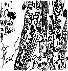
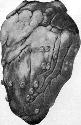
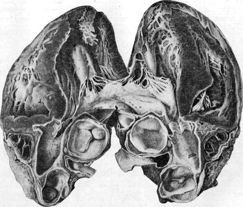
of the human heart. Surgery.-D nkanelidze Yu., Wounds of the heart and their surgical treatment, L., 1927 (bibliography); Zelenin V., Kulebyakin N., Revival of the heart in chloroform fainting, dissertation, SPB, 1913; Napalkov N., Surgery of the heart and pericardium (Russian surgery, edited by P. Dyakonov, L. Levin, et al., vol. III, SPB, 1902, bibliography); Constantini, De la chirurgie des plaies recentes du coeur, P., 1919; Koch W., Der funktionelle Bau des menschlichen Herzens, B., 1922; KiilbsF., Erkrankungen der Zirkulationsorgane (Handbuch der inneren Medizin, herausgegeben von G. Bergmann und R. Staehelin, B. II, T. 1, B., 1928, bibliography); Kiittner H., Die Operationen am Brustkorb (Chirurgische Operationslehre, herausgegeben von A. Bier, H. Braun u. H. Kiimmel, B. II, pp. 526-573, Lpz., 1923); Rehn E., Die Chirurgie des Herzens und des Herzbeutels (Die Chirurgie, herausgegeben von M. Kirschner u. O. Nordmann, B. II, T. 2, B.-Wien, bibliography); Sauerbruch F., Die Chirurgie der Brustorgane, B. II, B., 1925; ibid., Verletzungen des Herzens (Handbuch der praktischen Chirurgie, herausgegeben von C. Carre, H. Kiittner, u. E. Lexer, B. II, Stuttgart, 1931). See also bibliography for the article Cardiovascular system.
Related articles
Mentioned in
Cite this page
“Heart.” Soviet Medical Encyclopedia. English translation of Bolshaya Meditsinskaya Entsiklopediya, 1st ed. (Moscow, 1928–1936), ed. N. A. Semashko. https://sovietmedicalencyclopedia.pages.dev/article/heart/