Body Cavities
Historical document, translated for reference. It reflects medical knowledge of the 1920s–30s and is not medical advice.
Summary
This article from the 1928–1936 Soviet Great Medical Encyclopedia provides a comparative anatomical and embryological overview of body cavities, tracing their evolution from simple digestive cavities in invertebrates to the complex coelomic system of vertebrates.
Encyclopedia article (1928–1936)
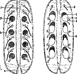
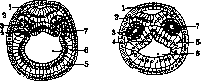
Body Cavities. Comparative anatomy and embryology. In coelenterate animals, as well as in flatworms, there is only one single internal cavity—the digestive cavity—which in these cases is usually provided with outgrowths or even passes into a peripheral system of channels (for example, in jellyfish). In flatworms, the space between the intestine and the integuments is filled with so-called mesenchyme (parenchyma), in which lie complexly constructed genital organs, containing a system of cavities and ducts, as well as excretory organs (protonephridia). In other worms, in addition to the intestinal cavity and the genital cavities, clear cavities appear in the surrounding parenchyma of these organs. For example, in roundworms there is an extensive cavity between the intestinal wall and the outer body wall. This cavity has no epithelial lining; it represents the result of the separation of mesenchyme cells and is called the primary body cavity. In higher worms (especially annelids), there is also a cavity between the intestine and the body wall, but this cavity, usually divided into right and left halves, and also in connection with the general segmentation of the body into a series of successive chambers, is lined with a flat epithelium and has a well-defined connection with the genital glands, which develop in its walls, and with excretory organs. This is the secondary body cavity, or coelom. Comparison with acoelomates and protocoelomates shows that the secondary body cavity represents the result of the proliferation of the cavities of the genital glands (Hatschek's gonocoel theory) (Fig. 1). The secondary body cavity is well developed not only in annelid worms, but also in enteropneusts, as well as in echinoderms. In mollusks it is poorly developed, being mainly in the form of a pericardial cavity, and in arthropods it is reduced and enters into connection with the circulatory system. The secondary body cavity acquires great importance in all chordates and in particular in all vertebrate animals, where it reaches especially complex differentiation. Embryologically, the secondary body cavity does not develop in all animals in the same way, and this gave rise to divergent views on the origin of the cavity itself. Sometimes it develops by the separation of cells ("schizocoel") of mesodermal strips growing from the blastopore lip area in a young embryo (annelids, mollusks), in other cases—simultaneously with the development of the mesoderm itself by separation from the primary intestinal cavity. This last, "enterocoelic" method of mesoderm and secondary body cavity formation is observed in some invertebrates (in hemichordate worms, in enteropneusts, and in echinoderms) and in chordate animals, although in vertebrates this process is somewhat modified and therefore not so clear. These observations allowed the conclusion to be drawn that the secondary body cavity represents the result of the separation of peripheral parts (outgrowths) of the intestinal cavity (the enterocoel theory of the Gertwig brothers). Since, however, comparative anatomical data give definite indications of the primary genital function of the coelom, the aforementioned gonocoel theory now has more supporters. The secondary body cavity, always paired in origin, may have metameric structure, subdividing into a series of successive chambers, the walls of which form transverse partitions or septa. The inner walls of the right and left parts of the body cavity cover the intestine externally and fuse both above and below it, forming dorsal and ventral mesenteries, on which the intestine is suspended inside the coelom. The inner wall of the secondary body cavity is in the form of the intestinal visceral leaflet, or splanchnopleure, the outer wall is called the cutaneous, parietal leaflet, or somatopleure. The latter gives rise to a significant part of the animal's musculature. In the walls of the coelom, as mentioned, genital glands develop, and its cavity communicates in higher worms with the external environment via segmental ducts serving as excretory organs (metanephridia), and via a similar series of shorter funnel-shaped ducts serving to expel mature genital products. In Amphioxus, the coelom develops by the separation of a whole series of paired sac-like outgrowths of the primary gut (Fig. 2). The secondary body cavity is then subdivided into a dorsal section—the cavity of the muscular segment (myocoel) and a ventral section—the cavity of the lateral plate or definitive body cavity (splanchnocoel). The latter loses its metamery, as the septa disappear, and thus develop continuous right and left
Fig. 1. Schemes of the formation of the segmented secondary body cavity of a higher worm from the genital parts of a lower worm: 1- foregut; 2- gonads; 3- protonephridia; 4- hindgut; 5- nephridium; 6- secondary body cavity.
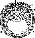
body cavities, which then, due to the partial disappearance of the mesenteries (especially the ventral one), merge into one unpaired cavity. In some cases in animals, cavities of ectodermal origin that to a greater or lesser extent envelop them also develop. Such is, for example, the peribranchial, or cloacal, cavity of tunicates, through which the respiratory stream of water is expelled, feces and excretions are removed, and also genital products (eggs often pass through a greater or lesser part of their development in it). The "epicel" developing from the outer body integuments has of course nothing in common with true body cavities. In vertebrates, the general body cavity develops more or less independently of the intestinal cavity, but traces of its original connection are often preserved, and in any case its limiting mesodermal walls develop in connection with the endoderm (Fig. 3). The general body cavity of vertebrates is laid down as a continuous, unsegmented structure and initially has the appearance of a narrow slit between the two mesodermal leaflets. Later, the dorsal part of the mesoderm, and consequently the cavity contained in it, segments, forming a series of primary segments, or somites, with small cavities connected by narrow ducts in the "somite stalks" and body cavity MITOV with a more extensive one in amphibians (transverse section through young
Fig. 3. Formation of meso-
triton embryo): 1- primary mouth; 2- ectoblast; 3- parietal mesoblast; 4- visceral mesoblast; 5- cavity of the primary gut; 6- entoblast. paired cavity of the unsegmented lateral plate (Fig. 4). The latter grows along the sides of the intestine until it meets over and under the intestine, where the visceral leaflets of the right and left body cavities, connecting with each other, form the aforementioned mesenteries. The part intermediate between the somite and the lateral plate, namely the somite stalk, gives rise to renal tubules, which in vertebrates also initially open into the secondary body cavity via funnels. Along the sides of the dorsal mesentery develops a paired thickening of the dorsal wall of the lateral plate, representing the primordia of the genital glands. The lateral plates themselves with their cavities give rise to the body cavity with its peritoneal lining. The ventral mesenter
1
%
9
ery largely disappears, and the two cavities join, so into one unpaired body cavity. The part of the visceral leaflet adjacent to the intestine gives rise, besides the peritoneal lining itself (through the process of mesenchyme formation or "coelenchyme"), also to connective tissue of its mucous membrane, blood and lymphatic vessels, and intestinal musculature. The somatopleure gives, besides the peritoneum, also numerous connective tissue elements. The main longitudinal blood trunks together with the heart also develop from the same mesenchyme in the form of longitudinal ducts in the ventral and dorsal mesenteries.
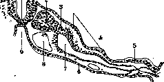
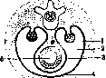
In adult vertebrates, the general body cavity is lined with flat epithelium and encompasses the intestinal canal (with stomach and esophagus) and its glands (liver, pancreas), the heart with a series of large vessels, and the reproductive organs. From the internal (visceral) walls of the body cavity, the serous lining of the intestinal tube and other internal organs and the mesentery, which suspends the intestine to the dorsal wall of the cavity in the form of a double leaf, are preserved in the adult animal. Only insignificant remnants of the abdominal mesentery remain in the region of the liver (lig. suspensorium hepatis). On the double folds of the peritoneum hang inside the body cavity other internal organs (glands, reproductive organs) (Fig. 5). The parietal leaf forms the lining of the outer wall of the body cavity. In all vertebrates, the anterior part of the body cavity, containing the heart (pericardial cavity), is separated from the posterior part or abdominal cavity. In lower vertebrates, a connection between these parts is still preserved through special canals. The lining of the pericardial cavity bears the name of the pericardium. The pericardial cavity, located initially in front of the abdominal cavity, moves back in terrestrial vertebrates and is pressed into the abdominal cavity, from which it is again separated in mammals with the formation of a transverse partition—the diaphragm. The latter develops from several sources. In all vertebrates, on the abdominal side, between the cardiac venous sinus and the stomach, a transverse fold of the peritoneum (septum transversum) develops in connection with a pair of large venous trunks (ducts of Cuvier). In mammals, in addition, a pair of dorsal folds develops, which, through connection with the abdomen, give rise to the diaphragm. Somatic myoblasts penetrate into these folds, which are then transformed into muscular fibers. In the thoracic cavity thus separated, in the middle lie the heart, surrounded by the pericardial cavity (lined with pericardium), and on the sides the lungs, surrounded by pleural cavities with their lining (pleura). Between the two lungs passes the esophagus from the dorsal side.
I. Schmalhausen. Pathological changes of the body cavities may consist in a disturbance of their shape and configuration, a change in volume, accumulations of various pathological products, etc. Disturbance of the shape and configuration of a particular body cavity most often occurs in various displacements, as well as in changes in the volume of organs and tissues bordering the body cavity; the same result can be produced by various pathological growths originating from neighboring parts. Partial and even complete obliteration of the body cavity is usually the result of connective tissue adhesion of the contacting walls of the body cavity, most often as a result of an inflammatory process; such partial or complete obliteration of the body cavity is called obliteration, whereas the formation of local connective tissue adhesions refers to synechiae. An increase in the volume of the body cavity is most often caused by the accumulation of pathological material in the body cavity. Sometimes the body cavity seems to be inflated by air or gas penetrating into it from neighboring parts or introduced intentionally, which most often occurs in pleural cavities (see Pneumothorax). The body cavities often serve as a place for the accumulation of serous fluid (see Ascites, Hydropericardium, Hydrothorax), which can to a significant extent increase the volume of the body cavity; the same consequence results from the accumulation of inflammatory exudate (see Pericarditis, Pleuritis, Peritonitis) in the body cavity, as well as from the effusion of blood into the body cavity. All the above-mentioned accumulations can lie freely in the body cavity, occupying the lower parts of the body cavity due to gravity (except for air, which is located in the upper parts), whereas in the presence of partial adhesions and obliterations the accumulation is located in a certain part of the body cavity, being isolated by adhesions (encapsulated accumulations). Large accumulations of air or fluids in the body cavity cause significant displacement of neighboring organs. Strong deformations of the body cavity are produced by tumors, both those primarily developing from the epithelium of the serous coverings of the body cavity (rare primary cancers of the pleura, pericardium, peritoneum), and those that grow into the body cavity from neighboring organs or metastasize into it and seed the cavity to a certain extent, spreading through lymphatic vessels and by implantation. In the abdominal cavity, sometimes there is a kind of seeding by mucus, which is secreted either from a perforated appendix or from an ovarian cyst (so-called pseudomyxoma peritonaei). Parasites sometimes occur in the body cavities, penetrating here most often from neighboring organs (ascariasis from a perforated ulcer of the stomach or intestine, echinococcus from the liver or lung). Foreign bodies in the body cavities are rarely encountered; these include bullets, fragments penetrating in gunshot wounds, needles penetrating through the coverings or from the gastrointestinal tract, surgical instruments, gauze swabs, cotton balls, rarely forgotten in the body cavity during operations. In the abdominal cavity, gallstones and intestinal stones, plant residues from food, sometimes enter through appropriate perforations; sometimes detached tumors of the ovary or uterus or simply detached appendices epiploicae (sometimes petrified) play the role of foreign bodies. The name corpora libera is accepted to denote small smooth clots appearing in the body cavities, more often in the abdominal cavity, consisting of fibrin and resembling the rice bodies of joints. When a foreign body is fixed to the wall of the body cavity, a process of encapsulation develops, usually with the formation of giant cells (see also Peritoneum, Pericardium, Pleura).
A- Abrikosov.
Related articles
Mentioned in
Cite this page
“Body Cavities.” Soviet Medical Encyclopedia. English translation of Bolshaya Meditsinskaya Entsiklopediya, 1st ed. (Moscow, 1928–1936), ed. N. A. Semashko. https://sovietmedicalencyclopedia.pages.dev/article/body-cavities/