Lungs
Historical document, translated for reference. It reflects medical knowledge of the 1920s–30s and is not medical advice.
Summary
This article provides a detailed anatomical description of the lungs in vertebrates, with special focus on comparative anatomy and human lung structure. It covers the evolutionary development of lungs from fish to mammals, including embryonic development, and details the physical characteristics, measurements, and properties of human lungs.
Encyclopedia article (1928–1936)
LUNGS. Lungs (lat. pulmones, gr. pleumon, pneumon), organ of aerial terrestrial respiration (see) of vertebrates. I. Comparative anatomy. Lungs of vertebrates exist as additional organs of aerial respiration already in some fish (in lungfish, crossopterygians) simultaneously with gills; however, only in terrestrial vertebrates they reach high differentiation and become the sole respiratory organs. L. develop in lower vertebrates (amphibians) as paired outgrowths of the wall of the anterior intestine directly behind its gill region. These outgrowths are connected with each other by a groove-like area of the anterior intestine, which then becomes increasingly isolated from the latter and forms the carrying lung sacs an unpaired division--the laryngo-tracheal chamber, connecting with the glottis to the abdominal wall of the pharynx. In higher vertebrates, this chamber grows into a long respiratory tube and develops very early embryonically, so that its primordium in the form of an unpaired groove-like outgrowth appears before the paired primordia of lung sacs. The latter then take the appearance of branches growing from the sides of the unpaired primordium. The progressive development of L. in the series of terrestrial vertebrates is accompanied by continuous increase of their respiratory surface. Almost smooth inner walls of lung sacs in some lower amphibians are supplied in most higher forms with numerous crossbars giving them a honeycomb character. On the walls of primary cells, secondary and tertiary crossbars then develop, so that already in reptiles the inner cavity of lung sacs is reduced to the degree of a relatively narrow central canal (intrabronchial), connected with numerous openings to a complex system of cells at the periphery of the lung. The walls of the latter thus acquire a spongy character. The complication of the structure of L. continues in the direction from outside inward, and with progressive development of the complex system of cells, only the central bronchus remains from the inner cavity of each lung sac, which in higher reptiles leads into a system of lateral bronchi communicating with numerous openings with alveolar tissue. In mammals, the process of isolation of respiratory pathways within the L. goes even further: higher-order bronchi develop, and their connection with alveoli is limited only to the branches of the finest bronchi ending in funnels with alveoli. In some contradiction to this central direction of the progressive differentiation of L. of vertebrates are the facts of their ontogenetic development in mammals, in which L. develop embryonically by progressive branching of their tubular primordia, i.e., centrifugally. These facts indicate accelerated development of airways within the L. itself, i.e., represent a particular case of heterochrony. (See also Respiratory organs.) I. Shmalgausen. P. Anatomy. L. of humans are constructed according to the type characteristic of all mammals and represent a complex paired honeycomb organ (fig. 1) with a widely developed apparatus of branching bronchi and a respiratory network of alveoli. L. are located in the thoracic cavity, closely adhere to its walls and are separated from the latter by the pleural fissure, while from each other by the organs of the mediastinum (see). From the abdominal cavity L. are bounded by the diaphragm (see).-The form and size of L. under normal conditions due to constant movement caused by breathing change during respiration and exactly repeat the form of the thoracic cavity containing them. Fixed with hardening liquids (formalin, chromic acid) and removed after hardening from the corpse, each L. with a certain degree of approximation resembles in shape half of a vertically cut cone. Its apex (apex) is directed to the supraclavicular fossa of the neck, the base (basis) to the diaphragm, the rounded lateral side to the ribs, the medial surface to the organs of the mediastinum. Corresponding to these three sides, 3 surfaces and 4 edges are distinguished in L.: sternocostal (facies sterno-costalis), diaphragmatic (facies diaphragmatica) and mediastinal (facies mediastinalis). Between them more or less sharply marked are the delimiting them 2 edges: vertical anterior sharp edge (crista mediastinalis ventralis, sive anterior), a blunter posterior one (margo mediastinalis dorsalis, s. posterior) and 2 horizontal ones at the base of L.: inner-to the mediastinum (crista mediastinalis caudalis) and outer-to the ribs (crista arcuata) (fig. 1). Dimensions and external configuration of each L. besides changes in volume connected with respiration, are in direct dependence on the size and shape of the thoracic cage (see), for which reason all constitutional, typal, age and pathological variations and changes of the latter are repeated also in the external appearance of L. (see below). The right L. is relatively wider than the left, which is already noted in the first phases of embryonic development (see vol. IV, art. 65, fig. A and B). This phenomenon is connected with different conditions of development and nutrition of L. of both sides due to the asymmetric, predominantly left-sided position of the heart and its rotation around the axis to the left side, which in turn affects the difference in the development of length of the vessels feeding the right and left L. (Braus). In adult state the left L. by its mass is 15% inferior to the right; the width is in the ratio 10:7; the extent of the sternocostal surface of the right L. considerably surpasses the left, since the ventral mediastinal edge of it goes mostly to the left beyond the median line. In the latest literature measurements (F. W. Müller; 1923) give the height of L. when lying: on the right-17.5 cm, on the left-20 cm; when standing: on the right-21 cm, on the left-23.5 cm. The difference corresponds to different height of standing of right and left halves of the diaphragm. The vertical dimension of L. is connected also with movements and deviations in the position of the spine. Dimensions of L. can present considerable fluctuations. On average in the resting state (intermediate between inspiration and expiration) the absolute figures according to Testut-Latarjet (Testut, Latarget) are the following. Vertical diameter in the largest posterior part of L..................... 25 cm Anteroposterior diameter at the base of L. ... 7 » Volume of lung at expiration (state on corpse at autopsy of thoracic cavity)--1,617 cm3 in men and 1,290 cm3 in women (i.e., female L. is 1/1 of its volume less than male). In the collapsed state in average L. in pneumothorax on corpse has only 1/3 of its size (Braus). L. of a non-breathing newborn occupy a very insignificant part of the thoracic cavity (fig. 2 and 3). This ratio to the volume of the latter in the embryonic state is expressed even more sharply and the younger the embryo the stronger [see separate table (art. 415-416), fig. 2]. With the beginning of the respiratory act the volume of L. rapidly increases. According to Gra-per'y, different parts of the thoracic cavity have the following volume (in cm3) (table on art. 415). Thus before birth L. occupy about half of the capacity of the thoracic cage, while in the period of lactation their share already amounts to about 2/3- In further growth L. is expressed in the following ratios: during the first year of extrauterine life L. increases in volume, according to Aeby, 4 times, by 8 years-8 times, by 12 years-10 times, after 20 years its size is 20 times greater than that of L. of a newborn.-The absolute weight of L. of a newborn, if it breathed and is not pathologically changed, is always less than water (according to Sappey, on average 0.49). Therefore a breathing L. with alveoli filled with air floats in water, while pieces of lung of a non-breathing child, stillborn, or L. with alveoli occupied by pathological detritus, exudate, blood, tumor etc., become heavier than water and sink in it. The specific gravity of such non-breathing L. of newborn on average is 1.06. The color of L. depends on the degree of filling of L. with air and blood. In a non-breathing fetus it is intensely pink, in a newborn it has different tones of pink. The weight of L. of a non-breathing child reaches 65 g. After established breathing-90 g. In adult the absolute weight of L. is 1000-1300 g. The color of L. of a newborn is pale pink. With years the color darkens and acquires a bluish tint. On the outer shiny smooth surface
of the lung
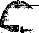
sterno-costal surface; C-mediastinal; 1- crista mediast. ventralis; 2- margo mediast. dorsalis; 3- crista mediast. caudalis; 4- crista arcuata.(After Peter)
different weight of L. of a non-breathing child reaches 65 g. After established breathing-90 g. In adult the absolute weight of L. is 1000-1300 g. The color of L. of a newborn is pale pink. With years the color darkens and acquires a bluish tint. On the outer shiny smooth surface
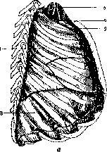
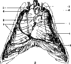
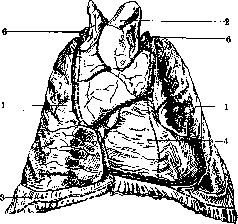
Fig. 2. Lungs of a non-breathing newborn (a-side view, b-frontal): 1-lung; 2-thymus; 3-diaphragm; 4-heart, dressed in pericardium; 5-a. subclavia; 6-v. anonyma. (After Peter)
different weight of L. of a non-breathing child reaches 65 g. After established breathing-90 g. In adult the absolute weight of L. is 1000-1300 g. The color of L. of a newborn is pale pink. With years the color darkens and acquires a bluish tint. On the outer shiny smooth surface
different weight of L. of a non-breathing child reaches 65 g. After established breathing-90 g. In adult the absolute weight of L. is 1000-1300 g. The color of L. of a newborn is pale pink. With years the color darkens and acquires a bluish tint. On the outer shiny smooth surface
different weight of L. of a non-breathing child reaches 65 g. After established breathing-90 g. In adult the absolute weight of L. is 1000-1300 g. The color of L. of a newborn is pale pink. With years the color darkens and acquires a bluish tint. On the outer shiny smooth surface
different weight of L. of a non-breathing child reaches 65 g. After established breathing-90 g. In adult the absolute weight of L. is 1000-1300 g. The color of L. of a newborn is pale pink. With years the color darkens and acquires a bluish tint. On the outer shiny smooth surface
different weight of L. of a non-breathing child reaches 65 g. After established breathing-90 g. In adult the absolute weight of L. is 1000-1300 g. The color of L. of a newborn is pale pink. With years the color darkens and acquires a bluish tint. On the outer shiny smooth surface
different weight of L. of a non-breathing child reaches 65 g. After established breathing-90 g. In adult the absolute weight of L. is 1000-1300 g. The color of L. of a newborn is pale pink. With years the color darkens and acquires a bluish tint. On the outer shiny smooth surface
aЩР^Ь
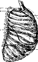
Fig. 3 L. p. p. (newborn breathed 1 day (a-view from the side, b-from the front, overview from above): 1-lungs; 2-thymus; 3-diaphragm; 4-heart; 5-a. subclavia; 6-v. anonyma. (According to Peter.) The average weight of the lungs is 1,100 g, of which 600 g accounts for the right L. and 500 g for the left. The specific gravity of any L. at any age L., moistened by the pleural fluid wetting it, thin or wider gray and black lines appear,
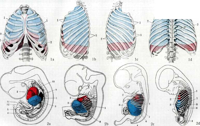
Figure 1 a-d. Relationship of interlobular grooves and lung edges to the chest wall; 1-upper lobe; 2-lower lobe; 3-middle lobe. Figure 2. Embryonic development of the lungs
in relation to other organs; according to Jackson): /-lung; 2-heart; 3-liver; ^-rectum; 5-cloaca; 6 in figs. a and b-urinary system, in figs. c and d-kidney; 7-spleen; 8-adrenal gland; 9-u. cardinalis, 10-v. jugularis, 11-a. umbilicalis, 12-v. umbilicalis; a, b, c and d-embryos 11 mm, 17 mm, 31 mm, 65 mm. To st. Lungs.
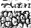
dots, black spots, which, increasing in quantity, merge with each other and create in places a mosaic pattern of polyhedrons, in places more or less extensive fields of black color of scalloped figures and spots (fig. 4). In adults by 30-35 years of age such a mosaic of pigmentation can spread over the entire surface of L., excluding certain areas of the diaphragmatic face or adjacent to the ribs, and is explained by the deposition in the inter--Hf fM TlfliWIJ/^ tochnoy interlobular tissue of dust, mostly coal particles. In these cases the color of L. approaches steel-gray and when alternating with preserved ro-Fig. 4. View of the lung peripheral zones (according to sometimes gives the surface of L. whimsical patterns of turtle shell and other figures (see Thoracoscopy). In old age the color becomes even more intense, especially in people who have long been in rooms rich in coal dust, and the color of L. acquires a blue-black shade, since deposition of dust particles also takes place in deeper layers, in lymph, nodes, along vessels, etc.
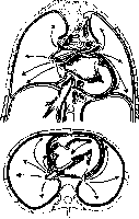
Physical properties. L. have a soft consistency. The visceral pleura covering them is intimately fused with them (see Pleura). L. can be easily compressed, but upon cessation of pressure it immediately restores its shape due to elasticity. Strong compression gives a sensation of crepitation, which is caused by the rupture of air bubbles through small ruptures of the pulmonary alveoli. When punctured with a syringe needle, L. gives easily aspirated clean blood; when cut with a knife, the surface of the section abundantly exudes frothy blood of a bright red color. In a healthy state, L. can offer significant resistance during strong tension, coughing, and experimental inflation; nevertheless, the delicate structure of lung tissue requires careful handling, because sharp compression and grasping with instruments and hand leads to small ruptures, mainly in the alveolar layer. Thanks to the abundance of elastic tissue in the visceral pleura, in the walls of the alveoli and bronchi, L. possesses exceptional elasticity. Stretched and pressed against the chest wall by atmospheric air, which has filled its alveoli since the first breath, the lung is under continuous influence of its "elastic traction" (see Respiration). This force of tendency to collapse and resistance to stretching of L. is not the same in different parts and depends on its mass and the degree of mobility of that section of the chest wall to which a particular part of L. is adjacent. In connection with these conditions, the elastic traction of the right L. is usually stronger than that of the left; it causes the retraction of the intercostal spaces and is balanced by the resistance of the unyielding part of the costal arches and diaphragm; upon its elimination, it is revealed in the reduction of the volume of L. due mainly to the reduction of its rib surface (see Thoracoplasty) or vertical size or both simultaneously (see Pneumothorax, artificial). In the apical region, the elastic traction manifests itself on the soft tissues of the supraclavicular fossa, affecting its depression and the blood filling of the large cervical veins lying nearby. During respiratory movements, the traction is uneven at different levels of L. and is stronger in the direction of the most mobile parts, therefore it is weaker above and stronger at the base below (see Respiration). The elasticity of L. also has a significant influence on the mediastinal organs. The elastic fibers of the mediastinal part of the lung-pleural apparatus cause traction in the direction from its mobile surface-mediastenum-toward the fixed one, i.e., the ribs. This dependence is reflected e.g. in the work of the atria, the walls of which are under the action of this traction, and promotes their expansion during diastole. This role of L. in the work of the heart appears with particular force and has practical importance when eliminating the elastic traction as a result of collapse of L. during opening of the chest cavity and collapsive therapeutic measures (fig. 5).
External appearance and position of L. The apex of L., bounded by a horizontal plane drawn through the I rib and forming the dome of the pleura (see), is blunt and rounded in shape (see separate table, fig. 1). In adults, varying greatly, the apex of L. protrudes and therefore is percussed above the clavicle to a height of 3 cm and in front above the edge of the rib by 5 cm, while behind it borders the level of the upper edge of the I rib. This protrusion is associated with the descent with age downward of the anterior part of the I rib, which until 7-10 years retains a more or less horizontal position (see separate tabl., fig. 1) (see Chest cage.)- neniya action elastich- On the surface up- skoy traction: above -on the knee joint Appmy- heart, below-on the right and left lung (according to Sauer-lynom L. clearly
ьтед'y). may be expressed 2 depressions, or grooves: horizontal - from pressure of the narrow ring of the I rib (subapical Schmorl's groove) and a notch at the very apex of the cone - trace of the adjacent subclavian artery. The apex of L., repeating the shape of the pleural cone, which is little mobile during breathing, does not encounter during its excursions reserve spaces, as in the lower parts of the pleura, and therefore represents a less mobile part of L. (frequency of inflammatory intrapleural adhesions).
Fig. 5. Diagram for explaining Facies stern o-c o s t a l i s (fig. 6) L. presents a convex largest surface of it. It adjoins the anterior outer and posterior wall of the chest cavity. Its lower boundary is formed by the sharp thin outer lower edge of L., level .1
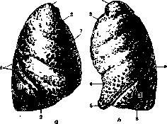
Figure 6. Facies sterno-costalis. View from the side: a and b-right and left lung; 1-apex; 2-crista me-diast. ventralis; 3-crista arcuata; 4-incisura cardiaca; 5-lingula pulm.;6 and 7-incisura inter-lobaris obliqua et horizontalis; /-upper lobe; II-lower; ///-middle. (According to Spalteholz.)
of which in relation to the ribs changes during respiration, filling the reserve spaces of the sinus phrenico-costalis (see Pleura) during inspiration and being displaced from it during expiration. In the last phase it has the highest percussive position relative to the sinus, namely on the VI rib along the mamillary line, on the VIII along the axillary line, on the X along the scapular line, and on the XI at the spine, or otherwise the posterior border can be established by a horizontal line drawn through the spinous process of DXI. During inspiration, the L., entering the sinus, however does not completely fill it. The degree of this inspiratory displacement is subject to very strong individual variations. On average it is calculated at 1-2 cm at rest and at 6-8-10 cm during forced respiration. (The relationship of the level of the L. at different respiratory forces-see Diaphragm.) The anterior border of the facies sterno-costalis is the anterior sharp edge of the lung. In its relation to the ribs of the anterior part of the chest wall it repeats the course of the transitional fold of the pleura. Its sinus reserve space is completely replaced by the L. entering it during inspiration. Corresponding to the curvature of the pleural border in the IV and V intercostal spaces along the parasternal lines, the L. forms a notch (incisura cardiaca), bordering on the cape-like projection of the L. (lingula), and at its greatest inspiratory stretching it does not completely fill this part of the pleural sinus [see separate table (pp. 415-416), fig. 1 a and b]. The level of the lower border of the L. varies considerably and descends in connection with and corresponding to the age-related descent of the diaphragm (see) and the skeleton of the chest (see). In early childhood the lower pulmonary border is at the following levels: posteriorly in a newborn who has not breathed-on the IX-X rib, after inspiration-on the X or XI; on the axillary line: before breathing-on the VII-VIII, after inspiration-near the VIII rib (Graper). In an adult this level is generally one and a half intercostal spaces lower than in a newborn, and in old age one intercostal space lower than in an adult (fig. 7). On the costal surface of an emphysematous L. the imprints from pressure of the adjacent ribs may be expressed with greater or lesser clarity in the form of wide, transversely oblique grooves. Both on the right and on the left the costal surface of the L. is crossed by the oblique interlobar notch (incisura interlobaris), to which on the right is joined the horizontal notch (incisura horizontalis). The aforementioned notches, or grooves, divide the left L. into two, the right-into three lobes-superior, middle, and inferior (lobi pulmonum-superior, medius, inferior). This division however is not limited to the surface of the organ, as the notch deepens into the tissue of the L. and reaches almost to the hilum of the L., so that the lobes remain connected to each other only by the branches of the bronchi entering them, the accompanying vessels, and the connective tissue fat with lymph nodes and nerve branches. The visceral pleura, lining the entire L., penetrates into the notches and thus, accompanying the lobes, makes them free and movable in relation to each other. Up to the bronchi and vessels connecting the lobes the pleura however does not reach and is thrown across the depth of the notch from the surface of one lobe to another, not reaching by at least 1 cm to the broncho-vascular bundle. Thus in surgical procedures on the 'hilum' of the lobe (ligation of the pulmonary artery, resection of the lobe, etc.) it is possible to reach the vessels and bronchi only after incising this transitional interlobar pleural fold. Both lobes of the left lung are usually of equal size. In the right L. the lower lobe considerably exceeds the size of the upper lobe; the smallest is the middle. The oblique notch in both L. posteriorly divides the upper lobe from the lower, on the left it begins higher than on the right [see separate table (pp. 415-416), fig. Id], namely in the IV intercostal space along the line of the oblique groove, further according to B03p.1CTax(m)Mehnert, y):
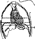
Fig. 7. Level of position of the diaphragm at different ages: 1-in a newborn; 2-in a 30-year-old man; 3-in a 72-year-old man; 4-diaphragma. on 2 cm outward from the costal angle crosses the V rib, enters the V intercostal space, passes it along an inclined line between the anterior axillary and mamillary lines, crosses the VI rib and on the mamillary line ends in the VII intercostal space or on the VII rib. On the right the same groove begins one intercostal space lower, namely-in the V, crosses it in the paravertebral section, in the region of the costal angle it goes obliquely across the VI rib, then for a short distance passes through the VI intercostal space, leaves it 3 cm laterally from the costal angle, reaches the VII rib and accompanies it forward until meeting the lower pulmonary edge, approximately on
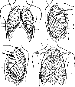
Figure 8. Working diagram of pulmonary-pleural borders in projection onto the chest wall (shaded areas correspond to sinus phreni-co-costalis): 1-upper lobe; 2-lower lobe; 3-middle lobe.
the mamillary line (Felix; 1928). These borders however can individually change, moving in the direction of elevation relative to the ribs and intercostal spaces. On the convex surface of the L. on the right to the oblique notch is added the horizontal groove (incisura horizontalis) separating the upper lobe from the middle. It begins on the axillary line from the oblique notch and goes horizontally forward, somewhat curving along the course of the IV rib, to the sternum, i.e. to the anterior edge of the L.-On the living projection of the described interlobar borders
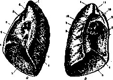
Figure 9. Diaphragmatic and mediastinal surfaces of the right (a) and left (b) lung: 1-apex; 2-crista mediast ventralis; 3-margo mediast. dors.; 4-crista mediast. cauda,lis; 5-crista arcuata; 6-fossa cardiaca; 7-sulcus oesophageus; 8-impression of v. cavae sup.; 9-lig. pulmon.; 10-impression of aorta; 11-impression a. car. comtti. sin. (According to Corning.)
are conducted as follows. The beginning of the oblique groove is led from the level of the end of the spinous process of the III vertebra, or (which is the same) the level of spina scapulae with arms lowered, and the point of intersection of this level with the margo vertebralis is connected with the intersection of the mamillary line with the VI rib or with the place where the VI rib passes into cartilage (fig. 8). To construct the horizontal notch a horizontal line is drawn from the place of attachment of the IV rib cartilage outward to the intersection with the constructed oblique line. Thanks to such a relationship of lobar borders the pulmonary field posteriorly both on the right and on the left is divided into 2 parts: the upper 1/4 belongs to the upper lobe, the lower 3/4 to the lower. Anteriorly however on the right there is a field of two lobes: the upper part (above the IV rib) is occupied by the upper, the lower-by the middle lobe; on the left almost the entire anterior field corresponds to one lobe-the upper. Along the axillary line on the right all three lobes are located, on the left-two: upper and lower. Facies diaphragmatica, or basis pulmonum (fig. 9), represents the concave, corresponding to the dome of the diaphragma
in
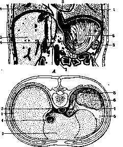
Figure 10. 1-right and left lung; 2-heart; 3-liver; 4-v. cava inf.; 5-stomach; 6-spleen; 7-esophagus; 8-kidney. the inferior surface of the L. The degree of concavity depends on the height of the excursions or position (e.g. in paralysis) of the diaphragm and its age-related positions. Along the inferior surface there is a groove crossing it obliquely-continuation of the oblique interlobar notch, which divides the base of the L. into two unequal parts: the smaller-anterior, belonging on the right to the middle lobe, on the left-to the upper, and the larger-posterior, on both sides corresponding to the lower lobe. The inferior surface of the L. is adjacent both to the muscular and to the tendinous parts of the diaphragm, which separates the L. on the right from the main mass of the liver, on the left-from its left lobe, the fundus of the stomach, the spleen and sometimes a larger or smaller segment of the transverse colon (fig. 10). 14 Facies mediastinalis. corresponds to the organs of the mediastinum, extends from the apex of the L. to the diaphragm and has 2 sharp edges-anterior and inferior-and one obtuse-posterior. The mediastinal surface can be divided into two unequal parts: the larger, free, lined with visceral pleura, movable within the pleural 'cavity', and the smaller, capable of moving to a lesser degree due to the organs of the mediastinum with which it is in intimate connection
in
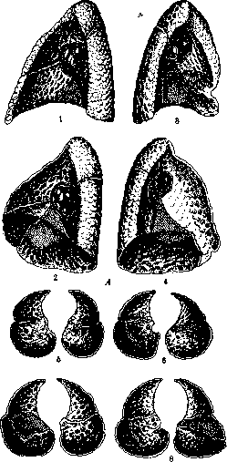
Figure 11. A-projection of lobus infracardiacus on the internal and diaphragmatic surfaces of the lungs: 1 and 2-right lung; 3 and 4-left lung; B-diaphragmatic surface of the lungs: 5-incomplete grooves in both lungs; 6-lobus infracardiacus on the left and variations of grooves on the right; 7-lobus infracardiacus; 8-lobus infracardiacus on the left and additional grooves on the right. with the help of a fixing apparatus, the root or hilum of the L. (radix, s. hilus pulmonis) and the pulmonary ligament (ligamentum pulmonale) (see separate table, figures 1 and 2). The free, mobile surface of the L. is adjacent to the organs of the mediastinum (hollow veins, aorta, esophagus, unnamed veins, heart, etc.). The trace of neighborhood with them clearly appears in the form of different depths of indentations and grooves (impressio cardiaca, etc.) on the inflated or emphysematous L. or on hardened in situ formalin or chrome preparations of the L. (see separate table, figures 1 and 2). The mediastinal surface is also intersected by interlobar incisions converging to the hilus of the L.: three on the right and two on the left. In relatively rare cases, both on the right and left, corresponding to the accessory bronchus (bronchus cardiacus) (see Bronchi), along with normal grooves, unusual additional grooves may also be found, delimiting accessory lobes (lobus infracardiacus), the location of which is schematically shown in Fig. 11. Deviations may also consist of partial and complete absence of incisions, their deformation, or the appearance of additional incisions, partially dividing the lobes or cutting them through to the root of the lungs (Fig. 12). The root, or hilus of the L. (hilus) is the point of entry into the L. of vessels and bronchi and has the shape of a rhombus or oval, occupying a central position on the mediastinal surface of the L. Their level in an adult corresponds to the intervertebral cartilage between Th11 and Th12; in early childhood it lies on average one vertebra higher. The upper part of the root of the L. is occupied by the pulmonary artery, bronchus, and lymph glands, in the lower part are the pulmonary veins, branches of the bronchial artery, and branches of the nerve plexus. The right hilus is wider than the left; in it, the bronchus is located highest; in front and below are the branches of the pulmonary artery. On the left, above and behind is the pulmonary artery, below and in front is the bronchus; the pulmonary veins are considerably removed from the artery and divide into two groups-upper and lower, which are separated by a large space filled with loose connective tissue. The pulmonary artery before entering the L. usually divides into 3 branches for its three lobes and is located for operative purposes where the posterior edge of the oblique interlobar incision approaches the root of the L. In the left root of the L., the organs are more crowded. Its vertically positioned oval is elongated downward and without sharp borders passes into the pulmonary ligament (see below). Usually the ratio is as follows: above-branches of the pulmonary artery, in the middle-main bronchus and branches of the upper pulmonary vein, below-the undivided lower branch of the pulmonary vein. Both hili are along their edge bordered by the transitional fold of the visceral pleura into the parietal. Its anterior and posterior plates, separated at the level of the root of the L. by its constituent parts, below the root again converge, but before turning back to embrace the L. as parietal pleura, over the entire distance from the root to the base of the L., they fuse with each other into a thin translucent sheet, which is called the pulmonary ligament (ligamentum pulmonale). Its shape is triangular; the ligament lies in the frontal plane intrapleurally and divides the lower part (below the root of the L.) of the mediastinal pleural cleft into anterior and posterior sections.
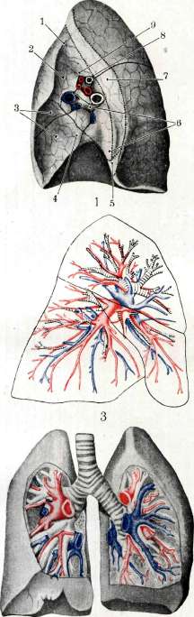
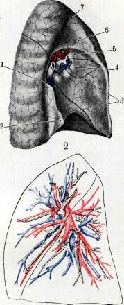

Figure 1 and 2. Facies mediaslinalis of the right and left lung (red color-a. pulmonalis; blue color-v. pulmonalis). Figure 1. Right lung: 1-groove of a. subclaviae; 2-groove of v. cavae sup.; 3- fossa cardiaca; 4-groove of v. cavae inf.; 5-groove of aorta; 6-place of attachment of the right atrium; 7-groove of esophagus; 8- bronchus; 9-groove of v. azygos. Figure 2. Left lung: 1-groove of aorta; 2-groove of esophagus; 3- fossa cardiaca; 4-place of attachment of the left atrium; 5-upper edge of fossae cardiacae; 6-groove of diaphragm; 7-groove of a. sagittalis comm. sin. Figure 3 and 4. Right and left lung. Ratio of bronchus, pulmonary arteries (red color) and veins (blue color) in projection on the surface of the lungs. Figure 5. Diagram of the ratio of bronchi and vessels in the central part of the lungs. Figure 6. Diagram of the distribution and ratio of the pulmonary artery, bronchus and pulmonary vein in different parts of the lungs (the right half of the circle corresponds to the left, the left-to the right lung; red color-pulmonary artery; white sector-bronchus; blue color-pulmonary vein). \Figure 1, 2, 5 and 6-after Felix.) 15. M. E. to the art. Lungs.
Internal structure of the L. Each lung lobe, taken separately, can structurally, according to Felix, be represented as consisting of two parts differing from each other: central, which includes predominantly denser formations, and peripheral, which as if covers the first with a sheath, of a more delicate structure, consisting mainly of the cellular tissue of lung vesicles (Fig. 13). The central core contains the root bronchus of the lobe, which gives off within it branches of the 1st, 2nd, 3rd and 4th order, the pulmonary and bronchial arteries and veins adjacent to the bronchus, pulmonary veins, lymph vessels and lymphatic glands, nerves and their ganglia, and connective tissue, and at the root-some amount of fatty tissue. The peripheral part has a thickness in larger lobes up to 4 cm and is divided into 2 layers: outer and inner. The latter is separated from the core of the central zone by a transitional layer of bronchial branches of the 3rd and 4th order and the lung lobules of rudimentary type lying between them. The most superficial layer is composed of a series of closely packed slender four-sided prismatic lobules (lobuli pulm.), having a height of 21-27 mm and a width of 9-21 mm and delimited from each other and from similar formations of the overlying layer by a thin layer of connective tissue. To each such lobule
14 V
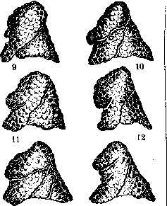
Figure 12. Variations of pulmonary grooves and lobes (A-of the right lung, B-of the left lung): 1 and 9-normal appearance, 2-8 and 10-14 - various types of groove development. (After Testut.) its own branch of the bronchus (bronchus tabularis, fig. 14 and 15a and b) and the accompanying twig of the pulmonary and bronchial (inconstant) arteries. The regularity of the rows of superficially located lobules may be interrupted by "insertion" lobules (fig. 16 a and b), repeating the structure of ordinary lobules and delimited by their connective tissue capsule. On the surface of the L. in \<\м-Л-M
Figure 13. Diagram of the structure of a lung lobe: 1-outer layer; 2-central core; 3-peripheral part. (After Felix-)
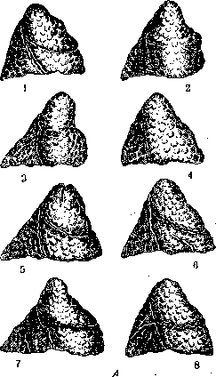
the area of their location, there may be observed as if retractions, simulating scar inflammatory changes. The inner layer also contains developed lobules, but they are located less densely and have a smaller size. The rows of lung lobules of the outer and inner layers are interrupted by more or less powerful connective tissue septa, which are a direct continuation of the subpleural connective tissue layer, extend from the periphery toward the central core, and divide both layers of the peripheral lung tissue (Lungenmantel) into wedge-shaped sections (sublobuli, according to Felix, or acini, according to other authors),
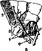
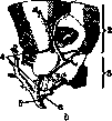

Figure 14. View of sublobus (acinus); a and b-view from two opposite sides. Relation of veins and bronchus to lobules: 1-outer surface; 2-outer layer of lobules; 3-inner layer of lobules; 4-vv. intersublobares; 5- branch of v. pulmonalis; 6-bronchus sublobarius. (After Felix.)
each of which has both rows of lobules of the outer and inner layers. In the thickness of these partitions are located lymph trunks and larger veins. The latter arise in the visceral pleura and, passing through the peripheral layers, flow into the larger vessels of the central lobar zone (Lungenkern). Structure of the lobule. Each lobule is built on the type of a lung lobe and can also be divided into central and peripheral parts. In the lung lobule there is its own bronchus, pulmonary artery and not always-bronchial artery. Blood flows out through 5
"^bronchial. Blood flows out through 5
venae intersublobares. The lobular bronchus initially gives off prism-shaped branches and breaks down into the lobule then into 2-4 terminal bronchioles, which no longer have cartilage (see Bronchi) and may be considered respiratory bronchioles: only at the beginning of the bronchus are the alveolar sacs located on one side and the other and, 5 ppiptpnp1,NYr £pk_ its lobular hov-ph parietal alveoli. Dividing into branches of the 1st and 2nd order, the terminal bronchioles as a rule acquire the type of respiratory bronchioles and again divide into branches of the 3rd and 4th order (up to 6-7 times), representing alveolar ducts (see Bronchi, color plate). The peripheral end of ductus alveolaris ends by dividing into 2-3 smaller alveolar ducts (ductulus alveolaris). Their continuation are the lateral and terminal funnels (infundibula) (fig. 17) and pulmonary vesicles, or alveoli (sacculi alveolares). The place where the alveolar duct divides into funnels is distinguished by some authors as vestibulum. The size of alveoli is not uniform: on average their diameter is about 250 [μ], their number varies and can reach 120 per lobule. The total number is determined in people from 150 million to 4 billion. The respiratory surface of all alveoli reaches 80-130 m2. Each lobule is considered as a collection of 12-18 acini, each of which contains up to 10-20 alveoli (Braus). With dense clustering of pulmonary vesicles, not all of them reach full development. In these cases, each alveolar duct at its end has only one pulmonary vesicle (Felix). These undeveloped areas together with rudiments of lobules of the outer part of the central zone of the lung lobes are rich material for regenerative compensatory processes in lung tissue when its areas are lost under the influence of various pathological conditions. The blood supply of the L. belongs to 2 systems: the system of pulmonary arteries and veins, performing the respiratory function,
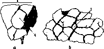
Figure 16. Metal cast of lung lobules from the outer 'sheath' of the lobe. View of the interlobular lobules (4) from the side (a) and from the surface (b): 1-outer surface of the lobe; 2-outer layer of lobules; 3-inner layer; 4-interlobular lobules. (After Felix.)
and the system of bronchial vessels (aa. and vv. bronchiales), intended for nutrition. However, it is not possible to make such a physiological distinction completely. Accompanying the bronchial tree with its branches, both systems connect in the capillary network surrounding the alveoli. The 'nutrient' branches of the bronchial artery can be traced b. ch. only to bronchioli respiratorii, and the nutrition of the terminal pulmonary alveoli is thus performed by the network from the pulmonary arteries. When
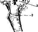
Figure 17. External appearance of acinus (in section): 1-v. interlobularis; 2-bronchiolus respiratorius; 3-ductulus alveolaris; 4-branch a. lobularis pulmonalis; 5-alveoli; 6-subpleural vein; 7-visceral pleura.
the vessels of the L. are injected through this network, the entire system of pulmonary arteries is perfectly filled. There are also observations establishing that the lymphatic glands of the pulmonary parenchyma can be supplied from the pulmonary, and not from the bronchial artery (Melnikov; 1923), and finally a large number of authors constantly found anastomoses between both arterial systems and pulmonary veins. Thus both systems are connected with each other, the capillary networks with which they are connected represent a neutral formation, the role of which can be twofold-respiratory and nutritional. The pulmonary artery can perform the role of a bronchial artery; however, the bronchial artery carrying arterial blood cannot take on the respiratory function after ligation, for example, of the main branch of the lobar pulmonary artery. The nutrition of the latter will be preserved, but the gas exchange role will be lost (Felix). System of pulmonary arteries and veins. Corresponding to the two possible types of bronchial division within the pulmonary parenchyma, the pulmonary artery, upon entering the lung, can branch either according to the main or according to the scattered type (Melnikov) (figure 18); transitional mixed forms are also possible. The main branches of the pulmonary artery between the lobes of the L. are distributed as follows. Left L. [see separate table (pp. 423-424), figure 4]. The upper lobe has 3 branches at the hilum, which break down into 5. Three of them go to the apex of the L., two-to the lingula. Lower lobe. With the embryonic type of division, the branches are distributed mainly on the ventral and dorsal surfaces. More often the pulmonary artery has 4 branches: 3 superficial and 1 deep. The first go in a descending direction radially toward the linea arcuata of the outer surface, one-to the middle part and two-to the anterior and posterior angles, while the deep one is directed to the internal mediastinal surface of the lobe. - Right L. [see separate table (pp. 423-424), fig. 3]. From the upper, bifurcated at the hilum of the L. branch, 3 trunks depart; at a right angle upward-a. apicalis, forward-a. ventralis. It supplies the entire anterior area of the upper lobe up to the interlobular incision. Posteriorly goes a. dorsalis, intended for the posterior part of the lobe; it departs together with a. apicalis from the upper branch of the pulmonary artery. Middle lobe. The artery with the bronchus is located in the posterior angle of the interlobular surface, divides inside the lobe into 3 branches: 1-to the posterior angle and 2-forward for its upper and lower half. In the lower lobe the artery retains the embryonic type of division and has 4 main branches: anterior and middle-descending, posterior ascending and one-descending direction to the mediastinal part of the lobe. In addition to these four, there is also one returning branch from the lower lobe to the upper, departing from the posterior ascending artery. - The system of pulmonary veins begins in the capillary network of the alveoli, which is in the closest connection with the extensive subpleural venous network. Thus both the subpleural and the adjacent superficial alveolar system of capillary networks of the outer zone of the L. receive blood from the superficial and deeper pulmonary acinar lobules. Their draining vessels, located at the periphery of the latter, do not coincide in position with the arteries concentrated in the center of the lobules and cross them (fig. 14). Further, leaving the acinar layers, the initial veins connect with each other into 1-2 larger trunks (without valves, like the entire venous system of the L.) and then follow along the bronchi together with the arteries, repeat their branching, and, approaching the hilum of the L., merge into large pulmonary veins (vv. pulmonales), which-appear in the lower half of the hilus in two groups: upper and lower. The relationships of the pulmonary veins and arteries with the bronchus inside the lung tissue change on the right and left depending on the side and lobe of the L. Usually the bronchus separates the vein from the artery. This relationship is visible in the schematic drawings proposed by Felix [see separate table (pp. 423-424), fig. 5 and 6].
I
II
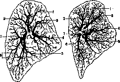
Figure 18. Diagrams with X-rays of the arteries of the left upper lobe: I - scattered type; II - main type; A - upper branch of a. pulm.; B - anterior; C - lower branch; 1 - ramus mediast. a ascend.; 2 - ram. costo-mediast.; 3 - a. obliqua sup.; 4 - a. transv. mediast.; 5 - a. obliqua inf.; 6 - a. obliqua margin.; 7 - a. transv. interlobaris. (According to Melnikov.) On the first of them are depicted the most large bronchi, pulmonary arteries and veins in the central zone of the L.; on the second the same radially diverging branches are presented schematically - in the form of colored sectors, the latter being located according to their position in the L. From these diagrams it is seen that the white sector, corresponding to the bronchus, always lies between the artery and the vein; moreover in the upper lobe at the top on both sides the artery occupies a medial position in relation to the bronchus, the vein - lateral. To solve the question of their relationships in other parts of the lung, it is only necessary to shift the entire group of 3 sectors along the circle, on the left - along the arrow, on the right - against the clock arrow. Thus in the lateral parts of the L. the artery is found cranially from the bronchus, the vein - caudally; in the lower part the artery is lateral, the vein - medial. This scheme can help the surgeon to understand these relationships when tying vessels in case of operations in the area of the L. lobes. The system of bronchial vessels - see Bronchi, insert table. The nutrition of the lung tissue, bronchial branches, vessel walls, lymph nodes, connective tissue and pleura is carried out by the system of bronchial arteries (aa. bronchiales). They depart mostly from the concave side of the aortic arch, but can also come from other sources. Variations relate predominantly to the right bronchial artery. Thus, cases are possible of its departure from a. oesophagea, very rarely from a. thyreoidea inferior, from the 3rd and 4th a. intercostalis, or a. mammaria interna, subclavia and intercostalis suprema. On each side for each L. two systems of bronchial arteries are distinguished: the anterior - from a. pericardiaco-phrenica (not constant) and the posterior - from the above-mentioned sources. Upon entering the L. the arteries divide according to the division of the bronchi [see separate table (p. 423-424), fig. 5], widely anastomosing in their branches, not only lobar, but also with the arteries of the other side (Melnikov; 1924). The bronchial arteries divide into the superficial and deep system (Konashko; 1926). The branches of the first go subpleurally in a radial direction from the hilum to the edges of the mediastinal and interlobar surfaces of the L., then go deeper and anastomose, as was indicated above, with the branches of the pulmonary artery. The deep system consists of branching arteries accompanying the bronchi from the hilum deep into the lung parenchyma, thin and tortuous. They widely anastomose with each other, give many lateral branches and form a wide-meshed, stretched along the surface of the bronchi network. The area supplied by them ends in most observations by the system of bronchioli respiratorii, where the terminal branches of the bronchial arteries anastomose with the network of pulmonary arteries. The venous outflow of the 'nutritive' system through the capillary network and small veins is directed into the pulmonary veins, while part of the blood is collected into separate trunks, which, gradually merging, form for each lung two to three bronchial veins, flowing on the right into v. azygos, and on the left - into v. hemiazygos. The lymph outflow occurs through 2 systems: 1) the superficial subpleural network, the flow of which washes the entire surface of the lung in the direction to its hilum, where this network of pulmonary vessels flows into the lymphatic glands of the hilus (Igl. broncho-pulmonales) (see Bronchial glands), and 2) the deep system, widely anastomosing in its initial origins with the superficial one. It consists of vessels merging radially from lymph networks surrounding the alveoli to the place of convergence of the branches of the bronchial tree; further these small vessels run along the enlarging bronchi and direct the lymph to the above-mentioned nodal lymph glands of the L. hilus (Igl. broncho-pulmonales). Along the way the deep system is interrupted by lymph nodes located in the lung tissue, in the angles between converging bronchi (Igl. pulmonales) [see vol. VIII (p. 158), fig. 6]. The innervation of the L. - see Vegetative nervous system, k Esipov.
III. Histology and embryology. Histology. The lung is usually compared to a complex tubulo-alveolar gland, in which the role of excretory ducts is played by the bronchi and their branches, and to the glandular cells correspond the alveolar passages and pulmonary alveoli. In details the description of the L. differs considerably in different authors, and to this day no fully established view on the structure of individual parts and uniform nomenclature exists. French authors (Charcot, Laguesse, d'Hardiviller, Grancher, Prenant) in describing the L. proceed from the anatomical unit - the pulmonary lobule (lobulus pulmonaris), clearly visible on the surface of the lung in the form of pulmonary fields of polygonal shape 0.6-1.0-2.8 cm in diameter, separated from each other by dark lines. German authors (Rindfleisch, F. E. Schultze, Kolliker, Ebner, Oppel) call these anatomical lobules secondary (sekundare Lappchen) and consider as the unit primary lobules (primare Lapp, acini) of smaller size (0.5-2 mm), clearly visible only in children and in some animals. For a complete understanding of the fine structure of the L. it is more convenient first to follow the path of the French authors. The pulmonary lobules (lobuli), bordering the surface of the lungs, have a pyramidal shape with the apex turned inward, while those lying deeper have a more complex, multifaceted shape; they are separated from each other by layers of connective tissue (septa inter lobularia), in adults usually black in color due to the presence of coal particles in them. The architecture of the lobule according to the scheme of Laguesse (Laguesse) is represented as follows (fig. 19). Into the apex of the lobule enters a bronchus of small caliber (bronchus lobularis or sublobularis), directed along its axis to its base (bronchus intralobularis); in the upper floor it gives off collateral bronchioles; on the border of the upper and middle third the bronchus divides into two branches of equal size, which dichotomously branch and on the border of the lower third give terminal bronchioles (bronchioli terminales Charcot) or acinar (bronchioli acinosi Grancher). Each such bronchiole gives rise to the terminal part of the L. - the acinus, or primary lobule of the German authors, which fill the lower floor of the lobule; the same acini depart from the collaterals forming directly or after division terminal bronchioles, and fill the free space of the upper and middle floors; the total number of acini in one lobule is 50-100. Of course the scheme of Laguesse, like any other scheme, only approximately conveys reality (see separate table, fig. 3 - photograph of a corrosion preparation of the lobule, according to Loeschke).
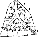
Figure 19. Diagram of the pulmonary lobule: 1 - bronchus intralobularis; 2 - collateral bronchus; 3 - acinus. (According to Laguesse.) The acinus, or primary lobule, is the essential part of the lung parenchyma: in it the gas exchange between the blood and air takes place; it has the form of a flattened cone and is separated from neighboring lobules by thin connective
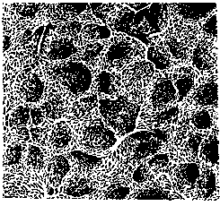
Figure 1. Syphilis of the lung: 1-whitish and pigmented scars; 2-sclerosis of the interlobular septa. Figure 2. Gumma of the lung; 2-large vessels and bronchi of the lung hilum. Figure 3. Pulmonary lobule: 1-site of a missing acinus (corrosion preparation according to Loeschke). Figure 4. Pulmonary alveolus: 1-network of elastic fibers; 2-capillary network. Figure 5. Leiomyoma. (Figure 34 from Braus.) To the article Lungs, Myoma, with connective tissue septa (Braus's scheme). Bronchiolus terminalis (acinosus, minimus) normally divides into 2 branches: bronchioli alveolares Braus (according to former terminology br. respiratorii Kolliker), on the walls of which hemispherical protrusions appear-pulmonary alveoli, or alveoli. Then the lumen of the bronchioles expands; they divide several times, forming so-called alveolar ducts (ductuli alveolares), which end in blind sacs (sacculi alveolares; according to old terminology-funnels, infundibula Rossignol). The walls of the alveolar ducts and sacs are completely covered with alveoli; the total number of sacs in one acinus reaches 60 (Braus). Some authors (Miller) give more detailed subdivisions of this alveolar tree (arbor alveolaris): the section directly adjacent to the bronchiole (vestibulum) passes after its division into an expanded space (atrium); behind it, after a ring-shaped narrowing, comes the alveolar duct. Others (Braus) deny the existence of such subdivisions in the normal lung, considering them pathological changes or artificially caused expansions. It should be borne in mind that all such studies were performed on injected lung preparations (see below), and their injection easily causes deformations; therefore, measurements of individual sections may give different figures. (Structure of bronchiole walls-see Bronchi.) The wall of pulmonary alveoli consists of a single-layer flat epithelium, so-called respiratory, basement membrane, and capillary network. The respiratory epithelium, the boundaries of which can be detected by treatment with silver nitrate, consists in mammals of two types of cells: small granular cells of cubic shape containing a nucleus, and large light anucleate cells having the appearance of thin plates; the first lie in the spaces between capillaries, the second-over capillaries and from their pressure they are flattened and lose their nuclei (fig. 20). In amphibians, only one type of cell exists, the nuclear areas of which lie between capillaries, and the parts covering capillaries are flattened into thin plates; according to Ostwald (Orpel), the epithelium of mammals is similarly constructed. The basement membrane in alveoli is either structureless or finely fibrous; individual nuclei have been described in it; in places where neighboring alveoli touch, between them is placed one common membrane. A characteristic feature of the basement membrane are networks of elastic fibers 1.0-4.5 μ in thickness, running in different directions (see separate table, figure 4); at the rounded edge of the alveolus they thicken and form closing rings. At this place, authors also note the presence of smooth muscle fibers; in larger quantities they are found between the walls of alveolar ducts, forming sphincters at the beginning of alveolar sacs. Branches of a. pulmonalis, going along the tract of alveolar bronchioles, form in the alveoli a dense capillary network (figure 21), inserted into the basement membrane in such a way that the endothelium of capillaries directly adjoins the anucleate plates of the respiratory epithelium. Venules do not follow the course of arterioles, but depart into the connective tissue at the periphery of lobules. Inside the acinus, connective tissue between alveoli and ducts is present in negligible amounts; there is more around bronchioles and at the periphery of acini; more significant accumulations are in the septa between lobules around bronchi and vessels. The connective tissue is always infiltrated with lymphocytes, forming follicles in places; histiocytes and macrophages that have absorbed black coal particles are encountered in it. Lymphatic vessels pass through the connective tissue, the beginnings of which lie between alveolar ducts.-Nerves of the lung, according to the descriptions of authors (Berkley, Retzius), form plexuses around bronchi, bronchioles, and between alveoli; their endings in alveoli have not been studied. In the lumen of alveoli, flat or round cells are sometimes encountered, often containing grains or coal particles, so-called pulmonary or dust cells (Staubzellen). Their origin is different: partly this is desquamated epithelium, partly emigrated leukocytes and histiocytes of the macrophage type; in pathological cases their number increases.-The question of the openings (stomata) between two neighboring alveoli has caused and continues to cause great controversy. Some authors describe them as a normal phenomenon (Malpighi, Magendie, Delafield, Roosevelt, Hansemann, Nicolas, Braus), others, on the contrary, deny their existence in the normal lung and consider them an artificial product or a pathological phenomenon (Moleschott, Rossignol, Kolliker, Fr. E. Schultze, Ebner, Miller). On ordinary lung sections, a picture is obtained that can be interpreted only knowing its structure: sections of alveolar ducts, alveoli, bronchioles form a tissue resembling lace. Relatively rarely, when the section passes along the axis of the terminal bronchiole, it is possible to see the branching off of alveolar ducts from it (fig. 22). In the thicker crossbars of connective tissue between lobules, sections of bronchi of various calibers, arteries and veins are encountered.
Figure 21. Capillary network of the human pulmonary alveolus (according to Kölliker).
The development of the lungs begins at a very early stage (human embryo of 3 mm) in the form of a sac-like protrusion of the entoderm of the pharyngeal intestine, which then branches, giving rise to the bronchial tree (see Pharyngeal intestine, Bronchi). The entodermal rudiment is soon covered by mesenchyme, forming rudiments of the lungs, which initially have a smooth surface. In an 8 mm embryo, grooves appear on the surface of the lungs, separating the lobes of the lungs; initially 3 lobes form on the left lung, 5 on the right, then their number decreases due to fusion; in a 3 cm embryo the lungs acquire their final external form [see separate table (art. 415-416), fig. 2]. Inside the lungs, as the bronchial tree develops, the amount of mesenchyme around the bronchi decreases, and it forms larger accumulations around the terminal branches, outlining the primary lobules. The development of pulmonary alveoli begins shortly before birth in the form of thickenings of the walls of bronchioles; alveoli in the form of protrusions form only after the first respiratory movements; at the same time, the cubic epithelium lining the terminal sacs transforms into respiratory epithelium. After birth, the growth of the lungs continues, associated with their restructuring: new alveolar ducts and sacs are formed, and the former ones are transformed into respiratory bronchioles (Vogt); at the same time, secondary lobules are formed by fusion of primary lobules.
Injections of the lungs. For studying the bronchial tree and the configuration of pulmonary lobules, the method of filling the lungs with a liquid mass capable of hardening, followed by the destruction of soft tissues, is widely used, as a result of which a cast is obtained-the so-called corrosion preparation [see separate* table (art. 431-432), fig. 31. The injection is usually performed through the trachea or large bronchi using a funnel or injection syringe; sometimes preliminary evacuation of air is practiced. As a corrosion mass, Wood's fusible metal is most often used (2 parts lead, 2-tin, 4-bismuth, 1-cadmium; melting point 75.5°), for which it is necessary to preheat the chest cavity. From other masses, a mixture of wax and mastic (Hyrtl), a solution of shellac in alcohol (Houel), collodion (Schifferdecker), celluloid (Storch) have been used, coloring the colorless masses with vermilion or Berlin blue. For dissolving the soft parts, antiformin, a warm solution of caustic potash are used; after collodion-pepsin with HCl. After injection of collodion, the preparation can be sectioned and the reconstruction method (Miller) can be applied. Loeschke achieved good filling of alveolar ducts with Wood's alloy, after pre-drying the inflated lung.
V.
Karpov. VI. Pathological Anatomy. Among postmortem changes in the L., postmortem hypostases (see) are most frequently encountered, which manifest themselves in the posterior parts of the L. (when the corpse is lying on its back) appearing dark and, on section, exuding an abundant amount of dark blood. Also of considerable importance is the postmortem acid softening of lung tissue (pneumomalacia acida), which occurs as a result of atonal or postmortal penetration of gastric juice into the L. Atonal penetration occurs through aspiration of vomit; after death, gastric contents can enter the L. either through the respiratory tract (e.g., during artificial respiration) or through the wall of the stomach, diaphragm, and pleura. Areas of acid softening stand out in the lung tissue as soft, moist foci of dirty gray-green color, sometimes with the formation of a liquefaction cavity in the center; they differ from rather similar foci of lung gangrene by the presence of an acid reaction, determined by litmus paper, and microscopically by the absence of any signs of inflammation and the presence of food residues. Developmental defects of the L. most often manifest as abnormal lobulation of the L. and 2 or 4 lobes in the right lung, 3 lobes in the left lung (see development of L.) - or else as incomplete division of the lung into the usual lobes; complete absence of division of the L. into the usual lobes may also be observed. More rarely, various types of accessory L. are observed, usually located in the costal pleura, less frequently - in the mediastinal connective tissue, and even less frequently - under the diaphragm in the abdominal cavity; accessory L. always have their own special bronchus, which branches off either from the trachea or from one of the two main bronchi. Very rarely, complete absence of formation of one or both L. is encountered (see Apneumia). In addition, cases of underdevelopment (agenesis) of lung tissue with good development of the bronchial tree of the lung occur, which is accompanied by significant expansion of the bronchi, i.e., the formation of congenital bronchiectases (see). A very rare developmental defect is congenital hyperplasia of both L., expressed in their large volume due to an increase in interstitial connective tissue, as well as in the quantity and volume of alveoli. Some cases of this kind are close to the so-called congenital cystic L., which, according to some authors, is a congen adenoma of the L. (Changes in the volume of pulmonary alveoli - see Atelectasis, Emphysema of the L.) Of endogenous deposits in the L., amyloid is very rarely observed, which in some cases of general amyloidosis manifests as deposition of amyloid along the course of the lung capillaries. Much more common is the deposition of salts in the L., and usually this is a matter of dystrophic petrification of various dead masses, most often tuberculous caseous foci; less commonly, there is deposition of lime salts of the metastatic lime type, expressed in the petrification of the walls of lung capillaries and small arteries. - Deposition of hemosiderin in the L. - see Brown induration of the lung. Of circulatory disorders in the L., hyperemia of their tissue is very often observed, and arterial hyperemia, manifested by an overflow of blood in the small arteries and capillaries, is most frequently encountered as a phenomenon related to the inflammatory process (at the beginning of inflammations of the L., in acute miliary tbc, when inhaling irritating gases, etc.). An influx of blood to the L. can also occur with rapid release of the L. from any pressure on it, for example, when emptying the pleural cavity of fluid that was pressing on the L., as well as with a decrease in atmospheric pressure, for example, when ascending to great heights, when quickly exiting from caissons. The result of rapidly developing arterial hyperemia can be hemorrhages into the lung tissue, sometimes of considerable size, as well as acute transudation of fluid into the alveolar cavities. Venous, congestive hyperemia of the L. is observed with difficulties in the outflow of blood through the pulmonary veins, which most often occurs with heart defects, mainly with defects of the mitral valve, especially if this is accompanied by weakening of heart activity and a drop in pressure in the pulmonary artery; as a result of rapid weakening of heart activity, acute blood congestion in the L. can develop, uniformly distributed throughout all parts of the L., which in such cases are somewhat increased in volume, heavy, and diffusely dark bluish-red in color; on section, a foamy dark blood flows out. If the difficulty in blood outflow exists for a long time (e.g., in the form of persistent narrowing of the mitral valve), then a long-lasting venous congestion, uniformly involving both L., may not be significantly expressed in terms of clearly noticeable hyperemia of lung tissue, but at the same time is accompanied by diapedesis of erythrocytes and deposition of hemosiderin in the lung tissue, which gives in the L. a picture of brown induration of the lung (see).-Weakening of heart activity, expressed in a gradually increasing decrease in the force of its contraction, leads to a weakening of blood entry into the left heart and consequently to the movement of blood in the vascular system of the small circle of blood circulation, which ultimately leads to blood retention, mainly in the lower parts of the L., which become dark bluish-red, dense, and on section give an outflow of an abundant amount of liquid dark blood; this type of congestive hyperemia is called hypostatic (see Hypostasis).-Microscopically, in venous hyperemia in the L., dilation and overflow of blood in the veins and capillaries are found; the latter, in addition, are elongated, snake-like, and curved, and as a result protrude into the alveolar cavities; one or another amount of erythrocytes that have entered the alveoli by diapedesis and signs of edema (see below), which very soon join the congestion, complete the picture. Hemorrhages in the L. can have very diverse origins. In addition to various types of traumatic injuries to lung tissue, the main significance here is the destruction of blood vessels by various destructive processes; in this regard, tbc comes first, followed by gangrene of the L., abscess, syphilis, malignant tumors. In the above cases, as well as in hemorrhages due to rupture of an aneurysm of a branch of the pulmonary artery in a tuberculous cavity, bleeding (hemoptysis) can be very significant, often fatal. Hemorrhages into lung tissue by diapedesis occur in congestive hyperemia of the L., in the formation of an infarct in the L. (see below), in many forms of inflammation of the L. (see Pneumonia), in various types of hemorrhagic diathesis, in reflex vaso-motor disorders of the state of the vascular wall, e.g., in lesions of the brain and its membranes. Foci of hemorrhages in the lung tissue appear as poorly defined areas of dark red color, of rather dense consistency; the microscope in such places reveals the presence of blood elements in the alveolar cavities. It should be borne in mind that blood in the L. can also enter secondarily by aspiration from extrapulmonary parts of the respiratory tract, from the oral cavity, from the nose; sometimes such aspiration of blood into the L. occurs with profuse bleeding from the stomach, from the esophagus. Embolisms in the L. are by no means rare; most often their source are detached parts1 of thrombi from the right heart and veins of the large circle of blood circulation (veins of the lower extremities, pelvis, genital organs); smaller emboli can originate from endocarditic deposits on the valves. In fat embolism, observed as a result of trauma to fatty bone marrow in fractures of bones, during surgical operations on bones, as well as as a result of damage to fatty tissue, less frequently - fat-rich liver, the capillaries of the L. are the main place of retention of the fat that has entered the blood. Cellular embolism often occurs in the L.; for example, after childbirth, syncytial elements of the placental villi are often found (about 70%) in the capillaries; as a result of body bruises, as well as in various infectious diseases, bone marrow cells, megakaryocytes, are carried into the L.; less frequently, embolism with liver cells occurs as a result of damage to the latter. Infectious, mycotic emboli most often occur in purulent inflammatory processes that spread to the veins (purulent otitis, parotitis, postpartum purulent processes in the uterus, parametrium, etc.); they can also be observed in ulcerative endocarditis. The consequences of embolism in the L. can be various. Obstruction of one of the main branches of the pulmonary artery by a large part of a thrombus usually leads to rapidly occurring death; smaller emboli are carried deep into the lung, where depending on their size they are retained in arterial branches of one or another caliber. Experience shows that usually emboli preferentially go to the lower parts of the L.; there is, however, an opinion (Kretz, Helly) that emboli are carried to the lower parts of the L. mainly from the system of the inferior vena cava, whereas from the superior vena cava they more often go to the upper parts of the lungs. With maintained good blood circulation in the L., embolization of small and medium arterial branches does not lead to any circulatory disorder; the embolus can only serve as the basis for further thrombus formation.
In those cases where there is a weakening of the circulation in the lungs, embolism of an arterial branch leads to the formation of an infarct, which in the vast majority of cases is hemorrhagic, very rarely ischemic (see Infarct). Small emboli, blocking individual capillaries, as occurs in cellular embolisms and in small fat embolisms, do not cause any subsequent changes, and subsequently the emboli are absorbed (the fat is previously saponified); however, with widespread obstruction of the capillaries of both lungs, the respiratory function ceases, and death quickly occurs, as is the case with abundant fat embolism. Infectious emboli with pus-forming bacteria cause the development of abscesses at their sites of deposition; if there was an embolism with a fragment of a thrombus containing pus-forming or putrefactive bacteria, then an infarct may first form, which then undergoes suppuration or gangrenescence. Thrombosis of the lung vessels may be a consequence of embolism or develops independently; the latter can be observed in patients with weakened circulation in the small circle as a manifestation of marantic thrombosis. Edema of the lungs manifests as an accumulation of transparent fluid containing a small amount of protein in the cavities of the pulmonary alveoli. The lungs in edema become heavier, lose their elasticity (when pressure is applied to the surface of the lungs, a pit remains); when the lungs are compressed from the surface of the incision, a more or less significant amount of fluid flows out, usually foamy due to the admixture of air bubbles, which are absent only in severe edema; with simultaneous plethora, the fluid is bloody due to the admixture of blood from the cut vessels. On microscopic examination, the presence of protein fluid in the alveolar cavities is found, often with an admixture of individual alveolar epithelial cells; usually there is also edema of the interstitial tissue. If edema affects a part of the lung that is in a state of atelectasis and plethora, then the tissue in such a place resembles the tissue of the spleen (splenization). Edema of the lungs most often develops as a result of weakened heart function, joining to stagnant plethora (stagnant edema) and in such cases being concentrated mainly in the posterior-lower parts of the lungs; at the same time, it can be a partial manifestation of general dropsy of stagnant origin or affect only the lungs. Stagnant edema of the latter kind is usually observed in acute cardiac failure (acute edema of the lungs) or in agonal irregular heart function (weakening of the contractions of the left heart with more or less satisfactorily continuing work of the right heart). In addition, edema of the lungs can develop as a result of toxic damage to the lung tissue and its capillaries (toxic edemas), which occurs when inhaling ether, nitrogen vapors, asphyxiating warfare substances, in sepsis, uremia. Inflammatory edemas of the lungs can be considered as a manifestation of serous inflammation of the lungs, which is also indicated by the higher protein content in the fluid in this type of edema and the presence of leukocytes in it; sometimes - deposition of fibrin; inflammatory edema occurs in the lungs either as collateral edema around areas of more intense inflammation or as an independent inflammatory process (e.g., in influenza, plague, anthrax, some septicemias). Inflammation of the lungs - see Pneumonia. Changes in the lungs in glanders - see Glanders. Pulmonary form of anthrax - see Anthrax. Pulmonary form of plague - see Plague. Fungal lesions of the lungs - see Bronchomycoses, Pneumomycoses. Dust diseases of the lungs - see Anthracosis, Pneumoconioses. Tuberculosis of the lungs - see Tuberculosis of the lungs. Actinomycosis of the lungs - see Actinomycosis. Lymphogranulomatosis most often secondarily spreads to the lungs from the lymph nodes of the mediastinum; usually in these cases, the formation of tumor-like nodes of granuloma with a characteristic structure occurs at the gates of the lungs or generally in their mediastinal parts (see Lymphogranulomatosis). It is also possible for lymphogranulomatosis to spread from the mediastinum retrogradely along the lymphatic pathways and for the formation of granulomatous nodes in parts of the lungs distant from the mediastinum. Primary lesion of the lungs by lymphogranulomatosis belongs to great rarities.
A. Abrikosov. X-ray Diagnosis. X-ray examination of the L., along with percussion and auscultation, is one of the main clinical methods for studying the L. Under normal conditions, the L., containing air and therefore only slightly retarding X-rays, appear on the film or screen as light, so-called lung fields; each lung field is bounded on the medial side by the mediastinal shadow, more accurately the shadow of the heart and large vessels, below by the dome of the diaphragm and subdiaphragmatic organs, and on the lateral side by the wall of the chest. The left lung field in its lower part is somewhat narrower than the right due to the asymmetrical position of the heart. The lung field is separated from the apex (or apical field) by the intense bony shadow of the clavicle. The ribs, crossing the lung field obliquely, stand out very contrastingly against the light lung background. The anterior inner ends of the ribs, specifically their cartilaginous parts, are permeable to X-rays; therefore, in the anterior position of examination (i.e., with dorsoventral ray direction), when the invisible cartilaginous ribs are adjacent to the screen or film, large areas of lung tissue are revealed, and this position of examination is more convenient than the posterior (ventrodorsal). The flat image of the scapula little interferes with the examination of the L. Very massive mammary glands in women, as well as strong pectoral muscles or thick fat coverings can significantly darken the corresponding areas of the lung fields. Pathologically unchanged pleura is not visible radiologically. Under normal conditions, the boundaries between individual lobes of the L. are not delineated. From a radiological point of view, the most accurate localization of some pathological formation is not according to lobes, but according to ribs and intercostal spaces or to zones. There are three such zones: 1) upper, or supraclavicular, extending from the clavicle to the horizontal line drawn through the attachment point of the II rib to the sternum; 2) the middle zone from this conventional line to the corresponding horizontal line of the IV anterior rib and 3) the lower zone, or supradiaphragmatic. The lung field is divided into three zones along vertical lines as follows: the clavicle within the lung field is divided into three parts; similarly, the diaphragmatic arch is divided into thirds, and the corresponding boundaries of these thirds on the clavicle and diaphragm are connected to each other by lines. The three zones resulting from this division, namely: 1) medial, or paravertebral, 2) middle and 3) axillary zones, are equal in width. The lymph glands in the root of the lung, as well as the large bronchi of the 1st and 2nd order, under normal conditions do not lie outside the hilus zone. The extensive part of the lower lobe of the L., located in front and especially behind and below, adjacent to the slopes of the diaphragm, is not visible at all when examining the chest in anterior and posterior positions, because under these usual conditions of radioscopy and radiography, the shadow of this part of the L. is projected onto the shadow of subdiaphragmatic organs. Therefore, examination in lateral and inclined positions is necessary here, when the central ray glides along the frontal plane of the body or parallel to the surface of the diaphragmatic slope. The areas of the L. covering the heart in front are also difficult to examine radiologically. The lung fields do not appear homogeneous; on their light background extend separate linear and branching shadows, diverging fanwise from the hilus of the L. to the periphery of the lung field and constituting the so-called lung pattern [see separate table (pp. 447-448), Figure 1]. In the area of the hilus of the lung on both sides, adjacent to the mediastinal shadow is a darker, dense shadow complex, which is designated as the hilus pattern or so-called hilar pattern. At present, it has been finally established that the paravertebral and lung patterns represent the projection shadow of intrapulmonary vessels, mainly branches of the pulmonary artery. Bronchi hardly participate at all in the formation of these normal shadows. The picture of the hilus of the lung and the lung pattern changes greatly depending on the degree of filling of the pulmonary vessels with blood. The dark round and oval isolated areas in the hilar shadow itself are the combined shadows of large vessels and mainly the projections of large, so-called orthoradientgenographic branches of the pulmonary artery, i.e., vessels that are located along the path of the rays, for example, those going in the usual anterior position of examination from front to back or from back to front. These shadows are constantly mistaken for lymph glands, which are not visible at all during radioscopy or radiography of the chest. Orthoradientgenographic large bronchi in the area of the hilus are outlined as ring-shaped shadows; with insufficient familiarity, they can simulate cavities (see). A complete radiological examination of the L. consists of radioscopy (fluoroscopy) and radiography (film or plate exposure). Fluoroscopy makes it possible to examine the L. on the screen in all positions, with oblique and inclined ray direction, to study the mobility of the diaphragm and ribs. The data obtained by fluoroscopy are supplemented by the radiograph. On the film, a more detailed structural pattern of the normal and pathological L. is obtained, and therefore the film has greater probative value. The analysis of pathological shadow images of the L. consists in determining all their elementary properties, namely: number, position, shape, size, intensity, pattern, contours and mobility. To these basic radiological signs can be reduced all the infinite variety of pathological changes in the L. In the clinic of abscess and gangrene of the L., the role of radiological examination is very great and has special significance in localizing the purulent process in the deep central parts of the L., in the area of the hilus of the lung and in the upper lateral sections, where clinical recognition is difficult. Radiological examination is completely irreplaceable when assessing indications for surgical intervention: the location, size and shape of the cavity are determined with precision on the film and screen, reactive changes in the surrounding lung tissue, the presence of a demarcation ridge, without which active intervention is contraindicated, pleural adhesions, etc. Radiologically, abscess and gangrene of the L. appear as more or less limited darkening of the lung field. Most often the focus is located in the middle zone of the lung field. In most typical cases, a lightened area is found in the center of the darkening. In an abscess, this cavity has a spherical shape with smooth inner walls, while in gangrene it has an irregular, map-like shape. In half of all cases of abscess and gangrene of the L., the cavity contains liquid decay with an upper horizontal level-indicating communication of the cavity with a bronchus [see separate table (pp. 447-448), Figures 6,7 and 8]. The antemortem recognition of multiple foci of decay, important due to the poor prognosis inherent in this form, is possible only with the help of X-rays. The radiodiagnosis is difficult in cases of complication of the basic disease by empyema, as well as in the first stage-pneumonic infiltration, when there is yet no decay or when the cavity does not communicate with the bronchial lumen. In actinomycosis of the L., the radiological picture is very diverse, little characteristic, and only in rare cases has decisive importance for establishing the etiological diagnosis. The actinomycotic infiltrate in lung tissue sometimes lies in the lung field as an isolated focus of darkening of large size, very intense, homogeneous or coarsely mottled, with uneven contours and radiating in all directions with pointed strands. In some cases, the infiltrate has lobar distribution and occupies a large area of the lung field with solid darkening, thus simulating on the radiograph a massive inflammatory infiltrate of any other origin or a lobar neoplasm. When the fungal process spreads into the L. from the esophagus, the radiological picture resembles a primary cancerous tumor in the hilus or a lymphosarcoma of the mediastinum. The undoubted significance for etiological recognition is the accompanying picture of multiple destructive processes in the ribs. The importance of this radiological sign, however, is diminished in those cases where there are already fistulas or superficial typical hard infiltrates, and the diagnosis of actinomycosis can be made more accurately on the basis of microscopic examination of pus or puncture. The radiological picture of syphilis of the L., as well as the clinical and pathological anatomy of tertiary pulmonary syphilis, despite the great interest in this disease, has not yet been sufficiently clearly studied. Of all forms of syphilis of the L., the greatest diagnostic value is the picture of the so-called sclerosis of the hilus of the L. in young patients, especially when the lesion has a unilateral localization (most often on the right side). On the radiograph, there is a marked intensification of the hilar and lung patterns, corresponding to the rich development of perivascular and to a lesser extent peribronchial connective tissue. This picture has some similarity with primary bronchial cancer and can be a cause of diagnostic error.
Significant difficulties are presented by the etiological x-ray diagnosis of gummatous infiltration of the L. The number of gummas in the lung tissue varies; both solitary and multiple infiltrates are encountered. Their sizes also vary widely: from miliary or submiliary gummas to large lobular and even lobar shadows. Extensive gummatous infiltrates are usually located in the middle or lower zone of the right lung, in the upper or middle zone. A correct diagnosis here is possible only when taking into account all the data of clinical research. Among the circulatory disorders of the L., practical significance is mainly attached to the x-ray examination in cases of congestion in the lesser circulation. In difficult clinical cases, especially in diagnosing the so-called central congestion, i.e., congestion mainly in some large branches rather than in the capillaries, x-ray diagnosis is irreplaceable. X-rays make it possible to determine the cause of congestion in the lesser circulation and to carry out differential diagnosis. Congested L. are determined radiologically by the general uniform darkening of the lung fields with enhancement of the lung pattern, and these phenomena are equally expressed on both sides. The shadow of the hilum of the L. is significantly widened and intensified. With significant congestion, individual irregularly shaped and round dense shadows with blurred contours appear, corresponding to the dilated arterial branches of the pulmonary trunk; these shadows are most densely located in the area of the hili and in the lower parts of the L. In emphysema of the L., the x-ray picture is characterized above all by unusually bright lung fields. There is no great difference in the degree of transparency of the L. during inspiration and expiration; emphysematously distended L. do not show it, i.e., in contrast to normal conditions, the lung fields do not lighten noticeably during deep inspiration. The chest cage, especially in its lower parts, is widened, and the lung fields and the retrocardiac field are also widened. The ribs and clavicle are positioned horizontally, the intercostal spaces are very wide, and in most cases the costal cartilages are calcified. The diaphragm is flattened, stands low, and the costophrenic angles on both sides are enlarged and approach a right angle. The respiratory mobility of the diaphragm is limited. The heart is in a vertical position and consequently has a reduced transverse diameter. The shadows of the lung hili are intensified. The x-ray examination plays an important role not so much for diagnosing emphysema itself, the diagnosis of which can be made quite reliably on the basis of percussion and auscultation data alone, but for detecting changes in the lung tissue, first of all tuberculosis and cancer of the L. Great difficulties are presented by the precise x-ray diagnosis of initial small degrees of emphysema. In tumors of the L., x-ray examination at present is one of the basic methods. Among the benign tumors of the L., the rarely occurring chondroma gives an extremely typical, almost pathognomonic picture on the x-ray, namely—a large spherical or ovoid shadow with smooth outer contours, and in the central parts of this shadow, separate amorphous calcifications are found. In the x-ray image, chondroma differs from the calcified echinococcus in that with echinococcus the calcifications are located superficially, not in the depth of the round shadow. Very rare fibromas and myomas of the L. are outlined radiologically as very voluminous homogeneous shadows with even contours, also of a regular, usually ovoid shape. Primary cancer of the L. is in the vast majority of cases primary bronchial cancer and as is known (see Bronchi, bronchial cancer) it occurs in the form of the so-called hilar cancer, lobar cancer, and the rarest primary miliary cancer of the lungs [see separate table (pp. 447-448), Figure 2]. When reading x-rays, it must be kept in mind that hilar and lobar cancer in their pure form are observed relatively infrequently; usually the x-ray picture is complicated by bronchostenosis with secondary obstructive emphysema or more often obstructive atelectasis, accumulation of fluid in the pleural cavity; large tumors as a rule undergo necrosis and disintegration in the center with cavity formation and subsequent gangrene, etc. Primary sarcoma of the L. is observed in x-ray examination either as an isolated spherical shadow in the lung field similar to benign tumors or as a lobar shadow indistinguishable from primary cancer [see separate table (pp. 447-448), Figure 3]. Metastatic tumors of the L. (cancer, sarcoma, hypernephroma) give an extremely characteristic x-ray picture of great practical diagnostic significance. Often metastases are determined radiologically earlier than any objective and subjective clinical symptoms from the L. or even from the primarily affected organ. In metastases of the L., against the background of light, slightly emphysematously distended lung fields, single or as a rule multiple—sometimes innumerable small and large round shadows appear. The contours of individual metastases are either sharply defined or—with infiltrative growth—blurred and notched [see separate table (pp. 447-448), Figs. 4 and 5]. Based on x-ray signs, differentiation between cancer, sarcoma, and hypernephroma is in most cases impossible.
s.
Reinberg. VI. Tumors. Among benign tumors of the L., chondromas are the most frequent; they are discovered as an incidental finding during autopsies in the form of small (0.5-2 cm in diameter), round, dense nodes, located either deep in the lung tissue or, more often, under the pleura, without any anatomical connection to the bronchi. Under the microscope, a lung chondroma consists of hyaline or reticular cartilage, often divided by connective tissue septa into several fragments; very often in these connective tissue septa and in the capsule of the chondroma, flattened slits covered with cubic or cylindrical epithelium are found. Chondromas of the L. apparently have a congenital origin, developing from detached parts of the bronchial wall during embryonic development. Chondromas sometimes undergo ossification. Subpleural chondromas may protrude into the pleural cavity, forming so-called free bodies.-Much less frequently, small fibromas, lipomas, and myomas are found in the L., sometimes multiple, located peribronchially or even protruding into the lumen of the bronchus; from the bronchial mucosa, round adenomas and papillomas may originate. If one of such tumors narrows the lumen of the bronchus, this serves as a cause for the development of chronic bronchitis, the formation of bronchiectases, etc. (see Bronchostenosis).-The existence of true osteomas in the L. is debatable. Most findings concerning the formation of bone tissue (usually with bone marrow) in the L. represent the result of metaplastic development of bone from connective tissue during its intermediate proliferation, during the encapsulation of caseous foci (e.g., of a tuberculous primary focus), during the formation of a scar. In particular, this includes the change designated as pneumopathia osteoplastica and consisting in that over a limited extent of the L. (more often in the lower lobes and in very elderly individuals) coral-like branching strands of bone tissue are found, embedded among little changed lung tissue. As isolated observations, gliomas and teratomas of the lungs have been described. . Of malignant tumors in the L., sarcoma rarely develops, Figure 1. Normal chest (female). Figure 2. Cancer of the right lung. The tumor occupies the entire upper lobe. Its lower edge is sharply defined. Figure 3. Primary sarcoma of the lungs. In the lower third of the left lung, directly above the diaphragm, there is a round-shaped tumor with sharply defined edges. The bronchial glands are also affected; they appear as dense, elongated formations with sharp edges. Figure 4. Metastasis in the middle third of the right lung in stomach cancer. Figure 5. Metastasis in the lower third of the right lung in cancer of the mammary gland in a male. The edges of the tumor merge below with the shadow of the right dome of the diaphragm. Figure 6. Abscess in the lower third of the right lung, in which there is air due to communication of the abscess with a bronchus; an air bubble and below it - a collection of pus with a horizontal level are visible. Figure 7. A large metapneumonic abscess of the left lung; a dense shadow with uneven edges, merging with the shadow of the vessels, is visible. Figure 8. Gangrene of the right lung; the darkening captures almost the entire lung and is uneven in density; small clearings are visible in the center and above. Figure 9. A round echinococcal cyst in the upper third of the left lung; its medial edge merges with the shadow of the vessels. The shadow of the cyst is clearly visible through the ribs. Figure 10. A round echinococcal cyst with sharp edges in the lower third of the left lung. Between it and the shadow of the heart, a strip of lung tissue is visible. The ribs under the shadow of the cyst are not sufficiently clearly defined (beginning calcification). Figure 11. Echinococcal cyst above the left diaphragm before fatty degeneration. Figure 12. Echinococcal cyst above the left diaphragm after fatty degeneration of the cyst. Figure 13. Multiple echinococcal cysts in the middle third of the right lung; the largest cyst is in the center. The shadows of the cysts are light, and the ribs are visible through them. Figure 14. A large echinococcal cyst in the right lung before its opening into a bronchus. Periapical inflammation in the lung tissue. (For illustration of the article Lungs). histologically usually belonging to polymorphocellular, more rarely to spindle-cell sarcomas; the tumor more often originates from the lower lobe of the L. and appears as an indistinctly delimited node of soft tissue resembling fish meat. Cancer is encountered much more frequently in the L. Cancers of the L. most often have their source of development in the epithelium of the bronchi and therefore belong to bronchial cancers (see Bronchi, bronchial cancer). Cancer from the alveolar epithelium, which is lung cancer in the narrow sense, is apparently encountered much less frequently. It should be noted that cancer from the alveolar epithelium does not possess clear pathological-anatomical and histological distinctive features; therefore, when it comes to cancer that has already reached considerable size, it is impossible to establish whether it originates from the alveolar or from the bronchial epithelium. Only in those cases where there is a small node, having no connection with a bronchus, located under the pleura and revealing the structure of a fine-cellular solid cancer from cells similar to alveolar epithelium, can one with a considerable degree of probability speak of cancer from the alveolar epithelium. Very often the L. are the site of formation of metastases of malignant tumors, which can penetrate the L. hematogenously (by the carriage of tumor cells in the blood stream) or lymphogenously due to the possibility of movement of tumor cells from the lymph glands of the mediastinum retrogradely into the lymphatic pathways of the L.; in addition, tumors can secondarily involve the L., spreading to them by contact of adjacent parts (e.g., spread to the L. of cancer of the esophagus, mammary gland, sarcoma of the mediastinum, cancer of the pleura). Hematogenously, sarcomas of various parts of the body, osteosarcomas, chondrosarcomas, malignant melanomas, chorionepitheliomas, malignant hypernephromas metastasize to the L.; comparatively more rarely - cancers. All these metastatic tumors form in the L. sometimes single, more often multiple nodes of round shape, of the most diverse sizes.-With lymphogenous spread of cancer in the L., no tumor nodes may form at all, and on a section only strands of whitish tissue along the course of the bronchi and along the interlobular septa are noticeable; under the pleura a network of whitish stripes corresponding to the pattern of the lobules of the L. is also often visible; at the intersections of the lymphatic pathways, small nodules visible under the pleura and on the section of the lung and which may be mistaken for miliary tubercles often form in such cases. This spread of cancer along the lymphatic pathways of the L. is often designated as cancerous lymphangitis of the L. (lymphangitis carcinomatosa); this term is incorrect, since in such cases there is no inflammation of the lymphatic pathways.
A. Abricosov. Clinic. Functional disturbances are observed only with malignant tumors of the L. They develop insidiously and begin to attract attention only when the tumor reaches significant sizes. The history usually provides no supporting data. The age of patients is usually 35-50 years, rarely older. Men (50-70% according to various reports) and hypersthenics predominate. The earliest and most persistent symptoms are dyspnea (often accompanied by stridorous breathing), dull pains in the chest and side (especially with sarcomas), and along the course of the nerve trunks of the brachial plexus (with pressure on it by the tumor), cough - often whooping-cough-like or else short but persistent and irritating to patients. Hemoptysis is very common: from a small admixture of blood in the sputum in the morning to prolonged discharge of gelatinous bloody sputum. Cases of sudden fatal hemorrhages have been described. The temperature is most often subfebrile with remissions and periods of abatement; with rapid tumor growth, especially with significant disintegration and complications of septic infection - febrile and even hectic. Examination: engorgement of the neck veins, venous collaterals on the corresponding side of the chest and back, puffiness of the face and neck (first described by Stokes), cyanosis of the face and mucous membranes (with compression of the superior vena cava), anisocoria with dilation of the pupil on the affected side, sometimes "drumstick fingers". Significant cachexia is often not observed, but the characteristic grayish-pallor of the face almost always attracts attention. The changes of the soft palate described by Neuda have not been confirmed by the author. Protrusion of the affected part of the chest wall is observed with sarcomas. Retraction of the affected half of the chest wall is always observed. - Palpation: at the sternal-clavicular-muscle junction, small metastatically affected lymph glands of cartilaginous density can often be found. Biopsy of them immediately decides the diagnosis. Sometimes fixation of the trachea is observed. Cases of compression of the subclavian artery and changes in the pulse on the corresponding hand are observed. - Percussion. In the area of the tumor, significant dullness or absolute dullness. Over adjacent areas with light percussion - tympany. Auscultation: almost always diminished breathing, often with bronchial sounds carried from the depth. There are no rales or they are few and not persistent. With disintegration and breakdown of the tumor into the bronchus, there are typical signs of a cavity. In rare cases of disseminated lymphogenous lung cancer - the auscultatory-percussive picture of emphysema (observation of Holzmann). With paralysis of the vocal cords (n. recurrens) - aphonia or hoarseness of the voice. Lung cancer is often accompanied by pleural exudate, usually sero-hemorrhagic or hemorrhagic. Persistently recurring after repeated aspirations, exudate without elevation of temperature even of a serous nature should arouse suspicion of a lung tumor. Laboratory studies [cytology of sputum (see) and exudate] sometimes provide valuable indications. Serological reactions (Abderhalden's, Davis's, Vilbushovich's, Kahn's, etc.) are unreliable and complex (see Abderhalden's reaction, Denis's reaction). Vidal's alimentary test also does not help in orientation. The red cell sedimentation reaction usually shows significant acceleration. Differential diagnosis of lung tumors must be made with tuberculosis, echinococcus, gumma, aortic aneurysm. The persistent absence of Koch's bacilli in the sputum and negative serological reaction with Besredka's antigen greatly help in the examination for tuberculosis. But often lung cancer is combined with destructive tuberculosis of the lungs, and this complicates the picture. The Wassermann reaction must be taken into account with great caution, as it is sometimes positive with lung tumors and in the absence of syphilis. To exclude echinococcus in doubtful cases, Weinberg's reaction (see Weinberg's reaction) or Casoni's test should be performed. Complication with abscess or gangrene of the lung greatly obscures the diagnosis, but with careful consideration of the history and the entire clinical complex, the diagnosis can still be made during life. Roentgenoscopy and roentgenography greatly facilitate and clarify the diagnosis. Atelectasis of the lung tissue around the tumor and in the system of the bronchus compressed by it gives diffuse, sometimes intense shadowing over a larger area than the tumor itself. Sometimes the borders of the tumor are sharply outlined and stand out against the transparent pulmonary pattern. With paramediastinal localization, it is necessary to examine the tumor from all sides to separate it from the aortic shadow and not confuse it with an aneurysm. For this, the patient should be turned in different directions and the screen and tube should be moved. Sometimes the tumor gives an irregular claw-like pattern or spreads in the form of massive shadows along the course of the bronchi. With exudate, the picture is masked by the latter, and in such cases, the application of artificial pneumothorax is of great help.--The prognosis is hopeless. Death - in 1/2-2 years; the outcome is often accelerated by complications from aspiration pneumonia or sepsis. The roentgenotherapy recommended by many has so far not given a convincing effect (see Bronchi, bronchial cancer). Surgical treatment - SEE BELOW.
B.
Holzman. VII. Abscess. An abscess of the L. forms as a result of disintegration or, more precisely, purulent melting of an infiltrate in one or another section of the L. with the formation of one or more cavities [see separate table (art. 463-464), fig. 5]. The infiltrate usually arises on the basis of acute pneumonia as a result of infection entering the lung either from the bronchial passages, or through the blood or lymphatic vessels, or as a result of damage to lung tissue in trauma in the form of compression of the chest in contusions of the chest and direct injury to lung tissue by cutting, piercing objects, as well as by projectiles (especially blind wounds). In the bloodstream, infectious agents can enter from various infected and purulent foci: in ulcerative endocarditis, in suppurations in the abdominal cavity, in the area of the uterus and appendages, the bladder, the rectum, in phlebitis, ulcers of the leg, in thrombosis of the sinuses, as a result of inflammatory processes in the middle and inner ear, in osteomyelitis, from surgical wounds, etc. In recent times, mention is made of causes of abscess of the L. from burns of the respiratory passages and lung from combat poison gases. In these cases, abscesses often arise after pneumonia caused by damage to the parenchyma. In trauma, infection can be introduced directly by a cutting instrument; at the same time, the injury can cause secondary infection of the lung parenchyma due to damage to the bronchial passages. Often an abscess occurs when foreign bodies enter the lumens of the bronchi. Finally, infection of lung tissue can occur as a result of the spread of infection by continuity, e.g., in suppuration of the glands of the root of the L., in subphrenic suppurations. In the latter case, pus makes its way through the diaphragm to the base of the L. This should also include the rarer cases of abscess of the L. in tuberculous abscesses of the spine, ribs, abscesses of the mediastinum and purulent pleuritis. All or many of these cases are in essence nothing more than particular cases of the spread of infection to lung tissue by lymphatic pathways. Relatively rarely does an abscess of the L. occur in general diseases, like glanders, more often in mycoses of the lungs. This variety of causal factors creates great diversity in path.-anat. and clin. manifestations. Various causes causing abscess of the L. give characteristic changes in lung tissue. In some cases the abscess is single-chambered, in others multi-chambered, in others multiple. Therefore, from a clinical point of view, it is extremely important to establish exactly the mode of origin of the abscess of the L. Thus, abscesses of the L. arising from suppuration in neighboring organs (liver, bronchial glands) are always single, as are metapneumonic abscesses. In embolic forms of pulmonary abscess, numerous foci are usually observed; the same is observed in grippe pneumonias and as a rule in aspiration pneumonias. In the development of abscesses, predisposing factors often play a major role: general weakening of the body, e.g., after bronchopneumonia, abscesses of the L. are more often observed in children, exhausted by measles or scarlet fever, and in adults - in alcoholism, emphysema, nephritis. In these cases, the processes of regression in bronchopneumonia proceed more sluggishly than in healthy people, and in the infiltrated area necrosis more easily develops with transition to abscess. This process occurs with greater ease when cardiac function is weakened. The fact is that in the affected areas of the L. blood circulation is very difficult, and only very intense blood circulation in the small circle can ensure the normal process of resolution. Favorable conditions for the onset of necrotic processes are also presented by changes in the L. in emphysema, in which vascular emptiness is observed. In addition to the general weakening of the body as a result of one or another prolonged diseases or chronic intoxications, special predisposition to the formation of abscess of the L. can also be created by factors that have developed acutely, for example infarcts or atelectases of the L. or various aspirations of the contents of the upper digestive tract. In view of the practical importance of the aspiration factor, especially in surgery, aspiration pneumonia with subsequent formation of abscess of the L. has become the subject of very interesting numerous works. According to the statistics of Cutler and Schliiter, out of 1,908 cases of abscesses of the L., 29.6% were postoperative abscesses and of them x/g - after tonsillectomy and tonsillectomy. This is explained by the fact that in America and England operations on the tonsils are performed under general anesthesia. In a series of experiments, introduction of infective material directly into the bronchi did not give an abscess of the L., which to a large extent undermined the generally accepted view of the bronchogenic development of abscess of the L. and highlighted the protective role of the alveolar epithelium and the underlying lung tissue. The experiments of the same authors with the introduction of infected material into the jugular vein in 100% gave a positive result, and in a number of cases extensive pneumonias developed, involving entire lobes. To form a limited experimental abscess of the lung, special conditions must be created; it is necessary to prevent the seeding of infective material over large areas of lung tissue and then create conditions for the slow penetration of infection into lung tissue. Despite the effectiveness of these experiments, it cannot be recognized as a general rule that abscess of the L. arises by the hematogenous route. Representatives of the opposite opinion consider aspiration of infectious material as the main factor in postoperative abscess of the L.; the following factors form the basis of the proof: 1) postoperative abscess of the L. most often occurs in operations on the upper respiratory passages; 2) abscess of the L. occurs in operations performed under general anesthesia, since at this time the cough reflex is destroyed; 3) postoperative abscess of the L. is most often observed in the lower lobes of the L. (up to 60% of all cases of postoperative abscesses, according to Moore). One of the important factors in the development of postoperative abscesses is gross violation of lung function with complete or relative obstruction of the lumen of one or another bronchi with subsequent atelectasis and with severe disturbance of blood circulation in the corresponding area of lung parenchyma. Of all the causes and conditions of the development of abscess of the L., the most important role is played by various types of pneumonias: lobar (pneumonia caused by the pneumococcus of Frenkel, Friedlander's pneumonia) and grippe pneumonia; Frenkel and Sello (1904) found abscess of the L. in fibrinous pneumonia in 1.5% of cases; Marchand found abscess of the L. in 72 cases out of 222 autopsies of those who died from influenza. In second place in frequency and practical importance, as indicated above, should be placed aspiration postoperative pneumonias and abscesses in blind wounds - up to 50% of all abscesses of the L. (Sauerbruch). This should also include cases of entry of foreign bodies, resp. contents of the oral cavity, pharynx, into the respiratory passages, as may occur in fainting, during loss of consciousness, in epileptic seizures, in paralysis of the soft palate of peripheral (diphtheria) or central origin, in alcoholic intoxication, in unhygienic and dangerous habits of holding in the teeth any small objects: metal objects, fragments of matches, pieces of straw, nuts; and finally in aspiration of food particles during laughter with a full mouth. In some cases of abscess in the patient, it is impossible to indicate the moment when a foreign body could have entered the respiratory passages, and only at autopsy are foreign bodies found that caused the suppuration. Other causal factors have less significance. The causative agents of suppuration are staphylo-streptococci, pneumococci, the bacillus of Friedlander, the bacillus coli sap or the bacillus of glanders, etc., as well as anaerobes. Cases of abscesses of the L. caused by dysentery amebas have been described. The pathogenesis and nature of the causative agent create specific features of the path.-anat. picture of abscess of the L.; necrosis in abscess of the L. occurs either rapidly (for example in thrombosis of vessels) or slowly; depending on this, a more or less sharply expressed demarcation of the necrotic area in the form of a layer of inflammatory-infiltrated lung tissue is observed. With sequential softening of the necrotic focus, a closed collection of pus is formed. The pus contains leukocytes, erythrocytes, cells of alveolar epithelium and tissue decay with an admixture of elastic fibers; it also contains fat droplets, pigment grains, and sometimes crystals of fatty acids, Ditchrich's plugs and crystals of cholesterol. The demarcation zone is not always a reliable isolation of the purulent focus from healthy lung tissue. The abscess can spread by continuity, involving more and more new areas, either in the direction toward the periphery - to the pleura, or toward the center - to the bronchi and hilus; in this way it can burst either into a bronchus or into the pleura, creating in both case the most severe complications in the form of pneumothorax and empyema, and sometimes also aspiration of pus into neighboring bronchi. Often, however, the rupture of the abscess into a bronchus leads to spontaneous healing of the lung abscess.
The lung tissue in an abscess of the L. rarely actually represents only a cavity filled with pus; here diffuse processes are most often observed, which is explained by the nature of the arrangement of the lymphatic pathways of the lung parenchyma (see above - anatomy). Due to the abundance of lymphatic vessels, their anastomoses, and glands, inflammatory processes in the lung, due to their vigorous reaction, present a picture of multiple lymphangitis and the breakdown of numerous glands lying along the lymphatic vessels. The resulting stasis of lymph promotes the retrograde passage of pathogens to peripheral areas of the L., to the subpleural layers, to the pleura, and back to the center. In limited or late-opening abscesses, the demarcation line turns into granulation tissue, forming a capsule around the abscess or the wall of its cavity. The clinical picture is very diverse depending on the pathogenesis, as well as on the size of the lung abscess and its position; this is further complicated in the presence of multiple small abscesses. The most constant and characteristic symptom is the type of fever. A shaking chill in the presence of difficult breathing in a metastatic abscess, the appearance or recurrence of fever after a drop in temperature in lobar or influenza pneumonia, or a prolonged fever of remittent or intermittent character in pneumonias are considered pathognomonic for a lung abscess. Physical symptoms are not constant: in single large abscesses, especially in the first stage of their development, signs of lung tissue consolidation are found: dullness of sound, diminished breathing, absence of rales. With softening of the infiltrate, rales appear, and when the abscess empties (rupture into a bronchus), when there is a cavity half-filled with pus and half with air, the cavity symptoms come to the forefront (see more in Cavities), which disappear when the cavity fills with pus with a tortuous course of communication between the abscess and the bronchial lumen, to appear again when it empties and disappear again when it fills. The emptying of the abscess sometimes lasts 6-8-10 weeks or occurs in several stages with intervals of several weeks. In healing of the abscess, cavity phenomena slowly disappear. In multiple small abscesses, the percussion and auscultatory findings are so scanty that it is impossible to base a diagnosis on them. One of the unquestionable symptoms in a lung abscess is the sudden discharge of a large amount of purulent sputum (from 1/3 to 2/3 l), sometimes with a sweetish odor, but more often without it. When standing, the sputum separates into two layers: below is thick pus, above it is an opalescent and sometimes brownish liquid. Its components are the same as those of the abscess cavity contents (see above). Great importance was attached to the presence of elastic fibers in the pus. This finding was considered pathognomonic for a lung abscess as opposed to lung gangrene (Traube). At present, no such importance is attached to the presence or absence of elastic tissue. In blood counts, marked changes are noted in both white and red blood cells. As a rule, there is leukocytosis (up to 10-20-30 thousand with a sharp shift to the left). From the side of red blood, a decrease in the number of red blood cells to 3-2 million, and Hb to 30-40-60%. Recognition of a lung abscess is not easy. It is necessary to differentiate a lung abscess from lung gangrene, cancerous lesions of the L., limited empyema, interlobar pleurisy, basal pleurisy (accumulation of pus between the diaphragm and the L.), from bronchiectasis, and finally from tuberculous cavities. Gangrene and lung abscess by many authors, due to the commonality of many clinical phenomena and the ease of transition of a lung abscess to gangrene, are considered together. From the point of view of practical, especially surgical measures, this is to some extent permissible, but it is incorrect because lung gangrene (at least bronchogenic) must be considered as a pathological process, in terms of etiology, in terms of the nature of destruction of necrotic tissue, in terms of clinical picture, and in terms of the effect on it of specific therapy (salvarsan), fundamentally different from a lung abscess (see below - lung gangrene).-As for other diseases, such as limited and interlobar purulent pleurisy, similar symptoms are observed both in the initial stage and in the final stage, during the emptying of purulent accumulations through the bronchus (see Pleurisy). Cancerous neoplasms often give complications in the form of purulent processes. In bronchiectasis (see), elastic fibers are absent in the sputum, which is sometimes also observed in abscesses. From many of the listed diseases, a lung abscess can be distinguished by X-ray examination (see above - X-ray diagnosis). Sometimes it is possible with the help of X-rays to establish the presence of multiple foci, both in lung abscess and in lung gangrene. When making a diagnosis, a trial puncture is absolutely contraindicated as a measure that could lead to serious complications (pleural infection, bleeding). The prognosis in a lung abscess depends on many circumstances: the general condition of the patient, the pathogenesis, the size of the affected area of the lung, the number of abscesses, the complications (pleurisy, empyemas, etc., septicopyemic processes). If when an abscess ruptures into the bronchial lumen there is no damage to adjacent areas of the lung, then self-healing can be expected within a few weeks. In these cases, for the quickest cleansing of the cavity, vaccine therapy should be tried (autogenous vaccine from the predominant microbe or polyvalent vaccine), which in a number of cases gives excellent results. In cases of small unilateral multiple purulent accumulations and after opening deep single abscesses, it is sometimes useful to create an artificial pneumothorax. Before the abscess opens, pneumothorax should not be used to avoid rupture of the abscess into the pleural cavity. At present, however, it must be recognized that for most lung suppurations, surgical intervention based on joint observation of the patient by the therapist, radiologist, and surgeon is the most appropriate treatment.-Surgical treatment-see below.
N. Burdenko. VIII. Gangrene. Gangrene of the lungs—a disease characterized by the destruction of lung tissue, caused by anaerobic microorganisms, and proceeding with a severe general condition of the patient, high temperature, and the discharge of foul-smelling sputum. The development of gangrene of the lungs can proceed in various ways: 1) the introduction of infection via the blood vessels or 2) through the bronchi and 3) the occurrence in the place of already existing destroyed parts of the lung parenchyma (acute pneumonias, tuberculosis). The last group is related to the second, as this gangrene is also of bronchogenic origin. Finally, the gangrenous process can spread from a neighboring destroyed organ (for example, in cancer of the esophagus). This form is also related to the bronchogenic one. Thus, there are basically two pathogenetically different forms: hematogenous and bronchogenic. 1. The hematogenous form arises as a result of the entry into the pulmonary artery from other parts of the body of septic emboli, causing inflammation and septic decay of the lung parenchyma (Traube). Operations on septic tissues can also cause such emboli. Out of 88 cases of gangrene of the lungs, in 8 cases (9%) the origin of the gangrene could be attributed to the entry of a septic embolus into the lung tissue (Tushinsky). Kislind gives 4.5%, Stegelin (Staehelin) 3% of the embolic form of gangrene of the lungs. The source here can be puerperal diseases of a septic nature, chronic mastoiditis, osteomyelitis, thrombophlebitis from leg ulcers, gangrenous appendicitis, cancerous diseases of abdominal organs, purulent inflammations of the skin, general septic diseases with an unidentified source, etc. After an operation for a perforated stomach ulcer, after extensive resections of the stomach or intestines, phenomena of pulmonary infarction may occur, which in some cases later give rise to phenomena of putrefactive decay of lung tissue. In such cases, in the lungs, one or more infarct-like foci located under the pleura are formed, which later soften, turning into a soft, dirty-green mass with a gangrenous odor. However, it is not always possible to determine whether the focus arose as a result of the entry into the pulmonary artery of an embolus already infected with putrefactive agents, or whether the phenomena of gangrene of the lungs arise as a result of infection of the infarcted area by the bronchogenic route (Stegelin). Usually, the gangrenous foci in this form are multiple, but they can also be single. The mortality rate is very high. Out of 8 similar gangrenes of the lungs observed by Tushinsky, 6 patients died (2 recovered after pneumotomy). 2. The bronchogenic form of gangrene of the lungs is predominant in number. It has its characteristic clinical features. Out of 88 cases, Tushinsky observed 79 cases where the origin of the gangrene could be attributed to a bronchogenic factor (89.8%). If in hematogenous gangrene of the lungs the main factor is the presence of a septic focus, then in bronchogenic one, many conditions contributing to the development of the gangrenous process can be noted. The weakening of the body is often noted, for example, as a result of starvation. V. Lubarsky (1922) noted that Russian prisoners of war in Germany, who were actually starving, more often contracted gangrene of the lungs than prisoners of war from other armies who were relatively well-fed. Many authors note the presence of alcoholism in the anamnesis of most patients with gangrene of the lungs. Interestingly, the first clinical example of gangrene of the lungs given by Laennec concerns an alcoholic. Of Tushinsky's patients, 12 people can be classified as drunkards. Some attach great importance to the factor of aspiration of the contents of the upper parts of the digestive and respiratory tracts (especially the oral cavity). For example, people who chew tobacco have the habit of leaving it in their mouth overnight and, in a state of deep intoxication, aspirate it, as a result of which gangrene of the lungs can develop (Kislind). However, such factors are absent in the anamnesis of most patients. In individual cases, there is gross aspiration: tracheotomy tube, part of a dental prosthesis, nut shell (Ivashentsov), fish bone, meat bone. Aspiration of food followed by gangrene of the lungs is also noted in the mentally ill. Gangrene of the lungs developing in patients with typhus and typhoid fever at the height of the typhoid state also suggests an aspiration origin. Bronchogenic gangrene of the lungs can obviously also develop as a result of aspiration after prolonged anesthesia. In the latter case, one should not forget the possibility of its hematogenous origin if the operated patient had a gangrenous focus in the body. In the anamnesis of patients with gangrene of the lungs, indications of trauma to the lung tissue in connection with profession are often encountered (brush makers, printers, bricklayers, plasterers, bridge builders, tobacco workers, basket makers). Among other professions, Tushinsky notes 3 teachers and one musician (wind instrument). The presence of pneumonia in the anamnesis (usually of a catarrhal nature) is of great importance. In 1918 and 1919, a sharp increase in cases of gangrene of the lungs was observed—from the usual average of 1.6% in relation to all autopsy cases to 5.3% (1919). According to the data of the Obukhovskaya hospital, out of 77 cases of gangrene of the lungs during these two years, 47 cases were associated with influenza infection. Some of these cases can be more or less linked to the influence of starvation, but here, of course, the prolonged influenza lung lesions also played a major role (Tushinsky and Tiggi; 1924). Thus, the simultaneous increase in the number of cases of gangrene of the lungs in France, which did not know starvation at that time, Tixier (Tixier; 1923) links to the same influenza pandemic. Finally, repeated pneumonias that do not give complete restoration of the affected lung area can lead to scarring of the lung tissue and create favorable conditions for the settlement and penetration of anaerobes. In such cases, one cannot always think of the simple introduction of putrefactive bacteria into the area of inflammation; for many cases, it is necessary to assume a preceding necrosis of lung tissue, arising as an expression of hyperergic inflammation during a relapse of pneumonia, and the subsequent putrefactive decay of the necrotic focus. Cancer of the bronchus often causes gangrene of the corresponding part of the lung tissue. Here, the narrowing of the lumen of the bronchus and the violation of its normal function are important. The lung loses its airiness; the bronchi are not cleared of the anaerobes often present in them, and the decaying tumor provides a good nutrient medium for the latter. It should be borne in mind that the secondary gangrene of the lungs in patients with bronchial cancer can begin acutely, without preliminary indications of a new growth, i.e., it can produce the impression of primary gangrene of the lungs (Frenkel). Active tuberculosis of the lungs usually does not contribute to the development of gangrene of the lungs: the tuberculous cavity usually does not give gangrenous sputum and is not complicated by gangrene of the lungs (Traube). But during the healing of a tuberculous lung in such an airless area, a gangrenous process can develop. Out of 88 cases of gangrene of the lungs, in 6 cases there was an indication of syphilis (Tushinsky). If one generally admits the role of syphilis in the origin of gangrene of the lungs in these cases, it can be assumed that the gangrene developed on the basis of a healing syphilitic process in the lungs. These cases belong to the tertiary period of the disease. Sometimes gangrenous processes spread to the lungs from neighboring organs: for example, in cancer of the esophagus, when foreign bodies are stuck in the esophagus. Finally, gangrene of the lungs can develop on the basis of lung damage by suffocating gas [Kontchalovsky (1924) and Vinogradov (1927)]. Special consideration is given to gangrene of the lungs in diabetes, where its development is obviously facilitated by the reduced resistance of tissues to the action of anaerobes. As a favorable factor for the development of gangrene of the lungs, as well as for its exacerbation, cooling is apparently important. Gangrene of the lungs most often occurs (or exacerbates) in the spring and autumn months. The focus of gangrene in the lungs appears as an area of dirty-green or black color, consisting of easily torn, soft, and extremely foul-smelling mass [see separate table (art. 463-464), fig. 6]; later a cavity, caverna, with sinuous, uneven walls is formed. The histology of gangrenous tissue—see Gangrene. Etiology. In the affected parts of the lungs, a large number of anaerobic microbes are found. Along with various cocci, the bacillus coli, Proteus, bacilli causing butyric acid fermentation, and anaerobic streptococci, various bacteria from the oral flora are encountered [Leptothrix, spirilla, fusiform bacilli, spirochetes, and in addition acid-fast pseudotuberculous bacilli (Frenkel)]. The flora in gangrene of the lungs is abundant. Buday established a regular distribution of bacteria in acute gangrene of the lungs in the gangrenous focus during its progressive spread. In the center of the focus in the decayed masses, there is a rich and diverse bacterial flora, coccal and bacillary. Towards the periphery of the focus, in the direction of healthy tissue, the diversity decreases: coccal forms disappear, the fusiform bacillus, comma-shaped bacilli, and spirochetes come to the fore. At the border with normal lung tissue, spirochetes are predominantly found in enormous numbers.
With the transition of gangrene of the L. to a chronic form, the flora changes sharply: spirochetes disappear, fusiform and comma-shaped bacilli are encountered in single specimens; long threads of Leptothrix, Cladothrix and Streptothrix appear. - Russian pathoanatomists (Bykova, Tsyngering; 1922, 1928), based on comparative observations of gangrenous processes and on the basis of experiments, come to conclusions identical to those of Budey regarding the etiological significance of the fusospirochete symbiosis in the origin of gangrene of the lung. In bronchogenic gangrene of the L., in the sediment at the bottom of the vessel in which sputum collects, in the lower of its three layers, in Dietrich's plugs, spirochetes, fusiform bacilli and comma-shaped bacteria are invariably found. They are well stained by diluted carbolic fuchsin upon heating or by 'blood' stains: Giemsa, Leishman. The absence of representatives of the fusospirochete symbiosis in this sputum does not yet indicate their absence in the focus in the lung. Budey's data precisely indicate that these microorganisms are usually not inside the cavity, but at the border with healthy tissue. On the other hand, their presence in gangrenous sputum does not yet allow one to speak of their etiological significance in this particular case. Vas. fusiformis and spirochetes easily settle in already dead tissues of the respiratory and digestive organs. Tushinsky observed cases of gangrene of the L. of undoubtedly embolic origin, in which it was possible to find both the fusiform bacillus and spirochetes in the sputum. - Data from sputum research, patho-histological findings, and sometimes the striking effect of neoarsphenamine on the course of individual cases of bronchogenic gangrene of the L. lead one to assume that in these cases spirochetes and fusiform bacilli are the causative agents of the disease, but to consider this indisputably proven cannot yet be done. - The finding in sputum in gangrene of the L. of bacteria of the Cladothrix, Leptothrix, Streptothrix type is quite understandable. These are cases of chronic gangrene with secondary proliferation of these microorganisms. Bronchogenic gangrene of the L. can be acute and chronic. The gangrenous process can develop in the form of one focus and give one cavity or develop in the form of many foci and give multiple cavities. Establishing one or another form has not only academic significance. - It indicates the course of the disease, determines the prognosis and paths of therapeutic intervention. - The acute form as a rule begins suddenly and menacingly with general phenomena, often interpreted as influenza, typhoid, pneumonia. Individual cases from the first day of the disease until the appearance of gangrenous sputum, in terms of the degree of intoxication and severity of the course, do not correspond to the local objective pulmonary symptoms. In many cases, one of the first symptoms is severe pain in the side. This symptom is very constant. At the beginning of the disease, the presence of blood in the sputum is often noted, either in smaller amounts (streaks, individual sputum) or in larger amounts. - The temperature curve in gangrene of the L. reflects the development of the process in the lung. In individual cases, there is a temperature curve reaching 39° and higher and ending after 2-3 weeks. Its fall coincides with clinical recovery. In other, more frequent cases, an intermittent course is observed. The appearance of new waves indicates the spread of the process to new areas of lung tissue. Individual rises in temperature often coincide with the retention of putrefactive sputum in the gangrenous focus. The secretion of abundant foul-smelling sputum coincides with a decrease in temperature. In individual cases of acute malignant gangrene of the lung, the temperature, having reached a high degree (39-40°), remains at these figures, indicating an uncontrollably progressing process. And in the recovery period, the temperature is not always uniform and reacts to any increased movement, etc. The chronic form develops more gradually, giving more or less sharp exacerbations. - The position of the patient is characteristic, forced. Since sputum in acute gangrene is abundant, liquid, and repulsive in odor and irritates the respiratory tract, the patient instinctively takes a position that prevents the sputum from flowing out. Therefore, he usually lies on the affected side. With localization of the gangrenous process in the lower lobes, the patient prefers a semi-sitting position. In gangrene of the upper lobes, the patient lowers his head. Such patients often refuse to sit up when they are being raised for examination. The establishment of tenderness in certain parts of the chest wall is important. Usually the patient himself during auscultation indicates this tenderness by movement, facial expression, or speaks of it. This should be paid special attention to: - this symptom may indicate the place where the gangrenous area is closest to the chest wall (Sauerbruch, M. Kryukov). Other symptoms are less characteristic. Percussion gives dullness of the percussion sound or muffling with tympany. Dullness is often not sharp, indistinct in relation to the borders. The area of the lung hilum is often involved. Vocal fremitus is usually enhanced over the area of dullness. With significant involvement of the pleura, it may also be weakened. With a cavernous cavity, it is sometimes possible, by changing the patient's position, to obtain symptoms of fluid displacement. In most cases, despite the presence of a cavity, amphoric breathing is not heard. A cavernous cavity with eroded walls, with hanging fragments of tissue does not provide physical conditions for the formation of amphoric phenomena. Over the gangrenous area, a peculiar 'sloshing' breathing (Kisling) is sometimes heard. The rales are of various characters and calibers. Both dry rales and crepitant and fine- and medium-bubble rales are noted. All these phenomena usually occur in the tissues around the gangrenous focus. Over the focus itself, the rales usually take on a sonorous character. One should remember the advice of older authors to carefully listen to pulmonary phenomena in the axillary fossa. - From the side of the cardiovascular system, there is tachycardia, dull tones. Blood pressure is lowered. Maximum is usually somewhat below 100 mm (95-90); minimum around 50 mm. The liver is usually somewhat enlarged, sometimes painful. The spleen is not clinically enlarged. - From the side of the blood, there are signs of secondary anemia. Its severity depends on the severity and duration of the suffering, being parallel to it. The number of red blood cells usually fluctuates around 4 million, sometimes dropping to 3.5 million. The more severe the phenomena, the sharper the anemia, the worse the prognosis, but even with a drop in the number of red blood cells below 3 million (2.4 million), recovery is possible. - The color index is below 1. In the acute period, leukocytosis from 12 to 18 thousand is noted, subsequently it drops to 8-6 thousand. A number of leukocytes above 20 thousand in 1 mm³ refers to cases complicated by (putrefactive pneumothorax, inflammation of the frontal sinuses). There is usually a shift in the nuclear formula of neutrophils to the left. - In the urine, usually traces of protein. An important symptom is the thickening of the terminal phalanges in the chronic form of gangrene, in prolonged, recurrent gangrene and in gangrene developing on the basis of putrefactive bronchitis (see Drumstick fingers). This symptom indicates the chronicity of the process, the multiplicity (usually) of cavities. It does not give certainty of complete cure of the process, either by the surgical method of treatment or by salvarsan therapy. The 'leading' symptom of gangrene of the L. is the character of the sputum. Its odor is repulsive. Usually it is the odor of a rotten tooth; in carious teeth, the fusospirochete symbiosis is also found, which explains the similarity of odors. Sometimes the odor has a sweetish character. Over time, sometimes only individual portions of sputum remain foul-smelling. Sometimes only the air exhaled by the patient after coughing is foul-smelling. Often, sputum, especially in the morning, is expectorated 'by the mouthful.' This is explained by the fact that the patient tries to irritate his respiratory tract less often. Sputum often fills the entire cavity, and the patient does not secrete but 'pours' it out entirely (A. Sternberg). This is the basis for the Quincke maneuver - to ask the patient to touch the floor with his palm. This causes suffocating cough and a large amount of sputum is secreted. The sputum is three-layered. The middle serous and lower crumbly layers are characteristic. With improvement of the general and local condition (especially with salvarsan therapy), the middle layer decreases. The sputum becomes more viscous and is expectorated with greater difficulty. In the lower layer, Dietrich's plugs must be sought. - Microscopically in the sputum, a large number of various bacteria are found, in particular Vas. fusiformis and spirochetes, detritus, pus cells in a state of decay, needles of fatty acids, fat droplets, elastic fibers. The absence of the latter does not speak against the presence of decay. They may be destroyed by the trypsin enzyme (discovered by Filehne), as Traubé already indicated. The elastic fibers appear swollen. Careful searches for elastic fibers usually meet with success. - The amount of sputum on average per day is 200-300 cm³, reaching in individual cases 800 cm³. It is not always directly dependent on the size of the cavity.
Sometimes with small multiple foci on the basis of bronchiectasis, a huge amount of sputum is separated, since in these cases the sputum comes not only from the gangrenous focus but also from the dilated bronchi. Foul-smelling sputum, along with the general state of poisoning, deprives the patient of appetite. The localization of the process is interesting. Number of cases. -16 6 30 l 14 18 2 Of these died . 5 l 10 l 8
6 2 Thus the right lung gives a larger number of cases (53 out of 87) with a mortality rate of 32%. The left lung gave only 34 cases with a higher mortality rate (47%). The upper lobes are affected less frequently (30 out of 87) and give a higher percentage of mortality (43.3). The lower lobes (including the middle lobes) gave 54 cases of gangrene of the L. with a lower percentage of mortality (31.5). It should be noted that it is usually not easy to localize the process by lobes. Here the clinical diagnosis often does not coincide with the sectional one. The embolic, hematogenous form of gangrene of the L. apparently affects both sexes equally. Bronchogenic gangrene of the L. is a disease predominantly of men aged 30-50 years; out of 74 men (92.5%) there are 6 women (7.5%) (Tushinsky). Out of 120 cases of Kischling, who does not separate in his material gangrene of the L. from abscess of the L. and does not differentiate gangrene of the L. by its origin (bronchogenic, hematogenous), there are 106 men and 14 women. Of the complications of gangrene of the L., phenomena from the pleura and pulmonary hemorrhages are frequent and very important. The pleura usually participates in the development of the process. The clinician should be interested in the question of whether adhesions of the pleural sheets have occurred corresponding to the gangrenous area, since the latter facilitates surgical intervention. Unfortunately, it is usually not possible to determine the presence of such adhesions. The presence of prolonged and distinct pain in the ribs, intercostal spaces, muscles speaks for the participation of the pleura, but does not give the right to conclude about the occurrence of adhesions. The matter may not be limited to dry pleurisy. Empyema may arise. It may also be non-putrefactive, of aerobic character. A formidable complication is putrefactive pneumothorax. The already severe condition of the patient quickly becomes desperate. Sharp pain in the side, weakness of the pulse, pallor, severe shortness of breath, chills, cold sweat, restless state appear. All signs of the presence of air, and soon also of fluid in the pleural cavity are present. Frenkel in his 85 cases had 11.8% pneumothoraces (1904). Tushinsky among 88 cases saw 9 pneumothoraces (10.2%). Small parenchymal hemorrhages are common for gangrene of the L.; they explain the dirty-brown color of sputum (from hemosiderin). Sometimes abundant hemorrhages are observed. Brugsch and Frenkel (Brugsch, Frankel) describe fatal pulmonary hemorrhage from a ruptured aneurysmal vessel passing through the gangrenous cavity. Recognition of gangrene of the L. in most cases is not difficult. The severe general condition, pulmonary phenomena, temperature, and mainly, foul-smelling sputum decide the question. It is usually not difficult to determine whether this gangrene of the L. has an embolic origin or bronchogenic. It is more difficult to decide whether there is one cavity or many. It is difficult to precisely localize it. These questions are fundamental in terms of prognosis and for surgical intervention. X-ray methods of research are completely irreplaceable here. Both fluoroscopy and radiograph are necessary. A single examination is insufficient. During the recovery period, with the help of X-ray, the possibility of a return of gangrene of the L. and the completeness of the cure are recognized. A completely unacceptable method for recognizing gangrene of the L. is puncture of the gangrenous focus. Through the puncture channel, healthy lung tissue, pleura, subcutaneous tissue are infected (Grekov). Puncture is also unnecessary and even harmful in putrefactive pleurisy. Puncture can only be performed in the operating room before the operation.
As indicated above, the gangrenous process usually does not complicate active cavitary tuberculosis, but can complicate a healing process. The reverse relationship is interesting: the development of tuberculosis on the basis of ending gangrene of the L.; 5 patients of Kischling and 3 patients of Tushinsky subsequently died from tuberculosis. The combination of gangrene of the L. with syphilis and cancer has been mentioned above. Drug treatment comes down to the use of creosote, thiocol, ol. There-binth. per os (Scoda). In chronically running cases, they achieve a reduction in the foul odor of sputum. Enhanced nutrition is indicated. All this, however, is only auxiliary means. A good support in the treatment of gangrene of the L. may be intravenous
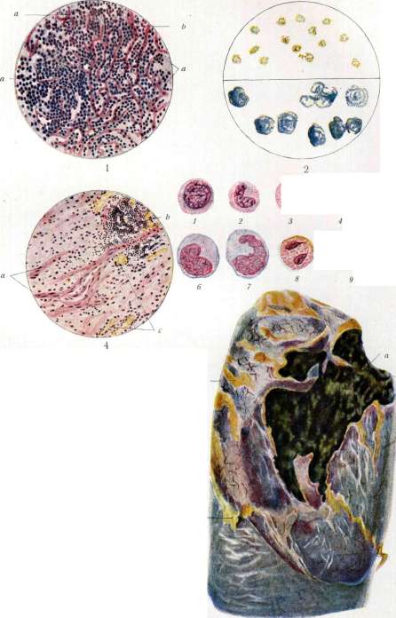
Fig. 1. Liver in myeloid leukemia; trabeculae (a) are atrophied, pushed apart by accumulations of myeloblasts (b) in the capillaries. Fig. 2. Reaction to oxidase with cells of the myeloid series: above - with benzidine (yellow granularity), below - with dihydroxyphenylalanine - blue granularity. Fig. 3. 1,5-neutrophils: 1-myelocyte; 2-young form; 3-band-nucleated: 4-normal segmented leukocyte; 5-hypersegmented; 6 and 7-monocytes; 8-eosinophil; 9-basophil; 10-lymphocyte. Fig. 4. Muscular cirrhosis of the lung in syphilis: a-bundles of smooth muscle fibers; b-round-cell infiltrates; c-vessels. Fig. 5. Multiple abscesses (a) of the lung in pyemia; b-abscesses that have ruptured into the pleural cavity. Fig. 6. Gangrene of the lung: visible (a) dirty gray-green decay of tissue: around - hyperemic pleura with fatty adhesions (b). See also Lungs, Leukemia, Leukocytes. Intravenous introduction of 25% glucose in 20.0 cm3 doses (Ilyashenko; 1929). Various medicinal substances are also administered intratracheally: 6% oil solution of eucalyptol and guaiacol in 10-50 cm3, 5% solution of thiocoll. Good results have been described. However, with extensive gangrene, results cannot be achieved by this method. In fusospirochetal gangrene, there is no certainty that the drug administered through the trachea will reach the pathogens located at the border of healthy tissue. Various serums, vaccines, and antiviruses are also used without particular success. The main methods of treatment of gangrene of the L. are neosalvarsan therapy and surgical intervention. Neosalvarsan is ineffective in embolic gangrene of the L. It makes no sense to use it in this form, and hoping for it is even harmful. In the bronchogenic form, neosalvarsan often brings great benefit and even cures the process. Neosalvarsan is used in 0.3-0.45 doses every 5 days. About 6-8 infusions are performed with a total amount of neosalvarsan administered of about 2.0, depending on the case. Usually, the nature of sputum changes sharply. The day after infusion, it usually becomes more abundant, but then its amount decreases. In addition, the odor of sputum decreases and then disappears. Temperature drops, appetite appears. Clinical observation, supported by X-ray, establishes improvement and even more or less complete recovery. Intravenous administration of urotropine has been proposed. Urotropine acts through the formaldehyde released from it. It should be remembered that neosalvarsan is a formaldehyde-sulfoxyl compound of salvarsan: In it, both arsenic and, according to Kravkov's opinion, formaldehyde act. When using neosalvarsan therapy, complications may occur. Neosalvarsan should be used immediately after dissolution. The temperature of the solution should be room temperature. In individual cases, more or less sharp increases in temperature are observed after infusion. Sometimes they force abandonment of this method of treatment. Sometimes the appearance of blood in sputum is observed after infusion. In these cases, infusions should be stopped and a smaller dose should be administered next time. In one case of Tushinsky, fatal bleeding was observed 3 days after the sixth infusion. Such bleeding is also observed without salvarsan therapy and after surgical intervention (Kisling described a case of death from bleeding after surgery).-In diabetic gangrene of the L., treatment with neosalvarsan and insulin is combined. In a number of cases, improvement from salvarsan therapy does not occur or only a transient effect occurs. In these cases, it is necessary to urgently decide on surgical intervention. With a convenient localization of the focus, the operation is brief and simple. Healing is achieved in cases that seem hopeless. According to Stögelin, surgical intervention is indicated: 1) in case of rupture into the pleura, 2) in case of spread of the disease to neighboring organs, 3) in the presence of large cavities, 4) in acute lightning-fast course, 5) in cases when other methods do not give clear results in a relatively short time. The operation does not preclude the subsequent use of neosalvarsan. The results of various types of treatment are visible from the table (according to Tushinsky's data). Method of treatment in and ?- v- o n p K2-S 5 and in og o l n o. <o S Mortality rate Neosalvarsan . . . Operation...... Without treatment..... 51 20 20 26 7 3 15 2 3 19.6 Total .... 38.5 Prognosis in embolic form depends on the primary focus, on the multiplicity of lesions, on the timeliness of treatment (surgery), on the age of the patient.-Prognosis in bronchogenic form depends on the same conditions. Here the main thing is the correct therapeutic tactic. The prognosis worsens in both cases with the advanced age of the patient (out of 12 patients of Tushinsky over 50 years old, 9 died). Alcoholism worsens the prognosis. The presence of elastic fibers in sputum, even in large quantities with their alveolar arrangement, does not give the right to make a poor prognosis.-Surgical treatment-see below.
M. Tushinsky. IX. Syphilis. Syphilitic changes in the Lungs. These are often found in newborns with congenital syphilis, whereas in adults with acquired syphilis they are comparatively rare; however, in recent years thanks to the work of Elizalde, Rossle and especially French authors (Letulle, de Jong and others), it has become clear that syphilis of the Lungs in adults occurs more frequently than was previously thought. In newborns with syphilis, changes in the lungs of two kinds are observed. 1. Limited gummas (see) in the form of slightly protruding rounded formations of a pale, grayish-pink appearance, sometimes with yellowish-gray spots on the surface. 2. Pneumonic processes diffusely affecting a portion of a lobe, an entire lobe, or even all the Lungs, designated as white pneumonia (see). In adults with acquired syphilis, changes in the Lungs usually appear in the late, gummatous period of the disease and manifest in three forms. 1. Most often, interstitial syphilitic pneumonia is observed, ending in sclerosis of lung tissue to one degree or another (indurative or sclerotic syphilis of the Lungs). The basis of this process is the intense proliferation of cells in the walls of the alveoli, around the bronchi and small vessels, as well as along the course of the interlobular septa, with miliary gummas also being encountered in places; subsequently, there is fibrous transformation of the cellular proliferation with the formation of scar connective tissue, the distribution of which in the Lungs may vary. Sometimes separate strands of white scar tissue form, which, usually following the course of the bronchi, vessels and interlobular septa [see separate table (pp. 431-432), Fig. 1], strongly retract the surface of the Lung, and the pleura in such places is thickened, sometimes adhered to the parietal pleura by blue spots. When several such strands are present in the Lung, the lung may appear as if divided into separate new lobes ('lobulated lung' - 'pulmo lobatus'). In other cases, fibrous strands seem to originate from the area of the lung hilum, distributing from them like rays. Finally, a more diffuse sclerosis of part or the entire lung often develops in the form of formation of an uneven network of connective tissue strands, more pronounced again along the course of the bronchovascular bundles and interlobular septa, or in the form of a dense consolidation of lung tissue resembling brown induration. In all these forms, the lung tissue between sclerotic areas is often emphysematous; atelectases and areas of carnification, bronchiectases often develop. - Microscopically, in the sclerotic areas, proliferation of connective tissue poor in carbon pigment is found, often rich in newly formed elastic fibers and especially bundles of smooth muscle cells - cirrhosis muscularis [see separate table (pp. 463-464), Fig. 4]; the latter, according to Davydovsky and Tanaka (Tapaka), can be considered characteristic of syphilitic proliferations of connective tissue in the Lungs. Miliary gummas are rarely encountered during this period of sclerosis. The pulmonary alveoli among the connective tissue are compressed, irregular in shape, with cubic epithelium; the bronchi are often dilated. Extensive or moderate sclerosis of lung tissue is the cause of impaired blood circulation in the Lungs, which leads to hypertrophy of the right ventricle of the heart, and subsequently can lead to disruption of cardiac compensation. 2. Solitary gummas [see separate table (pp. 431-432), Fig. 2] are less frequently found in the Lungs. They range in size from a pea to a large egg and are located, in contrast to tuberculosis in adults, predominantly in the lower lobes of the lungs. Undergoing rapid caseous necrosis, the Lung gummas at the same time have very little tendency to soften and disintegrate; they are usually surrounded by a powerful proliferation of connective tissue and gradually resorb, leaving retracted scars. Only with larger gummas does the outcome lead to disintegration and cavity formation (cavernous, ulcerative syphilis of the Lungs). 3. The rarest form of syphilis of the Lungs is nodular thickening along the course of the small bronchi, described by Kaufmann. There are clinical data indicating that during the eruption in the papular period, and according to some authors also during the gummatous period, specific catarrhal bronchopneumonias can develop in the Lungs. In the absence of pathological-anatomical data concerning these pneumonias, it is difficult to say anything definite about their nature.
A. Abrikosov. Clinic. Syphilis of the L. belongs to the late manifestations of acquired syphilis and is most often encountered in the tertiary period, 10-20 years after infection. Clinically, 1) acute syphilitic disease of the L. with elevated temperature, resembling acute tbc or acute tuberculous bronchopneumonia, with the appearance of individual gummas in the L. or diffuse infiltration of the L.; 2) slowly developing lesion of the L., resembling ordinary pulmonary tbc with the development of cavities; 3) sclero-gummatous or sclerotic form, so-called cirrhosis of the L., sometimes accompanied by bronchiectasis and dry or exudative pleurisy. In addition, syphilis of the L. is also observed as a manifestation of congenital syphilis in newborns from syphilitic parents, in the first years of life usually accompanied by corresponding changes in bones (periostitis), eyes (keratitis), ears (deafness), and teeth. So-called 'white pneumonia' of Virchow (pneumonia alba) is observed only in stillborn syphilitic children or in those who lived only a few hours. - Syphilis of the L. in acquired syphilis develops slowly and imperceptibly, giving almost no subjective or objective signs in the initial stage. Sometimes the disease begins in the form of chronic afebrile laryngo-tracheitis and bronchitis, with cough, difficulty breathing, feeling of pressure, heaviness, and painful sensations in the chest. In some cases, weight loss and feverish condition are noted. Physical signs in the initial period are either absent or not sharply expressed, especially in cases where the gumma or limited infiltration is located in the deep parts of the L. With further progression, the painful symptoms gradually intensify, general decline in nutrition is observed, cough becomes stronger, more pronounced shortness of breath appears, sputum is separated in larger quantities and sometimes with an admixture of blood. In relatively rare cases, quite profuse hemoptysis may even be observed. In most cases, the discrepancy between the size of the affected areas of the L. and the minor subjective symptoms attracts attention. The general condition and nutrition of patients with syphilis of the L. usually remain undisturbed for a long time. Sometimes evening temperature elevations and night sweats are noted, which is mainly observed during the period of decay of gummatous tumors in the L. and gummatous infiltrations and the resulting formation of bronchiectases, cavities, and pneumothorax in the L.; at this time the clinical picture of syphilis of the L. extremely resembles tbc of the L. Gradually all painful symptoms intensify; disturbance of nutrition, increasingly increasing, joins; patients lose weight sharply, and finally death occurs due to slow and progressive exhaustion. Shortness of breath is one of the frequent symptoms of syphilis of the L. Severe, disproportionate to the objective data shortness of breath should always arouse suspicion of syphilitic lesion of the L., since sometimes even the most limited syphilitic changes in the lungs cause severe shortness of breath, not corresponding to the extent of the pulmonary process. Individual symptoms of syphilis of the L. may not be equally expressed in all cases. Cough is not always a characteristic symptom for syphilis of the L. and sometimes is absent. Cases of significant syphilitic lesion of the L., in which there was absolutely no cough or it was extremely insignificant, have been described. Cough, if present, bothers patients most at night and is stronger in cases when the larynx, trachea, and bronchi are simultaneously affected by the process. Sputum in the initial stage of syphilis of the L. is sometimes completely absent. Sputum, initially usually scanty, of mucous character, with further progression of the disease becomes more abundant, mucopurulent character, sometimes with an admixture of blood. In case of cavity formation, sputum takes a purely purulent character and is excreted in the form of lumps of dirty and grayish-yellow color. The presence of miliary gummas in the sputum is considered characteristic but rare for syphilis of the L. (K. M. Popov, Ginz, Engel). In the literature, even isolated cases have been described in which the excretion of gummas weighing up to 20 g with sputum was observed. The examination of sputum for tubercle bacilli is of great importance, which is important not only for differential diagnosis between syphilis and tbc, but is also of great importance due to the frequent combination of syphilis with tuberculous lesion of the L. Some authors consider the absence of hemoptysis characteristic for syphilis of the L., but most observations show that hemoptysis in syphilis of the L. is not so rare and can be even so significant that patients die from it. The temperature in syphilis of the L. does not give any definite data. In some cases it remains normal, in others it is slightly elevated, and in third cases hectic fever with chills and profuse sweating is observed, as in hectic tbc patients. - Syphilitic lesion of the L. is most often unilateral and affects mainly the right L., mainly the middle or lower lobe. But syphilis can also affect other parts of the L., and in some cases the process is concentrated exclusively in the apices of the lungs. Diagnosis of syphilis of the L. is extremely difficult and with full certainty is possible only 1) in the absence of tubercle bacilli in the sputum after numerous careful examinations of it and 2) with the rapid therapeutic effect when using anti-syphilitic treatment. Syphilis of the L. can be suspected in cases if in the anamnesis of the pulmonary patient there is syphilis or any other tertiary manifestations of it are determined, e.g., periostitis, characteristic scars, aortitis, or there is a positive RW. Individual painful symptoms observed in syphilis of the L. do not give definite data for making an accurate diagnosis, although it should be borne in mind that in syphilis of the L. cough is less constant and not so persistent as in tbc of the L., sputum is less, sometimes it is completely absent, hemoptysis and temperature elevations are less common, and often there is severe shortness of breath, not corresponding to the pulmonary lesion. The localization of the process in the middle or lower lobe of one lung, considered characteristic by many authors for syphilis of the L., cannot serve as an exact point of support for differential diagnosis, since syphilitic lesion of the L. can also be located in other lobes, even in the apices of the L. or in the form of scattered small foci. The absence of tubercle bacilli in the patient's sputum is a very important circumstance speaking in favor of syphilis of the L., although on the other hand, due to the frequent simultaneous lesion of the L. by both syphilitic and tuberculous process, the presence of tubercle bacilli in the sputum does not exclude the possibility of the simultaneous existence of syphilis and tbc of the L. One of the important but rare signs of syphilis of the L. should be considered the presence of miliary gummas in the sputum and the finding of syphilis spirochete in the sputum. X-ray pictures can be useful for making a diagnosis, on which syphilitic processes usually appear denser, sharper, and more massive than tuberculous processes. The course of syphilis of the L. is usually chronic, lasting several years. In cases complicated by tbc or syphilis of other internal organs, syphilis of the L. can take a more rapid course. The prognosis in syphilis of the L., provided the other internal organs are in good condition, is favorable, and in most cases good results can be expected from anti-syphilitic treatment. In the literature, there are observations when even with significant destruction of the pulmonary parenchyma, recovery occurred as a result of anti-syphilitic therapy. - The prognosis is always serious if there is already amyloid degeneration of parenchymatous organs. Age has a great influence on the outcome of syphilis of the L.: in children with congenital syphilis and in old people, syphilis of the L. proceeds much more seriously and more often gives a fatal outcome than in people of middle age. Various diseases accompanying syphilis of the L. worsen the prognosis. Of the complications in syphilis of the L., tbc is most often encountered, and tbc that developed in the secondary period of syphilis sharply worsens the course of the main process, while tbc in the late period of syphilis proceeds relatively favorably. - Therapy in syphilis of the L. consists in carrying out specific treatment. In the simultaneous disease with syphilis and tbc of the L., it is also necessary to resort to the prescription of anti-syphilitic treatment, since observations show that when syphilis of the L. is cured, the tuberculous process subsequently responds more easily to treatment with appropriate measures. But on the other hand, if in a patient with syphilis of the L. tbc of the L. is also noted, then anti-syphilitic therapy should be carried out with caution, since in such patients with the use of vigorous specific treatment, hemoptysis and temperature elevations may appear. Treatment in a warm and even climate gives more favorable results.
D. Russian. X. Parasites. The true lung parasites of humans are the lung fluke (Paragonimus Ringeri), which encysts in the bronchi, and the nematodes Metastrongylus elongatus (in pigs, rarely in humans in the bronchi and trachea) and Syngamus Kingi (respiratory passages). In the lungs, along with other organs, various flagellates (Monas, Cercomonas, Trichomonas) can parasitize (in cavities); the larvae of Taenia solium develop relatively rarely in the L. compared to other organs; Sparganum proliferans, the plerocercoid stage of the tapeworm Diphyllobothrium proliferum, is found in the L.; the plerocercoid of Diphyllobothrium Mansoni has been found in the pleural cavity; Microfilaria nocturna retreat during the day (more precisely during the period of wakefulness) into the blood vessels of the L. The lungs serve as a stage in the migratory paths of certain parasitic worms, the larvae of which are brought by the blood via the a. pulmonalis and then actively penetrate through the alveolar lining into the respiratory passages. This is the path of Ascaris lumbricoides, Ankylostoma duodenale, Necator americanus in humans, Toxocara canis in dogs, probably T. cati in cats, etc. The diagnosis of parasitic infection of the L. is made by finding the parasites themselves in sputum (e.g., protozoa) or their eggs; the latter occurs when semi-adult parasites are present in the L. The eggs of lung parasites can also be detected in faeces, where they may arrive with swallowed sputum. It is possible to detect migrating helminth larvae in sputum. For examination, it is necessary to take large quantities of sputum. The presence of encapsulated and enclosed parasites (e.g., echinococci, etc.) is established by radiography and various indirect methods.
E. Pavlovsky. Echinococcus. Among animal parasites, the echinococcus is found in the lungs. The so-called unilocular, hydatid echinococcus is more common, which usually penetrates the lungs from the liver through the diaphragm or by the hematogenous route; however, primary echinococcus of the lungs is also observed. In the lungs, the echinococcus forms one or more vesicles with the typical structure of this parasite (see Echinococcus); around the vesicles, productive inflammation develops, sometimes with abundant formation of connective tissue and obliteration of the pleura. Suppuration often occurs, which can lead to rupture of the vesicles and their breakthrough into the bronchi with the excretion of chitinous membranes and daughter vesicles with sputum. Secondary entry of air into the emptied echinococcus cavity can result in the formation of a unique air cyst in the lungs. The multilocular, alveolar echinococcus in the lungs is much rarer, and in the lungs it also penetrates secondarily by contact from the liver, through the diaphragm, or by hematogenous metastasis (most frequently also from the liver through the inferior vena cava). Primary alveolar echinococcus is manifested in the formation of a node or (e.g., in metastatic origin) several, sometimes multiple, nodes of dense consistency with a characteristic spongy surface on cross-section. A. Abricosov. Echinococcus of the lungs is the third most frequent (after the liver and spleen) localization of this parasite in the internal organs. Lesion of abdominal organs by echinococcus accounts for 40-70% of all cases of echinococcus, while lesions of the lungs on average account for from 2% to 14%. The data below from three authors illustrate these relationships. Statistical studies also show that echinococcal disease is associated with certain geographical territories (for more details—see Echinococcus). The clinical signs of echinococcus of the lungs vary greatly depending on the stage of the disease process. Subjectively and even objectively, echinococcus of the lungs for a long time may not give any symptoms. Cases of asymptomatic course for many years (up to 20 years or more) have been described. Therefore, in the early stages, the echinococcal cyst of the lungs is often overlooked, since neither percussion nor auscultation in many cases give any definite changes. This is especially true for cases with central localization of the cyst. Among the general clinical symptoms accompanying the picture of chronic intoxication ('cachexie hydatique' of French authors), it is necessary to note emaciation, general weakness, loss of appetite, palpitations, sweats, and dyspnea on movement. One of the early clinical manifestations of echinococcus of the lungs is the sudden onset of hemoptysis, often erroneously attributed to tuberculous lesions of the lungs. A more characteristic symptom that allows for the undoubted diagnosis of echinococcus of the lungs is the excretion of serohemorrhagic sputum containing hooks of the echinococcus, daughter cysts, or pieces of the parasite's membrane. The latter occurs only in cases of rupture of the cyst into a bronchus. However, the main diagnostic method for early recognition of echinococcus should be recognized as radiographic examination. The latter plays no small role in the development of modern knowledge about the clinical picture (localization, course, etc.) of echinococcus of the lungs. Radiographically, echinococcus of the lungs is generally characterized by a spherical-shaped shadow. In those cases where the echinococcal cyst empties spontaneously, resp. ruptures into a bronchus or pleural cavity, conditions are created for the formation of an echinococcal pulmonary cavity (pneumocyst). The radiographic picture is very similar to the usual picture in a tuberculous cavity, while in cases of complete emptying, the shadow of the cyst becomes less distinct and sometimes mottled. Such open forms of echinococcus of the lungs represent considerable danger to patients, since along with the phenomena of general intoxication observed at this time, suppuration in the cyst cavity, resp. pleura associated with it (pyopneumothorax) usually develops as a result of subsequent infection. Only in rare cases does spontaneous emptying of the echinococcal cyst lead to spontaneous healing [see separate table (pp. 447-448), figs. 9-14]. The lower lobe of the right lung is a more common localization for echinococcus of the lungs, but cases of echinococcus of the lungs in the region of the root of the lung are also not uncommon. Finally, in rare cases, multiple cysts in both lungs are also observed. Among the indirect symptoms confirming the diagnosis of echinococcus of the lungs, it is necessary to note eosinophilia and a positive reaction of Weinberg (see Weinberg's reaction) and Casoni (on the value of these methods—see Echinococcus). In the diagnosis of echinococcus of the lungs, difficulties may arise in differentiating it from a number of similar (mainly radiographically) diseases. A distinctive feature of the shadow in hydatid echinococcus of the lungs is its transparency, which is reflected on the radiograph by sharp contouring of both the 'anterior' and those located behind the cyst ('posterior') ribs. This generally correct symptom, however, is not always confirmed. Cases of undoubted echinococcus of the lungs have been described where no transparency of the shadow was observed, and conversely, a typical radiographic picture of echinococcus of the lungs in the presence of a sarcoma in reality. In small cysts, the diagnostic value may sometimes be the change in the contour of the shadow from round to oval during respiration (Nemenov). The usual spherical shadow in echinococcus of the lungs is sometimes difficult to differentiate from interlobar pleurisy and tubercous infiltrates of the lungs, and when localized near the aortic arch—also from an aortic aneurysm. In cases of cyst emptying, as mentioned, the radiograph resembles other forms of pulmonary cavities, and in these cases, clinical data play a decisive role in diagnosis. Finally, in cases ending in scarring, the picture of pure sclerosis of lung tissue is very similar to that of healed tuberculosis of the lungs (Uspensky). Among diagnostic methods, puncture of the echinococcal cyst should be considered contraindicated. As for the treatment of echinococcus of the lungs, in rare cases, self-healing (scarring) is observed after emptying of the echinococcal cyst, but in most cases the only reliable method is surgical treatment, unfortunately not possible with every localization of the cyst. For methods and indications for surgical intervention—see below. XI. Surgical treatment of lung diseases. Surgery of lung diseases in recent decades has widely expanded its boundaries. The development of radiodiagnosis, in particular the use of contrast agents in radioscopy of the lungs and stereoscopic photographs, has refined diagnosis, placing it on an exceptional height of objectivity, and the introduction of methods for regulating intrapulmonary pressure and the construction of corresponding apparatus have made it possible to implement the boldest plans of surgeons. It must be said that the method of regulating pulmonary pressure has created an era of development in the history of pulmonary surgery. With this method, the question of combating the consequences of open pneumothorax, often leading to death, has been resolved. This method has its own history. On the basis of an analysis of the physical conditions maintaining the normal position of the lungs, chambers with rarefied air were initially created, with negative pressure in them equal to the negative pressure in the pleural cavity (7 mm Hg=9 cm H2O). The torso was placed in these chambers, while the head was outside the chamber. Opening the chest cavity under these conditions does not lead to collapse of the lungs. Collapse of the lungs can also be prevented in another way—by increasing the pressure inside the bronchial branches. The latter can be created either by placing patients in chambers with increased pressure or by using hermetic breathing masks, allowing to maintain the desired level of intrapulmonary pressure. At present, chambers with reduced pressure (Sauerbruch model) and with increased pressure (according to Brauer and Engelken) have almost gone out of use; in practice, apparatuses with hermetic masks are usually used. The main moment in the action of these apparatuses is the maintenance of a certain force of pressure in the inlet and outlet tubes. Technically this is achieved differently—either by means of oxygen cylinders or by pumping atmospheric air. The first principle is implemented most simply and practically in the Jena type apparatus (fig. 23), Figure 23 (diagram). 1-oxygen bomb; 2-connecting tube; 3-vessel for narcotizing substance; 4-mask; 5-vessel with water for maintaining pressure. somewhat more complex, but also more perfect in the Tiegel model (fig. 24). In the Jena and Tiegel apparatuses, increased pressure simultaneously allows to maintain anesthesia. During the war of 1914-1918, many simplified models of apparatuses for increasing intrapulmonary pressure were proposed with wide use of gas masks. Very simple models were proposed in the USSR by Bereznechovsky, Girgolav, and more recently by Gershe. Gershe's apparatus (fig. 25).
An ordinary foot bellows (M), with a capacity of 2-3 liters, with two valves (K and Kj), arranged so that the bellows acts as a force pump, is connected with a thick rubber tube to a rubber cushion (A). A T-shaped tube with one end connects to a water manometer (B), and with the other end connects to a short thin-walled rubber tube (I), onto which a Moro clamp (P1) is fitted. Further follows a T-shaped tube (T1), after it a long thick rubber tube (Hj), leading to a mask (B), covered with rubber and hermetically fitting to the face. In front of the mask is placed a valve (K2), opening towards the mask. The side arm of the T-shaped tube (Tx) is connected by means of a rubber tube and a fork with two bottles for anesthesia (/). One of the bottles is for chloroform, the other-for ether. The short metal tubes of these bottles are connected with the fork and further with the T-shaped tube (Tx). The long metal tubes of these bottles, immersed in the narcotic substance, are also connected with a fork, onto which a double rubber balloon is fitted, with the help of which vapors of the narcotic substance are pumped in. With the help of Moro clamps (P2 and P3) one can at will pump in chloroform or ether or both together. Into the mask (B) a second tube (E) is also fitted, which connects with a valve opening outwards, behind which follows a pressure regulator (D). The latter consists of a sleeve (D), in the bottom of which is a carved hole, and a valve (L), well ground to this hole. The valve is held by a spring, the pressure force of which is regulated by a screw (J). The regulator is preliminarily set so that its valve opens at the desired pressure according to the water manometer readings. In the upper wall of the mask a third tube (C) is fitted, onto which a short rubber tube is placed, closed by a Moro clamp (P). The opening of the bellows occurs automatically with the help of a spring.-Air, continuously pumped by the bellows, first fills the cushion, and then gradually increases the pressure in it and in all parts of the apparatus, and as soon as the pressure reaches the force with which the spring presses on the valve (L) in the regulator (D), the air will lift the valve (L), break through it and will exit through the regulator.-Further increase in pressure will no longer occur despite the constant pumping of the bellows. When pumping stops, there will be no decrease in pressure, as the valve will immediately close the regulator opening and stop the air from exiting the apparatus. The greater the pressure required in the apparatus, the tighter the regulator spring needs to be screwed with the help of screw J.-The average necessary pressure equals 7 mm of mercury or about 10 cm of water column. The maximum pressure (used when searching for lung tissue wounds) equals 10-11 mm of mercury or about 15 cm of water column. The minimum pressure (used when searching for foreign bodies in lung tissue and during transpleural operations on the esophagus) equals 3 mm of mercury, or about 4 cm of water column. Accordingly, agreeing with the water manometer readings, the regulator spring is loosened or compressed with the help of screw P. From the moment when the pressure throughout the apparatus reaches the required height and the air begins to exit through the regulator, in the mask a continuous flow of air in one definite direction is established thanks to the constant pumping of the bellows, namely: from the cushion into the mask and from the mask into the regulator, since the regulator is the final and only place in this entire closed system where air finds an outlet. Thus, into the mask a continuous inflow of fresh air arrives, and the air exhaled by the lungs is carried away by the air current and exits through the regulator. Since for normal breathing about 8-9 liters of air per minute are necessary, then with the capacity of a small bellows even of 1 liter, only 8-9 pumpings per minute are necessary, which will not be difficult not only for an adult man, but even for a woman or adolescent. Thanks to the constant air flow, the size of the cushion also loses significance, which can be taken of any size. However, the smaller the cushion, the greater pressure fluctuations the manometer shows during breathing and when pumping the bellows.
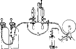
Figure 24. Tiegel's apparatus (diagram): 1- shut-off cock; 2- fork; 3- cock for removing the remainder of the narcotic substance; 4- vessels with narcotic substance; 5- cock for regulating the narcotic substance; 6- mixer; 7- bypass tube for oxygen; 8- cock for stopping the inflow of narcotic substance while maintaining the inflow of oxygen from the bomb; 9- tube for exhaled air; 10- vessel with water to maintain pressure in the mask at the desired height; 11- thin-walled rubber bag.
Surgeons have accumulated a rich technical and clinical experience, which has led to deepening research work on the anatomy, physiology and pathology of this organ. Although modern surgery is far from the ideal of Gluck, who in 1881 predicted that surgeons soon would be guided in relation to the L. by the general surgical principle: ubi pus, ibi incisio; ubi haemorrhagia, ibi ligatura; ubi tumor, ibi exstirpatio (where there is pus-there is an incision; where there is bleeding-there is a ligature; where there is a tumor-there is removal), nevertheless at present one can already speak of brilliant achievements in the surgical treatment of a number of diseases of the L. and the chest cavity. These include: 1) Congenital diseases: a) stenosis of the aperture-insufficiency of the upper opening of the chest cavity (see Chest cavity); b) funnel chest; c) hernias of the L., as well as splitting of the sternum; d) immobile expanded chest due to incorrect growth of cartilages with secondary
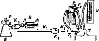
emphysema (Freund).
2) Traumatic injuries to the L. 3) Acute
Acute and chronic purulent processes in the L. 4) Complications in purulent processes in the form of pulmonary fistulas. 5) Chronic infectious processes (see Tuberculosis of the lungs). 6) Parasitic diseases of the L. (echinococcus, lung fluke, etc.). 7) Mycoses of the L. (actinomycosis, streptomycosis, aspergillosis). 8) Benign and malignant tumors. 9) Functional diseases (see Bronchial asthma). Indeed, with regard to certain forms of diseases, surgeons, having performed a series of interventions that do not go beyond elementary empiricism in their theoretical justification, have had negative experience, e.g., in the surgical treatment of bronchial asthma, primary emphysema (by means of Freund's operation). The cause of this was the insufficiently studied pathogenesis of these diseases. The operation proposed by Freund aimed to reduce the volume of the chest box by resection of the costal cartilages from both sides through a parasternal incision. This operation, after Freund's views on the pathogenesis of emphysema were not confirmed, lost its theoretical basis; moreover, the functional effects after the operation did not justify the hopes for collapse of the L.—it did not occur (Sauerbruch). Nevertheless, in certain forms of emphysema it gives subjective relief to patients, sometimes freeing them from severe asthmatic attacks, for which reason it is still performed in some cases. Its technical performance is very easy. Along the edge of the sternum, an incision is made under local anesthesia, extending from the I to VI costal cartilage. For better exposure of the costal cartilages, two lateral transverse incisions 4 cm long can be made at both ends of the longitudinal incision (Braun). Each flap together with m. pector. major is separated and pulled outward; the costal cartilages are exposed and from II to V, a piece 3 cm long is excised. It is recommended to remove them together with the periosteum, since the regenerative capacity of the periosteum promotes cartilage regeneration or the appearance of ossifying perichondritis (Seidel, Stieda), as a result of which the initial improvement disappears. Operation should be performed on one side and only if all symptoms of the disease recur, the costal cartilages should be excised on the other side. Almost the same surgical intervention has been proposed by Sauerbruch for the treatment of funnel chest. 'In this developmental defect, another method diametrically opposite (Kapp) has been proposed. An incision is made in the rib and a piece of bone is implanted between the ends of the incised rib. In case of insufficiency of the aperture, which was assigned a very important role in the development of pulmonary tbc, paravertebral resection of the upper ribs was proposed. This operation, however, did not become widespread. Traumatic injuries to the L. are rarely isolated from injuries to the chest; the latter is usually the site of application of the damaging force, as is especially observed in injuries without damage to the soft coverings and ribs. Traumatic injuries are distinguished as closed and penetrating-open. Closed injuries are observed in concussions and in compressions of the chest (in falls, collapses, when run over by a vehicle, contusion from explosion of combat projectiles, in factories, during blasting operations). The physical properties of the costal arches and the entire chest are such that under certain conditions it can change its shape and return to a normal position, and all tissues of the chest wall sometimes show no visible signs of damage. At the moment of chest compression, the L. receive contusion, and sometimes remain without visible gross damage. More often, however, hemorrhage, ruptures of the parenchyma are observed. This type of contusion without gross anatomical changes sometimes gives a picture of exceptional severity with a fatal outcome. A case of contusion without damage to the chest and L. has been proposed to be called commotio thoracis (concussion of the chest) by analogy with concussion of the brain, which is justified by the severe condition of such patients: paleness of the skin, cold extremities, uneven weak, very slowed pulse and similarly uneven superficial breathing. The symptoms are almost identical to those in concussion of the brain. Therefore, many authors believe that the clinical picture should be explained by both contusion of the chest and simultaneous concussion of the brain. But there is another opinion: the picture of concussion of the chest is the result of reflex phenomena from the vagosympathetic system, in particular from the nn. splanchnici, and an analogy is drawn with the experience of Golts. Cases of chest trauma with clear damage to the chest wall (fracture of ribs) or with severe bruising of the L. with violation of their anatomical integrity are called compressio thoracis (compression of the chest). The signs of trauma are pronounced here. In severe injuries, loss of consciousness is also observed. Sometimes the face and neck have a markedly cyanotic coloration; this is explained by impaired blood circulation in the small circle; in the pulmonary vein and right heart due to compression of the L., stagnation of blood occurs, and sometimes it leads to rupture of small-caliber vessels. In such cases, damage to only the L. without violation of the integrity of the chest wall is observed. In these cases, the damage to the L. is expressed either by hemorrhages or ruptures of the L., and both can be located either closer to the surface of the organ or in a central position. In most cases, damage to the L. is inflicted by fragments of ribs. These injuries, understandably, can be very significant and severe in their consequences. In injuries caused by rib fragments, severe complications are more often observed than in other types of chest injuries; pneumothorax, hemothorax, and sometimes hemopneumothorax. Often, in such cases, signs of progressive pneumothorax with emphysema of the mediastinum are observed. The outcomes of injuries to the L. in compressions depend on the severity of the damage to the pulmonary parenchyma, the presence and nature of complications with hemopneumothorax and the infection that sets in (empyema, lung abscesses). Penetrating wounds. Typical penetrating wounds are most often gunshot, then cut, punctured with cold weapons—sabers, daggers, knives, chisels, awls, more rarely—pikes, spear-shaped plates of iron gratings, stakes. The type of injury (weapon, damaging force) also determines the anatomical nature of the wound to the L. and the severity of the course of such injuries. Cases of injury with a broom handle, a shaft with enormous crushing of the L. have been described. Burdenko observed injuries with Cossack pikes and German and Russian bayonets. The former were of exceptional severity. Bayonet injuries (Russian) do not cause such severe injuries. Gunshot wounds are inflicted either by small-caliber firearms in peacetime or by large-caliber military firearms during battles. In this type of injury, penetrating wounds with relatively minor damage to lung tissue are usually present, and their severity is mainly determined by the location of the bullet channel. Through wounds at the edges sometimes proceed unusually easily. Wounds passing close to the roots of the L., on the contrary, are very dangerous; here fatal bleeding, injuries to large bronchi and hence the exceptional danger of secondary infection can occur. In bullet wounds in the L., it is sometimes very difficult to trace the course of the bullet channel, and for a time, field surgeons were too optimistic about lung injuries, especially when observing them in rear institutions. Wounds to the L. by fragments of grenades, shrapnel, hand grenades belong to the most severe injuries. Wounds to the L. by shrapnel bullets occupy a middle position in terms of severity of course, but the wounds to the L. in this case are very severe, and the severity is determined by both the immediate damage and complications in the near and distant periods. This should include cases of injury to the L. and abdominal cavity with penetration of the diaphragm, as well as cases of blind wounds. On battlefields during the imperialist war, 30-35% of the killed had lung injuries. The immediate complication of penetrating lung injuries are pneumothorax and hemothorax. The severity of the complication with pneumothorax depends on the anatomical conditions of its occurrence and the severity of the functional disorders it causes. Pneumothorax is distinguished: 1) open outward, 2) open inward, 3) closed, and 4) the most severe form of pneumothorax with progressively increasing pressure. The latter in turn is subdivided by mechanism of origin into inspiratory and expiratory pneumothorax (fig. 26). This form of pneumothorax assumes the presence in the damaged L. of conditions that reproduce the mechanism of a valve (see Pneumothorax). Expiratory pneumothorax occurs in convulsive closure of the glottis due to painful sensations or during coughing. The situation soon complicates with mediastinal emphysema with its spread to the neck and throughout the body. The second in severity is open pneumothorax. Closed pneumothorax is usually observed in cut and punctured wounds with a narrow canal. Air can enter in these wounds both from the outside and from the bronchi.
Thanks to the narrowness of the wound channel, the latter quickly collapses, and the matter is limited to the amount of air that entered immediately after the injury. The entered air is fairly quickly absorbed, and the functional severe phenomena rapidly disappear. In open external pneumothorax, the functional severe phenomena in the form of disturbances in the activity of the lungs and heart do not subside, but persist stubbornly (without weakening). Here a very complex clinical picture develops, which is based on the collapse of the L., cooling of the pleural cavity, evaporation of moisture from the pleural walls, irritation of the endings of the nerve trunks of the sympathetic and parasympathetic systems, displacement of the heart, or rather,
Figure 26. Pneumothorax: a-broadly open externally; b-valvular; c-closed, with negative pressure; d-closed, with high positive pressure (Kienbock positive); e-surgical, widely open to the outside.
the mediastinum with each inspiration, which in turn leads to a violation of the suction of blood from the system of hollow veins: the thin-walled right atrium cannot withstand the pressure of the air (Glück). With pneumothorax with progressively increasing pressure, functional disorders continuously increase: with each respiratory movement, new portions of air are introduced into the pleural cavity, and consequently each respiratory movement aggravates the condition of the patient. Relatively often observed in injuries to the L. is subcutaneous emphysema, which must be distinguished from mediastinal emphysema; it is limited to a small area near the entrance opening or extends to larger areas. In these cases, air penetrates either from the entrance opening from the outside or from the lungs, which is sometimes observed in fractures of the ribs. First, the air collects in the pleura, then it enters the subcutaneous tissue if the wound channel creates favorable conditions for this. The second complication-hemothorax (accumulation of blood in the pleural cavity) can occur when the vessels of the chest or the vessels of the lung are damaged;
Figure 27. True hernia
Fig.28.Hernia according to Hertzberg'y. a-schematic
view of a gunshot wound: 1-edges of the lateral incision: 1-lung
rot; 2-necrosis is not uncommon; 2-hernial sac
of the hernia wall; pleura; 3-chest
ney wall; 3-edge of the damaged
rib; 4-V rib. b-hern
rib; 5-IV rib. (According to
ial gates (1) between
IV and V ribs (2).
Hertzberg'y.)
very rarely in injuries to the L., bleeding from the external wound of the chest is observed. The severity of this symptom is determined by the amount of blood that has flowed out and the degree of functional disturbances in the activity of the heart and the undamaged lung. The amount of blood that has flowed out depends on the caliber of the damaged vessel. Therefore, with edge wounds, relatively little blood flows out and vice versa. The vessels of the chest walls (a. mammar. int., aa. intercostales) can sometimes also give very profuse bleeding. Bleeding here occurs under exceptional physical conditions, due to the fact that in the pleural cavity the pressure is negative: it is as if blood is being sucked in. Sometimes intrapleural bleedings quickly lead to death, on the other hand, cases of small bleedings are also observed, as well as cases of spontaneous cessation of bleeding with large hemorrhages into the pleural cavity-occurs from compression of the lung by the blood that has flowed into the pleural cavity; it sometimes collects up to 2-3 l.-The two main severe symptoms described determine the nature of the clinical picture and even the fate of the wounded in the coming hours and days. Among other symptoms, bloody sputum separation, severe cough, and occasionally symptoms of injury to the sympathetic trunks are very often observed. As distant complications, empyema, traumatic pneumonia, abscesses and gangrene are most often observed, and finally-secondary pneumothorax or hemothorax. The course and outcomes of lung wounds vary. Undoubtedly, of all types of wounds, the most easily penetrating revolver wounds (small-caliber bullets) also proceed easily, military bullet wounds (jacketed pointed bullets) also proceed easily; shrapnel wounds and blind bullet wounds proceed more severely. Exceptionally severe course in grenade wounds. At present, the following data, which can illustrate the just stated position, are considered more or less reliable: 1) those who died from chest wounds make up to 29-30% of all those who fell on the battlefield; 2) those who remain alive die in a significant number, at different times and at different evacuation points; especially many die in field infirmaries located close to the forward positions. The following figures give an idea of mortality in these infirmaries: a) penetrating bullet wounds give 20% mortality, b) bullet and shrapnel blind-40%, c) grenade damage-45%. Mortality in stage infirmaries and corps hospitals with penetrating bullet wounds-15%, with grenade wounds-20-25%. The overall mortality is calculated by various authors at 60-80%. Treatment of lung wounds is carried out in most cases conservatively, while some forms require mandatory surgical intervention and moreover more or less urgent. Indications for intervention and its nature are determined by the presence and severity of symptoms. 1) cases without pronounced hemothorax and pneumothorax, 2) cases with simple closed pneumo- and hemothorax are carried out conservatively.-The conservative method requires absolute rest and excludes evacuation for 6-9-10 weeks. Patients must be under long-term control; any deterioration in the general condition due to secondary bleeding or pneumothorax or the onset of infection raises the question of surgical intervention. At present, the following principles have been developed in relation to field and generally gunshot wounds
2-pleura; 3-lung; 4-ribs; 5-muscles.
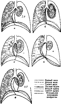
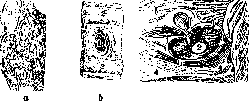
indication for direct surgical intervention: one should operate in those cases when there are present 1) increasing pneumothorax, 2) rapidly increasing hemothorax, 3) open pneumothorax, 4) open pneumothorax with greater or lesser damage to lung tissue (rupture, crushing); 5) mediastinal emphysemas, 6) penetrating wounds through the diaphragm accompanied by damage to the gastrointestinal tract. Intervention depending on the nature of the wounds is reduced either 1) only to puncture and aspiration of blood, 2) to thoracotomy and tamponade, 3) to ligation of bleeding vessels with subsequent tamponade or blind suture, 4) simply to tamponade of the pleural cavity with fixation of the lung to the edges of the chest wall wound or finally 5) to partial resection of parts of the lung. In some cases of open pneumothorax without extensive damage to the L., it is necessary to suture the chest wall wound, and with simultaneous significant damage to the L.: sometimes a suture, but in most cases-tamponade. With simultaneous wounds of the chest and abdominal cavities, it is usually necessary to intervene actively either through the pleural cavity, through the diaphragm, or by laparotomy. In the subsequent course, surgical intervention is indicated for infectious processes in the form of empyemas, single and multiple abscesses, very similar to bronchiectases, late abscesses near foreign bodies in blind wounds (see below). A comparatively rare remote consequence of both gunshot and blunt injuries are hernias of the L. (hernia pulmonum, pneumocele). In blunt subcutaneous injuries, a fissure may form in the intercostal muscles, and finally in fractures of the ribs, bone fragments deprived of periosteum may be resorbed and give yielding areas in the chest wall wall; an even simpler representation is the formation of hernia-like protrusions (Lejars) of the lungs in tangential wounds and resections of ribs. Here the term hernia-like protrusions is most appropriate; here there is no hernial sac, and extensive adhesions are always present. The first types of protrusions may have all the elements of the concept of hernia: hernial sac, hernial gates and contents of the hernia (fig. 27 and 28). In this respect they resemble true hernias of the L.; congenital and so-called spontaneous hernias in emphysema and arteriosclerosis. Treatment of pulmonary hernias is carried out either conservatively-with pelottes and corsets-or surgically according to the general rules for operating on hernia, and for closing the hernial gates, periosteum taken from adjacent ribs is used (figures 29 and 30). Surgical treatment of purulent processes. These include abscesses, gangrene of the L. and closely related congenital or acquired bronchiectases, as well as bronchial fistulas, which are most often a consequence of pneumotomy. In abscesses and gangrene of the L., most therapists and surgeons consider surgical intervention fully indicated. The questions about the urgency of intervention are less definite, of course if we are not talking about rapidly progressing violent infectious processes or putrid intoxications in gangrene of the L. Among surgeons there is a tendency to perform intervention as early as possible, which does not find recognition from therapists and a certain part of surgeons; also there is no complete unity of views among surgeons on the methods of operating. As for the method of operating at present, there are proposals along with the classic operation of pneumotomy to perform aspiration of pus using bronchoscopy and to apply methods of collapse therapy (phrenico-exeresis, thoracoplasty). Supporters of expectant therapy are based on observations of self-healing of both pulmonary abscesses and gangrene of the L. Especially in this respect, the course of abscesses in young subjects has always been impressive. In them, due to the peculiar elasticity of tissue and the rapid healing of all kinds of defects in tissues, cavities remaining after spontaneously emptied abscesses collapse exceptionally easily and are quickly eliminated. This circumstance has always justified the legitimacy of expectant therapy, and the outcomes of surgical intervention with a high percentage of mortality have little encouraged therapists to refer patients to surgeons. Mortality in surgical intervention in gangrene of the L. and in various abscesses is very high. According to Quincke's combined statistics of 1901, the overall percentage of mortality in surgical treatment of pulmonary suppurations reached 35%, and according to the statistics of 1903-25%. Kiesling's statistics of 1906 regarding outcomes in gangrene of the L. gave 40% mortality (in 120 cases). During internal treatment, mortality for the same period was calculated at 60-90%. In subsequent times, the percentage of mortality has decreased, but remains unstable. Combined statistics for the period 1903-12 give the following percentage ratio: out of 148 cases of abscess, 34 ended fatally (17.5%), out of 281 cases of gangrene-84 (29.3%). But on the other hand, a recently published statistic by Sauerbruch gives less favorable results: out of 140 cases of abscess, 55 cases ended fatally (39.2%), out of 53 cases of gangrene-30 (56.6%). In particular, out of 140 cases of abscesses, there were 33 cases of acute with 36.3% mortality. In recent years, the percentage of mortality has decreased to 30%, and according to the statistics of 1920-to 16.3%: The outcome of the operation is greatly influenced by the severity of the cases and the preliminary treatment, often erroneous (unsystematic punctures) and the timing of intervention. Among surgeons, there has been a strong trend recently in favor of early intervention. Here it is necessary to establish the true meaning of the concept of "early operation" in relation to pulmonary surgery. In purulent processes in the L., intervention is possible only after the expiration of a certain period, which is necessary for the formation of a reliable barrier zone. At one time this period was calculated at 5-6 weeks from the initial stage of the purulent process, and it was not recommended to wait more than 8 weeks. The success of surgical intervention besides the terms depends on the form of the abscess and the ease of access to it. In this respect, abscesses located closer to the root of the L. are almost inaccessible and require very careful study of the access routes to them and complex operative techniques in the form of such preparatory operations as thoracoplasty, plombage. In any case, surgical intervention should be undertaken only after preliminary testing of general therapeutic treatment and in particular treatment by Quincke's method (so-called positional drainage). Finally, an extremely important condition for success is the prevention of pleural infection. Therefore, in operations for purulent processes, it is necessary to accurately determine the state of the pleural cavity and specifically-the presence of adhesive inflammation in the area of the planned operation. In the absence of adhesions, they need to be induced artificially if the circumstances allow, the nature of the infection (rapidly increasing septic phenomena, gangrene of the L.) and the condition of the patient's strength. If it is necessary to perform urgent intervention in the absence of pleural adhesions, measures are taken to protect the pleural cavity. For this purpose, it was proposed to proceed as follows: after opening the pleural cavity, to suture the lung into the pleural
Figure 31.
the wound in such a way that the site of the proposed incision over the lung abscess is located in the center of the pleural wound (Roux) (fig. 31). Naturally, the sutures should be placed in areas of healthy tissue and the suture should create a hermetic closure of the pleural cavity. However, due to the mobility of the lung, this is very difficult to achieve: the lung often tears, and the goal is not achieved. A simpler but at the same time cruder method is the tamponade of the free pleural cavity with gauze around the proposed incision. Both of these methods are far from ideal, and in addition they can lead to very dangerous phenomena in the form of pleural shock. In view of this, in one-stage operations, the use of general anesthesia is mandatory, and it must be remembered that general anesthesia in cases of lung abscesses is sometimes complicated by the aspiration of pus into the bronchi of adjacent areas of the lung, as well as by aspiration by the healthy lung. Therefore, the use of anesthesia should be limited to the shortest possible time; for this purpose, rib resection is performed under local anesthesia; then the patient is asked to cough and general anesthesia is begun with the body lowered and the head hanging down (Quincke). The application of increased intrapulmonary pressure (3-5 mm Hg) is mandatory to prevent serious complications - embolism, aspiration. The lung tissue is not cut but separated by the tip of the Pakelen apparatus. In the presence of adhesions, the course of the operation is significantly simpler: as extensive a resection of the ribs as possible is performed, and it is recommended to remove the intercostal muscles with nerves and vessels. This creates two favorable moments for the future period of healing: the ease of collapse of adjacent tissue areas into the cavity and through this its faster closure. It is always useful after resection to perform a trial puncture of the supposed abscess, but necessarily with a thick needle, and then along the course of the needle, if pus is obtained, a path to the abscess is laid, either with a knife or preferably with the tip of the Pakelen apparatus. The opening is made with either a wide or a small incision, resp. by dissection. Both methods have their advantages and disadvantages. With wide opening, it is easier to cope with possible bleeding, with narrow opening - it is easier to create the possibility of conducting the postoperative period with siphon drainage. However, wide opening should still be preferred. It is often necessary to examine the opened cavity with a finger or thoracoscope, and if it is not available - with a cystoscope, in order to open adjacent abscesses that are sometimes connected with the opened focus or located near it. Subsequently, the opened focus is tamponaded or a siphon drainage is established. In the absence of indications for urgent intervention in cases of free pleural cavity, surgical intervention in purulent processes is performed in two stages. In the first stage, as extensive a resection of the ribs as possible is performed; if the absence of adhesions is established, the operative wound is tamponaded with gauze for 6-8 days. In recent years, Sauerbruch has vigorously propagated the method of plombage. The plomb is left in place for 2-3-4 weeks, after which it is removed; its receptacle, resp. the parietal leaflet now fuses over a large area with the visceral pleura, and the floor of the receptacle is the place where the focus is opened. The author of this method points out two more very important points in plombage in cases of lung abscesses. 1. In one category of patients, the plomb not only causes adhesions but also leads to the closure of the cavity according to the same laws as is observed in the compression of tuberculous cavities. In these cases, the second stage is eliminated, i.e. there is no need to open the abscess, as its cavity has already disappeared. The second stage in these cases is the removal of the paraffin plomb, if there are special indications for it. 2. In another category of patients with signs of sharply expressed tissue infiltration, the plomb causes necrosis in the lung tissue and leads to the opening of the abscess in the bed of the plomb in one or many places. Usually this is preceded by adhesions in the pleural cavity, and thus the danger of pleural infection disappears. In addition, Sauerbruch attributes to plombage the value of an exclusive method in the treatment of deeply located abscesses and especially abscesses located near the roots of the lung. But in these cases, despite the compression of lung tissue, it is sometimes necessary to create a path to the abscess in several stages due to the danger of severe bleeding. Complications in operations for lung abscesses are pleural reflexes, hemorrhages, and pulmonary reflexes arising from the mucous membrane of the bronchi. All these complications sometimes lead to a fatal outcome. Especially severe is the clinical picture in pulmonary reflex: here progressively one after another threatening phenomena appear - cessation of breathing, fall of blood pressure, cessation of heart. In the further period, complications of lung abscesses and especially gangrenous foci are bronchial fistulas, which sometimes represent cavities of considerable volume, lined with epithelium originating from the bronchi and communicating with them. Surgical treatment of bronchiectasis. Surgical intervention for bronchiectasis is indicated only in certain cases and at a certain stage of the disease. Regardless of whether the bronchiectasis is congenital or acquired, operative intervention is indicated mainly in cases of unilateral bronchiectasis and rarely in bilateral cases; in the latter case, only pneumothorax and phrenic exeresis (see below) can be considered. The clinical manifestations giving indications for intervention are a large amount of sputum, its foul odor due to the decomposition of purulent mucus, which is the usual content of bronchiectatic cavities, and repeated hemorrhages. Pulmonary hemorrhages in bronchiectasis due to poor contraction of the vessels passing in the wall of the bronchi, which have lost their elasticity, are a very severe symptom both in frequency and intensity. Sometimes they take a profuse character and lead to a fatal outcome. Surgical intervention for bronchiectasis consists of 1) aspiration from the lumen of the bronchi of accumulations, 2) the use of collapse therapy methods, 3) direct intervention on the lung tissue. Bronchoscopic treatment is especially widely carried out in America and France, less in Germany and as individual attempts in the USSR; it is also used in lung abscesses. It consists of repeated (every 7-8 days) bronchoscopy, aspiration of secretions, washing and introduction into the bronchial cavities of various substances, such as iodoform (10% emulsion on mineral oil), bismuth, 1% solution of carbolic acid with Lugol's solution, etc. In Germany, this method did not meet with approval: it is complex, difficult for patients, unsafe (fatal hemorrhages) and does not give brilliant results. The methods of collapse therapy are the same as in tuberculosis of the lungs: pneumothorax (no stable positive results have been noted in the literature, nor are they found in the practice of Soviet medical institutions), unilateral and bilateral phrenic exeresis, extrapleural thoracoplasty according to Sauerbruch or Brauer, plombage of the lung. Intervention on the lungs themselves in recent years has been very diverse; these include: 1. Opening of isolated single cavities of the type of lung abscesses (always two-stage). 2. In case of involvement of individual lobes - ligation of the artery supplying the affected lobe. This is easier to achieve in case of involvement of the lower lobes (Walter, Schumacher). 3. Displacement of the affected lobe with compression by means of fixing sutures (Garre). 4. Intrathoracic compressive tamponade. 5. Removal of the affected lobe of the lung. The listed methods of surgical intervention require extremely precise topographic localization of the disease, which has become possible only in recent years with the use of lipiodol and iodipin in the roentgenoscopy of the lungs. Ordinary clinical examination - percussion, auscultation - does not give especially characteristic signs. Slightly dullness of the tone over certain areas and weakened bronchovesicular breathing are noted. Sometimes breathing has an amphoric character, sometimes abundant catarral sonorous rales are heard, sometimes hard, dry rales. The pathognomonicity of these auscultatory phenomena is confirmed by the data of the anamnesis, the characteristic features of the dynamics of the process. Particular attention is required to the contrast between the persistence and expressiveness of the phenomena, the stability of their localization and the absence of a picture of a progressive infiltrative or pneumonic process. Additional and most precise data are obtained by roentgenoscopy and in particular by bronchography. According to Schroder, cylindrical and saccular bronchiectases give shadows with narrow, sharply defined contours and central enlightenment; a reticular pattern of shrunken tissue, wide intense streaks - in secret-filled bronchi or wide light bands when the cavities are emptied of their contents. Saccular bronchiectases and bronchiectatic cavities can give a picture of cavities with a horizontal fluid level. More precise topographic data are obtained by bronchography (see.).
The pictures obtained through these methods make it possible to establish 1) the presence of bronchiectasis, 2) their nature, 3) their topography, and 4) to choose the appropriate type of intervention. When selecting an operative method, the following considerations are followed, which are presented schematically here. For bilateral bronchiectasis affecting several lobes of the lung, pneumothorax (Hedblom) or bilateral phrenicectomy (Mukhin's case; Yalta) can be applied. Cases of single large sac-like bronchiectasis should be treated from the standpoint of therapy as abscesses; cases of isolated bronchiectasis located in the lower lobes, whether congenital or acquired, can undergo radical therapy, i.e., removal of a segment or resection of a significant portion of it. Diffuse bronchiectasis with predominant dilation of small bronchi, most commonly occurring after pneumonias, should be treated by extensive thoracoplasty. Here it is necessary to create favorable conditions for the development of retraction phenomena throughout the lung. This process can be accelerated by combining thoracoplasty with phrenicectomy or plombage of the lung. The performance of such operations as pneumothorax, phrenicectomy, thoracoplasty is almost the same as in tuberculosis. In these cases, thoracoplasty is recommended to also remove the intercostal muscles, which is not usually done in tuberculosis. The operations just listed are performed in some forms of bronchiectasis as independent procedures, and in other forms as preparatory to more radical interventions, such as ligation of branches of the a. pulmonalis, compression tamponade, resection of the lung. Ligation of branches of the pulmonary artery is easiest in the case of the left lower lobe and right upper lobe (figure 32). It should be said that this operation is little effective in its results and has been performed very rarely in bronchiectasis to this day. Compression tamponade consists of the gradual necrosis of lung tissue under the influence of constantly increasing pressure through tampons. Usually this intervention is preceded by plombage with extensive thoracoplasty. The affected lobe in this case is found surrounded by adhesions and isolated from the free pleural cavity. Released from adhesions up to its base, it is tightly packed to the point of compression with gauze tampons. Each subsequent tamponade is done more and more tightly, and thus compression of the lung vessels is caused, nutrition is disrupted, followed by more or less rapidly occurring necrosis. A more delicate intervention is the displacement of a lung lobe, also combined with tight tamponade according to the method of Garre. After very extensive resection of the lower five ribs (up to XI), the edge of the lung is sutured to the lowest of the remaining ribs, whereby the lobe strongly shrinks. The enormous cavity freed above the diaphragm is tightly tamponaded (figure 33). The most radical intervention should be considered the removal of an entire lobe or resection of a significant portion of it. In this operation it is necessary to reach the base of the lobe, which is easily accomplished in the absence of adhesions, but in this operation, for its successful performance, the isolation of the affected lobe from the free pleural cavity is an essential condition for success. It, as was indicated above, should be preceded by preparatory operations - phrenicectomy, thoracoplasty and plombage - in all those cases when there are no independent adhesions. From adhesions, once they have been obtained by one method or another, the lobe is carefully dissected free, at its base the vessels are ligated and divided
Figure 32. Ligation of a. pulmonalis of the left lower lobe: 1-pericardium; 2-upper lobe; 3-v. pulmon.; 4-left bronchus; a-a. pulmon.; 6-lower lobe; 7-n. phrenicus.
From adhesions, once they have been obtained by one method or another, the lobe is carefully dissected free, at its base the vessels are ligated and divided
of the segments of the lung lobes fig 33. This operation often presents exceptional technical difficulties and is interrupted at one moment or another; it is performed exclusively under general anesthesia. All the listed operations are of relatively recent times, and the summaries of authors operate only with two-digit figures. The most valuable material is available from Garre, Sauerbruch, Graham (Graham). The immediate mortality, common to all types of radical operations, is 39%, for resections 52% (for 48 cases, according to Graham); in Sauerbruch's cases, mortality was noted only in 11.6% out of 26 cases. The long-term results for non-radical operations are not encouraging: complete recovery is noted in rare cases, more often there are only indications of improvement; in pneumothorax, improvement is in 26%; in thoracoplasty, recovery is in 19%, improvement in 66%, died 9.6%. In bronchoscopic treatment, recovery is noted only in individual cases. One of the serious complications after radical operations are fistulas with very large sacs lined with bronchial epithelium. Bronchial fistulas in most cases are a complication after surgical intervention in gangrene of the lung, in abscesses, in bronchiectasis, and in resection and extirpation of lung segments. Two types of fistulas are distinguished—small and large. Small fistulas are passages of small diameter, sometimes corresponding to the lumen of the bronchi. Large fistulas, remaining after opening of large abscesses, bronchiectasis, resection and extirpation of lung segments, usually over time turn out to be lined with bronchial epithelium, which sometimes in the number of 4-5-6 open into the lumens of the fistulas. In small fistulas, surgical intervention is relatively simple: it comes down to a multi-layered suture of the tissues mobilized adjacent to the fistula. First, with a circular incision along the edge of the bronchial lumen, the mucous membrane of the bronchus is separated. Very carefully it is dissected in the form of a free cylinder from the muscle-cartilaginous layer of the bronchial tube. The dissection goes 1-2 cm deep into the lumen of the bronchus, where it is resected. The bronchus deprived of the mucous membrane in turn is dissected from the lung parenchyma also approximately 1-2-3 cm. The bronchial tube thus freed is cut lengthwise with two cuts of straight scissors; thus two half-cylinders are obtained, which with slight pressure easily fold along their inner surfaces against each other and in this position are fixed with sutures. The wound in the lung parenchyma is tamponaded, or better yet, sutured, if at all possible. In advanced cases, when a mass of scar tissue has formed around the fistula, in which it is very difficult or almost impossible to dissect the wall of the bronchus, it is more advantageous to use plastic surgery in the form of implantation of living tissue into the fistulous tract, taken from the nose from neighboring areas. The fistulous tract is preliminarily prepared for implantation: the mucous membrane from the bronchial lumen is removed by scraping, the fistulous tract is refreshed. It should be said that in these operations it is rarely possible to immediately obtain the desired effect. In large fistulas with many bronchial lumens, surgical intervention is somewhat more complicated and requires certain conditions under which positive results can be expected—this is the complete epithelization of the cavity and, if possible, the complete cessation of inflammatory processes in it. Surgical intervention consists in dissecting from the lung tissue the mucous membrane with some layer of underlying tissue and suturing it, and the suturing is done with the utmost care and with some peculiarities: the insertion and exit of the needle pass only through the tissue lying under the mucous membrane. No suture penetrates into the lumen of the inverted mucous membrane. After this, a suture is placed on the refreshed lung tissue and, if possible, the altered pleura is dissected along the edges of the wound and a 3rd row of sutures is placed on it; finally, from neighboring areas, a muscle-fascial flap on a pedicle is dissected, which covers the operative field, fixing it. The operation is completed with skin sutures. The performance of this operation requires very careful handling of the mucous membrane of the cavity and very meticulous suturing. Finally, some cases of fistulas require resection of lung lobes. Surgical intervention for parasites (echinococcus, etc.). In recent years, many attempts have been made to apply radiotherapy for echinococcus of the lung, but very convincing results have been obtained. They hoped by this method to cause inflammation and secondary shrinkage of lung tissue. From this point of view, there are more chances to achieve the set goal in the case of small-cystic multichambered echinococcus. For single large-volume cysts, attempts at radiotherapy have been abandoned, and at present the only rational method of treatment is surgical intervention. In addition to radical surgical intervention for the treatment of echinococcus, repeated punctures of cysts and injection of substances causing tissue shrinkage have been proposed. This method, however, despite its attractive simplicity and apparent safety, has not found recognition due to the sad outcomes, which amount to 68% in 47 cases, and fatal outcomes occurred as a rule in the first 24 hours and were explained in part by anaphylactic shock when the contents of the cysts poured into the pleural cavity. Bleeding was also assigned an important role. Subsequently, infections of the pleural cavity and its seeding with echinococcus were observed. Surgical intervention for echinococcus of the lung consists of extensive resection of the ribs over the area of the cyst location. The further course of the intervention is determined both by the condition of the pleural cavity and by the suppuration of the cyst, since about both one can form a more or less clear idea. The presence of pleural adhesions is a guarantee of success of surgical intervention. Pleural adhesions ensure the removal of the cyst contents without the danger of anaphylactic shock and seeding of the pleural cavity or infection in case of suppuration. In the absence of pleural adhesions, some authors consider it absolutely necessary to achieve them artificially—whether it is a matter of suppurated or non-suppurated echinococcus cysts. Thus, a two-stage intervention is proposed, completely analogous to the treatment of lung abscess. In suppurated cysts, in 50% of cases, adhesive inflammation of the pleural sheets is observed. Regarding the infected echinococcus, the two-stage method is accepted almost unanimously by all, while in the absence of infection, a number of authors, as has already been indicated, propose a one-stage method. After resection of the ribs and opening of the pleural cavity, the cyst is located and removed entirely, and the remaining cavity is closed (Walter, Garre, Guimbellot). At present, Sauerbruch is a sharp opponent of this method, considering the two-stage method mandatory for all types of echinococcus. As a basis, he points to the danger of intoxication and seeding of the pleura due to the special fragility and easy vulnerability of the wall of the sac. In the two-stage intervention, pleural adhesions are formed either by means of fixing sutures (the lung is sutured into the lumen of the pleural wound—the parietal sheet), or tamponade is done, i.e., the same as is done in the two-stage opening of a lung abscess. Sauerbruch rejects both for non-infected cases and proposes extrapleural plombing of the lung; extrapleural plombing by his method after 4-6 weeks gives exceptionally strong and extensive adhesions in the pleural cavity, which ensure safe removal of the cyst or removal of its contents. The ideal method is removal of the cyst; its enucleation in some cases does not present great difficulties, but it is significantly more often very difficult due to more or less pronounced reactive inflammatory processes in the lung. In these cases, they limit themselves to opening the sac, removing its contents, and careful removal of the shell. For bleeding, depending on the case, ligation, cauterization or tamponade are used. Simple and relatively safe in terms of bleeding and air embolism is the intervention for echinococcus cysts located more or less close to the surface, and conversely, very difficult and very dangerous—in cases of central location of cysts. Here a complex technique is applied, which comes down to the following: 1) extensive resection of the ribs, 2) extrapleural plombing for several weeks, 3) removal of the plomb, 4) staged laying of a path to the echinococcus cyst through the lung parenchyma during several sessions, 5) application of increased intracellular pressure. In cases of removal of non-infected cysts, postoperative treatment is conducted either with tamponade and drainage of the cavity or without it, which is largely determined by the relationship of the cavity to the lumen of the bronchi.
It is difficult to formulate a general rule in this direction; observations by authors give various results, which apparently depend less on the method than on the peculiarities of one case or another—on the presence of communications with the bronchi, on previous pulmonary diseases of the patient undergoing surgery for echinococcosis. One of the frequent complications of the postoperative period is pneumonia, and later—fistulas. The final results of surgical intervention for echinococcosis of the L. in general appear quite favorable; according to statistical summaries, the percentage of cure ranges between 80-87. Expectant therapy, as is known, gives a mortality rate of 64%. In cases that ended successfully without surgical intervention, self-healing occurred either due to emptying of the cysts during coughing or due to the death of the parasite. In this case, secondary phenomena of degeneration of the cyst contents are observed, up to the calcification of the capsule. In cases of spontaneous emptying of the echinococcal cyst through the bronchus, surgical intervention is sometimes required later, precisely in those cases when a purulent cavity forms in the freed space or it persistently does not collapse. A picture is obtained that is completely analogous to the cystic form of bronchiectasis. In the first case (with suppuration), the intervention is analogous to the treatment of a lung abscess, in the second—to the treatment of bronchiectasis. Finally, with expectant therapy for echinococcosis, a number of complications are observed that require urgent surgical intervention—rupture of the echinococcal cyst into the pleural or abdominal cavity, into the pericardial sac, and finally suppuration of the echinococcus. With timely recognition, immediate surgical intervention is limited to opening the cavity into which the rupture occurred and its cleansing, drainage, or tamponade. Sometimes, however, very rarely, when an echinococcal cyst ruptures into the pleural cavity, after thorough removal of the spilled contents, removal of the remaining cyst from the lung tissue, and washing or wiping the wound with formalin solution, the wound was closed tightly. Of other parasites that gave cause for surgical intervention, Distomum pulmonale is occasionally mentioned, and as a very great rarity, Schistosoma haematobium. These parasites caused lung abscesses, and the true cause of the latter was established only at the autopsy table. Cases of ascarids crawling into the respiratory tract have been described repeatedly, which caused severe phenomena of suffocation; tracheotomies were undertaken to alleviate the latter. Mycoses of the lungs (see Actinomycosis, Aspergillus). Of mycotic pulmonary diseases, cases of actinomycosis are most often the object of surgical intervention, and among them, in the first place, cases of primary disease of the L. with direct infection through the respiratory tract (during field and agricultural work and carious teeth), precisely—in the stage of formation of abscesses and fistulas in the walls of the chest cavity. Both are the result of a long and very complex process in the lung tissue in the form of infiltration, granulations, shrinkage, and sclerosis. Impairment of blood supply due to shrinkage and hardening of lung tissue sometimes leads to disintegration and abscessation of large areas of lung tissue and, as a result, to the formation of cavities. As this process approaches the surface of the L. and the visceral pleura, the latter adheres to the parietal, and the process further proceeds in the direction of the superficial coverings. The wall of the chest cavity is, as it were, undermined by a whole series of foci, and fistulous passages and ulcers are formed here and there with a characteristic appearance of the bottom, walls, and, above all, the discharge: the latter has the character of a serofibrinous pus, in which sometimes yellowish grains can be noticed. At this stage, exudative pleurisy can be observed (first serofibrinous—reactive, later purulent and bloody—specific). With early recognition of the disease, surgical intervention has some chances of success, if there are no metastases or too great a spread of the process both in the plane and in depth of the lung tissue. Usually, operations are performed in the stage of involvement of the chest wall and superficial coverings. The duration of the process varies greatly; sometimes it proceeds violently, like miliary tuberculosis, and the end comes in several weeks or months; others last for 1-2-3 years. Unfortunately, the diagnosis of actinomycosis is generally difficult—during life it is established in approximately 55% of cases (statistics by Neuberg). Surgical intervention consists in the wide removal of affected areas—both of the chest wall tissues and of the lung. However, to accomplish this task technically is very difficult, and along with possibly radical intervention, one has to resort to auxiliary means in the form of carbolic acid solution, alcohol, and tincture of iodine. These means are used to treat the operative field after the surgical intervention has been performed: resection of ribs, removal of the bordering connective tissue scar tissue, opening of fistulous passages, scraping of suspicious foci in the wall and at the bottom of the tunnel continued into the lung tissue. After the operation, in addition, potassium or sodium iodide is prescribed up to 10.0 daily. In the postoperative period, solutions of Hg salts (25% HgCl) and copper (1% Cuprum sulfuros.) are sometimes used for injection into the tissue, intravenously—silver salts (Argent, colloid.) and others (see Actinomycosis). In recent years, radiotherapy has been recommended (Perthes' clinic). Cases of lung lesions with actinomycosis are relatively rare, and the surgical literature is neither rich in statistical summaries nor in casuistic considerations. Thus, for example, Sauerbruch had only 4 cases of surgical intervention by 1930; Opokin collected in the Russian literature by 1909 reports of 7 operated cases with a good outcome. At present, surgical intervention is as a rule combined with radiotherapy. Very close in origin and in the anatomical changes in the L. stands the relatively rare disease caused by a fungus of the type Strepto-thrix or Aspergillus fumigatus. These diseases give cause for surgical intervention insofar as they are the cause of the formation of abscesses.
Related articles
Cite this page
“Lungs.” Soviet Medical Encyclopedia. English translation of Bolshaya Meditsinskaya Entsiklopediya, 1st ed. (Moscow, 1928–1936), ed. N. A. Semashko. https://sovietmedicalencyclopedia.pages.dev/article/lungs/