Pleurisy
Historical document, translated for reference. It reflects medical knowledge of the 1920s–30s and is not medical advice.
Summary
This historical article from the 1928–1936 Soviet Great Medical Encyclopedia discusses pleurisy (inflammation of the pleura), covering its etiology, classification into primary and secondary forms, tuberculosis as the leading cause, and its pathogenesis and pathology.
Encyclopedia article (1928–1936)
396 Pleurisy (pleuritis), inflammation of the pleura, was first clinically and anatomically distinguished by Laennec. Before him, pleurisy was described only together with pneumonias under the name of peripneumonia. Etiology. Pleurisy is a very common disease affecting people of all ages. Statistical data concerning pleurisy are almost absent. There are indications that pleurisy occurs most frequently between the ages of 25 and 50 and more often in men. That pleurisy is a very frequent disease is shown by the fact that autopsies very often reveal traces of fresh or previously suffered pleurisy. Even in individuals who died during the war, i.e., in people who were generally healthy and died at the height of their youth, pleural adhesions were found in almost 50% (Weinert). There is a fairly widespread division of pleurisy into "primary" and "secondary." "Primary" pleurisy was considered to be that in which no causal connection with a lesion of the lungs or other organs could be discovered; this pleurisy was also called idiopathic. The remaining cases of pleurisy, arising as a result of other lesions of the organism or individual organs, were called secondary. This division is incorrect, because at present it can be considered established that from the standpoint of the relationship of pleurisy to other lesions of the organism and individual organs, any pleurisy is secondary. But on the other hand, having begun as a secondary disease, pleurisy itself often becomes the source of a number of processes, causing, for example, peripneumonic pneumonia, atelectasis, pneumosclerosis, bronchiectasis, and so on, i.e., in relation to these processes, pleurisy becomes a primary disease. Essentially, the more important question is not whether it is "primary" or "secondary," but whether pleurisy in a given case constitutes the main disease, determining the principal objective and subjective changes, or is merely one of the manifestations of some other underlying disease, e.g., a malignant neoplasm of the lungs, stomach, subphrenic abscess, and so on—then this pleurisy can be considered symptomatic. Most common are tuberculous pleurisies. They make up no less than half of all pleurisies. At the same time, pleurisies often represent the first and only clinical manifestation of tuberculosis. The pathway of tuberculous infection in these cases usually proceeds from peribronchial or mediastinal lymph nodes giving rise to tuberculosis, or—much more rarely—from superficial tuberculous foci in the lungs, as may be observed, for example, in subpleurally lying primary affects. Tuberculous pleurisy can also develop as a result of far-advanced tuberculous changes in the lungs; in these cases, a tubercular dissemination of the pleura is sometimes formed. That pleurisy is most often of tuberculous origin is proven by the following data. In the pleural exudate, according to Silberschmidt, tubercle bacilli can be detected in 50% of cases. Those same exudates in which tubercle bacilli cannot be found often reveal their tuberculous nature in experiments on animals when the exudate is injected into the peritoneal cavity of a guinea pig; in doing so, however, it must be taken into account that in infected guinea pigs, tuberculous changes in the regional lymph nodes appear only after several months, while these guinea pigs sometimes die even later than a year. Finally, the tuberculous origin of the majority of pleurisies is indicated by the fact that patients with pleurisy subsequently frequently turn out to be afflicted with pulmonary tuberculosis. In second place in the etiology of pleurisy must be placed pneumonia. Croupous pneumonias are always accompanied by a greater or lesser lesion of the pleura, and pleurisy often develops in connection with focal pneumonias. True, it is not always correct to represent the relationship between pneumonia and pleurisy as a relationship between cause and effect, since often both pneumonia and pleurisy are the simultaneous consequence of one and the same infection. Eichhorst's assertion that pleurisy sometimes develops as a result of acute bronchitis is doubtful; it is more probable to think that in these cases as well, an inflammatory process (perhaps insignificant) took place in the lung tissue itself. Pleurisy can also be caused by other processes in the lungs, namely abscess, gangrene, bronchiectasis, actinomycosis, carcinoma and sarcoma, echinococcus, syphilis of the lung, and pulmonary infarction. Among extrapulmonary processes, pleurisy can be caused by mediastinitis, aortic aneurysm, mediastinal tumor, and decaying esophageal cancer. Penetrating cancer of the breast, and chronic caries of the ribs and vertebrae can also cause pleurisy. Pleurisy can also be caused by the spread of an inflammatory process from neighboring serous membranes: the pericardium and peritoneum. In this way, pleurisy arises in pericarditis and in connection with diseases of the abdominal cavity organs: subphrenic abscess of the liver, spleen, paranephritis, gastric cancer, and so on. Finally, pleurisy can also be a manifestation of polyserositis (see). How often rheumatic pleurisies occur has not yet been definitively clarified. Older clinicians abused the diagnosis of rheumatic pleurisy too much, and the majority of such pleurisies were actually of tuberculous origin; recently, on the contrary, the role of rheumatism in the etiology of pleurisy has often been underestimated. Rheumatic pleurisies undoubtedly exist and do not appear to be a very rare disease. Those rare pleurisies that occur during articular forms of rheumatism cannot be considered the only ones, since non-articular forms of acute rheumatism are also frequent, as is known. Pleurisy can also develop as a result of embolisms occurring in the postpartum period, after major operations, and during septic processes. Combining all the aforementioned etiological moments, we can say that up to now we have been speaking generally about the infectious etiology of pleurisy, if we do not count cancerous pleurisies and pleurisies associated with aortic aneurysms; however, pleurisy in lung cancer also often develops as a result of secondary infection from infectious-inflammatory foci formed in the lungs in connection with the neoplasm. Thus, the overwhelming part of pleurisy is associated with infection, in connection with which various microorganisms can be detected in pleural exudates. In pneumonic pleurisies, diplococci are often found in the exudate, and streptococci less often; the latter are usually observed in purulent exudates. It was already pointed out above that microorganisms are often not found in tuberculous exudates; they are also not found in the effusion in rheumatic pleurisies, and often in other (non-tuberculous and non-rheumatic) pleurisies, perhaps because microorganisms vegetate only within the thickness of the pleura itself, but furthermore perhaps also as a result of the bactericidal properties of the effusion. As for pleurisy with a non-infectious etiology, apart from the aforementioned carcinomatous and aneurysmal pleurisies, these arise as a result of irritation of the pleura by a bone end or bone callus in a rib fracture, as a result of irritation of the pleura by traumatic hemorrhage, and during repeated artificial pneumothorax. But in these cases too, secondary infection often plays a role. Traumatic pleurisy can, of course, turn out to be infectious from the very beginning if the trauma caused a breach in the integrity of the integument, for example, in an open rib fracture, gunshot wound, stab wound, and so on. Finally, it must be pointed out that pleurisy can develop as a result of toxic effects caused by nephritis, as a result of vascular changes in scurvy, and as a result of deposits of uric acid salts in the pleura in gout. A cold and general weakening of the organism in connection with suffering from a severe general infection, such as typhus, sometimes have significance for the onset of pleurisy. But in the majority of cases, these pleurisies usually turn out to be tuberculous, i.e., caused by a tuberculous focus in the lungs. Consequently, it must be thought that both a cold and general post-infectious weakening of the organism merely create a favorable soil for the exacerbation of tuberculosis. Pathogenesis and pathological physiology. Both tuberculous and other infections can reach the pleura via various pathways. There may be a direct spread to the pleura of inflammatory processes localized on the subpleural surface of the lung, or the infection reaches the pleura via hematogenous or lymphogenous routes from both the lung and extrapulmonary foci. As a result of the penetration of the infection, inflammatory exudation begins; the exudate, rich in fibrinogen from the very beginning, easily clots on the surface of the pleura. Fibrinogenous deposits thus form on the pleura; if further exudation ceases or continues to a minor extent, then only a fibrinogenous (dry) pleurisy is formed. But the process often does not stop at this stage. The walls of the pleural capillaries under the influence of inflammation become more permeable, in connection with which inflammatory exudate begins to pour into the pleural cavity. The nature of the arrangement of the exudate in the pleural cavity depends mainly on three factors: the retractile force of the lungs, negative pressure in the pleural cavity, and hydrostatic pressure. The accumulating exudate will collect most where the lungs undergo the greatest volumetric changes during respiration, i.e., in the lateral parts (Tendeloo).
This predominant accumulation of exudate in the lateral parts is explained chiefly by the retractive capacity of the lung tissue, which is not expressed equally everywhere. The retractive capacity is most pronounced in the points most remote from the root of the lung (Damoiseau, Sokolov), i.e., in the direction of the costo-diaphragmatic sinus, namely in the lower-lateral sections of the pleural sac, wherefore the exudate will accumulate there the most. But with such a distribution of exudate, negative pressure persisting in the pleural cavity even after the accumulation of exudate (Gerhardt) may also be of significance, which must be explained by the compensatory influence of the thoracic musculature causing enhanced expansion of the chest. The chest assumes an enhanced inspiratory position even on the healthy side, but in the presence of exudate, negative pressure persists in the pleural cavity not everywhere, but only on the surface and above the exudate, which apparently explains the fact that the exudate, especially if it is small, does not immediately descend into the phrenico-costal sinus, but remains standing above it (Aschoff). For large exudates, hydrostatic pressure also acquires significance, reaching a considerable degree sometimes in the lower parts of the exudate. The influence of exudative pleurisy on the organism reduces mostly to the mechanical action of the exudate, unless this exudate is purulent and unless another serious disease is concealed behind it, e.g., pneumonia, cancer, etc. But this mechanical influence of the exudate cannot be understood in a simplified way. The hydrostatic pressure of the exudate will first of all affect the lower parts of the lungs and the diaphragm. Compression of the lower parts of the lungs quickly leads to atelectasis, while pressure on the diaphragm meets resistance caused by intra-abdominal pressure. However, with large exudates, even protrusion of the diaphragm into the abdominal cavity is sometimes observed. Compensatory expansion of the chest leads to some lowering of the diaphragm on the healthy side as well. The hydrostatic pressure of large exudates leads to displacement of the heart and the inferior vena cava, but compensatory expansion of the chest on the healthy side also plays a significant role in the displacement of the mediastinum. Bulging of the chest wall on the affected side is also caused primarily by the fact that normal negative pressure persists only in the upper parts of the pleural cavity, which creates different conditions for the stretching of individual parts of the chest. Of course, with large exudates, the chest, especially if it is pliable, will also bulge under the influence of the pressure of the exudate. The influence of pleuritic exudate on respiration is caused by several factors. Here, the reduction of the respiratory surface of the lung and the displacement of the heart and vessels are of significance; sometimes even displacement of the trachea may occur. The febrile state caused by pleurisy also affects respiration; it is also affected by the pain of respiratory movements, and finally, the inspiratory position of the chest makes breathing difficult. All this leads chiefly to difficulty in inspiration, but also in maximal expiration. It is clear that with all these changes, the vital capacity of the lungs decreases. The influence of pleuritic exudates on circulation is caused
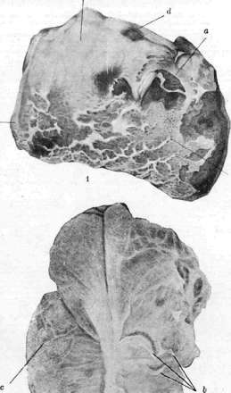

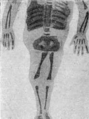
Figure 1. Diaphragmatic pleurisy: a - uneven layered deposits of fibrin; b - purulent coating; c - apex of the lung; d - anterior edge of the lung. Figure 2. Scars of the visceral pleura upon absorption of exudate in pleurisy; most of the scars (a) are free, on some areas (b) adhesions with the parietal pleura are torn; c - lungs. Figure 3. Lung tissue with hemorrhages (infarct) in pleurisy: a - fibrinous coating on the pleura (b); c - individual leukocytes. Figure 4. Sireniform monster. (After Shubina.) Figure 5. Synecephalus monoprosopus, distomus, thoraco-gastropagus, tetraurachius, tetrapus. (After Semyannikov.)
not primarily by the narrowing of the pulmonary circulation pathway, which only in the case of large exudates leads to an increase in pressure in the right ventricle, but by the lowering of the suction power of the thoracic cavity and the compression of the large veins caused by the displacement of the mediastinum. Compression and even kinking of the inferior vena cava at the site of its passage through the diaphragm are also facilitated by the displacement of the latter under the influence of the exudate. Deep breathing could counteract all these circulatory disturbances to a significant degree, which is indeed induced by this very circulatory disturbance, but deep breathing encounters a number of obstacles in a patient with exudative pleurisy, as already indicated (see above); in addition, the state of a certain paresis of the respiratory muscles apparently also has significance in this respect (Staehelin). Encapsulated pleuritic exudates usually do not cause circulatory disturbances, bulging of the chest wall, compensatory expansion of the chest wall on the healthy side, etc.; of course, here too everything depends on the size of the exudate and the compliance of the chest wall. Pathological anatomy. Pathological-anatomical changes in pleurisy are characterized initially by hyperemia of the pleura, soon accompanied by swelling, proliferation, and desquamation of the surface epithelium. The pleura ceases to shine and becomes dull and opaque. Subsequently, inflammatory exudation develops, and fibrinous deposits form on the surface of the pleura, which, in the case of large amounts of exudate, lie in continuous sheets; in smaller amounts, when the direct attracting action of the chest wall on the deposits makes itself felt, the latter, by virtue of adhesion to the parietal pleura in localized areas, are drawn out in the form of papillae running perpendicular to the pleura [fence-like appearance, see separate table (figures 1 and 3)]. The process ends with fibrinous deposits in dry pleurisy. However, an exudate having a serous, hemorrhagic, purulent, or putrid character may also form. Along with this effusion, fibrinous deposits and significant thickening of the pleura are formed, i.e., plastic pleurisy. Thick scars consisting of homogeneous, partially hyalinized, and sometimes locally edematous connective tissue occasionally form on the pleura. In these scars there is a greater or lesser number of newly formed vessels, sometimes with clear differentiation of arteries and veins. The pleuritic process frequently spreads to the neighboring connective tissue of the chest wall (peripleuritis) as well as of the lung (interstitial pneumonia), as a result of which bronchiectasis may develop. In purulent pleurisy, the spread of the process to the chest wall can cause a peripleuritic abscess, while spread to the lung causes purulent infiltration of the latter. Pleuritic adhesions in the region of the apices and upper lobes, where the mobility of the lungs is limited, having the character of broad (planar) deposits, do not undergo special changes in the future, whereas in the lower and lateral parts, adhesions under the influence of extensive respiratory movements are sometimes drawn out into strands, and sometimes resolve completely, leaving irregularly stellate whitish cicatricial fields on the pleura (see separate table, figure 2). In tuberculosis, tubercles are sometimes found on the pleura in greater or lesser numbers; they can be very small, but sometimes merge into larger conglomerates and can undergo caseous necrosis. As for the relationship between the character of the effusion and the etiology, each of these forms of pleurisy can be caused by various etiologies and, conversely, the same etiology can cause forms of pleurisy that differ in the character of the exudate. However, despite all the resulting diversity, certain regularities associated with the etiology of pleurisy can be noted. Thus, hemorrhagic pleurisies are most often associated with tuberculosis or malignant neoplasm, but they can also be caused by large pulmonary infarcts, trauma, and scurvy. Purulent pleurisies are frequently caused by purulent processes in the lungs: abscess, gangrene, bronchiectasis, purulent bronchitis, and peribronchitis. Not infrequently, purulent pleurisies form as a result of the transition of a purulent process from neighboring organs and tissues to the pleura. Purulent pleurisy can also be caused by a metastatic embolus from any septically affected organ [e.g., appendix, ovaries, heart (valves)]. But at the same time, purulent pleurisy is often formed even without the presence of a purulent focus, e.g., in pneumonia, tuberculosis. On the other hand, it is known that sometimes under the influence of purulent processes, e.g., subdiaphragmatic abscess, purulent peritonitis, etc., not a purulent, but only a sero-fibrinoid, so-called sympathetic pleurisy is formed. Fibrinoid and sero-fibrinoid pleurisy are most often connected with tuberculosis, pneumonia, and rheumatism, but they can also be of any other etiology. Of the above-mentioned, rheumatism, as far as we know, causes mainly sero-fibrinoid pleurisy, but it is possible that it also causes purely fibrinoid (dry) pleurisy. As for rare chylous exudates, they are most often caused by a tumor proliferating into large lymphatic ducts or compressing the latter. Dry pleurisy (fibrinoid), pleuritis sicca, only rarely acquires the significance of an independent disease. This occurs mostly in the case of a tuberculous origin of dry pleurisy, which is based on some tuberculous focus in the lungs or bronchial glands, but usually of low activity. Dry pleurisy in such cases represents almost the sole manifestation of the activation of tuberculosis. Such dry pleurisy begins mostly suddenly with side pain, fever, cough. The fever is usually low, rarely exceeding 38°. The pains are often very pronounced, intensify with deep breathing and coughing, sometimes reaching such an intensity that they hinder deep breathing, thus causing shortness of breath. The patient often achieves relief from pain by pressing their hands on the painful area and thereby restricting the mobility of the chest during breathing; this also explains why the patient often prefers to lie on the affected side. These pains usually do not last long; after a few days they diminish and soon disappear altogether, which, however, does not always indicate the liquidation of the process. The pains persist as long as there is a fresh inflammatory process in the not-yet-severely altered pleura, in which well-functioning nerve endings are still preserved, but when subsequently the activity of the inflammatory process of the pleura has already decreased and, on the other hand, the pleura has significantly changed and become covered with fibrinous deposits, the function of the nervous apparatus of the pleura is also impaired, as a result of which its sensitivity decreases. Cough in fresh dry pleurisy is observed mostly as dry, sometimes very agonizing due to the fact that it significantly increases the pain. General phenomena of weakness, malaise, poor appetite, and night sweats are also observed. Objectively, a certain lagging of the affected half of the chest during respiratory movements is noted. Upon percussion, there is limited mobility of the lung edge and slight dulling of the percussion note in the area of fresh pleurisy due to reflex tension of the external chest muscles. Upon auscultation, a pleural friction rub is heard, initially very soft, subsequently often acquiring a rough character resembling the creak of new leather or rough scratching. The pleural friction rub is often heard from the very first day of the disease, and sometimes appears somewhat later. It is heard best in the places of greatest mobility of the lung, i.e., in the lower parts and laterally. The pleural friction rub is easily confused with buzzing, especially crackling, and sometimes even crepitating rales. Therefore, it is important to know that the pleural friction rub is often distinguished as an intermittent sound, heard sometimes sharper during inspiration, but more often equally well during expiration, and not always strictly connected with respiratory phases. Pressure with the stethoscope usually causes an intensification of the pleural friction rub; this friction is often felt by the hand; in the place of the heard friction, the patient often feels pain, and finally, after a few deep breaths, the friction rub often weakens and sometimes disappears altogether. Taking into account all these features, it is mostly easy to distinguish the pleural friction rub, but these distinguishing features are not always clearly expressed. Upon X-ray fluoroscopy, fresh dry pleurisy is revealed only by limited mobility of the diaphragm, while later a diffuse darkening of the lung field can be noted. Dry pleurisy lasts a short time, usually a few days, sometimes 1–3 weeks. An intensification of general phenomena in dry pleurisy in the form of increasing weakness, persistent elevation of temperature, widespread pain, and a widespread pleural friction rub—all this indicates the further development of dry pleurisy into exudative. Sometimes dry pleurisy takes on a chronic, recurring character; with each exacerbation, an elevation of temperature is observed, and the process can drag on for months, even years. But even from these forms, progressive tuberculosis rarely develops; mostly these patients are found to have a poorly active, generally benign apical or glandular process, which ultimately ends in recovery. Such is the clinical picture of those dry pleurisies which have independent significance.
Most often, dry pleurisy has no independent significance, representing merely a reactive change of the pleura in response to the most diverse processes originating from the lungs and organs adjacent to the pleura (pericardium, aneurysmatically expanded aorta, liver, etc.). The addition of dry pleurisy to these underlying diseases is usually manifested by the appearance of pleural friction rubs and pain. This pain is sometimes very severe, for example, in the first days of acute pneumonia, or sometimes little pronounced. Dry pleurisy, which appears very frequently, almost constantly, in chronic disseminated pulmonary tuberculosis, chronic purulent processes in the lungs, and lung cancer, often does not cause pain sensations, but sometimes even in these chronic processes patients complain of constant pain sensations of greater or lesser intensity. Sometimes such dry pleurisy, appearing in diseases with unclear diagnosis (for example, certain forms of subphrenic abscess), facilitates the path to a correct diagnosis. In individual cases, manifested dry pleurisy serves as a precursor of perforation of cancer of the esophagus, stomach, or cancer of the flexura lienalis, subphrenic abscess, and so on. The diagnosis of dry pleurisy is based exclusively on the presence of a pleural friction rub. Other manifestations, such as pain and cough, although they have a number of characteristic features when present, may nevertheless be completely absent. But sometimes the pleural friction rub may also be absent, and then it is not always easy to distinguish dry pleurisy from intercostal neuralgia or muscular rheumatism. In these cases, it is necessary to remember that in intercostal neuralgia and muscular rheumatism, pain is poorly related to respiratory movements, and moreover, in intercostal neuralgia, characteristically located pain points are observed in the intercostal spaces in the axillary region and near the sternum. The detection of dry pleurisy necessarily requires further search for the causes of this disease. Prognosis is good in so-called independent pleurisy (tuberculous, rheumatic, traumatic). This, of course, does not apply to those pleurisy cases that develop in disseminated severe forms of tuberculosis; the prognosis of such pleurisy, like other chronic pleurisy, depends on the underlying disease. Chronic recurrent pleurisy may be associated, as already indicated above, with chronic benign tuberculosis of the apices, and then the prognosis remains good, but it must be remembered that behind any other chronic pleurisy, serious diseases are often hidden. Treatment of dry pleurisy must be directed against the underlying disease. Sometimes it is necessary to resort to symptomatic treatment—application of iodine, mustard plasters, dry cupping, which have a pain-relieving effect. Dry diaphragmatic pleurisy (pleuritis sicca diaphragmatica), inflammation of the diaphragmatic pleura, can also occur as an independent disease, but more often it is associated with a disease of neighboring abdominal organs. Diaphragmatic pleurisy is characterized by certain features arising from its localization. Patients complain of pain in the hypochondrium and lower ribs. This pain intensifies very sharply not only with coughing, but also with hiccups and eructation; sometimes swallowing is painful. Abdominal breathing is especially painful, in connection with which patients have almost exclusively costal, superficial, and accelerated breathing. The lower parts of the chest participate almost not at all in respiratory movements. On auscultation, changes are not detected. Diaphragmatic pleurisy is sometimes accompanied by a whole series of pain symptoms. The aforementioned soreness in the hypochondrium intensifies upon pressure on the epigastric region. In addition, Mussy described a number of pain points characteristic of diaphragmatic pleurisy, which are called Mussy's points. These include the point between both heads of the sternocleidomastoid muscle, because the phrenic nerve passes here, the point at the edge of the sternum in the region of the first intercostal space, the region of the cervical plexus and over the spinous processes of the cervical vertebrae, the region of attachment of the diaphragm to the chest wall, and the intersection of the parasternal line and the line representing the continuation of the tenth rib; the latter point is named the "diaphragmatic button" ("bouton diaphragmatique"). All these points are far from constant. Sometimes pain is obtained by pressing only on some of them, but even the absence of soreness in the region of all these points does not exclude diaphragmatic pleurisy. Often patients with diaphragmatic pleurisy complain of spontaneous pain in the shoulder region; sometimes these patients exhibit the respiratory reflex of the rectus abdominis muscles described by Schmidt (R. Schmidt), which reduces to the fact that when attempting to take a deep breath on the affected side, lightning-fast twitching occurs in the region of the upper part of the rectus abdominis muscle. Radiologically, patients with fresh diaphragmatic pleurisy show limited excursion of the affected diaphragm, insufficient opening of the sinuses, and sometimes high standing of the diaphragm; all these are uncharacteristic data. Later, however, with the formation of adhesions, more characteristic changes are noted, namely: irregularities of the diaphragm, bulging or, conversely, flattening of the diaphragm, which sometimes takes the form of a straight line, and partial or sometimes even complete obliteration of the sinuses. Exudative pleurisy (sero-fibrinous), pleuritis exsudativa (sero-fibrinosa), just like dry pleurisy, is often an independent disease, i.e., sometimes no pathological processes are hidden behind it either in the lungs or in other organs, or else (much more frequently) these pathological processes are such that, having played the role of a causal factor, they subsequently have very little significance in the clinical picture of the entire disease. Previously, the term "idiopathic" was applied to these pleurisy cases; such a term, if understood as an indication of the absence of an etiological factor, is of course completely incorrect—these are not "idiopathic" pleurisy cases, but pleurisy of a completely definite etiology, which, admittedly, in each individual case is not always easy to identify. Most of these pleurisy cases represent a manifestation of a relatively benign exacerbation of a tuberculous focus localized in the lung tissue itself or, much more frequently, in the bronchial glands; part of these pleurisy cases are of rheumatic etiology and finally, behind some, clinically poorly manifested pneumonic (non-tuberculous) processes are hidden. Another large group of exudative pleurisy cases, to which the incorrect name of secondary pleurisy (see above) is often applied, is more correctly called symptomatic, because they represent only one of the symptoms of the underlying disease. The clinical picture of symptomatic pleurisy for the most part recedes into the background in the clinical picture of the underlying disease (e.g., pleurisy in progressive pulmonary tuberculosis, nephritis, etc.).
Symptomatology. Exudative pleurisy sometimes develops as an acute disease, accompanied from the first days by a rapidly accumulating effusion; the limiting capacity of the pleura is determined by Gerhardt at 2-4 liters, and by Sokolovsky even at 6-10 liters. Sometimes the disease develops gradually; although the exudate begins to accumulate from the first days, it subsequently increases slowly, gradually. Finally, often exudative pleurisy develops from a preceding dry pleurisy that ran a more or less prolonged course. The general manifestations of this disease are very diverse. The exudate may be very small in general, or very small because it has not yet had time to accumulate; sometimes so small that it cannot be detected even upon careful objective examination, while the general manifestations of this disease at that time may already be strongly expressed. The temperature can reach high figures, even 41°, and have a remittent, sometimes even hectic character; general weakness, significant sweating, poor appetite, and rather rapidly developing emaciation may be observed. But more often this disease proceeds with negligible general manifestations. The temperature in these cases is subfebrile, slowly rising, and after a few days of bed rest begins to fall lysis-like; there is still only insignificant shortness of breath, while objective examination may reveal a large exudate. Patients with exudative pleurisy frequently complain of pain in the sides and back, intensifying with coughing, deep breathing, movements, sometimes very strong, colic-like, radiating to the shoulders and epigastric region.
This irradiation usually indicates the involvement of the diaphragm, a more pronounced lesion of which sometimes causes painful swallowing and the remaining pain symptoms characteristic of diaphragmatic pleurisy (see above); on the other hand, in exudative pleurisy, pains are often insignificant or absent altogether, and the patient frequently speaks of pains that occurred only in the first days of the disease.
Cough usually does not bother patients much, but a dry, painful cough is sometimes observed, especially with a deep breath—this cough is caused by irritation of the pleura. Cough in pleurisy may also often be caused by concomitant bronchitis developing in atelectatically collapsed parts of the lungs; such cough may be accompanied by scanty sputum production. Finally, in symptomatic pleurisy, the cough may be caused by the underlying disease, upon the character of which the nature of the cough will also depend.
Objective data. The patient mostly assumes a semi-lateral position on the affected side, with a large exudate—on the back, but sometimes even with a small exudate the patient prefers a position on the back, rarely even on the healthy side, when a position on the affected side causes pain and cough. Asymmetry of the chest is revealed: the affected side is protruded and most of all in the lower-lateral part, the shoulder on the affected side is raised, from the side of the spine there is scoliosis with a convexity toward the affected side, but subsequently the affected half of the chest sinks, even if the fluid from the pleural cavity has not yet completely reabsorbed (Stehelin).
The skin on the affected side is sometimes shiny, tense, and thickened—edematous—apparently due to impaired lymph circulation (Bönniger). The intercostal spaces are widened, smoothed, sometimes even protruded. The affected side lags during breathing, Litten's sign is absent. The apex beat is displaced. Swelling of the neck veins is frequently observed.
Palpation always reveals a weakening or absence of vocal fremitus in the area of the exudate, increased vocal fremitus over the exudate often, and normal vocal fremitus still higher.
Percussion may fail to detect dullness in very small exudates not exceeding 400 cm3 (Vierordt, Yanovsky), although, according to Damoiseau, careful percussion can determine even very insignificant effusions (60–90 cm3). In pleuritic exudates on the affected side, dullness is noted, passing downward into absolute dullness, and upward—sometimes into a zone of tympanites due to the relaxation of the lung tissue compressed by the exudate. The upper boundary of this dullness has a characteristic outline known as the Ellis-Damoiseau line; this line goes from the spine outward and upward, reaching the highest point on the posterior axillary line, and descends anteriorly, so that on the anterior chest wall dullness is found in a small space or is not found at all.
The character of this line was previously explained by the fact that, in accordance with the patient's position on their side, the fluid is also fixed in such a position by adhesions formed at the border of the fluid. This explanation is incorrect, since it is known that the same outline of the dullness boundary is observed regardless of whether the exudate shifts or not upon changing position, and regardless of whether the patient lay on the healthy or the affected side. (For the correct explanation, see above.)
With very large exudates, this typical boundary of dullness is absent; the latter then occupies the entire half of the chest, and perhaps only above and below the clavicle, in the first intercostal space anteriorly, above the scapula, and in the interscapular space posteriorly is the dullness less intense. With large exudates, tympanites is revealed under the clavicle, often intensifying when the mouth is opened, i.e., Williams' tracheal tone is revealed, and with deep percussion this tympanites acquires the characteristic cracked-pot sound. Large left-sided exudates lead to the disappearance of tympanites in Traube's space, while small exudates lead to dullness in the upper part of this space.
With right-sided exudates, dullness is often noted anteriorly, near the right border of the heart, which resembles the dullness in exudative pericarditis, i.e., it is located from above downwards along the sternum, while lower down it moves outward. This dullness is apparently explained by the penetration of the effusion into this area, since negative pressure is well expressed here due to the significant mobility of the lung margin (Stehelin).
With large exudates, percussion reveals a paravertebral triangular dullness of Rauchfuss-Grocco on the healthy side, located near the spine in the lower part of the chest (Fig. 1). This dullness is explained by the displacement of the mediastinum, causing atelectasis of the lung on the healthy side (Rauchfuss, Sahli, Goldscheider, and others), but it is possible that resonance of the dull sound transmitted adjacently from the affected side also plays a role in the origin of this dullness. It must be remembered that, although rarely, this triangular dullness is also observed in croupous pneumonia (Hamburger, Matthes, Hochhaus).
Of great diagnostic significance is the triangular area of clearing of the percussion sound of Garland, located posteriorly on the affected side between the spine and the line of pleuritic dullness (i.e., the Ellis-Damoiseau line); this phenomenon is apparently explained by the fact that the lung compressed by the exudate (large bronchi) nevertheless gives a less dull tone than the exudate. Features of dullness in exudative pleurisy include: 1) Rauchfuss's triangle; 2) Garland's triangle. Auscultation reveals a weakening, sometimes even a complete absence of respiratory sounds at the site of greatest fluid accumulation. Above the dullness, i.e., above the exudate, bronchial breathing or breathing with a bronchial tint may be heard, passing higher into harsh, normal, or weakened vesicular breathing. At the border of the dullness, a pleural friction rub is often heard; hearing this sound at the site of dullness where it was not heard before is of great significance, as it indicates the reabsorption of the exudate. At the borders of the dullness, atelectatic rales are frequently heard, as well as moist rales indicating bronchitis developing in the atelectatic parts of the lungs. Bronchophony in the area of dullness is weakened, and above the dullness it may be somewhat enhanced. X-ray examination (see separate table, figures 1, 2*, 4, and 5) with small exudates shows a laterally located homogeneous, triangular-shaped shadow, the upper-inner boundary of which rises from below and inside upwards and outwards, forming a concave inward, not entirely sharp line. With large exudates, this line represents a straight line and approaches the horizontal. With very large exudates, the entire lung field is darkened. With small exudates, the shadow fills only the phrenicostostal sinus and is better revealed standing, but may be completely absent. Upon changing position, the shadow boundary almost does not change. Diaphragm contours are often poorly outlined, but sometimes a high standing of the diaphragm can be noted, which may be due to its paralysis on an inflammatory basis. The rest of the lung field is usually darker than on the healthy side, which is due to compression of the lung. With a very large exudate, the lung field on the healthy side is mostly darker than usual, which is connected with compression, partly with hyperemia. Fluoroscopy also shows the degree of displacement of the mediastinum, in particular the heart. *In the explanations to the radiographs on the separate table, read: Figure 1. Right-sided exudative pleurisy; Damoiseau's line is well expressed. Figure 2. Encapsulated pleuritic exudate.
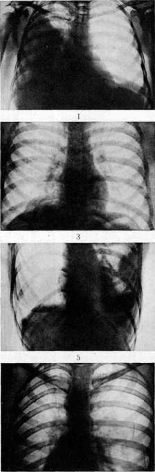
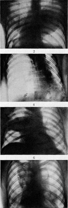
Fig. I. Encapsulated pleuritic exudate. Figure 2. Right-sided exudative pleurisy (from behind); the Damoiseau line is well expressed. Figure 3. Right-sided diaphragmatic pleurisy; fixation of the inner half of the diaphragm. Figure 4. Left-sided [pyothorax] during resorption of the exudate; sharp retraction of the chest and displacement of the cardiovascular [shadow] to the left; high standing of the diaphragm. Fig. 5. Encapsulated interlobar pulmonary pleurisy; adhesion of the diaphragm. Fig. 6. Interlobar right-sided pleurisy. Fig. 7. Artificial right-sided pneumothorax: complete resolution of the process. Fig. 8. Bilateral pneumothorax: hanging heart. S. St. Ischtin. Pneumothorax. of the heart displaced to the left retain three arcs. With a large exudate, a clarification caused by the displaced trachea may be noted in the shadow of the large vessels (mediastinum) displaced to the right or left. When the shadow of the mediastinum is displaced to the right by a large left-sided exudate, the shadow of the superior vena cava stands out especially due to its overfilling with blood. Test puncture of the pleura should be performed every time there is a suspicion of the presence of an effusion in the pleural cavity; it finally confirms the presence of fluid, and most importantly, it makes it possible to study the nature of this fluid, facilitates the solution of the question of etiology and rational treatment of pleurisy. T e c h n i q u e of test puncture. First of all, the puncture site is chosen; it is outlined where there is the most pronounced dullness and weakening of vocal fremitus. If the exudate is large enough, the puncture is best done in the VIII or IX intercostal space between the posterior axillary and scapular lines; with a small exudate, the puncture is best done in the IX intercostal space along the scapular line; with encapsulated exudates, the test puncture has to be done at the site of maximum dullness, but one must remember the danger of injuring the a. intercostalis. Posterior to the axillary line, this artery is protected by the lower costal edge, while anteriorly it is unprotected and near the sternum, at 0.5-1 cm from the sternal edge, runs the a. mammaria interna. The best position for the patient is sitting, with a hand placed on the head, which achieves the expansion of the intercostal spaces. The syringe (10-20 g) with which the puncture is made must have a well-fitted piston and a sufficiently long (not less than 7 cm) and not very thin (not thinner than 1 mm) needle. The syringe with the attached needle is taken in the hand like a pen, and the needle is injected into the intercostal space above the upper edge of the underlying rib. After inserting the needle, movements of the syringe make sure that the needle is in the pleural cavity, and then by pulling the piston, the pleuritic fluid is aspirated. Pleuritic exudate is usually a light yellow or yellowish-green liquid containing about 4-6% protein; the specific gravity of the exudate ranges between 1.015-1.020. Already with this study, it is usually possible to distinguish whether the obtained fluid has the character of an exudate or a transudate, and the appearance of the fluid alone makes it possible to determine its serous, hemorrhagic, or purulent character. The cytology of the obtained exudate has a fairly large diagnostic significance. Vidal and Ravaut were the first to establish that the predominance of lymphocytes in the exudate is characteristic of tubercular pleurisy, and the predominance of neutrophils is characteristic of pleurisy caused by coccal infection. This proposition does not always correspond to reality, since in the first days even in a tubercular exudate, neutrophils often predominate, and on the other hand, by the time of recovery, lymphocytes may begin to predominate in a non-tubercular exudate as well. Thus, the cellular composition of exudates is determined not only by the etiology, but also by the duration of the process. Modern knowledge about the cellular composition of exudates can be formulated as follows. In tubercular pleurisy, in the first days of the disease, the cellular composition of the exudate is not very characteristic: it consists of lymphocytes, neutrophils, and endothelial cells, and sometimes neutrophils may even predominate. But soon (after 7-10 days) the number of neutrophils and endothelial cells begins to decrease, endothelial cells may even disappear completely, and erythrocytes and lymphocytes begin to distinctly predominate. In protracted tubercular pleurisy, the number of exudate cells noticeably decreases, degeneration of neutrophils begins; neutrophils shrink, contain pyknotic or decayed nuclei (Kuniger). Thus, in a tubercular exudate, sometimes lymphocytes predominate even at the beginning, but very often they predominate only at the height of the disease. In acute non-tubercular pleurisy, only at the very beginning of the disease there is a fair amount of both lymphocytes and endothelial cells in the exudates, but very soon neutrophils begin to predominate, by the height of the disease the exudate contains almost exclusively neutrophils and erythrocytes, and finally by the beginning of resorption of the exudate, lymphocytes increase in it again. In protracted exudates and non-tubercular ones, lymphocytes often predominate, and degenerative changes are found in neutrophils, which often differ from the degenerative changes of neutrophils of tubercular exudates (see above) in that the nuclei appear swollen, stain poorly; the protoplasm containing vacuoles quite often stains poorly as well. Exudates caused by neoplasms often have a hemorrhagic character, which happens, however, not only with neoplasms, but can also be with tuberculosis, as well as with infarcts and diseases accompanied by a tendency to bleeding (scurvy, purpura, etc.). On the other hand, carcinomatous exudates often have the character of serous effusion. Tumor exudates often contain a large number of endothelial cells, and sometimes characteristic tumor cells. Interlobar pleurisy (pleuritis interlobaris) presents a number of features. Accumulating between the lobes, the exudate, due to adhesions formed at their border, may not break into the free pleural cavity. The clinical picture is various, mainly depending on the size of the exudate. With a small exudate, even the most careful percussion and auscultation often reveal nothing. Sometimes a slight dullness is discovered in the form of a strip stretching according to the border of the lobes from behind upwards to the front and downwards; this strip of dullness is sometimes located only behind or only in the axillary region, or only in front in the region of the IV rib. With large exudates, both upwards and downwards from this strip of dullness, a zone of tympanitis can be noted, caused by the compressed lung. With an increase in exudate, the dullness also increases; in this case, with large exudates in the region of the interlobar border behind, and especially in the axillary region, absolute dullness of the percussory sound may be obtained, caused by the effusion coming close to the chest wall. Absolute dullness downwards turns into dullness stretching to the very lower border of the lung; this dullness is caused by significant compression of the entire lower lobe of the lung. Data from auscultation in interlobar pleurisy are less characteristic; in the area of dullness and dullness, usually weakened breathing is heard, sometimes a friction rub of the pleura, while above or below the dullness, sometimes in a limited area, sometimes more widespread, bronchial breathing is often heard. A large interlobar exudate can break into the free pleural cavity, which is usually accompanied by an increase in temperature, pain, and the appearance of a pleural friction rub. The X-ray picture of interlobar pleurisy (see separate table, Fig. 6) has great diagnostic significance; it is characterized by sharp boundaries of the shadow and its uniform intensity; its shape depends on the size of the exudate and whether it is located more anteriorly or posteriorly, inwardly or outwardly. With small exudates, a characteristic ribbon-like shadow is often visible, having the appearance of a long shadow in an oblique position, corresponding to the lobar border. With large exudates, the upper boundary is usually straighter, while the lower one is more convex, but the reverse can also happen. The shadow of the interlobar effusion is usually separated from the diaphragm by a light zone, which, however, may be absent if the exudate is located in the lower part of the interlobar region; fluoroscopy in a sharply expressed lumbar-lordotic position helps to figure out these cases. Test puncture in interlobar pleurisy should be done with a long needle in the axillary region at a place corresponding to the interlobar border. A large interlobar effusion causing significant compression of the lower lobe of the lung and the dullness in the subscapular region caused by this compression sometimes simulates an exudate located not between the lobes, but freely in the pleural cavity, but in these cases a test puncture in the subscapular region does not reveal effusion, and it is easily detected by puncture in the axillary region. Mediastinal pleurisies (pleuritis mediastinalis) also have a number of features. Media
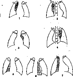
Figure 2. Various forms of mediastinal pleurisies: 1—pl. mediastinalis ant. sup. dext.; 2—pl. mediastinalis post. sup. dext.; 3—pl. mediastinalis ant. sup. sin.; 4—pl. mediastinalis post. sup. sin.; 5—pl. mediastinalis inf. ant. dext.; 6—pl. mediastinalis post. inf. sin.; 7—pl. mediastinalis post. sup. et inf. dext.
Mediastinal pleurisy represents the encystment of an effusion that has accumulated between the mediastinal and pulmonary pleura. Depending on the location of the effusion, a distinction is made between anterior (left-sided and right-sided) and posterior mediastinal pleurisy (Fig. 2). In anterior mediastinal pleurisy, regardless of whether it is left-sided or right-sided, subjective complaints may be identical, specifically substernal pains, which can vary greatly in intensity, and dyspnea, usually mild. Objectively, in left-sided mediastinal pleurisy, percussion reveals dullness on the left along the upper part of the sternum, which, depending on the size of the exudate, extends to the left as far as the midclavicular or even the axillary line; the heart may be displaced to the right, and sometimes to such an extent that the heart pulsation is felt only to the right of the sternum. In the area of dullness, vocal fremitus and respiratory sounds are usually diminished. In right-sided mediastinal pleurisy, dullness is found on the right along the sternum, the heart may be displaced to the left, and marked cyanosis, caused by compression of the superior vena cava, is frequently observed. Posterior mediastinal pleurisy is often accompanied by paroxysmal cough, sometimes stridor breathing due to compression of the trachea, and sometimes difficulty in swallowing due to compression of the esophagus. The diagnosis of mediastinal pleurisy is difficult; it is very often facilitated by X-ray examination, although frequently X-ray examination is also unable to distinguish mediastinal pleurisy from mediastinal tumors. Diaphragmatic exudative pleurisies present the same clinical picture as dry diaphragmatic pleurisies (see above); only radiologically can a supradiaphragmatic shadow caused by the effusion be detected (see separate table [p. 367-368], Fig. 3). The course of exudative pleurisy depends on many factors. If the pleurisy is symptomatic, it is clear that its course will depend on the nature of the underlying disease, while for the most part this pleurisy has little effect on the course of the underlying disease. Thus, the majority of parapneumonic pleurisies proceed so mildly that they do not affect the duration of the pneumonic process and the course of the convalescence period; due to the rapid resorption of these exudates, they very often pass even unrecognized. Most metapneumonic pleurisies also usually run a mild course; they manifest themselves by a subfebrile temperature for several days, with corresponding percussion and auscultation data. Metapneumonic pleurisies proceed with almost no subjective complaints, are rapidly absorbed, and usually do not affect the favorable course of the post-pneumonic convalescent period, which is characterized by the rapid restoration of health. But sometimes both para- and metapneumonic pleurisies exhibit a protracted course, and then a suspicion of a superimposed tubercular infection always arises. Occasionally it happens that an exudative pleurisy, arising in the very first days of pneumonia, rapidly increases and gives clinical manifestations that dominate the general picture of the disease and push aside the manifestations of the pneumonia itself, which in these cases usually has the character of a poorly disseminated focal pneumonia. Exudative pleurisies having the character of the primary disease, the majority of which belong to tubercular pleurisy, proceed for the most part relatively favorably. The elevated, mostly subfebrile, less often remittent temperature usually lasts for 2 to 3 weeks; after this, the temperature drops lyrically, and gradual resorption of the exudate and recovery begin. The onset of exudative resorption is not always easy to detect because percussion and auscultation changes are often poorly expressed due to the formation of pleural adhesions. Of greatest importance for establishing the beginning of resorption is the appearance of a pleural friction rub in those areas of percussion dullness where it was not previously auscultated. Resorption can also be indicated by an increase in diuresis and a positive water test; the latter, conversely, proves negative in the stage of exudate accumulation. Finally, good indications of exudate resorption are provided by careful measurements of each half of the chest at the nipple level and at the level of the xiphoid process (Stehelin). But exudative pleurisies do not always run an easy and rapid course; sometimes they proceed very severely, resembling an acute septic disease. These hypertoxic forms of pleurisy run a high, sharply fluctuating temperature, often acquiring the character of a hectic fever, with phenomena of clouded consciousness and increasing cardiac weakness. The clinical picture resembles empyema, but in reality, the exudate has a serous character. In these septic forms of pleurisy, death of the patients sometimes occurs suddenly, but often these patients also recover. Finally, sometimes exudative pleurisies have a chronic course, when the exudate is not absorbed for a long time, over many weeks. The temperature is mostly elevated, but sometimes it becomes normal for a short time, usually for several days; poor appetite persists for weeks, patients become significantly exhausted, the affected half of the chest begins to sink, which, however, does not exclude the presence of exudate remnants. Most of these patients ultimately recover anyway, but some retain an elevated temperature, general weakness, and rapid fatiguability for a long time. On the other hand, prolonged exudative pleurisies sometimes suppurate, which of course can also occur with acutely developing pleurisies. Occasionally suppuration develops following a puncture. Suppuration of the exudate is often indicated by a deterioration in the general condition, chills, and a large rise in temperature; however, these signs of deterioration do not always appear, which is why in all cases of chronic exudative pleurisies proceeding with a high temperature, it is necessary to resort to repeated trial punctures. It is necessary to single out those tubercular pleurisies that are called by some clinicians secondary tubercular pleurisies, i.e., those that occur in the presence of an already severe, widespread pulmonary tuberculosis due to the spread of tuberculosis to the pleura. These pleurisies, which are, however, observed quite rarely, usually make themselves known by chills, a high rise in temperature, a sharp deterioration of the general condition, and are accompanied by the progression of the underlying tubercular process in the lungs, as a result of which these patients usually perish soon. Some authors note that when an exudative pleurisy is added to pulmonary tuberculosis, causing compression of the diseased lung, an improvement in the pulmonary process sometimes occurs (Forlanini, Kuniger), but this takes place mainly in mild forms; in severe pulmonary tuberculosis, this apparently occurs only in exceptional cases. In the elderly, the course of exudative pleurisies presents certain features. Firstly, when establishing exudative pleurisy in an old person (and in advanced age), the question always arises as to whether this pleurisy is caused by a malignant neoplasm; it must be remembered here that these tumor pleurisies can be both serous and hemorrhagic, less often purulent. Secondly, non-tumor exudative pleurisies occur more slowly in the elderly than in the young, often with a slight increase in temperature or with a normal temperature; at the same time, dullness is not always intensely expressed, while vocal fremitus on the affected side often differs little from vocal fremitus on the healthy side; all this significantly complicates diagnosis and necessitates resorting to a trial puncture more often than in the young. The diagnosis of exudative pleurisies is not difficult in typical cases, but it must always be confirmed by a trial puncture, especially since the first stage of diagnosis comes down to establishing an effusion, but this is the easiest question, after which arises the question of the nature of the effusion (exudate or transudate, and if an exudate, what kind—serous, hemorrhagic, purulent, putrid, chylous) and most importantly—the etiology of the pleurisy. The question of the nature of the effusion can be definitively resolved only with the help of a trial puncture, the results of which are of no small importance for understanding the etiology (see above). But the results of punctate examination alone are of course insufficient to resolve the question of the etiology of pleurisy; for this, a thorough study of the entire organism and especially a careful examination of the lungs are required first of all. The differential diagnosis of exudative pleurisies is of some importance only with respect to croupous pneumonia, since in the latter, diminished vocal fremitus and diminished respiration may also sometimes be observed. Of differential diagnostic significance in these cases is the boundary of dullness, which in croupous pneumonia often corresponds to the lobar boundary, whereas in pleurisy it has the character of Damoiseau's line; the marked triangular dullness of Rauchfuss-Grocco, dullness in Traube's space in left-sided pleurisy, and the presence of displacement of neighboring organs are also of significance. It is more difficult to resolve the question of whether there is an exudative pleurisy along with pneumonia. In these cases, suspicion of the presence of exudate arises with very intense dullness increasing downwards; this question is finally resolved only by trial puncture, and all precautions must be observed so as not to injure the lung itself during the puncture.
Diagnosis is further complicated when, alongside pneumonia, there is an encapsulated exudate; in such cases, even a trial puncture does not always yield a positive result. The prognosis varies depending on the nature of pleurisy. Where exudative pleurisy has the character of a primary independent disease, and where aside from this pleurisy there is no other severe disease in the organism, the prognosis is mostly favorable. On rare occasions, with very large exudates, sudden death of patients occurs, apparently in connection with paralysis of the intercostal respiratory muscles, sometimes (very rarely) from pulmonary embolism, and finally, in cases of protracted non-absorbing exudates, general exhaustion leading to death may sometimes develop. Prolonged exudative pleuritides, especially recurrent ones, leave large pleural adhesions that prevent the expansion of the lung and cause shortness of breath, and are accompanied by a slow restoration of working capacity (sometimes only after 2-3 months). For the prognosis of these chronic and especially recurrent exudative pleuritides, it must be borne in mind that even more late consequences of these pleuritides can be bronchiectasis and pneumosclerosis. The prognosis of those pleuritides that developed on the basis of some other severe disease, e.g., pulmonary tuberculosis, nephritis, heart disease, etc., is almost always poor; the prognosis here is essentially determined not by the pleurisy itself, but by the underlying disease, which usually causes the formation of pleurisy already in very far-advanced stages. The treatment of patients with exudative pleurisy boils down first of all to rest. In the initial acu...
PLEURISY
During the first period of pleurisy, when the inflammatory process is still spreading along the pleura and leading to the accumulation of exudate, absolute rest is necessary. Rest, by limiting the movements of the chest, leads to a decrease in pain and the subsiding of the inflammatory process. Early disruption of rest, early getting up, and the transition to movement often lead to a return of the febrile state and a new accumulation of exudate. Fears that rest will contribute to atelectasis and greater formation of adhesions are unfounded, since in the first acute period of pleuritic exudate the main thing is the progressive inflammatory process in the area of the affected pleura. The nutrition of patients should be frequent, in small portions, and of sufficiently high caloric value; it must be remembered that exhaustion of patients can favor the development of pulmonary tuberculosis. In cases with protracted exudate, one can try treatment according to Karel (see Karel's method). Local heat (warming compress), as well as so-called counter-irritants (mustard plasters, cupping glasses), often act as pain relievers, but hardly contribute to accelerating absorption. Among drug therapy, one must first of all point out salicylates, which since the works of Fiedler and Aufrecht have been very willingly prescribed by many doctors in the treatment of patients with exudative pleurisy, although this treatment is still poorly justified. The majority of authors view salicylates merely as a symptomatic remedy that lowers temperature, reduces pain, and sometimes creates a temporary euphoria, and deny a favorable effect of salicylates on the course of the process itself. They base this on the fact that the majority of fresh exudative pleurisies proceed favorably and in the same time frames both with and without the use of salicylates. An exception is made only by rheumatic pleurisies; here salicylates, prescribed in a sufficient dose (4.0-8.0), have a good effect. The administration of diuretin, if it has some meaning, is only in the stage of resorption of the exudate, perhaps accelerating the process of absorption; in this same stage, the administration of calcium chloride can also be justified by the diuretic effect, which L. Blum recommends giving in a concentrated solution (30 g to 100 g of water) in teaspoons hourly (at the rate of a daily intake of 1.5-30 g of calcium chloride) for several days. Some authors recommend using autoserotherapy. This method, which consists of injections of pleuritic exudate immediately after its extraction through the same skin puncture under the skin (the needle is withdrawn from the pleural cavity and inserted into the subcutaneous tissue), was first proposed by Gilbert. He considered it sufficient to make two injections of 1.0 within 2-3 days; after this, he observed the resorption of the exudate within 6-10 days. Some authors (Senator, Glinchikov, and others), who frequently resorted to a larger number of injections (3-5), and sometimes in larger doses (up to 3.0), confirmed the favorable result of this treatment method, while others (I. V. Zavadsky, Stehelin, and others) consider these results doubtful, and individual authors even warn against this treatment method, pointing out that it can even worsen the course of the process, exacerbating pulmonary tuberculosis. In exudative pleurisies, one sometimes has to resort to narcotic drugs in the presence of painful dry cough and sharp pains in patients. But it must be remembered that narcotic drugs should not be prescribed in doses that depress respiration, since rapid breathing in pleurisy patients sometimes compensates to a certain extent for circulatory disorders. One must be careful with large doses of narcotic substances also because they can weaken the reflex increase in the tone of the chest muscles present in pleurisy patients, especially with significant exudates, and paralysis of these muscles can turn out to be very dangerous, even fatal, for patients. Drug therapy may also be needed for phenomena of cardiac weakness, which in pleurisy patients can be caused by sharp displacements of the heart, and sometimes also by toxic influences. In phenomena of acute heart weakness, camphor, caffeine should be used, while in patients with organic heart lesions, early administration of digitalis is useful. In pleuritic exudates, one often has to resort to thoracentesis for the purpose of releasing the pleuritic fluid. Technique of thoracentesis. Thoracentesis for releasing pleuritic fluid must necessarily be preceded by a trial puncture (see above). The mere introduction of a trocar or a thick needle into the pleural cavity is for the most part insufficient for releasing the pleural fluid, since in the pleural cavity, even in the presence of exudate in it, negative pressure is almost always maintained. Thoracentesis with subsequent aspiration of the exudate was first proposed by the American doctor Bowditch in 1863. This method began to be widely used especially after Dieulafoy described his aspiration apparatus, which is a large syringe equipped with cocks that, as needed, can establish communication of the syringe either with a needle put on a rubber tube or with a second rubber tube. Having inserted the needle into the pleural cavity and, with the help of the cock, established communication of the syringe with this needle, the pleuritic fluid is aspirated into the syringe; when the syringe is filled with this fluid, it is removed by repositioning the cock from the syringe through the second rubber tube, after which the pleuritic fluid can be sucked into the syringe again by repositioning the cock, etc. At present, Potain's aspiration apparatus is used much more often. Regarding the place where to perform thoracentesis, what is said about the place of the trial puncture remains in force (see above). The question of how much fluid to release must be decided depending on the condition and reaction of the patient. If the patient begins to complain of weakness during the release of the fluid, becomes pale, if his pulse begins to quicken, cough appears, and especially if foamy bloody sputum begins to be released with the cough, the pumping out of the fluid must be immediately interrupted. Usually, it is possible to release 1-1.5 liters without difficulty, and sometimes almost completely empty the pleural cavity. It is very important at the same time to release the fluid slowly, for example, releasing 1-1.5 liters no faster than within half an hour. During thoracentesis, a number of phenomena can develop that can become dangerous for the patient. Phenomena of cardiac weakness often develop: frequent pulse, shortness of breath, even collapse—these phenomena can often be avoided by a slow release of the exudate. Rapidly passing dizziness and even losses of consciousness, usually caused by air embolism, often occur. Occasionally, cerebral embolism occurs, caused by the detachment of thrombus from the pulmonary veins or from the auricle of the atrium. Sometimes blood begins to mix into the fluid, caused either by hyperemia due to the rapid emptying of the pleural cavity or by the rupture of adhesions or minor damage to the lung tissue, which usually does not lead to any bad results. An insignificant admixture of blood does not present a danger, while a significant one requires the termination of the puncture. Very rarely does one have to observe hemoptysis caused by the rupture of a tuberculous cavity, or a pneumothorax formed from the same cause. Finally, a particularly dangerous phenomenon should be considered a cough accompanied by increasing separation of foamy "albuminoid," "serous" sputum ("expectoration albumineuse"). Sometimes the immediate termination of the puncture leads to an improvement in the condition of the patient. This dangerous complication, which can lead to death, is apparently caused by pulmonary edema; it is observed mainly in exhausted patients, patients with heart weakness and severe pulmonary disease; to prevent this complication, it is very important to release the exudate as slowly as possible. It is also important to take care of the comfortable position of the patient during the puncture. In very weak ones, it should be done in a semi-recumbent position. Indications for pumping out the exudate can be twofold. The first indication is the so-called vital indication (indicatio vitalis), when excessive accumulation of exudate threatens the life of the patient; having strongly displaced the heart and hindered blood circulation, pleurisy causes shortness of breath, cyanosis, poor pulse. Such circulatory disturbance is not always necessarily caused by a very large exudate; it can also be caused by an exudate that is not very large, but located in such a way that it causes a strong displacement of the heart (for example, displacement of the heart by mediastinal pleurisy). Increasing shortness of breath, even with a good pulse, indicates the pumping out of the exudate, and it is better in these cases to release the fluid once more than to bring the patient to a very severe, life-threatening condition. Coinciding for the most part with this first indication is Trousseau's indication, which comes down to the fact that it is always necessary to pump out the exudate if it is very large, if the dullness reaches the 1st intercostal space. The second indication is sluggish absorption of the exudate. If after the drop in temperature or with very minor temperature increases the exudate does not resorb well, it must be pumped out.
This necessity is dictated by the fact that prolonged retention of exudate promotes the formation of adhesions, which can subsequently hinder the expansion of the lung. It is important to perform this extraction of the exudate when the inflammatory process has already subsided and new exudate is no longer accumulating. Finally, some practitioners practice extracting the exudate in the acute period of the disease even in the absence of vital indications, hoping thereby to achieve accelerated resorption of the exudate, but such early evacuation of the exudate makes little sense, since it is more often accompanied by a new accumulation of exudate. Replacement inflation of the pleural cavity with air or nitrogen in place of the evacuated exudate may have some significance in exhausted patients or in patients with severe pulmonary tuberculosis, when replacement inflation of air may perhaps prevent the rapid expansion of the lung, which in these cases can sometimes entail an exacerbation of tuberculosis or pulmonary edema. Purulent pleurisy (empyema) (pleuritis purulenta, empyema). Children contract purulent pleurisy more often than adults, and among adults, men more often than women. Etiologically, empyemas are mostly associated with a lesion of the lung, and most often with pneumonia. Often empyema develops in connection with widespread pneumonia, but sometimes there is only a small pneumonic focus or insignificant metastatic suppuration in the lungs. Less commonly, purulent pleurisy is caused by pulmonary suppurations (abscess, gangrene, bronchiectasis, putrid bronchitis) and suppurations associated with lung tumors, actinomycosis, echinococcus. Tuberculosis plays a smaller role in the etiology of purulent pleurisy than in that of serous pleurisy. Hedblom notes tuberculous etiology in 20-26%. More likely in these cases there is not a pure tuberculous, but a mixed infection, especially streptococcal. Purulent pleurisy can also develop by the spread of a suppurative process in the vicinity, e.g., from a subdiaphragmatic abscess, from a decaying cancerous neoplasm of the stomach, esophagus, from purulent paranephritis, and so on. Finally, such empyemas are observed that seem to be primary, not associated with a lesion of any other organ; these empyemas are observed more often in children, they are usually caused by pneumococcal or streptococcal infection; the anamnesis sometimes reveals a previous tonsillitis in these cases, but more often some pneumonic focus lies at the base of these empyemas. Cultures of purulent exudates most often detect pneumococci or streptococci, sometimes a mixed infection, very rarely staphylococci, and finally sometimes the exudate turns out to be sterile. The symptomatology of purulent pleurisy, unlike serous pleurisy, is usually characterized by more severe general phenomena: chills, rigors, large temperature fluctuations, a frequent and often small pulse, rapidly developing pallor, and general nutritional decline. Leukocytosis is typically noted in the blood. Of the local symptoms, skin edema in the chest area is much more common in empyema than in serous exudate. But often the general symptoms in empyema are also mildly expressed, and since the physical data of percussion and auscultation do not distinguish empyema from serous pleurisy in any way, the nature of the exudate is clarified exclusively by trial puncture. Often a persistent lack of appetite, increasing weight loss, and loss of strength are the main symptoms raising suspicion of the purulent nature of the exudate. Occasionally, in some empyemas, pulsation of the upper intercostal spaces above the exudate is observed; such empyemas are called "pulsating" (empyema pulsans). Isolated cases of serous "pulsating pleurisies" have also been described, but usually "pulsating" pleurisies turn out to be purulent and almost exclusively left-sided. According to Eichhorst, this pulsation is caused by the heart's pulsation transmitted through the purulent exudate to the paretic intercostal musculature. Other authors consider strong compression (airlessness) of the lung to be a mandatory condition for a "pulsating pleurisy." At the same time, according to Comby, if there are adhesions of the pericardium to the pleura, the heart's pulsation is transmitted to the chest wall through this compressed lung; according to Bard, the arteries themselves, compressed by the exudate of the lung, pulsate. Another, more frequent feature 88 of purulent pleurisy is a great tendency to encapsulation. Depending on the localization, interlobar, mediastinal, and diaphragmatic purulent pleurisies are particularly easily formed, which lead to the same local symptoms as the corresponding serous pleurisies (see above), differing from them usually (but not always) by more severe general manifestations. Encapsulated empyema can of course also develop in any other place, forming parietal, sometimes difficult to diagnose multilocular purulent exudates. The clinical picture of purulent pleurisy often reveals certain regularities associated with the etiology of the given disease. Thus, tuberculous empyemas develop mostly gradually, proceed with mildly expressed general phenomena, slight temperature elevation, and sometimes last for months, very slowly exhausting patients; however, sometimes tuberculous empyemas also exhibit a turbulent course from the very beginning. Pneumonic empyemas represent a complication of croupous, less often lobular pneumonia. They can develop at the end of pneumonia—metapneumonic empyemas—and during pneumonia—parapneumonic empyemas. Metapneumonic empyemas are observed more often, developing in croupous pneumonia usually following the crisis, while in lobular pneumonia they appear sometimes later in connection with necrotic foci formed in the lungs. In these cases, at the end of pneumonia, the temperature usually does not drop to normal, but, remaining subfebrile at first, then gradually begins to rise again, parallel to which the characteristic symptoms of empyema usually begin to appear. Empyemas associated with focal pneumonia are particularly often encapsulated. Parapneumonic empyemas, developing during the febrile period of pneumonia, often have little effect on the fever and on the general condition of patients. Mostly these are insignificant exudates, which are often even overlooked. However, sometimes they also assume significant proportions, causing the general manifestations characteristic of empyema. Much more malignant are empyemas associated with some suppurative process in the lung, especially streptococcal ones. Although these empyemas sometimes remain even unrecognized, more often they constitute the main picture of the disease, complicating the diagnosis of the underlying disease. Course of purulent pleurisy. Some purulent pleurisies proceed very benignly, and small parapneumonic exudates often even spontaneously resorb. Spontaneous resorption of purulent exudates is often observed in children, while in adults it is significantly rarer. Sometimes purulent pleurisies proceed very turbulently, with very severe general phenomena, leading to death within a few days. More often, however, the disease leads to prolonged exhaustion, sometimes to the development of amyloidosis, peripneumonic pneumonia, or pyemia. Exhibiting sometimes a prolonged course, purulent pleurisy in individual rare cases flows very sluggishly, sometimes for years, having little effect on the general condition of patients, even on their working capacity, but in the end, general weakness and increasing exhaustion of strength begin to manifest themselves in these cases as well. Often, as a result of the destruction of the parietal pleura by the suppurative process, pus begins to seep into the external tissues, forming what is called empyema perforans s. necessitates. This is an abscess in the area of the soft tissues of the chest wall, which begins to rapidly increase in size and leads to thinning of the skin and the formation of a fistula in it, through which pus discharge occurs. Pus sometimes seeps downwards and is detected in the abdominal region. Recovery very rarely occurs in this way, since the fistulas often close, but even when they are open, the discharge of pus is often insufficient; patients therefore continue to run a fever, become exhausted, and ultimately perish with the picture of generalized amyloidosis. More often, especially in encapsulated empyemas, pus from the pleural cavity seeps into the lung; then the patient begins to cough up a huge amount of purulent fluid. Sometimes up to 1 liter or more of pus is discharged at once, and if this happens at night unexpectedly for the patient, death from asphyxia can occur. The discharge of purulent sputum in a reduced amount continues for several more days; the sputum begins to acquire a putrid character caused by the decomposition of pus in the respiratory tract, but at the same time the temperature usually begins to drop and the patient begins to recover. If this was a meta- or parapneumonic empyema, recovery usually occurs in this way; empyema associated with a suppurated echinococcus can also be cured in this way. However, sometimes the outflow of pus through the lung can stop due to the blockage of the opening formed in the lung, or the outflow may turn out to be insufficient because there are several encapsulated purulent cavities in the pleura that do not communicate with each other; but even then, new breakthroughs of pus can ultimately lead to recovery.
Tuberculous empyemas rarely break through into the lungs, but in cases where this breakthrough occurs, it does not lead to a cure, because infection of the pleura usually continues from the lungs. In empyemas, pus sometimes breaks through not into the lung, but into the posterior mediastinum or into the esophagus, trachea, or pericardium; such an outcome is little favorable. The diagnosis of purulent pleurisy is not difficult when physical phenomena characteristic of exudative pleurisy are present, which proceeds with a high irregular temperature, severe general symptoms, and high leukocytosis. But even in these cases, definitive certainty in the diagnosis is given only by trial puncture. All the more is it necessary to resort to trial puncture in any febrile condition accompanied by unclear changes on the part of the respiratory organs. Differential diagnosis is sometimes especially difficult in encysted empyemas, which are sometimes hard to distinguish from a pulmonary abscess. An encysted diaphragmatic purulent pleurisy is difficult to distinguish from a subdiaphragmatic abscess. Differentiating these two diseases is helped by the fact that diaphragmatic purulent pleurisy often causes a whole series of symptoms characteristic in general of diaphragmatic pleurisies (see above), which does not happen in a subdiaphragmatic abscess, in which, on the other hand, it is usually possible, in contrast to the first disease, to ascertain a, albeit limited, excursion of the lower pulmonary edge. Sometimes pus upon puncture reveals a more increased discharge during inspiration, which indicates the pleural origin of the pus; pus from a subdiaphragmatic abscess, however, sometimes reveals a stronger discharge during expiration (Stehelin). The prognosis is poor in tuberculous empyemas. Taking into account each time the individual characteristics of the patient, it can be said that meta- and especially parapneumonic empyemas, diagnosed in a timely manner and subjected to proper treatment, often give a not bad prognosis, with pneumococcal empyemas giving a more favorable prognosis than streptococcal empyemas. Treatment of purulent pleurisies—see below. Putrid pleurisy (empyema putridum). Etiologically, putrid pleurisies are most often associated with putrid processes of the corresponding lung (gangrene), putrid bronchitis (bronchiectasis), or some putrid processes located in the vicinity of the lungs (e.g., disintegrating cancer of the esophagus, less often of the stomach, subdiaphragmatic abscess or liver abscess, and so on). Less often, putrid pleurisy is connected with a tuberculous cavity, open pneumothorax, or a wound of the chest wall. Finally, sometimes a pleurisy that was previously purulent, less often serous, can pass into a putrid pleurisy upon the addition of a corresponding putrid infection. When sowing putrid exudate, the colon bacillus is sometimes grown, but more often sowings by standard methods do not yield growth because putrid pleurisies are caused for the most part by anaerobic infection. Symptomatology of putrid pleurisies is characterized mainly by the foul odor of the pus obtained by trial puncture, which usually has a more fluid consistency than the exudate in purulent pleurisy, and frequently a brownish color. This pus sediments very easily, which is usually responsible for the considerable transparency of the upper layers of the exudate, sometimes resembling even serous exudate in appearance, but of course differing from it by the foul odor. Microscopically, only fatty-degenerate leukocytes are usually found in the exudate, and sometimes only the detritus of disintegrated leukocytes is visible. Putrid pleurisy is distinguished by a special malignancy of its course; with a usually low, sometimes even normal temperature, a sharp deterioration of the general state, poor appetite, general weakness, and general exhaustion develop very quickly. The diagnosis of putrid pleurisy is made on the basis of the putrid odor of the exudate obtained by trial puncture, and of course will always be insufficient until the underlying disease that led to the putrid pleurisy has been diagnosed. Upon the prognosis of this underlying disease depends in essence the prognosis of putrid pleurisy as well. If one takes into account the etiology of the given disease (see above), it is clear that this prognosis is for the most part poor; it is generally worse than in purulent pleurisy. Treatment of putrid pleurisies must generally be based on the same principles as the treatment of purulent pleurisies (see below). Chylous and pseudochylous pleurisies are pleurisies with a white or whitish exudate resembling milk in appearance. Chylous effusions are due to the pouring of lymph and chyle into the pleural cavity, which can happen as a result of a disruption of the integrity of the thoracic lymphatic duct. This disease does not always represent pleurisy in its essence, since the pouring out of chyle is not necessarily accompanied by inflammation of the pleura; in relation to these cases, it is more correct to use the term "chylothorax," however, often the cause causing chylothorax simultaneously affects the pleura as well—then it will be completely justified to call this disease chylous pleurisy. Pseudochylous pleurisies, however, are diseases completely unrelated to a lesion of the thoracic lymphatic duct. In pseudochylous pleurisies, the exudate also has a whitish color, but the latter is due for the most part to a significant content of fat droplets that originate not from lymph, but are formed within the exudate itself as a result of fatty degeneration of its cellular elements, i.e., these are essentially fatty pseudochylous exudates, called by some authors chyliform exudates. Sometimes, however, the pseudochylous nature of the exudate is explained not by fat droplets, but apparently by very small protein formations which, resembling fat droplets in appearance, in fact do not give the reactions characteristic of fat—these are non-fatty pseudochylous exudates. Thus, pathogenetically, chylous exudates seemingly have nothing in common with pseudochylous ones, but etiologically they are not always different, since both chylous and pseudochylous exudates are often caused by malignant neoplasms. A malignant neoplasm of the lung or lymph gland can compress or grow through the thoracic lymphatic duct without affecting the pleura, then chylothorax is obtained; this process can also affect the pleura, then chylous pleurisy will be obtained; finally, the same process, without affecting the lymphatic pathways, but only the pleura, can cause ordinary serous or purulent pleurisy, which, having undergone further changes, can take on the character of pseudochylous pleurisy. In the etiology of chylothorax, as well as chylous pleurisies, trauma (e.g., a wound) entailing a lesion of the thoracic lymphatic duct can also play a role. In the etiology of pseudochylous pleurisies, besides malignant neoplasms, tuberculosis also often plays a role; but also in any chronic exudative pleurisy in which thick scars have formed that destroy the possibility of the absorption of the exudate, favorable conditions can be created for the fatty, and possibly also proteinaceous, degeneration of the cellular elements of the exudate, consequently for its transition into a pseudochylous exudate. Thus, the main conclusion for the clinic and symptomatology of these pleurisies follows: pseudochylous pleurisies are essentially old, chronic pleurisies with gross changes in the pleura itself and usually not with a very large exudate; chylous pleurisies, however, can manifest themselves sooner as a fresh disease of the pleura, usually accompanied by a large exudate that, after being released, very quickly accumulates again. Diagnosing chylous and pseudochylous pleurisies is possible only on the basis of the appearance of the exudate obtained by trial puncture, but it is far from always possible to distinguish whether this exudate has a chylous or pseudochylous character. The distinction is built mainly on the following. In a chylous exudate upon standing, a considerable upper cream-like layer and a lower layer resembling skimmed milk very quickly form; under the influence of the addition of ether, the exudate clearly clarifies, and a lot of fat is found in the ethereal extract; under the microscope, there are the smallest fat droplets of the same size and often the absence of any other cells. In a pseudochylous exudate, if it is fatty, upon standing there also forms, albeit a less significant, cream-like upper layer; under the influence of the addition of ether, the exudate will also clarify, but less intensively, and the ethereal extract usually contains little fat, while under the microscope, besides fat droplets of usually uneven size, there are also leukocytes, for the most part degenerated, and cellular detritus. Thus, chylous exudate differs from pseudochylous, on the one hand, purely quantitatively, i.e., by a large content of fat droplets, which is rather difficult to use diagnostically, and on the other hand, by the absence of cellular elements, but this second difference is characteristic only of chylothorax, and not of chylous pleurisy. Some pseudochylous exudates do not exhibit the above-mentioned features characteristic of chylous exudate, namely, upon standing, neither a cream-like nor a "skimmed" layer forms in them, under the influence of ether they do not clarify, and under the microscope, although the smallest droplets are visible, these droplets are not stained by osmic acid and do not dissolve under the influence of heating, i.e., they do not exhibit the features inherent in fat droplets. It has already been pointed out above that these droplets are apparently of protein origin, which, however, cannot yet be considered definitively proven (Gandin).
Thus, these forms of pseudochylous exudates are easily distinguished from chylous ones. From the foregoing, it is clear that the treatment of pseudochylous pleurisy coincides with the treatment of chronic, mainly purulent pleurisy, while the treatment of chylous exudates should be reduced to the treatment of the underlying disease and repeated removal of the exudate under appropriate indications. Pleural adhesions (adhaesiones pleurae) to a greater or lesser extent always remain after exudative pleurisy. It is not always possible to note a parallelism between the duration and prevalence of pleurisy and the amount as well as the character of the remaining adhesions; but nevertheless, the greater the formation of adhesions, the longer and more widespread the exudative pleurisy was. Pleural adhesions may also remain after dry pleurisy. Depending on their location and, mainly, on their number, pleural adhesions often cause significant disturbances in the function of the organism, principally in the function of the respiratory organs and the heart. Therefore, when there is no longer any exudate in the pleural cavity and the inflammatory process of the pleura has already ended, this does not yet create the conditions for complete recovery and full restoration of the patient's working capacity if significant adhesions remain. Pleural adhesions have been thoroughly studied by A. Schopf, who distinguishes between adhesions causing complete obliteration of the pleural cavity and limited adhesions: flat (flächenförmige), ribbon-like (bandförmige), and in the form of cords (strangförmige). Limited adhesions can be between the lungs and the chest wall, between the pulmonary lobes, and between two surfaces of the chest wall itself. Symptomatology. Subjective complaints of patients with insignificant adhesions may be absent, while with significant ones they most often reduce to shortness of breath, increasing with movement and work, and to pains that are either constant or appear only at times. Objective changes reduce to the following. Widespread adhesions cause retraction of the corresponding half of the chest. Some retraction of the chest may be due to thickening and wrinkling of the costal pleura, but a more significant one is caused by the fact that the lung is unable to expand due to pleural adhesions; often the lung cannot expand either due to cirrhotic changes in it that developed as a result of interstitial pneumonia spreading from the pleura. Chest retraction is usually most pronounced on the side at the level of the VI-VII ribs, and sometimes also in front. Pleural adhesions cause a scoliotic curvature of the spine to the affected side; in connection with this scoliosis, as well as with the shortening of the corresponding half of the chest, the lowering of the shoulder occurs. The intercostal spaces on the affected side narrow, sometimes the ribs even overlap each other in a tile-like manner, the shoulder blade is often displaced, turning anteriorly. The affected half of the chest participates little, and sometimes even does not participate at all in respiratory movements. Upon percussion, greater or lesser widespread dullness is detected on the affected side, and auscultation usually gives a significant weakening of respiratory sounds, sometimes in places a rough pleural friction rub is heard. Sometimes, in connection with adhesions, especially limited ones, and even more so in the presence of cirrhotic changes in the lung, bronchiectasis (see) subsequently develops. - Neighboring organs are pulled over to the affected side. The heart is sometimes shifted especially strongly, the impulse of which with left-sided shrinkage can reach the axillary line, and with right-sided shrinkage, the entire heart is sometimes moved to the right half of the chest; the impulse can then be felt to the right of the sternum, but a true dextrocardia is never obtained, because the heart never rotates around its sagittal axis and the impulse always remains located on the left. Circulatory disturbance is sometimes manifested by cyanosis, appearing especially during physical exertion. This cyanosis can also be caused by impeded outflow of blood into the chest cavity. Disturbances of pulmonary circulation usually lead to hypertrophy of the right heart, which is manifested by the strengthening of the second tone on the pulmonary artery. Hypertrophy may, as a result of overwork, sooner or later join myodegeneration of the heart. The mediastinum is also sometimes shifted to the affected side, which is recognized by the appearance of a clear percussion sound in the area of the upper part of the sternum and dullness at the edge of the sternum. Sometimes the trachea is also shifted to the affected side. Radiologically, diffuse shadowing is usually noted, sometimes in the area of the entire lung field, but it may be absent if the pleura is only fused but not thickened. Narrowing of half of the chest and, in particular, narrowing of the intercostal spaces is often visible. Sometimes calcium deposits are visible in the altered pleura. With limited adhesions, if they are located only between the lobes, which happens more often between the upper and middle lobes, a band of shadowing is visible, which usually changes its size and intensity when turning the patient, causing a change in the direction of the X-ray rays through the chest. With diaphragmatic adhesions, a shadowing of the phrenic-costal sinus not opening during breathing can be obtained, the diaphragm can form a straight line, but more often unevenness and serration of the diaphragm are noted, standing out especially relief-deep during a deep breath. With mediastinal adhesions, an uneven, serrated mediastinal shadow penetrating into the lung field is visible. The treatment of pleural adhesions boils down mainly to breathing exercises, but under no circumstances can the use of these exercises be recommended until the inflammatory process in the pleura has completely ended, which must be doubly remembered in relation to tuberculous pleurisy. Early gymnastics can exacerbate pleurisy, just like pulmonary tuberculosis. When the exudate has already completely resolved, but no earlier than a month later, it is best to recommend muscular work associated with deep breathing, for example, walking, climbing stairs with a gradually accelerating pace, as well as systematic breathing exercises: several times during the day the patient makes deep breathing movements, and the strengthening of the respiratory movements of the affected side can be achieved if the patient performs this gymnastics lying on the healthy side or standing, leaning somewhat to the healthy side and holding the healthy side with a hand. Under the influence of systematic breathing exercises, pleural adhesions decrease and can even resolve.
E. Helshtein. The surgical treatment of purulent pleurisy, which dates back to Hippocrates—who already knew how to recognize purulent pleurisy and treat it by opening the pleural cavity with a red-hot iron—has undergone a significant shift in recent decades. The reasons for this were the accumulation of clinical experience, which was enriched especially after the epidemic of Spanish flu frequently complicated by purulent pleurisy, and the expansion of our knowledge in the field of the physiology of the pleural cavity. The latter is characterized by the inflexibility of its outer wall, representing a rigid framework of ribs; the elastic pull of the lung tissue, which strives, according to the apt description of Sauerbruch, to contract and occupy a minimal volume in the direction of the lung hilum; and finally, the negative pressure existing in the pleural cavity. All these factors determine the peculiarity of the course of purulent pleurisy, the therapy of which has accordingly recently followed a path that does not always correspond to the principle ubi pus, ibi evacua. Thoracotomy with rib resection, which until recently was the operation of choice, did not yield sufficiently satisfactory results. In many of the operated patients, the process passes into a chronic state that is difficult to cure and often leads to the death of the patient from general exhaustion and amyloid degeneration of parenchymal organs. Among the causes of such a course of purulent pleurisy, the main one can be considered the formation of a non-healing cavity that continues to secrete pus. The non-healing of the cavity depends, on the one hand, on the inflexibility of the parietal layer of the pleura, held back by the chest wall, and on the other hand, on the densification of its visceral layer, upon which masses of fibrin accumulate over time, reaching considerable thickness and often a cartilaginous consistency. These deposits, enveloping the lung in a dense, inflexible shell, limit its mobility, thus preventing the contact of both pleural layers. Naturally, the lung itself, being as it were in a vise, loses its elastic properties over time. There are other explanations for the frequent transition of purulent pleurisy into a chronic state. Aschner, who autopsied 69 patients who died of chronic purulent pleurisy, found abscesses in the lungs in 47 cases, and bronchiectasis and gangrene of the lung in 22. This gives Aschner grounds to consider the indicated processes as the causes of the transition of the disease into a chronic state. His assertion is hardly acceptable for all cases. The reverse causal connection is also possible: the development of pathological processes in the lungs as a reaction to chronic irritation from the pleura. Often the cause of the transition to a chronic state is the presence of a bronchial fistula (according to Kohler in 60%, according to Hedblom in 28.4% of cases) remaining as a result of the perforation of a pulmonary process (e.g., in an abscess) into the pleural cavity. Especially often, purulent pleurisy of a tubercular character passes into a chronic state. For them, Aschner's explanation, which considers the cause to be the process in the lung losing its elastic properties, is fully applicable. To this is added another peculiarity of tubercular patients, whose organisms cope poorly with infections, which easily assume a chronic course in them. In a number of statistics (Garré, Domanig, Melchior, Bauer, Sauerbruch, Anikin, Kovalsky, Maksimovich, and others) based on extensive material, the percentage of transition to a chronic state is indicated on average as not lower than 15–20%.
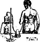
The second important factor that forced a review of the issue of the surgical treatment of purulent pleurisy was high mortality, reaching up to 50% and even higher according to some statistics (Schoedel). Particularly unfavorable results are noted by surgeons who operate extensively on children. In addition to unsatisfactory long-term results, open-type surgical treatment also entails a number of dangers arising for the patient's life both during the operation and in the first hours and days after it. Among them, pneumothorax arising at the moment of opening the pleural cavity should be placed in first place. Even more dangerous is the displacement of the large vessels and the atrium, arising as a result of the rapid emptying of the pleural cavity and sometimes leading to instantaneous death. The danger of hemorrhage usually cited in textbooks of private surgery, resulting from hyperaemia ex vacuo arising after the rapid release of fluid from the cavity, is neutralized in the pleural cavity by the pneumothorax compressing the lung. All of the above fully explains the increasing spread of another method, the so-called closed aspiration method, which is gradually displacing the open method or being used in combinations with it. The method itself is not new and dates back to Dieulafoy, who used it for aspirating effusions in serous pleurisy and constructed a special device for this purpose. This method in purulent pleurisy is carried out in the form of frequent repeated evacuations of pus in small portions or rarer evacuations of large amounts of pus. For this purpose, the apparatus of Dieulafoy and Potain is used. As a result, intoxication is rapidly reduced and phenomena of displacement of large vessels are eliminated. Abroad, the aspiration method according to Bülau has become widespread. His method of permanent aspiration consists of the following: a puncture of the pleural cavity is performed with a trocar having a large-diameter opening; after removal of the stylet, a drainage tube is inserted through the trocar into the pleural cavity, the free end of which is immersed in a vessel suspended from the edge of the bed and filled with a weak solution of some antiseptic substance; the pus gradually flows out into this vessel. Along with Bülau's method, some authors recommend the methods of Perthes-Hartert, Kirschner, and others (drain en syphon), the action of which is based on active suction by means of a siphon system, while in Bülau's method, the suction of pus occurs by means of passive aspiration. The Perthes-Hartert method and a number of others similar to it are less widespread, partly due to some complexity of the apparatus, and partly due to its lack of safety (cases of pulmonary hemorrhages from lungs stretched by suction using the Perthes-Hartert apparatus have been described). In the USSR, permanent aspiration by Bülau, Perthes-Hartert, and others has not become widespread.
The advantages of the aspiration method lie in the fact that it presents no danger of displacement of the mediastinum and large vessels from rapid emptying of the pleural cavity and does not result in pneumothorax. These two points fully explain the wide dissemination of the closed method, which is winning more and more adherents. The closed method has become particularly widespread in pediatric practice, where purulent pleurisy, which is generally a frequent disease of childhood, yielded very poor results under the open method. Krasnobaev and Freydin, Petrov, Stamm, Hudson, and others, having made the aspiration method the rule and turning to thoracotomy only in case of its failure, sharply reduced mortality in their material. Krasnobaev and Freydin, who had a 35.7% mortality rate with the open method, obtained 7.7% mortality with exclusively closed treatment, and 9.6% with combination treatment. I. I. Grekov and many others moved to mandatory preliminary application of the aspiration method in adults as well, which sharply reduced mortality. These same authors point out, however, that in adults it was still only rarely possible to cure patients using the closed method exclusively. A number of authors (Petrov, Lockwood, Stamm, Bauer, and others) combine aspiration with washing out of the pleural cavity using various antiseptic liquids (vucin, rivanol, Dakin's solution, etc.). This proposal has not become widespread in view of a number of dangerous complications described by individual authors: exacerbation of the process, intoxication, pleural shock, air embolism, etc. Thus, at present, the method of choice in the treatment of purulent pleurisy in adults is thoracotomy with preliminary repeated aspirations. In children (especially under 1 year of age), the aspiration method is the main one, while thoracotomy should be used only in rare cases of failure of the closed method. In cases proceeding very severely from the very beginning—in putrid, septic purulent pleurisy—wide thoracotomy is indicated as an urgent intervention. As for the technique of thoracotomy, there is some divergence of views among authors in this regard. For a very long time, thoracotomy with rib resection was universally accepted. The most convenient place for this is the VIII-IX rib along the posterior axillary or scapular line. A higher opening of the pleural cavity can create unfavorable conditions for the outflow of pus, while at a lower one there is a danger of opening the abdominal cavity. To avoid such errors, it is recommended before making the incision, already on the operating table, to perform a puncture again and only after this proceed to the operation. In localized, mainly interlobar abscesses, the incision of the pleura should be made without removing the needle inserted for trial puncture, since in such cases (especially with small abscesses) it is not always easy to find the abscess. The site of the incision is chosen here, of course, according to the location of the abscess. Pediatric surgery introduced a more conservative method into practice, replacing rib resection for the majority of cases with simple intercostal thoracotomy. This modification, which reduces surgical trauma, sharply reduced mortality. It makes it possible to preserve the tightness of drainage for a longer time, which plays a large role in terms of protection from pneumothorax and creates better conditions for recovery. Thus, at present, intercostal thoracotomy is used predominantly in pediatric practice, while thoracotomy with rib resection is used in operations on adults. The question of the timing of the intervention is extremely important. Most authors at present adhere to the expectant method, intervening quickly only in cases of putrid purulent pleurisy. Bauer asserts that in children, for example, early intervention (in the first days) gives a 100% mortality rate. The improvement in results with later intervention can be explained by the decrease in the virulence of the pathogen occurring over time, the increase in the immuno-biological properties of the organism, and the formation of adhesions that prevent the development of pneumothorax and eliminate the danger of displacement of the large vessels and mediastinum. The waiting period must necessarily take place in a hospital setting with careful observation of the general condition of the patient. Periodic blood tests, repeated fluoroscopy, etc., are mandatory. Most authors (Bauer, Sauerbruch, Lockwood, and others) consider the 3rd week to be the most favorable moment for intervention in an adult; some wait even longer (until the end of the 4th week). During the waiting period, however, periodic aspiration of pus must necessarily be performed. To illustrate, on page 389, the table of Krasnobaev and Freydin, compiled from pediatric material, is given. Kruzhkov had the lowest mortality after surgery at the end of the first to the beginning of the second week. Duration of disease before surgery: Total number of cases 68, 34, 48, 36, 90. Percentage of mortality: 3rd week ..., 4th week ..., Over 4 weeks ..., 26.8, 29.0, 16.5, 43.0, 31.0. Technique of performing intercostal thoracotomy. The operation is performed mostly under local anesthesia (in very small children under narcosis) with the patient in a sitting position, with the corresponding upper limb highly elevated. The skin, subcutaneous tissue, muscles, and especially carefully the pleura are anesthetized. Sauerbruch observed cases of sudden cessation of respiration and heartbeat during the operation as a reflex from the pleura due to its insufficient anesthesia during the operation. Layer-by-layer incision down to the pleura; bleeding vessels are caught and ligated. The edges of the wound are spread apart with sharp hooks, and the pleura is additionally anesthetized. With a small incision, the pleural cavity is opened, into which a drainage tube is quickly inserted, tightly embraced by the edges of the pleural wound. The release of pus through the drainage, the end of which hangs down from the operating table. The described technique eliminates the danger of pneumothorax and contamination of the wound with pus. The operation is completed by suturing the skin wound around the drainage tube and securing the latter. When draining pus through the tube, it is necessary to carefully monitor the pulse, respiration, and general condition of the patient and, at the very first alarming signs, stop the release of pus, transfer the patient to a supine position, and administer stimulants subcutaneously. At the end of the operation, the patient is given a supine position in bed. The end of the drainage tube is immersed in a vessel suspended from the edge of the bed with a weak solution of an antiseptic substance (lysol, boric acid). (Special siphon devices of the Perthes-Gaharter type, etc., can also be used to improve drainage.) In most cases, it is thus possible to maintain the tightness of the drainage for a fairly long period (up to 2 weeks), which creates a whole range of advantages: it eliminates the danger of pneumothorax and thereby promotes more rapid and final healing, provides enormous savings in dressing material and linen, and facilitates the care of the wound, which heals under these conditions per primam. The leakage of pus past the drainage worsens the course of the disease, infects the wound, and makes it necessary to immediately remove the stitches and spread the edges of the wound in order to avoid the development of phlegmon of the chest wall. The described method of intercostal thoracotomy, used mainly in children and in very weakened adult patients, does not achieve the goal, however, in very narrow intercostal spaces, as well as in very thick pus with a large amount of fibrin clots. The presence in the pleural cavity of adhesions encapsulating the abscess and detected by means of fluoroscopy or thoracoscopy [for example, using a cystoscope inserted into the pleural cavity (Hertzen)] also requires a wider opening of the pleural cavity for its inspection, destruction of adhesions, removal of clots, etc. For this, it is necessary to perform thoracotomy with rib resection. In any case, in children, this has to be resorted to only in exceptional cases, and intercostal thoracotomy remains the operation of choice for them (Paschlau and Lehman, Stamm, Krasnobaev and Freydin). For thoracotomy with rib resection, the skin incision is made not in the intercostal space, but over the rib. When making the incision, it is necessary to fix the soft tissues with the index and thumb of the left hand. It is better to make the incision immediately through all layers of tissue right down to the rib. This technique easily eliminates the displacement of soft tissues leading to the erroneous removal of the lower or upper...

Figure 4.
Figure 5. The edges of the wound are retracted with sharp hooks, and the periosteum on the anterior and posterior surfaces of the rib is additionally anesthetized. The latter is performed by introducing an anesthetic substance under the posterior surface of the rib through injections over the upper edge of the rib. Incision of the periosteum and its detachment with a raspatory upward and downward along the entire length of the rib segment intended for resection (Figures 4 and 5) both on its anterior and posterior surfaces. Special care is required when detaching the periosteum at the lower edge of the rib, behind which the neurovascular bundle is located. Exfoliation of the rib from the periosteal sheath is facilitated and accelerated by using a special Doyen raspatory (Figure 6). To avoid the subsequent development of osteomyelitis in the ends of the resected rib, the periosteum should be detached strictly within the limits intended for resection. Hudson recommends wrapping the ends of the resected rib with a cuff of periosteum to prevent their subsequent osteomyelitis. The posterior blade of the rib shears is carefully introduced behind the rib (Figure 7), and while pulling the soft tissues upward, the rib is resected. The pleural cavity is punctured again and then opened (Figure 8). The further course of the operation does not differ from the intercostal thoracotomy described above. During the first days after thoracotomy, a sharp drop in temperature is often observed, sometimes below normal, which is apparently explained by shock and a large loss of fluid. Then, from the 3rd–4th day, the temperature usually rises again and remains at this level for a fairly long time (up to 10 days), then gradually reaches normal. By this time, with a favorable course, it begins to decrease both
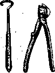
Figure 6. Doyen raspatory and Piston scissors.
the amount of discharge and to change its physical properties. The pus becomes more liquid and transparent, and under conditions of good pus drainage and improvement of the general condition of the patient, by the end of the second, third, or beginning of the fourth week, the caliber of the drainage and its length can be reduced to a small tube led out into the dressing. Under favorable conditions, on average 35–45 days after the operation, the outflow of pus stops and the fistula closes. Often, however, the closure of the fistula is only apparent, and a few days after the closure has taken place, the pus again breaks its way through the fresh scar. Such previously closed fistulas sometimes have to be widened surgically to re-introduce a drainage tube into the pleural cavity, after which final healing may subsequently occur. Considering primary drainage to be one of the main causes of prolonged non-healing of the fistula, Petragayevskaya, Gorodkov, Kruzhkov, and others suggested that after releasing the pus, no drainage should be introduced into the pleural cavity. The soft tissue cavity is filled with gauze tampons or even tightly sutured. The named authors presented at the XVI Congress of Russian Surgeons a number of cases cured extremely quickly in this way. It must be assumed, however, that these were milder cases with weakly virulent pathogens. It is possible that simple aspiration would have given the same result in these cases. Finally, Anikin proposes treating purulent pleurisy with autopyotherapy and cites 6 cases cured in this way. Other authors recommended vaccination with specific vaccines. This is permissible, but only as a preliminary act to radical intervention.
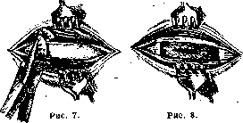
Outcomes. The accumulated rich material on the treatment of purulent pleurisy yields very uninspiring results. Mortality percentages are determined as follows: Schede—62%, Melchior—48%, Garré and Sauerbruch—40%, Domshig—25%, Anikin—40%, and Maksimovich—22.5%. In children: Stamm—35%, Krasnobaev and Freydin—30.5%, Kruzhkov—29.7%. Krasnobaev and Freydin also provide mortality statistics by patient age. Age (years): Total operated—0–1: 51, 1–4: 162, 4–9: 96; Mortality percentage—0–1: 56.8%, 1–4: 33.95%, 4–9: 19.8%, 9–13: 11.3%. Significantly better results were obtained by the same authors on material treated by more conservative methods: thoracotomy with preliminary aspiration (52 cases) gave only 9.6% mortality, and with the exclusively closed method (26 cases)—7.7%. In Schede, who also switched to this method, mortality dropped to 20%. Some correction in the direction of an increase in mortality should probably be made in these statistics, since it must be assumed that predominantly milder cases were treated in this way. The etiology of the disease and the flora of the pus also have great significance in terms of the outcome of the disease.
.
According to statistics by Krasnobaev and Freydin, in 28.6% of cases (out of 105 cases) diplococci were found in the pus, in 14.3% of cases (out of 21 cases)—staphylococci, in 35.3% of cases (out of 34 cases)—streptococci, in 42.4% of cases (out of 33 cases)—mixed flora. These authors indicate that streptococcal purulent pleurisy and pleurisy with mixed infection give the highest mortality. According to Kruzhkov, streptococcal (41.7%) and diplococcal (23.7%), followed by mixed infection (diplococcus + streptococcus, diplococcus + staphylococcus, streptococcus + staphylococcus). In rare cases, the pus turns out to be aseptic. Puschlau and Lehmann deny the influence of pus flora on the disease outcome. They attach greater importance to the etiology of the disease, believing that a large percentage of poor outcomes are given by purulent pleurisy arising as a complication of infectious diseases (so-called Spanish flu, typhus, scarlet fever). However, the most severe outcomes are given by septic forms. Bilateral purulent pleurisy is often observed in this regard. Fabrikant collected 118 cases of bilateral purulent pleurisy from the literature. Surgical approach in these cases requires special care and should be performed without particularly long waiting, but in any case not simultaneously on both sides. It is recommended to start with the more affected side, relieving the other side by aspiration. Sauerbruch, who suggests operating on all purulent pleurisy in general with a high-pressure apparatus (see Lungs—surgical treatment of lung diseases), especially recommends this for bilateral cases. Purulent pleurisies left to themselves often end with spontaneous evacuation of pus (empyema necessitatis) through a passage that the pus carves out through the chest wall! In this case, the pus can spread between the endothoracic fascia and the parietal pleura (peripleuritis according to Billroth's terminology) (see Pleura). Breakthrough of pus outward occurs more often in the lowest parts of the chest, but it also has to be observed in its higher parts (e.g., below the clavicle, in the nipple area, etc.). Differentially-diagnostically, these fistulas must be distinguished from fistulas formed in rib osteomyelitis. In the case of a low location, the treatment of the fistula can be limited to its expansion and drainage of the pleural cavity; at a high position, it is necessary to perform the operation in a typical location. A special position in terms of treatment is occupied by purulent pleurisy of a tuberculous nature. Experience has shown that their surgical opening leads in most cases to death from sepsis due to secondary infection rapidly joining after the operation. Some authors indicate that surgical treatment also leads to an exacerbation of the process in the lung, freed from compression by pus. Sauerbruch considers surgical intervention in tuberculous purulent pleurisy a gross error (Kunstfehler). Complications depending on the opening of the ple
ural cavity
and rapid evacuation of the pleural cavity, in tubercular purulent pleurisy proceed significantly more severely. Treatment of tubercular purulent pleurisies must therefore proceed exclusively by the closed method. Here, asepsis rules must be observed especially strictly during infections, since tubercular purulent pleurisies are very easily infected. Gaza and Schmidt therefore even advise against using the aspiration method, and directly proceeding to extrapleural operations (thoracoplasty, extrapleural plombage - see Lungs, surgical treatment of diseases of the lungs). When a secondary infection joins, treatment is conducted according to the general rules for acute purulent pleurisies. The prognosis in these cases is poor. The severe course and high mortality obtained in cases of purulent pleurisy of a tubercular character subjected (mostly erroneously) to surgery make the question of their correct diagnosis especially acute. However, it is not always easy to establish a differential diagnosis between acute infectious and tubercular purulent pleurisy. The main points distinguishing the latter are: a gradual onset, subfebrile temperature, absence of leukocytosis, and the character of the pus—richness in lymphocytes and tubercular bacilli. The latter, however, are frequently absent in the pus, and then only the injection of pus into the abdominal cavity of a rabbit can decide the issue one way or the other. Chronic purulent pleurisy (chronic empyema, old empyema, pleural cavity fistula). If within 2-3 months after the operation the fistula has not closed, practically such a case should be considered as having passed into a chronic state. The theories explaining such frequent transition of purulent pleurisy into a chronic state have already been outlined above. To these can be added a number of causes of a purely technical nature. Among them, a frequent one is a drainage tube that has fallen into the pleural cavity (drain perdu), supporting suppuration. This cause was discovered by Maksimovich, for example, 5 times out of 20 cases of non-healing fistulas. Cases of finding 2, 3, and even 5 (!) rubber drains in the pleural cavity of a single patient have been described. The fact of a drain slipping into the pleural cavity often goes unnoticed by both the medical staff and the patient. This is especially often observed in outpatient conditions when changing attending physicians. To avoid this complication, it is necessary to take measures for firmly securing the drain in the wound, tying it with a dense silk thread passed around the chest, attaching a safety pin to its outer end, etc. Any new patient suffering from a non-healing fistula after purulent pleurisy is subject to mandatory fluoroscopy or radiography. The latter is more reliable, since a drain surrounded by fibrinous masses may go unnoticed during fluoroscopy. If a drain is discovered, surgical intervention is indicated for its removal, which is sometimes very difficult both technically and from the standpoint of the trauma inflicted on the patient. It frequently leads to the patient's death from shock caused by the intervention or from an exacerbation of the infection harbored in the thickness of the granulation masses surrounding the drain. Cases of non-healing of the fistula are observed depending on osteomyelitis of the ends of the resected rib. Surgical treatment—resection of the affected rib segments. Non-healing of the fistula sometimes depends on excessively prolonged drainage. Irritation of the fistula walls by a foreign body—the drain—and pus leads in these cases to the formation of a dense, non-collapsible fistulous tract, the walls of which are lined with dense scar tissue. Deep in the fistula, there is usually an accumulation of flabby granulation tissue. Treatment consists in excision of the entire fistulous tract and curettage of granulations. Surgical methods of treating chronic purulent pleurisies are divided into 1) extrapleural, 2) intrapleural, and 3) operations on the lung aimed at freeing the latter from adhesions enveloping it. The simplest method of the extrapleural type is the excision of small (4-6 cm) segments of several (2-4) ribs above and below the fistula. This low-risk intervention often yields success, especially with relatively small cavities. It should in any case be applied as a transition to more extensive and dangerous operations. The most widespread type of such an extrapleural operation is the method of Simon, Küster, and others, improved by Estlander—subperiosteal resection of the IV-VIII ribs over the greatest possible extent. The idea of the operation is the mobilization of the chest wall with the aim of its retraction and contact with the visceral pleura. With a very dense parietal pleura with large deposits (Schwarten), the effect of the operation may be unsatisfactory. Only a change in the configuration of the cavity is obtained, while its volume remains unchanged. Subbotin modified the operation by proposing to remove pieces of ribs in the anterior and posterior sections of the chest wall. Significantly greater success was achieved by Schede, who proposed in 1885 to remove the ribs together with the corresponding section of the parietal pleura. Ribs, intercostal muscles, nerves, arteries and veins, and the parietal pleura are excised (operation technique—see Thoracoplasty). The Schede operation, which is widespread and often yields a good result, is also not free from a whole series of drawbacks. The extent of the intervention frequently leads to death from operational shock. According to Sauerbruch's observations, infection (phlegmon, erysipelas) of the huge wound cavity often develops in the postoperative period. Finally, extensive rib resection often leads to significant chest deformity with all its consequences (see Thoracoplasty), to paralysis and atrophy of the upper sections of the abdominal wall due to the transection of intercostal nerves. Mortality, according to Yudin, reaches 20%, according to other authors significantly higher. To reduce the operational risk, H. Braun proposed performing the Schede operation in several stages (up to 4). To achieve greater collapse of the cavity, Sauerbruch performs paravertebral thoracoplasty. Among extrapleural methods, plombage is used (see Lungs, surgical treatment of diseases of the lungs) with various materials: paraffin, fat, muscle, etc. A number of authors recommend the intrapleural method of Kirschner—plombage of the cavity with a muscle flap on a pedicle. Proceeding from the fact that the least compliant part of the pleural cavity in total purulent pleurisy is its upper aperture, Kirschner fills it with a flap formed from the peripheral section of the pectoralis major muscle. This muscle plomba, hanging on a pedicle, is inserted through an opening made in the chest wall (Fig. 9, 10, 11 and 12) into
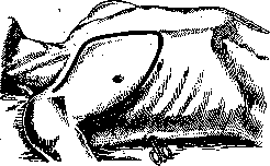
Figure 9.
the pleural cavity, where it is fixed with sutures. After the flap takes, its pedicle is severed, and the opening in the chest wall is closed with a skin flap. The upper section of the thoracic cavity is thus obliterated, after which the closure of the fistula and complete recovery soon ensue. In addition to the operation, Kirschner proposes performing phrenicectomy (see). The advantage of the Kirschner operation is the absence of deformity after the operation,
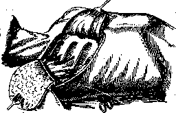
Figure 10.
the negative side is the complexity of the intervention and possible extensive suppuration that can nullify the plasty. Considering the main cause of the formation of chronic purulent pleurisy to be the loss by the lung of its ability to expand, depending on the thickening of the visceral layer of the pleura, Delorme proposed the operation of decortication. After wide opening of the pleural cavity, these deposits and the thickened pleura are removed partly by sharp, partly by blunt methods. Significant hemorrhage, arising mostly during the peeling of the thickened pleura, which is very intimately fused with the lung tissue, the danger of pleural and pulmonary reflexes leading to shock, the danger of air embolism, etc., have not given this operation wide distribution, especially since the therapeutic effect obtained from the operation was not very significant. Modifications proposed by Kümmell, Braun, and others also did not have great success. In cases of chronic purulent pleurisies depending on the presence of a bronchial fistula, one has to add special interventions leading to the closure of the bronchial fistula to one or another operation performed for the obliteration of the cavity. These cases are particularly difficult to treat (see Lungs, surgical treatment of diseases of the lungs). The cure of purulent pleurisy is achieved by obliteration of the pleural cavity, which can lead to a whole series of severe changes. Of these, the most important is the limitation of lung excursion and chest deformity, especially markedly expressed after total empyemas that have passed into a chronic state and been operated on according to Schede. The ensuing severe chest deformity and scoliosis

1
Figure 11.
of the spine sometimes reach colossal dimensions at that. Subsequently, secondary severe changes also occur on the part of the heart (hypertrophy). To combat these severe consequences which make patients invalids, prolonged and energetic use of medical gymnastics and various types of physiotherapy are recommended. Set forth
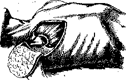
Figure 12.
The above-mentioned principles of treatment for purulent pleurisy, with careful observance of the indications for operations, proper selection of the latter, and the greatest possible conservatism, should contribute to an easier course and a rarer transition into a chronic state that is difficult to treat and leads to disability.
N. Blumenthal. Pleurisy in children is a frequent and all the more severe affliction, the younger the children. The statistical data on morbidity and mortality cited in the literature are less than the actual figures. This is explained by incomplete registration and difficulties in diagnosis, especially in infants (according to Comby, empyema is not diagnosed in them in 25%). The indication that pleurisy occurs more often in children aged 3–5 years is inaccurate; data from the State Scientific Institute for the Protection of Motherhood and Infancy of the People's Commissariat of Health show that younger children exhibit no less morbidity. Regarding the etiology of pleurisy in children, pneumococcus is most frequently found among microorganisms (39% according to Netter in 80.7%, according to Blecher in 94%), and less frequently streptococcus (18.2–19.7%). In older children, tuberculosis bacilli account for 5.5–7%. In individual cases, bacilli of Eberth, paratyphoid, Loeffler, and others are found. Direct mechanical irritation of the pleura as a result of trauma is rare in children. Primary diseases vary at different ages. In newborns and children up to 3–4 months of life, sepsis occupies first place, followed, according to some authors, by irritation of the lungs by masses aspirated during birth, and less frequently by lesions of the upper respiratory tract, abdominal organs, and skin. At the age after 4 months, the main disease causing pleurisy is pneumonia. According to Kochmann, every fifth pneumonia patient has pleurisy; according to the Institute for the Protection of Motherhood and Infancy, every third. (Pleurisy is especially often observed after pneumonias complicating measles, scarlet fever, and whooping cough.) In older children, tuberculosis plays the predominant role in the occurrence of pleurisy, followed by rheumatism. Those cases where pleurisy in children arises as if primarily can be explained by incomplete examination of the patient or the inconspicuousness of the manifestation of the primary disease. Engel believes that all so-called genuine primary pleurisies are of tuberculous origin. In children, the primary disease determines not only the occurrence of pleurisy, but also its course and outcome. Pleurisy, in turn, sharply worsens the course of the primary disease in a child and often becomes a source of new complications. Thus, pleurisies in children can be divided into symptomatic and independent, and further into concurrent with the primary disease and subsequent, arising after a certain interval. From the standpoint of the character of pathological and anatomical changes on the pleura in children, just as in adults, one can distinguish dry and exudative pleurisies. Dry pleurisy as an independent disease in young children is not described. It occurs at an older age. The disease begins with pain in the side, a dry, often agonizing cough, and an elevated temperature. The child spares the affected side, trying not to breathe deeply. Percussion on the affected side reveals nothing pathological; on the healthy side, a box sound is often present, which is evidence of compensatory expansion of the lung. Auscultation reveals a pleural friction rub, more delicate than in adults, which is covered by bronchitis rales. When the diaphragmatic pleura is affected, the diaphragm is immobile and stands high on the X-ray; pains are localized in the epigastric region; hiccups are noted. In mediastinal dry pleurisy, there are pains in the heart region, a feeling of fear, suffocation, sometimes dysphagia, and spasmodic cough. The disease lasts from several days to several weeks. Cases of mass outbreaks of dry pleurisy in schools have been described (Attlee); in all such cases, children showed lesions of the pharynx. The outcome is usually recovery or sometimes a transition into an exudative form. Treatment consists of bed rest, pain-relieving ointments, warming compresses, iodine tincture, and internal salicylate preparations.
A frequent form of pleurisy in early childhood consists of fibrinous and purulent-fibrinous pleurisies, which usually accompany pneumonia. They constitute 59% of the total number of pleurisies noted at an age up to 3 years (State Scientific Institute for the Protection of Motherhood and Infancy). Clinically, they proceed against the background of pneumonia and are not always diagnosed during life. They are usually discovered at autopsy in the form of deposits on the pleura. In purulent-fibrinous pleurisy, the deposits cover the lung with a thick, greenish-yellow, greasy-dirty coating that extends far beyond the boundaries of the lung area affected by the inflammation. Purulent-fibrinous pleurisy sharply worsens the course of pneumonia: the phenomena of toxemia intensify, the child becomes grayish-pale, the skin doughy, cardiac activity and respiration are further disturbed, and the temperature shows large swings. Locally, sometimes, especially in focal pneumonia, dullness begins to stand out more distinctly, and the skin over it becomes clearly edematous. Upon auscultation, pleural sounds are heard; pulmonary inflammatory rales are somewhat muffled. Sometimes, however, local findings remain unchanged. On the X-ray, there is more or less uniform clouding of the lung, obscuring the small inflammatory foci that previously stood out clearly. On the radiograph, the thickened wall of the affected side of the chest is sometimes clearly visible. The outcome in purulent-fibrinous pleurisy is poor and is related to the severity of the underlying disease—pneumonia. According to the State Scientific Institute for the Protection of Motherhood and Infancy, out of 78 cases, 53 died. Treatment, as in pneumonia, is predominantly symptomatic and aimed at supporting the child's strength. There are no special means of acting on the affected pleura. Exudative pleurisies in children are sharply demarcated diseases with characteristic symptomatology. They are observed up to 3 years of age somewhat less frequently than the symptomatic ones described above (State Scientific Institute for the Protection of Motherhood and Infancy—46%). The character of the exudate may be the same as in adults. Only the distribution of the effusion is of special significance. Quite often, it accumulates not along the Damoiseau line, but envelops the lung in a thin layer from the base to the apex. This especially occurs in children of the first months of life, during severe pneumonia, in rachitics, and others, entailing a more severe disorder of respiration, cardiac activity, and bowel functions than in adults. The absorption of toxic products affects the general condition of the child's organism more strongly. To varying degrees, depending on age, constitution, and the bacterial factor, the body's resistance decreases, and the child falls victim to secondary infections, which is particularly characteristic of children of the first years of life. Among the various forms of exudative pleurisy, purulent pleurisies—empyemas of the chest cavity—occupy first place in early childhood. Up to the age of three, they make up 83% of all exudative forms of pleurisy (data from the State Scientific Institute for the Protection of Motherhood and Infancy). According to Netter, purulent pleurisy in children is 5 times more frequent than in adults, and in relation to the total number of all pleurisies up to a year old, it constitutes 63%, from a year to 5 years—51.8%, and from 5 to 10 years—20%. Purulent pleurisy is most often of pneumogenic origin. According to Maslov, every 8th to 9th pneumonia patient develops purulent pleurisy, and according to the State Scientific Institute for the Protection of Motherhood and Infancy—every 9th to 10th. Moreover, the majority of purulent pleurisies are parapneumonic and most often the result of focal pneumonia. Purulent pleurisies of other origins are less frequent. According to Maslov, in 11% a connection with tuberculosis is noted, and in 36% the etiology is not elucidated. According to Nobécourt, after measles, scarlet fever, and other infections, aseptic purulent pleurisy can arise. Clinically, purulent pleurisy can sometimes develop completely imperceptibly, especially during the severe course of the underlying disease—pneumonia or sepsis. Local symptoms are poorly manifested; there is often little pus, and therefore it is not always easily determined. Sometimes the only indicator of an added purulent pleurisy in these cases is a noticeable deterioration in the underlying disease. Subsequent, so-called metapneumonic purulent pleurisies develop after the child has suffered from pneumonia, sometimes after a certain clear interval. The seemingly recovering child suddenly or gradually begins to run a temperature, weakens again, turns pale, loses appetite, and sleeps poorly; small children become capricious, tearful, and older ones complain of a number of painful sensations. In the case of rapid development, there may initially be convulsions and vomiting. Sometimes purulent pleurisy is imperceptibly included in a slowly resolving pneumonia without visible changes in the general condition of the child. Such cases are often regarded for a long time as protracted resolution of pneumonia, while empyema is overlooked. Diagnosis of variously onseted purulent pleurisies is possible only with very careful repeated examination of the child. Simple inspection yields a lot. With minor movement of the child, severe dyspnea may sometimes be revealed. Sputum is scarce or is swallowed. The chest is asymmetrical. The healthy side often protrudes more due to vicarious expansion of the unaffected lung and curvature of the body toward the effusion. Pain in children is most often near the nipple, in the intercostal space, in the epigastric region; when the right side is affected—at the site of the cecum. Dullness over the effusion is by no means always determined. Even with quiet percussion, recommended in children, in cases of diffuse parietal purulent pleurisy, the sound may remain completely unchanged or only slightly blunted with a tympanic shade. The same may occur with large accumulations of pus and the more frequently the younger the child. In the case of the presence of dullness, even occupying the entire half of the chest, in small children one can find a band of dull-tympanic sound paravertebrally behind (Eichhorst's groove, Hamburger's groove). An important symptom of purulent pleurisy in children is the sensation of a peculiar resistance under the finger acting as a pleximeter. Therefore, during percussion, it is necessary not only to listen, but, which is sometimes more important, to be able to feel the area under examination. Due to the greater compressibility of the mediastinum in children, Rauchfuss's triangle appears brighter and more often. Traube's space, on the contrary, in the majority of cases, especially in infants, remains free. This finds an explanation in the distension of the stomach by swallowed air and the intestines by gases, which causes the displacement of the exudate, the conductivity of the tympanic sound, and possibly the early development of adhesions in the sinus. In interlobar effusions, dullness may be determined in the form of a band along the course of the fissure (Dietlen's ribbon). In mediastinal pleurisies, it sometimes takes the form of an hourglass with expansion at the top and bottom and a relative constriction at the root of the lungs. Auscultatory data in small children can be even less pronounced than percussion data. If the chest is narrow, the exudate is small, and the bronchi are free of mucus and permeable to air, a completely distinct bronchial breathing may be auscultated. In other cases, it may be somewhat muffled. Almost unchanged puerile breathing may also be auscultated. Pleural sounds are most often heard at the border of dullness; in fresh cases, they resemble pulmonary crepitation. Filatov attached great importance in the diagnosis of purulent pleurisy to the absence of bronchophony and considered this symptom most characteristic of children's empyemas. In some cases, Pitres' phenomenon and the resonance phenomenon described by Leiper can help. In the presence of cavities in the lung, which are not uncommon in purulent pleurisy, amphoric breathing may also be auscultated; the same sometimes occurs with a narrow chest and free bronchi, which sum up when the lung is compressed as if into a large resonating cavity. The data of X-ray examination are of important, but not always decisive, significance. On the fluoroscopic screen, the initial stages of purulent pleurisy can be determined in the form of clouding of the costophrenic angle, and parietal effusions appearing in the form of a narrow strip of shadow along the costal border. Sometimes, unexpectedly in the absence of direct physical data, the X-ray gives a solid clouding occupying a significant portion, sometimes an entire half of the chest cavity. Moreover, X-rays make it possible to discover interlobar, mediastinal, subdiaphragmatic, and small encysted pleurisies that are poorly accessible to other research methods. Difficulties on the X-ray are created by the circumstance that in children with large effusions, the intercostal spaces stand out well, and the costophrenic sinus is relatively light. Pleurisy can be missed in bilateral minor exudate or coexisting emphysema of the lung. To use the diagnostic advantages of X-rays, it is necessary to use them more often and to perform not only fluoroscopy, but also radiographs. Final confirmation of the diagnosis of purulent pleurisy is given only by a positive diagnostic puncture, which should be performed in children in all suspicious cases. At the same time, it is only necessary to observe a certain caution and to traumatize the patient less, since cases of sudden death during this operation have been described.
Some explain this by a reflex from the pleura; more often, however, the child dies from phenomena of cardiac failure, and autopsy reveals marked degenerative changes in the heart muscle. In the case of a small effusion, and even more so with a parietal effusion, the puncture should be made in a sagittal direction in order to hit the space tangentially. It is necessary to avoid wounding blood vessels and the periosteum. The course of purulent pleurisy is extremely diverse. It can end in recovery or the death of the child within a few days or drag on for months. In the latter case, children become markedly emaciated, reach various degrees of hypotrophy and atrophy, look sharply pale, with a characteristic waxy tint of the skin, which is edematous in places—especially on the face and legs. The severity of the course and duration are not proportional to the magnitude of the effusion. Small interlobar, mediastinal, and apical encysted empyemas often drag on heavily and stubbornly. There are cases when, in the presence of a large amount of pus, the child feels generally not bad; they remain only markedly pale and bloodless. This occurs when the process is encysted. Local symptoms change with the development of the disease. Respiration sometimes becomes calmer, more even, and shortness of breath appears only upon movement. The data of percussion and auscultation often do not correspond to one another: in the absence of dullness, weakened breathing is auscultated, sometimes bronchial with an amphoric tint, moist rales, and rattling. The heart moves back to its place, and with the development of adhesions it is pulled toward the affected side despite the presence of a large amount of pus. Diagnosis in prolonged cases of purulent pleurisy is therefore always difficult. Confusion is possible with pneumonia, tuberculosis, and sometimes, in the case of weak local symptoms, with malaria or typhus; in some cases, diseases of neighboring organs also provide grounds for an incorrect diagnosis. These include subdiaphragmatic abscess, tumors of the liver, kidneys, spleen, large hydronephrosis, echinococcosis of the lungs, and bronchiectasis filled with pus. The course of purulent pleurisy is especially aggravated by the addition of a number of complications. According to data from the State Scientific Research Institute for the Protection of Motherhood and Infancy, in children under 3 years of age, purulent otitis was noted in 40%, antritis in 20%, sepsis in 16%, pericarditis in 12%, phlegmonous mediastinitis in 8%, purulent meningitis in 10%, followed by isolated cases of peritonitis, thrombosis of the venous sinuses and vessels of the pia mater, purulent arthritis, etc. Pneumothorax was diagnosed in 26%, and the formation of purulent cavities in the lung parenchyma in 28%. The breakthrough of pus from the pleural cavity into the bronchi—empyema perforans interna—is difficult to determine in children. They do not exhibit the major sign of Dieulafoy; the pus is discharged in small portions and swallowed. Empyema perforans externa, the breakthrough of pus under the skin, is observed in neglected cases, as well as with large effusions following a puncture. The addition of other diseases, especially infectious ones, sharply alters the course of purulent pleurisy. The course of the latter is in turn poor. Even ordinary chickenpox or banal influenza take on a severe course and often decide the fate of the patient. Treatment. First of all, it is necessary to create conditions ensuring proper care, nutrition, and constant medical supervision. In the regimen, fresh air, cleanliness of the skin, upper respiratory tract, and oral cavity come first; an appropriate position in the crib ensuring easier breathing (elevated position of the shoulders with the head slightly thrown back). A patient with pleurisy loses a lot of water and protein, is emaciated, and therefore must be persistently fed. The diet must correspond to the age; it is necessary to avoid transferring the child to a monotonous diet. Infants need to be given at least a small amount of breast milk; a mandatory addition is fruit juices, fresh berries.
oats, vegetables, fruits. To increase resistance and raise immunity, general strengthening and tonic therapy is used. In some cases, hemotherapy, vigantol, fish oil. Locally, at present, the majority use the conservative method of treatment instead of the previously widely practiced resection operation. (See above regarding the treatment of purulent pleurisy.) As an adjunct to puncture (in cases where pus collects again after its removal), washing of the cavity with a physiological solution is used. In addition, optochin has been proposed, with caution in cases of cardiac weakness and in young children. (According to Woringer, optochin is effective only in pneumococcal empyemas.) Other authors recommend rivanol, methylene blue, gentian violet, ox bile, sodium taurocholate, diathermy, etc. As an agent preventing adhesions and increasing the content of lipolytic enzyme, oil infusion is used. To limit the amount of exudate, novasurol is given. Specific therapy is also used—serum, vaccine, Besredka's antivirus. Autopiotherapy and protein therapy have been tried. The effect of all these remedies is generally small. If puncture is unsuccessful and toxic symptoms increase, one should resort to thoracotomy with drainage according to the method of Bülau or Perthes. Particularly careful care must be organized by the child's bed for the drainage to avoid bedsores and phlegmon formation. Denis recommends for this purpose covering the wound with gauze moistened with sterile paraffin. Thoracotomy is contraindicated in the presence of incomplete pneumonia. Resection is used in older children, and in very exceptional cases in young ones. The results of conservative and surgical treatment of pleurisy in early childhood are currently completely unsatisfactory; mortality reaches extremely high figures. In individual authors it ranges from 9.09% to 88% (Marconi). Age has a great influence on the outcome. According to Holt, children under one year die in 65%, from one to two years in 29%, from 0 to 3 years in 40% (Institute for the Protection of Motherhood and Infancy). The highest mortality (up to 100%) is given by pleurisy in newborns resulting from sepsis; further, pleurisy in young children with focal pneumonia (according to Kochmann, 75%; according to the Institute for the Protection of Motherhood and Infancy, 84%); with lobar pneumonia it is lower (according to Kochmann, 15%; according to the Institute for the Protection of Motherhood and Infancy, 8%). Along with purulent pleurisy, the deceased children in the overwhelming majority have a number of complications (Institute for the Protection of Motherhood and Infancy, 100%). After 3-4 years, mortality from purulent pleurisy drops. According to Nassau, mortality from diplococcal, streptococcal, and staphylococcal purulent pleurisy relates as 1 : 2 : 3. Among pneumococci, type I is the most favorable. According to Krasnobaev's statistics, diplococcal purulent pleurisy gives a mortality of 28%, staphylococcal 14.0%, streptococcal 35%, mixed 14.8%, of unknown etiology 21%. Serous pleurisy is rarer the younger the child. According to Mazzeo, it occurs at the age from 0 to 2 years in 0.13%, from 2 to 4 years 0.8%, from 4 to 6 years 0.97%, from 6 to 8 years 1.46%, from 8 to 10 years 1.3%, from 10 to 12 years 1.37% of all diseases. In relation to the total number of pleurisies in children under 3 years old, it is 45% (Institute for the Protection of Motherhood and Infancy). According to Kiselev and others, all serous pleurisies are of tubercular origin; according to Shkarin and Baginsky, only in one third of cases. Nobechi considers that serous pleurisy can be a syndrome of pleuropneumonic tide or pleural reaction to pneumonia. Some authors point to cases of serous pleurisy in infectious diseases—measles, scarlet fever, diphtheria, influenza. In older age, rheumatism is also of importance. Clinically, serous pleurisy manifests itself more often as an independent disease. The onset can be completely unnoticeable. The child loses appetite, becomes sluggish, moody, runs a slight fever in the evenings, and sometimes diarrhea, vomiting, and a slight cough appear. Then, after some time, the clinical picture of the disease becomes clearer. Shortness of breath, pain in the side appear, temperature settles at significantly higher figures, and local symptoms from the chest appear. Sometimes serous pleurisy begins turbulently, resembling rapidly developing pneumonia, and is accompanied by vomiting, convulsions, severe pain in the side, in the abdominal area, high temperature, delirium. Locally in the developed stages of the disease, the same physical phenomena are observed as in purulent pleurisy. Only the performed puncture yields a different fluid—transparent, yellowish-greenish color, with a specific gravity within 1.010–1.023 and with a relatively small protein content (3-9%). Cytological data vary depending on the etiology of serous pleurisy. Serous exudate must be distinguished from transudate, which can occur in cardiac weakness, edema, and also from echinococcal fluid in pulmonary echinococcosis. Serous pleurisy on average lasts from 1 to 3 weeks. The outcome is generally much more favorable than that of purulent pleurisy. Mortality directly from serous pleurisy is small; children subsequently die mainly from tuberculosis as the cause of serous pleurisy (according to Maslov, in 15%, Nobel, Steinenbach, 16%). General treatment is the same as in purulent pleurisy. Then treatment is directed at the underlying disease that caused serous pleurisy, and primarily tuberculosis. Locally, for large effusions, puncture, then administration of calcium, dry diet, periodically administered subcutaneous injection of serum. Furthermore, painting with iodine, rubbing with green soap, dry and moist compresses are recommended. Internally, salicylate preparations, diuretin, adrenaline.
Pleurisy, especially exudative, leaves behind various degrees of changes in the pleural cavity. Most of these changes are insignificant and are discovered accidentally, most often on X-ray, in the form of pleural thickenings, interlobar adhesions. In these cases, the child usually does not complain of anything. Sometimes there may be complaints of pain, especially with movements, coughing, which can persist relatively persistently. At the same time, objectively, some lagging of one side of the chest, its flattening, and less elasticity are noted. On percussion, slight dullness; on auscultation, dry pleural rales; on X-ray, varying degrees of homogeneous or patchy clouding. Severe cases of post-pleuritic changes with sharp changes in the chest, displacement of organs, formation of cavities, emphysema, etc., are of great importance. The child himself, with the duration of the suffering, looks retarded, infantile. Complaints of shortness of breath, fatigue, headaches, sometimes voice changes (involvement of the recurrent nerve), dysphagia, whooping-cough-like cough; cardiac disorders, cyanosis; dilation of neck veins, parasternal edema; diarrhea, abdominal distension, enlarged spleen, liver. The condition may remain stationary, but may also progress. Treatment must be long and systematic. Heliotherapy, aerotherapy, water and mud therapy, diathermy, massage are recommended. Breathing exercises according to Goldscheider are of great importance; in young children, moderate singing, crying, blowing up a balloon. Sometimes fibrolysin and, in severe cases, surgical intervention., S. Borisov.
Related articles
Mentioned in
Cite this page
“Pleurisy.” Soviet Medical Encyclopedia. English translation of Bolshaya Meditsinskaya Entsiklopediya, 1st ed. (Moscow, 1928–1936), ed. N. A. Semashko. https://sovietmedicalencyclopedia.pages.dev/article/pleurisy/