Pneumonia
Historical document, translated for reference. It reflects medical knowledge of the 1920s–30s and is not medical advice.
Summary
Pneumonia is an acute infectious disease that occurs independently or as a complication of other diseases, primarily infectious ones, and is characterized by inflammation of the lung parenchyma and bronchi. The article distinguishes between two main types: lobar (croupous) pneumonia and catarrhal (bronchopneumonia), with detailed discussion of etiology, particularly the role of pneumococcus and other microorganisms.
Encyclopedia article (1928–1936)
682 VI. II. animals . . . :................ 683 Pneumonia, an acute infectious disease that occurs independently or as a complication of other diseases, mainly also infectious ones, and is accompanied by inflammation of the lung parenchyma and bronchi in a large or significant extent. However, not every inflammation of the lung-bronchial tissue is inflammation of the lung, P. in the clinical-anatomical sense; inflammatory processes that arise, for example, in the place of infarcts or in the place of lung injuries, along the course of a bullet channel, etc., are not included in the concept of P. In all these cases, we only have a demarcation zone of inflammatory reaction surrounding one or another local process. This distinction is also necessary because the same infarct or injury, under certain conditions, can give not only or not so much zonal inflammation as typical pneumonia, i.e., an effect of general pathological significance, far exceeding the limits of the regularly developing local reaction. It is customary to distinguish two main types of P.-lobar and catarrhal (or bronchopneumonia)-and within these types to make further subdivisions. The subdivision of P. into lobar and catarrhal is however not something strictly principled that would draw a sharp line between them in all respects,-bronchopneumonia can be even a complete equivalent of lobar P. If such a subdivision is nevertheless preserved, this occurs mainly for the convenience of analyzing phenomena in clinical-anatomical and pathogenetic relations. The classical, most vivid form is considered to be lobar P.; it also under the name "pneumonia" usually figures among the main acute infectious diseases.
I. Lobar pneumonia. Etiology. The vast majority of cases of lobar pneumonia in humans are caused by the pneumococcus of Fränkel - Wesselbaum [see separate table (pp. 623-624), fig. 6] (see Pneumococcus). In favor of the etiological significance of pneumococcus, besides its constant isolation from the affected lung tissue, there are also experimental observations by a number of authors, especially Blake and Cecil, who succeeded in causing monkeys to have the typical clinical and anatomical picture of lobar pneumonia by intratracheal introduction of minimal doses of pneumococcal culture. Pneumococcus is however not the only causative agent of lobar P. Besides the Friedländer bacillus, the etiological role of which in certain forms of this disease has long been generally recognized, other microorganisms, such as streptococcus, staphylococcus, Pfeiffer's bacillus, can undoubtedly also cause lobar lobar inflammation of the lungs. Thus, in the material of the clinic of the Rockefeller Institute in New York, the causative agents of lobar P. were distributed as follows: Pneumococcus 454 cases, Bac. Friedlander-3, Bac. influenzae-6, Streptococcus pyogenes-7, Streptococcus mucosus-1, Staphylococcus aureus-3, mixed infections-6. Systematic bacteriological examination of sputum and blood in patients with lobar P. in the hospital named "Medsantrud" in Moscow during 1929-31 gave the following results regarding the frequency of isolation of individual microorganisms: Pneumococcus 361 cases, Bac. Friedlander-4, Bac. Friedlander-Pneumococcus-6, Streptococcus haemolyticus-7, Streptococcus viridans-3, Staphylococcus haemolyticus-2.
Cases of lobar P. caused by the microorganisms listed above represent, depending on the causative agent, certain anatomical, as well as clinical, peculiarities. These peculiarities have been studied in most detail in relation to Friedländer's P. As shown by the observations of Neufeld, expanded and deepened by Dochez, Gillespie, Avery and Cole and other collaborators of the Rockefeller Institute in New York, there are a number of varieties of pneumococcus differing from each other in their immuno-biological properties. According to these authors, the most common (in 70-80% of all pneumococcus isolates) in lobar P. are three varieties of pneumococcus (the so-called I, II and III types-see Pneumococcus). As for the remaining1 varieties of pneumococcus, which on the basis of observations by Park and Cooper number more than 20, they are encountered so rarely that their isolation into separate types with the preparation of corresponding sera for their identification is complicated and impractical. At the suggestion of the collaborators of the Rockefeller Institute, all strains of pneumococcus that do not give specific immunity reactions with the sera of the three types mentioned are attributed to the fourth type. The latter thus does not represent a separate variety with definite biological properties, but is a collective group uniting the numerous varieties of pneumococcus that do not belong to the three fixed types mentioned.
The differentiation of pneumococcus into specific biological groups besides theoretical interest also has practical, clinical and epidemiological significance. Almost all authors who studied lobar P. from the point of view of the type of pneumococcus note the dependence between mortality and the type of pneumococcus with which a given patient is infected (see below prognosis). Determination of the type thus to a certain degree predetermines the prognosis of the disease. For the successful application of serum treatment, it is also necessary to determine the type of pneumococcus beforehand, since specific serum is used mainly in relation to type I. Establishing the type of pneumococci is also of great importance in solving epidemiological problems. Recent observations have shown that the type of pneumococcus to a certain degree also leaves its mark on the clinical picture of lobar pneumonia (temperature curve, nature of the termination of the infection, etc.) (Ettinger, Viktorov, Mazel). Until recently, the study of lobar P. from the point of view of the type of pneumococcus was carried out mainly by American and English authors. Although the frequency of isolation of individual types of pneumococcus in patients with lobar P. varies among different authors depending on the season and geographical location of the given locality, in most series of observations, approximately one third of all cases falls on types I and II, type III-10-15%, and the remaining 25-30% on type IV.
The dependence of the type of pneumococcus on the geographical location of the given locality is vividly illustrated by the observations of Lister, according to whose research the ratio of individual varieties of pneumococcus in South Africa sharply differs from that in North America. Thus, about V» of the strains he isolated in patients with lobar P. falls on the variety of pneumococcus rarely observed in our latitudes, which belongs to type IV. In Moscow, in the hospital named "Medsantrud", systematic determination of the type of pneumococcus in patients with lobar P. for the period since 1929 gave the following results: t "
"Number", "cases", "j I type", "16,5 I III TYPE", "9,5 I IV type"
33.0 What is characteristic of the Moscow pneumococcus, at least during the period indicated, in comparison with data from American authors, is thus the sharp predominance of Type I over Type II. The frequency of cases of Types III and IV in Moscow and America approximately coincides (Ettinger, Viktorov, Mazel). The pneumococcus, and in rare cases other microorganisms listed, are thus the main factor that, on the one hand, causes infection, and on the other hand, determines the specificity and cyclicity of its course. The circumstance, however, that the pneumococcus is extremely widespread as a saprophyte inhabiting the mucous membrane of the oral cavity and upper respiratory tract, indicates that the entry of this microbe into the human body by no means yet means inflammation of the lungs. Some predisposing factors are therefore required. Among the latter, meteorological factors apparently play a predominant role. Working and living conditions and constitutional properties of the body, understood in the broad sense of this word, related to age, sex, previous diseases, intoxications, etc., are also of great importance. - During the year and meteorological factors. Statistical data from various authors show that cases of lobar P. are not distributed evenly throughout the year. In temperate geographical zones, the largest number of cases of lobar P. is observed in winter and early spring months (December-April). During this period of maximum morbidity, fairly sharp rises and falls in the morbidity curve are in turn observed. In summer and early autumn months, the morbidity of lobar P. usually reaches minimal figures. It has not yet been established from which meteorological factors the sharp fluctuations in the morbidity of lobar P. depend in connection with the seasons and weather. In addition to fluctuations in temperature, barometric pressure, and humidity, the so-called movements of air masses (cyclones, anticyclones) are apparently also of importance. Older authors who claimed that frigus pneumoniae unica causa undoubtedly exaggerated the importance of cooling in the process of developing lobar P. To completely reject cooling as a factor predisposing to lobar P., as some authors do, however, there are no grounds: even a single sharp cooling, as numerous observations show, can be a factor triggering lobar pneumonia, provided, of course, that the corresponding causative agents are present in the body. Prolonged stay and work in the open air are undoubtedly also a predisposing factor. Among patients with lobar P. admitted to hospitals, a large percentage are representatives of professions working in unprotected premises or in the open air and constantly exposed, as a result, to temperature and other atmospheric influences. Heavy physical labor, severe fatigue apparently also predispose to lobar P. German and French statistics show that lobar P. is more often observed among soldiers than among the civilian population. This especially applies to recruits, who are not yet sufficiently trained and accustomed to the conditions of military life. According to data from American authors, lobar P. is observed less frequently among the rural population than among the urban population. In cities, lobar P. is more often observed in areas with dense populations and poor sanitary-hygienic conditions. Among the adult population, the largest number of cases falls on the age group from 20 to 40 years and older than 60 years. These extensive indicators, however, have very limited significance, as they do not take into account the number of persons in the corresponding age groups. Among patients with lobar P., men sharply (3-4 times) predominate over women according to hospital statistics of various countries. This is partly explained by the greater tendency of men with lobar P. to seek hospital care. Statistics covering non-hospital material, although also showing a predominance of the male sex, do so to a lesser degree. The lower morbidity of women with lobar P. is apparently not caused by special properties of their organism, but by those exogenous influences to which representatives of both sexes are not exposed to the same degree (work in the open air, alcoholism, etc.). In childhood, both sexes suffer from lobar P. with equal frequency. Lobar P. belongs to those diseases that do not leave any lasting immunity. On the contrary, in the anamnesis of these patients, there are often indications of having had lobar P. in the past. In the literature, cases of tenfold and even twenty-eightfold illness with lobar P. by one person are described. These observations indicate the importance of individual predisposition. When analyzing these cases, however, the possibility of the effect of the same external factors during individual illnesses should be taken into account. A frequent predisposing factor is general exhaustion on the basis of previous severe chronic diseases. P. is often observed in patients with severe heart and kidney diseases, severe diabetics, in malignant neoplasms and other cachectic conditions. In these diseases, lobar or catarrhal P. often becomes a terminal process. Among acute infections of adult age, influenza, typhoid fever, and typhus fever are often complicated by lobar or catarrhal P. Among chronic intoxications, alcoholism plays a significant role as a factor predisposing to lobar P. The latter in alcoholics usually proceeds especially severely. Among acute intoxications, anesthesia, especially ether, is of importance. The significance of chest trauma as a predisposing factor in the development of lobar P., overestimated by older authors, after the discovery of the Fraenkel pneumococcus, became almost completely ignored. Meanwhile, observations by various authors have undoubtedly established that some time after severe bruises of the chest wall, even in the absence of external injuries, lobar P. can develop in a small, albeit small, percentage of cases; the incubation period according to various authors ranges from 10 hours to 18 days. According to observations by Proust, out of 100 cases of severe chest bruises, about 4 cases of subsequent so-called traumatic P. occur. The latter is apparently caused by a decrease in the resistance of the traumatized lung tissue. It is very possible, however, that trauma first causes a violation of the integrity of the lung, hemorrhage into it. 20 Epidemiology. Until recently, lobar pneumonia was viewed as an autogenous infection. The fact that the pneumococcus very often vegetates in the oral cavity and pharynx of healthy people caused some indifferent, fatalistic attitude towards the possibility of fighting this infectious disease along the line of microorganisms. Main attention was paid to the predisposition of the macroorganism and the effect of various exogenous factors on it, such as cooling and other meteorological factors, trauma, etc. The view of the autogenous nature of pneumococcal infection, however, was in contradiction with the repeatedly observed mass outbreaks of lobar P. in barracks, camps, prisons, dormitories, steamships, etc. Such outbreaks of lobar P., especially of large size, were observed among workers during the construction of the Panama Canal, as well as among black miners in South Africa. Supporters of the autogenous nature of pneumococcal infection always emphasized the fact that transmission of the disease from a patient to others, even under the most unfavorable sanitary-hygienic conditions, is never observed. American authors, employees of the Rockefeller Institute in New York, approached this problem from a new point of view. Confirming the long-known fact that the pneumococcus as a saprophyte vegetates on the mucous membrane of the oral cavity and pharynx in approximately half of the population, they showed, however, that the ratio of individual types of pneumococcus in healthy people sharply differs from that in patients with lobar P. As we have already seen, in patients with lobar P., Types I and II account for about 2/3 of all isolated strains, while in healthy people not in contact with patients with lobar P., Types I and II pneumococci are encountered extremely rarely - less than 1%. Almost all strains isolated from them belong to Type IV or III. A significantly higher percentage of pneumococci of Types I and II, although far from reaching the corresponding figures in patients with lobar P., is found in healthy people in contact with patients with lobar P. (about 12% of carriers of corresponding types of pneumococci according to the data of the mentioned authors). As a rule, among people in contact with pneumonics of Type I, carriers of Type I pneumococcus are exclusively observed. The same applies to Type II. In convalescents after lobar P. of the first two types, the corresponding pneumococci can usually be found in the oral cavity and pharynx during the first 3-4 weeks after the end of the infection. After this period, they disappear or are replaced by Type IV pneumococcus.
The disappearance of pneumococci of the first two types after some time after the crisis indicates that they are not permanent inhabitants of the oral cavity in these individuals. The fact that pneumococci of the first two types are extremely rarely found in healthy persons who have not been in contact with patients with lobar P., and are almost exclusively isolated from patients with this infection during the period of illness, as well as during recovery, or from persons who have been in contact with the corresponding patients, indicates that these types of pneumococcus occupy a special place among the varieties of this microorganism. The listed epidemiological facts indicate that pneumococci of the first two types are closely associated with pneumonic infection, and in contrast to other varieties of pneumococcus (types III and IV) cannot be classified as saprophytes. On the basis of the epidemiological observations presented, a number of American and European microbiologists and clinicians come to the conclusion that lobar P. caused by pneumococci of types I and II is not an autogenous, but a contagious infection. As for cases of lobar P. caused by pneumococci of types III and IV, which are extremely common as saprophytes, these authors admit the old view of the autogenous nature of the infection in relation to these varieties of microorganisms. The undisputed fact that direct transmission of lobar P. from a patient to those around is observed extremely rarely by no means speaks against the contagious nature of the infection in the broader sense of the word. At present, no one denies the contagiousness of cerebrospinal meningitis, and yet the danger of direct infection with this infection is extremely small. The complete similarity of the phenomena observed in the study of the epidemiology of cerebrospinal meningitis (see Meningitis) and lobar P. of types I and II speaks for an analogy in the routes of spread of these infections. However, for final proof of the contagious nature of lobar pneumonia of types I and II, further epidemiological observations are still required.
1. Ettinger. Statistics and geographical distribution of P. 1. Mortality. Among the main causes of death, P. occupies one of the first places along with tuberculosis, cancer, and organic diseases of the heart. In most European countries and the USA, mortality from pneumonia ranges from 50 to 100 people per 100,000 population per year (Table 1). Table 1. Mortality from P. (all forms) for the period from 1925 to 1929 (per 100,000 population). Countries 1925 1926 1927 1928 1929 Austria .... 1 114.0 120.0 129.0 122.0 England . . 95.1 62.8 94.8 78.6 Belgium . 89.1 99.4 105.5 95.9 - Hungary . 160.0 160.0 195.0 184.0 - Germany. 93.0 88.0 96.0 93.0 Holland - 80.0 91.0 87.0 - Denmark . . 121.0 130.0 139.0 131.0 - Norway . 43.4* 73.0 82.0 - France . 43.3 47.5 32.2 - Switzerland - 76.0 74.0 77.0 Sweden . . 62.0 70.0 82.0 - 93.5 103.0 - ~ Czechoslovakia * - 170.2 176.8 163.5 * Mortality from lobar P. For the five-year period 1925-29, mortality was highest in Hungary, Denmark, and Czechoslovakia, and lowest in France. When comparing mortality rates in individual countries, however, it is necessary to take into account the diversity of systems for registering causes of death and the different degree of provision of medical personnel in different states, as well as the insufficient delineation of the disease forms covered by the term P. from other diseases of the respiratory organs. In addition, it is necessary to take into account that P. is a frequent complication of many other diseases that directly lead to death. In the statistical practice of individual countries, the classification of causes of death in cases of multiple causes is carried out far from uniformly, with significant deviations from the rules developed by international conferences. Fluctuations in the mortality rate from P. in individual years are largely associated with the level of mortality from influenza. Every increase in influenza mortality is immediately reflected in the mortality curve from pneumonia and to a lesser extent in the mortality curve from other diseases of the respiratory organs (Figure 1 shows mortality from pneumonia, other diseases of the respiratory organs and influenza in Germany from 1921 to 1929, see also the influenza curve of mortality from pneumonia and influenza in England for 1853-1926). The following data can give an idea of mortality from pneumonia in the USSR, "Г . _ I...I ' pneumonia influenza ' \ / v i i . \ i i i \ Mortality from pneumonia /all forms influenza in Leningrad for 1917-1928 per 100,000 population 1 . \ / ,/ 257, }- V2 J07.8 02,8 ?м « / \ ,-' h,We«^18W«9leS0192l192219231924,925le2e,e27l8281829 Figure 2.
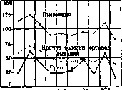
Figure 1. Mortality from pneumonia, other diseases of the respiratory organs and influenza (per 100,000 population).
relating to Leningrad (Table 2 and Figure 2); for comparison, the mortality rate from influenza is also given. Table 2. Mortality from P. (all forms) and influenza in Leningrad for 1917-1928 (per 100,000 population). Years Pneumonia Influenza Years Pneumonia Influenza 1917' 1918 1919 1920 1921 1922 1923 284.6 544.0 1084.1 453.4 307.8 302.8 176.0 12.6 94.2 257.9 91.6 35.2 26.5 25.3 1924 1925 1926 1927 1928 1929 1930 188.4 145.5 158.2 172.0 167.5 158.9 175.2 15.9 11.4 .41.4 42.0 32.5 30.7 25.3 For the last 8 years, mortality from pneumonia in Leningrad has fluctuated from 145.5 to 188.4 per 100,000 population compared to 301.8 in 1911-13. This significant reduction in mortality is mainly due to the sharp decrease in mortality from pneumonia in infancy (Table 9). For Moscow, where mortality from lobar pneumonia is registered, the following data are available (Table 3 and Figure 3). Lobar P. in Moscow in recent years gives a mortality rate 2-3 times lower than in the pre-revolutionary years. During the years of the influenza pandemic (1918-19), the highest mortality from lobar P. was observed in Moscow, 21/2 times exceeding the mortality of previous years. Table 3. Mortality from lobar P. and influenza in Moscow for 1903-1930 (per 100,000 population). Years Pneumonia Influenza Years Pneumonia Influenza 60.7 , 1917 62.8 8.6 69.6 - 141.7 25.7 55.3 -. 153.8 39.9 52.5 - 76.0 18.4 70.8 ' - 56.7" 10.1 68.7 - 53.2 10.4 62.3 - 29.2 13.2 72.7 22.1 26.4 12.3 74.5 . 13.8 24.0. 11.3 75.0 16.7 25.1 22.9 74.8 15.2 25.1 19.7 75.9 16.8 25.1 . 2t.0 ' 58.4 12.6 23.6 16.5 62.7 12.2 27.8 14.7
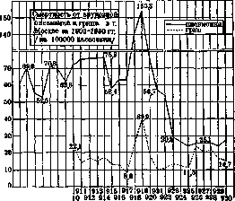
Figure 3.

2. Mortality from P. in cities and rural areas is not the same. Data from Leningrad and Leningrad Province (Table 4) indicate that mortality from pneumonia in rural areas is higher than in cities, while at the same time the mortality coefficient from pneumonia in small cities is lower than in Leningrad. Table 4. Mortality from P. (all forms) in cities and other localities of Leningrad Province (per 100,000 population). Cities Years Rural Leningrad Other cities localities 176.0 151.3 188.4 175.5 - 115.5 122.6 231.2 158.2 107.8 167.8 172.0 171.1 - 167.5 - - 158.9 - - In Germany, the mortality coefficient from pneumonia in populated areas with 15,000 or more inhabitants is lower than the corresponding coefficient for the entire country (Table 5). 3. Mortality by sex and age. Table 6 and Figure 4 present mortality from all forms of P. by age and sex in Germany in 1930, Table 7 and Figure 5 give the same data for Leningrad in 1923. From these tables it is evident that mortality among men in all age groups exceeds that of women; the only exception is the age group of 60 years and older, which in Leningrad in 1923 gave the opposite relationship. From the same tables, the extremely sharp differences that are observed in the magnitude of mortality from P. for different ages are apparent. These differences significantly exceed those that exist in relation to the age-specific coefficients of general mortality. Extremely high mortality from P. in the first year of life sharply decreases in the age group from 1 to 4 years with the same Age Men Women All ages
Figure 4. Mortality from pneumonia by age and sex in Germany in 1930.

Age Men
to reach its minimum at ages 10 to 14 years; after the age of minimum mortality, mortality begins to increase first slowly and then at a faster rate, again giving high coefficients in old age and especially in senile age. From Table 8 it is seen that the main mass of deaths from pneumonia falls on the age up to 5 years and on the age of 50 years and older. Among the causes of death in the first year of life, pneumonia occupies one of the first places. Table 7. Mortality from P. (all forms) by age and sex in Leningrad in 1923 (per 100,000 population of the corresponding group). Age Men Women Both sexes 0-12 m....... 3,550.6 3,124.1 3,343.8 1-4 years..... 858.3 763.1 745.2 5-9 years...... 42.3 40.6 41.4 10-14 years..... 23.8 19.9 21.8 15-19 years..... 40.4 32.3 36.8 20-29 years..... 47.0 22.3 34.0 30-39 years..... 43.9 40.6 42.4 40-49 years..... 101.0 43.6 71.6 50-59 years..... 190.4 150.6 167.7 60 years and older . . . 483.7 540.0 524.3 ) Table 8. Mortality from P. (all forms) by age in Leningrad in 1921-1923 (per 1,000 deaths in all ages). Age 0-12 m..... 1-4 years..... 5-9 years..... 10-14 years..... 15-19 years..... 20-29 years..... 30-39 years..... 40-49 years..... 50-59 years..... 60 years and older 1921 1922 1923 5a
Age-specific mortality from lobar P. is illustrated in Table 9. In general, age-specific mortality from lobar pneumonia has the same character as mortality from all forms of pneumonia with the difference that here senile age gives maximum mortality, not infant age (Fig. 6). Table 9. Mortality from lobar P. by age in Moscow in 1931 (per 100,000 population of the corresponding group). Age Mortality 0-4 years..... 33.0 5-9 years..... 12.3 10-14 years..... 4.4 15-19 years..... 9.1 20-29 years..... 8.1 30-39 years..... 16.8 Age 40-49 years..... 50-59 years..... 60 years and older All ages. Mortality 59.0 111.1 32.1 4. Morbidity. Registration of cases of P. began comparatively recently and in very few countries. In 1928, P. was registered in England, Scotland, Ireland (N.), Ireland (Free.), Denmark, Iceland, Norway, USA (28 states), Canada, N.Zealand and some other states. Usually, summary data on all cases of P. are published without division into individual forms. For Moscow and Moscow Province, morbidity from lobar P. is distinguished. In 1925 in the city of Moscow, 13.6 cases of lobar pneumonia were registered per 10,000 population, in 1926-15.2, in Moscow Province in 1906-1908, 31.0 cases were registered, in 1925-34.0, in 1926-25.6. Table 10 gives an idea of the morbidity from lobar P. by age and sex. Table 10. Morbidity from lobar P. by age and sex in the city of Moscow and Moscow Province (per 10,000 people of the corresponding group) in 1926. Moscow Age M-12 mos. 1-4 years..... 5-9 years..... 10-14 years..... 15-19 years..... 20-29 years..... 30-39 years..... 40-49 years..... 50-59 years..... 60 years and older Moscow Province M. | F. 48 46 19 11 16 9 10 17 15 20 16 67 30 20 21 18 19 25 27 23 11 ' Mortality from lobar pneumonia by age in Moscow in 1931; per 100,000 population of the corresponding group) The age group most affected by lobar P., both in the city of Moscow and in Moscow Province, is childhood (up to 10 years), especially infancy (up to 1 year), with some increase in old and senile age. In all ages (except for the age of 1 year in the city of Moscow), men suffer from lobar P. more often than women. The average morbidity in the province's population is higher than in Moscow.
5. Seasonality. The maximum mortality from P. in most countries falls on the first quarter, as seen in Table 11, where the number of deaths from pneumonia in some countries and cities for the last three years are summarized by quarters; the minimum falls on the third quarter
O O MS O £ O
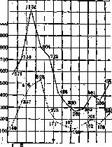
Fig. 6. Mortality from pneumonia observed in the first quarter of 1929 in European countries and the USA finds explanation in the significant influenza epidemic that these countries experienced at the beginning of 1929. Fig. 7 shows the monthly distribution of cases of lobar pneumonia in Moscow and Moscow Province for 1926. The maximum number of cases in the city of Moscow fell in April, while in Moscow Province it falls in March; the minimum number of cases of lobar pneumonia and in
Moscow Province, city of Moscow
* Figure 7. Monthly distribution of cases of lobar pneumonia in 1926 in the city of Moscow and in Moscow Province falls in August.

I. Blokh Pathological anatomy. From the pathological-anatomical side, lobar P. is an acute fibrinous inflammation of the bronchopulmonary tissue with the deposition of abundant masses of fibrin in the air passages or in the air sacs (hence the sometimes used synonym - fibrinous pneumonia). Four stages of the pulmonary process are distinguished: the first stage - the stage of congestion - is characterized by sharp hyperemia of the vessels. This hyperemia over significant areas passes into stasis of blood, while the permeability of the capillaries of the corresponding areas of the lung sharply decreases, and in some places apparently completely disappears. Already at this time, the affected parts of the organ become somewhat dense, darker. The second stage - red hepatization (hepatization) - is characterized by a rapidly increasing diapedesis of erythrocytes and the exudation of plasma proteins, especially fibrinogen, into the air-containing parts of the lung parenchyma. The lung 'hepatizes' and clearly increases in volume. Its weight increases sharply - 2-3 times and more. Table 11. Mortality from P. (all forms) by trimesters of the year (absolute figures). States and cities Germany (49 cities) Holland....... Spain ........ Scotland* ..... Czechoslovakia . . . . Berlin .......... Calcutta....... Leningrad....... London*........ Moscow **....... New York ....... Paris*........ Rome* ......... Tokyo* I........ 1928 g. 1930 g. 4 865 1239 2 641 2 047 7 161 912 747 628 1 619 192 3 451 1 328 429 1125 II 3 835 888 2 247 1367 6-?41 1 013 10C5 3 405 804 356 4й5 2 295 3 219 8 882 2 584 2 900 3 812 3 748 3 964 5 754 10 030 1 614 ,. 732 1 014 3 484 2 074 4 599 2 161 1 139 3 987 1(20 2 527 1S81 5 546 2 127 2 327 481 1495 773 3 109 387 336 494 465 75 981 489 179 181 3 228 695 2 383 1240 4 628 513 502 421 770 124 1975 629 308 313 4 923 1 108 2 953 2C95 6 C19 2 858 3 965 2 324 5 186 2 341 771 288 241 2 273 371 1 539 ' 748 3 627 339 342 681 399 1026 410 145 130 3 370 2 3C2 782 195 313 Including mortality from bronchopneumonia. Mortality only from lobar pneumonia. quarter. This table once again demonstrates the close connection that exists between pneumonia and influenza. A sharp increase in mortality (up to 2,000:-2,500 cases with total lesions). On section, the lung is red, fine-grained, somewhat dry. The granularity is sometimes very fine, sometimes coarse, depending on the size of the alveoli; in lobar P. in emphysematous patients, the granularity is often especially coarse. The grains themselves are compact fibrinohematic masses of exudate, which as molds fill the alveoli, and often the bronchi as well. The third stage - gray hepatization - is a direct continuation of the previous one, and the transition itself is either rapid or somewhat drawn out and uneven; that is why sometimes they speak of gray-red hepatization. The essence of the changes in the third period lies in the subsidence of the diapedesis processes of erythrocytes and in the enhancement of desquamative and proliferative phenomena on the part of the alveolar epithelium, but mainly in the emigration of leukocytes, the mass of which, having a gray-greenish color, gives the section of the lung the corresponding shade. The granularity is clear, the hepatization of the lung is pronounced, the weight increases. The hepatized lung, especially gray, tears easily even with slight violence, for example, with careless puncture with a needle. The fourth stage - the stage of resolution - is characterized by the onset in the exudate of proteolytic and autolytic processes; liquefaction and dissolution of fibrin occurs, as well as fatty degeneration, disintegration of leukocytes and desquamated alveolar epithelium. This fatty degeneration can give a slightly yellowish tint to the entire section of the affected lung, and then they speak of 'gray-yellow hepatization'. The granularity weakens or disappears, the phenomenon of hepatization gradually weakens. The lung in consistency and color rather resembles the spleen ('splenization'). Finally, complete resorption of the exudate occurs, the organ takes on a soft and even flabby consistency with significantly less elasticity than normal. At the end of the fourth period, leukocytes finally disintegrate, and in their place appear cells of lymphoid type with broad protoplasm, in which it is not difficult to recognize the regenerating alveolar epithelium. This regeneration, however, proceeds unevenly, and alongside restored alveolar structures, for quite a long time one can still find in some places accumulations of free cells in the alveoli. In general, the period of resolution should be regarded as the most extended in time; in any case, it extends to the first 5-7 days of the afebrile state of the disease. In the vast majority of cases, the entire exudate is resorbed without a trace; only a relatively small part of it is expectorated; the lung anatomically returns to normal; it is true that functionally it is still for some time deprived of its usual perfect elasticity. Histological, and often already macroscopic, examination reveals in some cases peculiarities in the morphology of the exudative processes or in the succession of individual phenomena. Thus, diapedesis of erythrocytes in its intensity sometimes resembles acute parenchymal hemorrhage, as if a continuous infarction of the lung. In other cases, a weak or extremely uneven fibrinous exudate, insignificant granularity of the section and even its absence are noted. In these cases of so-called smooth pneumonias, the exudate filling the alveoli consists mainly of serous fluid, alveolar epithelium, leukocytes, and sometimes exclusively the latter, the lung appearing as if impregnated with liquid pus. Purulent exudate in large quantities is also removed from the section of the organ. It would be incorrect, however, to interpret such cases as purulent P. or as an outcome in suppuration, since the melting of lung tissue and the formation of abscesses are by no means necessary in this case. French authors therefore distinguish two types of 'gray hepatization' (see separate table, figure 4): 'suppurée', or ordinary gray hepatization, and 'suppurée'-pus-like transformation of the intra-alveolar exudate. In these same cases of 'pus-like' P., the stage of red hepatization often completely falls out; the general development of the exudate thus proceeds as it were along the lines of banal catarrhal P.--To all the above it is necessary to add that the development of the pneumonic process in each specific case and even in the same lobe of the lung can have its own details and peculiarities, not only in the sense that the very unfolding of the changes by stages takes place in different places at somewhat different times - this is a common phenomenon (see below), but in the sense that in some places the processes unfold quickly and typically, in others - slowly and not quite typically (little or no fibrin at all, sharp diapedesis of erythrocytes, pus-like character, massive thrombosis of vessels, etc.). In other words, both the very beginning of the process and its evolution, even in the same lobe of the lung, can be different in different places; in this one cannot but see the prerequisites for this or that focal complications, often observed in lobar P. (see below).-As indicated, the exudate usually also fills parts of the bronchial tree, and1 cases are not uncommon when this filling reaches the medium and large bronchi, and from the lumen of the latter one can pull out entire casts of the bronchial pathway (to be examined in water). P. with diffuse exudation into the bronchial tree is also designated as massive P.-On the part of the interstitial tissue of the affected area, phenomena of serofibrinous impregnation are also noted, and in the lymphatic vessels sometimes pictures of lymphangitis and thrombosis. In most cases, an entire lobe of the lung undergoes hepatization - hence the synonym 'lobar pneumonia'. Not so rarely, two and three lobes are affected, bilateral P. with hepatization of the main mass of both lungs are also noted. The right lower lobe is most often affected, and in general the lower lobes. Pneumonia of the upper lobes is not so rare; the comparative frequency at autopsies of exactly such upper and even 'apical' P. apparently speaks of their particular severity (compression of the vagus nerve? thoraco-abdominal?). Lobar P. is not always lobar in the full sense of the word: individual parts of the lobe often remain non-hepatized, for example only edematous, but aerated or even emphysematous. This lobular principle is especially often violated in childhood and old age. In children, especially small ones (up to 5 years), typical lobar P. often does not occur at all; also often in exhausted subjects and marasmic patients. Sometimes the exudate, at different times and with significant intervals, captures separate pieces or entire lobes of the lung - so-called migrating P. (for example, the right upper lobe has gray hepatization, while the left lower lobe is only undergoing red hepatization, etc.).
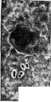
So-called smooth pneumonias are characterized by the absence of typical fibrinous exudation. The exudate in them is mainly serous, containing a large number of leukocytes and desquamated alveolar epithelium. Macroscopically, the lung is dense, dark red or grayish-red, with a fine granularity. Microscopically, the alveoli are filled with serous fluid with cellular elements. The course of such pneumonias is usually milder. In some cases, the exudate may become purulent, but this does not necessarily indicate a transition to purulent pneumonia. The resolution of smooth pneumonias occurs more quickly and without significant residual changes. The term 'smooth pneumonia' is not widely used in modern medicine but was employed in the early 20th century to describe these atypical forms. The pathogenesis of smooth pneumonias is still not fully understood, but they are believed to result from a less intense inflammatory response compared to typical lobar pneumonia. Clinically, smooth pneumonias may present with fewer systemic symptoms and a more localized process. The prognosis is generally better than for typical lobar pneumonia, especially in debilitated patients. Histological examination reveals less fibrin deposition and more cellular exudate, with prominent alveolar epithelial desquamation. The inflammatory process tends to be more limited in extent and severity.
Figure 1. Catarrhal pneumonia; a-airless (flooded with exudate) parts; o-emphysematous swollen area; n in the middle and on the right-airless parts; s-pleura; d-exudate; e-bronchus; f-bronchus blocked with exudate. Figure 2. Interstitial pneumonia (interstitial lung sclerosis): o-flattened alveoli with markedly thickened walls. Figure 3. Carnification of the lung in pneumonia as a result of organization of bright exudate within the alveoli (a); o-elastic fibers outlining the alveoli. Fig. 4. Lobar pneumonia (gray hepatization) of the lower lobe (a) of the right lung; o-partial airlessness; s-middle lobe; d-upper lobe. Figure 5. Desquamative pneumonia; in the alveoli-a large amount of 'thickened epithelium'. Figure 6. Pneumococci. Central P., i.e., those located in the depth of the lung parenchyma without the focus extending to the pleura, are more often observed in children, but in general they are quite rare; most often we are dealing here with a transitional state, for example, in the course of development of ordinary *forms, which eventually reach the surface of the lungs. Deviations in the course of lobar P. are numerous and frequent, and they depend, on the one hand, on the peculiarities of the infection, and on the other hand, on the peculiarities of the reaction of lung tissue, previous diseases, on the state of cardiac activity and the entire organism. The above-described classical course of periods of consolidation with resolution is a property of most P. with pneumococci of types I, II and IV. Pneumococci of type III are usually found in atypical cases (absence of lobar distribution, absence of granularity on the cut surface, purulent nature of gray consolidation, true suppurations, hemorrhages, sequestra, etc.). In general, these cases have great similarity with Friedländer's pneumonias. Friedländer's forms of P. almost always show significant deviations in morphology and evolution. Lobar distribution is very often absent in them, although this may be because this form more often occurs in old age and in individuals with impaired nutrition, in alcoholics. Usually, groups of foci of liver consistency and even individual, sometimes completely round, gray nodes scattered in different parts of the lung are observed. Sometimes such nodes merge, and pseudo-lobar foci arise. Lesions of the upper lobes in Friedländer's forms are particularly frequent. The evolution of the pneumonic foci in Friedländer's P. is very often atypical: a great tendency to form necroses, sequestra; on this basis, acute hemorrhages, development of large cavities with dirty uneven walls, gangrene of the lung, etc., are possible. The most striking feature of the same P. is the nature of the exudate scraped off or even spontaneously flowing from the surface of the cut of the foci: it is extremely abundant and consists of turbid, thick, mucous masses, emitting a characteristic smell of burnt meat. Under the microscope, leukocytes, abundant cellular detritus and masses of mucus with a large number of diplobacilli [see separate table (to art. Lung), figure 8] are found; the usually weak staining of the latter depends on their strong envelopment by a powerful mucous capsule, and on the other hand, on the pronounced plasmolysis of the bacilli themselves. The abundance of bacilli is also striking in histological examination of the lungs; sometimes they are the exclusive component of the intra-alveolar content. Friedländer's 'mucous' P. are distinguished by particularly intense toxic, and often septic, or pyemic phenomena with septic jaundice, hemorrhages into the skin, mucous membranes, acute serositis, etc. Their mortality is apparently the highest among all P.-All of the above can be applied to pneumococcal P. of type III; only in the exudate, bacilli are not found, but chains of pneumococci, which are also usually weakly stained due to their envelopment in mucus and plasmolysis, are found.-As special microbiological varieties, lobar P. should be distinguished, in the exudate of which ordinary or hemolytic streptococcus, staphylococcus, as well as mixed infection, especially with Pfeiffer's bacillus, are found. In both cases, the peculiarity is the tendency to hemorrhages: the consolidated lung has a mottled appearance, somewhat resembling the variegated grippe P., and also the tendency to prolonged suppurations, carnification (see below) is significant here too.-Parts of the lung that remain free from the lesion also undergo certain changes, mainly in the sense of the development of acute vicarious emphysema in them. Lobar P. as a rule is accompanied by pleurisy. Pleural processes usually arise in the very first days of the disease (parapneumonic pleuritis); less often, the appearance of exudate is noted only during the course of the disease, sometimes at its end as a complication (metapneumonic pleuritis, especially empyemas). The exudate most often has a fibrinous [see separate table (to art. Lung), figure 7] or sero-fibrinous, rarely purulent nature. Complications. The wide possibility of development of various complications in lobar P. follows from a whole series of prerequisites relating to the morphology, dynamics and microbiology of the process. The questions of general nutrition, age, condition of the respiratory pathwaysg especially the lungs, before the disease P.; P. developing under conditions of stagnation in the small circle (in heart defects), in various intoxications (alcoholism, uremia), etc., are often very atypical. One should, however, distinguish atypical courses of P. from complications in them. As complications, only those cases should be designated when the course of the disease at one or another stage of its development makes a sharp retreat in essence of the process. But if the essence of the process remains within the indicated regularities and only features of a topographical nature (e.g., upper P.), or volumetric (non-lobar P.), or chronological nature (rapid or, conversely, slow evolution), or finally if there is a falling out of some stages, resp. brightness of others, then in all these cases it is more correct to speak not of complications, but of atypical forms of P. The most important complications of P. are gangrene, abscess (see Lung, abscess, gangrene), purulent lymphangitis and carnification of the lung. From typical gangrene, the so-called putrid form of it (pneumomalacia) should be distinguished, when there is no putrefactive decay and corresponding smell; in this case, aspergilli, sarcinae are often found in abundance in the decay. Putrid forms are relatively frequent in diabetics, in Friedländer's P. If putrid gangrene of the lung in the vast majority of cases ends in death, this cannot be said about putrid forms, in which a favorable outcome is sometimes observed, namely, liquefaction of necrotized masses, their removal through the bronchial tree with subsequent epithelization of the cavity formed in place of the sequestrum. Such cavities resemble lung cysts, smooth cavities. Purulent lymphangitis of the lung and pleura is a serious complication [see separate table (to art. Lung), fig. 9]. Macroscopically, in these cases, a strong thickening and gray-green color of the interstitial tissue of the lung, and under the microscope, lymphatic pathways strongly stretched by purulent exudate and loosened cellular tissue are found. Purulent lymphangitis usually is combined with the development of sequestra, abscesses of the lung, empyema of the pleura, purulent lymphangitis and lymphadenitis of the mediastinum, mediastinitis; sometimes the process along the coronary ligament of the liver or along the lymphatic system of the diaphragm descends downward to the peritoneum, causing peritonitis. Streptococci are a common finding in all these cases.-Carnification of the lung is observed in those cases when the usual processes of resorption of fibrinous exudate are weakly expressed or fall out and the so-called organization of exudate takes place, resembling the organization of a thrombus. Microscopically, the process is expressed at first in the growth of the exudate by individual mesenchymal elements; and later by ordinary granulation tissue with vessels; intra-alveolar fibrinous plugs gradually turn into dense clumps of fibrous tissue, organically fused both with the walls of the alveoli and with each other through pores and terminal funnels. In late stages of carnification, the corresponding parts of the lung become almost unrecognizable; however, with the help of staining for elastic tissue, it is possible at this time to detect elastic fibers, which in some places continue to outline the contours of the lung alveolar parenchyma, but later this characteristic detail also disappears. Carnified parts of the lungs have the consistency of meat, are airless, gray-rose, and later gray in color, [see separate table (art. 623-624), fig. 3]. Very often in the same areas, bronchiectases, as well as abscesses, are found; the combination with the latter is more common than rare. Not however is carnification always caused by an intra-alveolar process of organization of the effusion. Cases are observed when sclerotic changes go predominantly interstitially [see separate table
(p. 623-624), figure 2); the entire stroma of the lung sharply coarsens, in particular the alveolar and interinfundibular septa thicken; the airiness of such a lung also sharply decreases. The basis of interstitial sclerosis of the lung is usually its lymphangitis, sometimes accompanied by thrombosis of the lymphatic system. About 3% of all lobar pneumonias are complicated by serofibrinous pericarditis. Peritonitis is rarely observed, mainly in childhood. The mechanism of development of such peritonitis can apparently be twofold. On the one hand, the inflammatory process from the diaphragmatic pleura can penetrate along the intermuscular layers of the diaphragm (microperforation) and emerge under the peritoneum; this is why a matte, i.e., covered with fibrin, peritoneum in the area of the diaphragm and a matte surface of the liver are not uncommon in pneumonia, especially on the right. On the other hand, embolism of the peritoneal vessels by diplococci is indicated (hematogenous peritonitis, see below). Diplococcal purulent metapneumonic peritonitis often occurs without the usual bright phenomena generally characteristic of peritonitis; this is why most of them are only an accidental finding at autopsy. Endocarditis is found in 4-5% of all autopsies in pneumonia; most of these cases, however, occur in patients who previously had a heart defect (rheumatic), which only worsened under the influence of pneumonia. The infarcts of the lungs observed in endocarditis sometimes lead to the development of typical lobar-pneumonic processes. On the other hand, in all such cases, it is necessary to clarify the sequence of events, since infarcts can also arise as a complication of lobar pneumonia. Pneumonias in patients with heart defects have certain features of a local and general nature. Usually developing against the background of already existing congestion in the lesser circulation, for example in mitral stenosis, they are often accompanied by violent bleeding per diapedesin. An extremely characteristic finding in the hearts of pneumonia patients is the presence of large, light fibrin clots in the heart cavities, emphasizing hyperfibrinosis of the plasma and increased blood coagulability in these patients. Among the complications of lobar pneumonia, meningitis often occurs (about 6% of all cases). Macroscopically, impregnation of the soft meninges with exudate is found, especially of the brain vault, and the exudate itself can be serous (slightly cloudy), serous-purulent, and purulent; the yellowish tint of the exudate in diplococcal meningitis attracts attention. Upper lobes are particularly often affected in meningitis. Degenerative and inflammatory changes in the sympathetic ganglia, especially cervical ones, are a fairly common phenomenon in pneumonia. It is possible that these changes have some significance for the development of such symptoms as hyperemia and swelling of the face of pneumonia patients, hyperemia of the brain and its meninges, etc. Childhood pneumonias. The change of phases of hepatization in pneumonia in children proceeds faster, the amount of fibrin is less than in adults, but there is always an abundance of desquamated pulmonary epithelium, so that sometimes the histological distinction of such pneumonias from banal bronchopneumonias becomes difficult, all the more so since the volume of the foci themselves most often remains lobular. Migratory forms are relatively common. In children, the paravertebral parts of the lung parenchyma (especially in children under 1 year) and the upper lobes of the lungs are often preferentially affected. Statistical observations show that in small children, the right upper and left lower lobes of the lungs are particularly predisposed to the disease; there is even mention of almost complete immunity of the left upper lobe. Compared to adults, herpes is more often noted in children. Several cases of lobar (lobar) pneumonia in newborns (so-called congenital pneumonia) have been described. In all such cases, lobar pneumonia in the mothers was observed. I. Davydovsky. Pathogenesis. The pathways of penetration into the lung parenchyma of pneumococcus or other microorganisms causing lobar pneumonia have not yet been finally clarified. Most authors adhere to the viewpoint of bronchogenous penetration of the infection. In addition to clinical observations, which will be discussed later, experimental studies speak in favor of this view. As we have already indicated, Black and Cecil caused in monkeys a disease completely analogous to human lobar pneumonia by intratracheal introduction of small amounts of pneumococcal culture (0.001-0.000001 cm³). Intravenous administration of the same animals with pneumococcal culture did not cause lobar pneumonia, even if the lungs had been previously traumatized. The observations of these authors indicate that pneumococci, when introduced into the respiratory tract in the manner described, do not directly enter the alveoli. Studies of the organs of infected monkeys show that pneumococci penetrate through the walls of rather large-caliber bronchi into the lung tissue, where, spreading perivascularly and peribronchially along lymphatic pathways, they cause an interstitial inflammatory process, in which the alveolar epithelium is involved only secondarily. Pneumococci thus, according to the observations of Black and Cecil, penetrate into the alveoli not directly through the airways, but from the lung parenchyma. The described method of penetration and further spread of infection, which is based on the primary infection of lymphatic pathways, makes the "lobar" character of the process partially understandable, i.e., the tendency to affect entire lobes. A number of authors, especially French ones (Widal, Calmette), hold a different viewpoint regarding the pathogenesis of lobar pneumonia. They believe that pneumococci penetrate into the lung parenchyma by the hematogenous route and that lobar pneumonia, similar to typhoid fever, is thus a primary bacteremia. Proponents of this viewpoint point out that pneumococcus can be found in the blood in the very early periods of the disease, when there are still no symptoms (including those observed in X-ray examination) indicating lung damage. However, these arguments are not convincing, since the absence of corresponding symptoms during clinical examination obviously does not exclude the presence of the initial stage of the inflammatory process in the lungs. It has been proven that X-ray examination does not always capture the first phases of the pneumonic process, especially the stage of hyperemia. Numerous clinical observations speak for the primary localization of the process in the lungs, when lobar pneumonia begins suddenly in complete health and when immediately after the initial shaking chill or even simultaneously with it, typical stabbing pain in the side and cough appear—symptoms indicating lung damage right from the start of the disease. The bronchogenous penetration of pneumococci into lung tissue is also indicated by those frequently observed cases when lobar P. is preceded for a longer or shorter period by catarrhal phenomena of the upper respiratory tract. Primary bacteremia is also contradicted by repeated blood cultures in the same patient, which showed that pneumococcus cannot be found in the blood in most cases, regardless of the stage of the disease (Ettinger, Viktorov, Mazel). The cases described in the literature of intrauterine infection of the fetus with lobar pneumonia indicate that the hematogenous origin of this infection is nevertheless possible under certain conditions. Multiplying rapidly in the affected lung tissue, pneumococcus releases toxic substances at the same time. The latter, being absorbed and penetrating into the general circulation, cause disturbances in various systems of the body. In a number of cases, the matter does not limit itself to one toxemia. From the affected lung tissue, pneumococcus penetrates into the circulatory system to a greater or lesser extent. As shown by the observations of a number of authors, cases with bacteremia proceed especially severely, give a significantly higher percentage of fatal outcomes compared to the average mortality in lobar pneumonia. Circulating in the blood, pneumococcus, by localizing in various organs, can cause sharp changes in them, giving a number of severe, often fatal complications, such as purulent meningitis, ulcerative endocarditis, purulent pericarditis, peritonitis, etc. The respiratory insufficiency observed in lobar pneumonia (dyspnea, cyanosis) is only partially explained by the exclusion of the affected area of lung tissue from the respiratory act. Heart failure and especially the vascular system play a major role in the occurrence of anoxemia. As shown by Romberg and Pussler on the basis of experimental studies, toxins in pneumonia act mainly not on the heart, but on the vasomotor centers. The basis of the collapse often observed in lobar pneumonia, which often leads to death, is the acutely occurring sharp decrease in vasomotor tone, leading to the accumulation of blood in the organs of the abdominal cavity and the associated insufficient blood supply to the central nervous system and heart. The processes underlying the cyclic course of lobar pneumonia, in particular the mechanism of crisis, have not yet been sufficiently clarified.
A number of authors (Klemperer, Romer, Neufeld, Handel) have shown that shortly before the crisis, and especially during and after it, protective bodies appear in the blood of patients with lobar P., which protect white mice when infected with highly virulent pneumococcal culture in amounts far exceeding the lethal dose. It has been proven that these protective bodies possess type specificity, protecting mice only when infected with a homologous strain, i.e., pneumococcal culture of the same type with which the given patient was infected. As some authors (Baldwin, Rhoades) have shown, these protective bodies are generally absent in blood samples in which pneumococci can be detected. These authors repeatedly observed the disappearance of pneumococci from the blood with the appearance of protective bodies. In cases ending fatally, protective bodies are absent from the blood until death itself. In the process of preventing and hindering the generalization of pneumonic infection, these antibodies apparently have great importance. However, their significance in the mechanism of the crisis itself cannot be considered finally clarified, since on the one hand, it is not possible to detect these substances in the blood of all convalescents after lobar P., and on the other hand, their appearance does not always coincide with the crisis. Often a high titer of these substances can be detected several days before the temperature drop. In the mechanism of the crisis, in addition to the appearance of these antibodies, other little-studied processes, probably of a cellular nature such as phagocytosis, etc., play a major role. In the process of eliminating the infection, in addition to the reaction of the macroorganism, the changes that the microorganism undergoes during the pneumonic process are undoubtedly also of importance. As Lubarsch showed, the pneumococcus possesses the greatest virulence in the initial stages of the disease, but as the crisis approaches, its virulence decreases. The parallel processes occurring on the part of the macro- and microorganism 31 ez'2 are closely related to each other and constantly influencing each other, at a certain moment of infection they cause the turning point that forms the basis of the crisis. Clinic. Typical course of lobar P. In typical cases, lobar P. begins suddenly, abruptly, in the midst of complete health. Patients often become ill during work. In the medical history, there is often an indication even of the hour of onset of the disease. Soon after the appearance of the first painful symptoms in the form of general weakness, malaise, aching in the limbs and headache, a shaking chill occurs, lasting from 10-15 minutes to 1-2 hours. The temperature rises rapidly and within the first hours after the chill usually reaches1 39° and even higher figures. In a number of cases, pleuritic pain already appears during or shortly after the chill, which usually increases with inspiration. Cough, painful and therefore suppressed by the patient, also often appears already on the first day of the illness. The patient's condition progressively worsens, appetite disappears, thirst appears. Due to severe headache, general excitement, painful cough, the patient usually spends the first night without sleep. In typically progressing cases of lobar P., a number of characteristic symptoms appear on the first or second day of the illness. There is a sharp increase in respiration (36-40 and more per minute), not corresponding to the temperature and pulse; herpes appears on the lips or wings of the nose, characteristic redness of the cheeks appears. At this time, it is often already possible to determine lag in the breathing of one half of the chest and the participation of the so-called auxiliary muscles in respiratory movements. Due to pleuritic pain and painful cough, breathing is shallow, irregular, speech is difficult. Examination of the blood usually already at this period reveals neutrophilic leukocytosis, and examination of the urine reveals a sharp decrease or almost complete disappearance of chlorides. The typical rusty sputum, which finally confirms the diagnosis, rarely appears before the third day of the illness. Symptoms on physical examination that definitely indicate the localization of the inflammatory process in the lungs (see below) often appear already on the 2-3rd day of the illness, in a number of cases however they can only be detected in the following days. Along with signs indicating a pneumonic focus, scattered dry and moist rales are often observed as a manifestation of accompanying bronchitis. The temperature curve in typical cases has the character of continua (see below). In a number of cases, 2-3 days before the crisis, and sometimes in the first days of the illness, a sudden drop in temperature is observed, often by 3-4°, to normal and even subnormal figures. This temperature drop usually lasts only a few hours, followed by a new rise. During such so-called pseudocrises, the patient's condition often improves, but breathing and pulse remain frequent, which usually allows one to distinguish these temporary temperature drops from the true crisis. On the 4-5th day of the illness, especially in more severe cases, phenomena from the cardiovascular and nervous systems begin to come to the forefront. The pulse becomes softer, more frequent, filling less, shortness of breath increases, cyanosis appears. From the nervous system, various degrees of disturbance are observed, ranging from drowsiness and mild delirium to severe soporose and even comatose states. In typical cases, lobar pneumonia ends with a crisis, which usually occurs at the end of the first or beginning of the second week of the illness. The temperature drops within 12-24 hours, and often reaches subnormal figures. The crisis is usually accompanied by profuse sweating. Within a few hours, a sharp turning point occurs in the patient's condition. Breathing and pulse become less frequent and return to normal figures. During the crisis, the patient often falls into sleep, from which he wakes up with normal temperature and a marked improvement in his condition. In a number of cases, a sharp rise in temperature directly precedes the crisis, with worsening of the patient's condition and general state. This phenomenon (so-called perturbatio critica) contributes to an even greater range of the critical drop in the temperature curve. Not all cases of lobar P. end with a crisis. Often a lytic drop in temperature is observed over 2-3 days. The return of temperature to normal does not coincide with the disappearance of the inflammatory infiltrate in the lungs. Symptoms on physical examination indicating consolidation of lung tissue (dullness of percussion sound, bronchial breathing, crepitant, resonant moist rales, bronchophony) may persist after the temperature drop for a week, and sometimes longer, usually having little effect on the patient's condition and general state. The duration of the convalescent period and the time to restoration of working capacity depend on the severity of the illness, the age of the patient1 and individual reaction. In a number of cases, patients feel quite recovered after 1½-2 weeks. Usually the convalescent period until restoration of working capacity lasts no less than a month. In some casesu of lobar P., a few days after the crisis, a new rise in temperature is observed with a new flare-up of the pneumonic process. These so-called relapses of pneumonia (ge-kurrierende Pneumonie of German authors, pneumonie a rochute of the French) are observed very rarely, according to the statistics of some authors in less than 1% of all cases. The inflammatory process may be localized in the site of the first lesion or in a new area of lung tissue.
k Special symptomatology. The sudden onset of the disease is observed in far from all cases of lobar P. Often, the disease is preceded for several days by more or less pronounced prodromal phenomena—malaise, headache, poor appetite—aches in the back and extremities, and in a number of cases also manifestations of catarrh of the upper respiratory tract (pharyngitis, laryngitis, runny nose). Some authors propose to isolate into a separate group secondary lobar P. those cases that arise against a background of preceding catarrh of the upper respiratory tract. Even in cases that proceed with prodromal phenomena, the onset of the pneumonic process is often marked by a shaking chill. The latter, however, is not an obligatory symptom of lobar P. Often, patients note only more or less pronounced repeated chills in the first days of the disease. "S3 Temperature in lobar P. (figs. 8-10) usually rises quickly, reaching 39-40° within the first few hours. In cases arising against a background of preceding influenza infection, the addition of P. usually* is reflected in the temperature curve in the form of a 39.5 39-38,-5--88- 'fi--37- 86,5- Я8- Days of illness- Шг' У* .......-«!- p £* Days of illness ££.456789 10П 39^fflrilllllTIfTl :^"6Ь:-7----------ay * -: гИ -_ di& - _. -_:t4~___:_ ..-----------------L.---------- Г------------------ 37 ЦД1 JJJ -----------------=t-- Figure 8. more or less sharp rise of it. The temperature curve in lobar P. in approximately 2/3 of all cases has the character of continua, while in the remaining cases it gives fluctuations of remittent and even intermittent type. The character of the temperature curve depends in part on the type of pneumococcus with which the patient is infected. Thus, lobar P. type I in more than 70% of all cases proceeds with a temperature curve of the continua type with daily fluctuations of less than one degree. Type III P., on the contrary, in 2/3 of all cases gives a remittent and intermittent curve. Cases of lobar P. types II and IV occupy an intermediate position in relation to the temperature curve between types I and III. A drop in the temperature curve down to subnormal figures is observed during the disease in so-called pseudocrises, as well as in collapses. The final drop in t° in lobar P. in some cases has the character of a crisis, in others - of a prolonged crisis or lysis. A crisis is spoken of when the drop in t° continues for no more than 12-24 hours. With a duration of the period of drop in t° from 24 to 48 hours, one speaks of a prolonged crisis or accelerated lysis. Even longer periods of drop in t° are referred to as lysis. The study of the clinical picture of lobar P. in connection with the type of pneumococcus shows that the character of the drop in the temperature curve depends in part on the variety of the causative agent. Thus, lobar P. type I in approximately 75% of all cases ends with a crisis, while approximately the same percentage of cases of lobar P. type III gives a lytic termination of the infection. Types II and IV occupy an intermediate position in relation to the character of the drop in t° between types I and III (Ettinger, Mazel, Viktorov). The period of convalescence usually proceeds with normal t°. Only in comparatively rare cases is a subfebrile t° observed, apparently connected with the absorption of the alveolar exudate. A prolonged t° or a new rise of it after a crisis is usually caused by the observed complications during the pneum
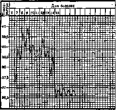
Figure 10.
Pneumonia with complications—empyema, abscesses—or else the non-resolution of the pneumonic process (carnification of the lung). In the elderly, as well as in individuals exhausted by severe preceding diseases, lobar pneumonia can proceed with a subfebrile temperature or even with a complete absence of a temperature reaction. General appearance of the patient. Lobar P. often gives the patient's face a certain characteristic appearance, so that some authors directly speak of facies pneumonica. Attention is drawn to the more or less intense hyperemia of the cheeks with a bluish tint, often more pronounced on the side of the affected lung. The picture of the pneumonic face also includes the frequently observed participation of the wings of the nose in respiratory movements. In more severe cases, already in the first days of the disease, a sharply expressed cyanosis of the lips and ears appears. Herpes is observed by no means in all cases of lobar P. (according to various authors, the frequency of this symptom ranges from 13% to 43%). Most often it appears on the lips or wings of the nose, less frequently on the neck and earlobe, and very rarely on the external genital organs or in the area of the anus. Some authors view herpes as a toxic phenomenon, associated with damage to the corresponding nerve elements. Other authors, based on the fact that in some cases pneumococcus can be isolated from the vesicles, speak of the localization of pneumonic infection in the skin tissue. From the point of view of authors who adhere to the view of herpes as a manifestation of a special filterable virus, lobar P. should be considered one of the infections that activate this virus under certain conditions. A number of authors consider the appearance of herpes as a prognostically favorable symptom. Cases that proceed without herpes allegedly give a higher percentage of mortality. Pain in the side, associated with the involvement of the pleura in the inflammatory process, is observed in the vast majority of cases of lobar P. and is often one of the main complaints of the patient. Pain in the side, which usually intensifies with deep breathing and coughing, is especially intense in the first 2-3 days of the disease, and then gradually subsides. It is usually localized at the level of the nipple on the affected side. In some cases, the pain radiates into the abdominal cavity or is exclusively concentrated there, which can simulate an acute disease of the abdominal cavity (appendicitis, cholecystitis, perforative peritonitis). In rare cases, patients feel pain not on the affected, but on the healthy side—this paradoxical phenomenon some authors explain by the presence of anastomoses between the intercostal nerves in these cases. Cough is often an early symptom, appearing already on the first day of the disease. In a number of cases, however, it appears later. Expectoration of sputum in lobar P. is usually difficult due to its viscous consistency. As the crisis approaches, the sputum becomes more liquid, which facilitates its expectoration. In senile and asthenic P. and in P. of alcoholics proceeding with delirium tremens, cough is often absent. Sputum in lobar P. is extremely typical. Its quantity is usually small. In different cases, variations are observed from 2-3 tablespoons to 200 cm3 per day. The coloring of the sputum can have different shades (brick-red, yellowish, yellowish-green, the color of prune decoction), which depends on the nature and degree of change in the leached Hb. The brownish-red shade observed in the vast majority of cases, reminiscent of the color of rusty iron, is so characteristic that it is almost pathognomonic for lobar P. This so-called rusty sputum is usually not an early symptom. In the first days of the disease, the patient either does not expectorate sputum at all, or it has a mucopurulent character. In the days preceding the crisis, the rusty color of the sputum often disappears. At this time, the sputum becomes completely discolored or acquires a yellowish-greenish tint. The sputum has a particularly pronounced green tint in so-called bilious P. (proceeding with jaundice), as well as in cases of lobar P. with an outcome in abscess formation or carnification. Sputum in all phases of the disease often contains as separate expectorations an admixture of red unchanged blood. More abundant hemoptysis is not characteristic of lobar P. It is observed only in so-called variegated P. of Spanish influenza, as well as in some severe cases of Friedländer's P. Pneumonic sputum in some cases contains macroscopically visible, dichotomously branched casts of small bronchi, consisting of fibrin. Sputum, both in the first days of the disease, when it usually does not yet have a characteristic color, and at the height of the disease, differs in viscosity, is difficult to expectorate, and adheres tightly to the walls of the vessel containing it. During convalescence, the expectoration of sputum continues for a more or less prolonged period. At this time, it is usually no longer colored, has a mucopurulent character, and is easier to expectorate. Sputum contains proteins and nucleic acids in solution. The latter mainly determine the viscous nature of the secretions. Under the microscope, in addition to erythrocytes, partly unchanged, partly leached, in the sputum of patients with lobar P., with appropriate staining, diplococci of Fränkel can be detected. The latter are usually observed in large numbers at the height of the disease, decreasing as the crisis approaches. In those cases where pneumonia is caused by the Friedländer bacillus, it is also usually possible to detect it upon microscopic examination. Sputum in lobar P. may be completely absent; this is especially often observed in children, the elderly, or in individuals exhausted by severe preceding diseases. In relatively rare cases and even with ordinary forms of lobar P., sputum is completely absent throughout the entire course of the disease, which makes it difficult or completely impossible to determine the type of pneumococcus with which the given patient is infected. In some cases of lobar P., the sputum throughout the entire course of the disease has a bronchitic, mucopurulent character.
Phenomena from the side of the lungs upon physical examination, since the time of Laennec, are associated with anatomical stages. Already in the period of engorgement, a dulling of the percussion sound with a peculiar tympanitic tint can usually be detected. These percussion changes are apparently associated with a decrease in the elasticity of the hyperemic lung tissue. The respiratory noise in this period usually retains its vesicular character, but due to the sparing of the affected side, it is often weakened. A characteristic auscultatory symptom of the stage of engorgement is fine crepitant rales, most clearly audible at the height of inspiration (so-called crepitatio indux). Often, crepitation appears clearly only after coughing jolts. In a number of cases of lobar P., the phenomenon of crepitatio indux cannot be detected even with careful daily examination of the patient. Along with typical crepitation on a limited area, scattered dry and moist rales, associated with accompanying bronchitis, are often heard. As the process transitions to the stage of hepatization, the dulling becomes more intense, but usually does not acquire the character of a femoral tone, characteristic of exudative pleurisy. Even at the height of the disease, the dull sound usually retains a tympanitic tint. Auscultation in this period gives typical bronchial breathing and intensified bronchophony. At the same time, intensified vocal fremitus usually appears over the surface of the consolidated lung tissue. The latter phenomenon is not according to—
PNEUMONIA
«38
constant in lobar P. In a number of cases, vocal fremitus is even weakened. This is especially frequently observed in the so-called massive P., when the inflammatory exudate fills not only the alveoli but also the conducting bronchi. The intensity of bronchial breathing in lobar P. can be very varied. In cases where, due to severe painful sensations, protection of the affected side is sharply expressed, or in cases of so-called massive P., the respiratory sound over the consolidated area may be sharply weakened, but usually retains the character of bronchial breathing. The latter in these cases seems to be heard from a distance. Bronchial breathing is often more clearly revealed upon direct auscultation. The crepitant rales, associated with the presence of liquid exudate in the alveoli, usually1 disappear during this period. They may appear in adjacent areas, later involved in the inflammatory process. Over the affected lung lobe, along with bronchial breathing, dry and moist sonorous rales of various calibers are usually heard, which is a reflection of the accompanying catarrhal process in the bronchi surrounded by consolidated lung tissue. At the height of the disease in the hepatization stage, however, rales may be completely absent, so that the only auscultatory symptom is bronchial breathing and intensified bronchophony. In the days preceding the crisis, the amount of moist rales usually becomes more abundant. Along with the indicated auscultatory phenomena, the friction sound of the pleura is often heard, indicating accompanying pleurisy. The beginning of resolution, P. and the associated liquefaction of the inflammatory exudate in the alveoli is noted in auscultation by the new appearance of crepitation (so-called crepitatio redux). The crepitant rales in this period are usually more abundant and have a coarser character compared to the similar phenomenon of the stage of congestion. As the exudate is resorbed, bronchial breathing gradually passes into vesicular. At the same time, the intensity of bronchophony and vocal fremitus decreases. The dullness of the percussion sound also gradually disappears. It should be borne in mind that from a clinical point of view, drawing a sharp distinction between the three Laennecian stages is usually not possible, since individual areas of the affected lung tissue are often in different stages of inflammation, as a result of which the picture upon physical examination has a variegated character. In the blood in lobar P., in most cases, a sharply expressed neutrophilic leukocytosis is found. The number of leukocytes often reaches 30,000-40,000, and in some cases even higher figures. Neutrophils usually constitute 80-90% of the total number of white blood cells. Eosinophils are almost always absent, especially in severe cases. A weak leukocytic reaction or even its complete absence is observed, on the one hand, in very severe courses of the infection, and on the other hand, in very mild cases. As Chatard showed on the basis of extensive material, cases of lobar P. proceeding with a leukocyte count below 10,000 give enormous mortality-55-60%. According to the observations of Norris and Farley, out of 108 cases of lobar P. that proceeded without a leukocytic reaction, 94 ended fatally. However, a single blood examination can be misleading regarding the prognosis of the disease. Thus, during the transition of the process from one lobe to another, a temporary decrease in the number of leukocytes is often observed, which however is not a prognostically unfavorable symptom. A gradual and steady increase in leukocytes during the disease usually indicates a favorable course of the process. After the crisis, the number of leukocytes quickly returns to normal. A prolonged leukocytosis after the elimination of acute phenomena from the lungs usually indicates a complication with a purulent process (empyema, lung abscess, etc.). During the recovery period, proceeding without complications, an increase in the number of lymphocytes is often observed (so-called post-infectious lymphocytosis). At the height of the disease, the number of platelets usually falls. After the crisis, on the contrary, an increase in their number is observed. The erythrocyte sedimentation rate in patients with lobar <P. is as a rule accelerated. It returns to normal only after a more or less prolonged period after the crisis. In rare cases, throughout the disease, normal erythrocyte sedimentation is observed. The amount of fibrinogen in the blood is in almost all cases more or less sharply increased. Blood coagulability is usually decreased. The accelerated erythrocyte sedimentation rate, along with decreased blood coagulability and increased amount of fibrinogen and number of leukocytes, forms the basis of the so-called crusta phlogistica-phenomenon, well known to older authors, who widely used bloodletting in lobar P. In a number of cases, especially with severe courses, a culture of pneumococcus can be isolated from the blood, and in cases ending fatally, the number of microorganisms in 1 cm3 increases until death. The number of cases of lobar P. proceeding with bacteremia, according to different authors, varies from 25% to 80%. Lobar P. is accompanied by sharply expressed disturbances in metabolism, and the most characteristic disorder is the retention of sodium chloride in the body. Usually from the very beginning of the disease, the amount of NaCl excreted in urine sharply falls. The daily amount reaches 1 g or less instead of the usual 10-15g. Silver nitrate solution, added to the urine, gives a slight turbidity or even complete absence of reaction. The retention of chlorides in the body, although observed in some other infectious diseases, is in lobar P. more constant and very sharply expressed, as a result of which it has definite diagnostic significance. It is impossible to explain the retention of NaCl* in the body solely by its accumulation in the inflammatory exudate, since the NaCl content in the affected lung lobes is not sharply increased. All tissues participate in the retention of NaCl. In the first days after the crisis, its excretion in urine sharply increases, reaching 30-35 g per day. The retention of NaCl in the body in lobar P. is not accompanied by edema (so-called dry retention). In lobar P., protein and purine metabolism are as a rule increased. During the illness and in the first days after the crisis, the amount of excreted urea and uric acid is sharply increased. In the stage of resolution? 64 the enhanced excretion of end products of nitrogen metabolism is associated with autolytic processes occurring in the alveolar exudate. As shown by Fr. Müller, in the process of resorption of exudate, enzymes with trypsin-like action play a major role, apparently coming from leukocytes. The autolyzate contains amino acids (leucine, tyrosine, lysine, histidine, etc.) as well as purine bases (xanthine and hypoxanthine). The appearance in some cases in urine after the crisis of albumoses is also associated with the indicated enzymatic processes. The urine is usually saturated, has a high specific gravity, contains increased amounts of urobilin, urea, and uric acid. A positive diazo reaction is often observed, a small amount of protein, as well as individual cylinders. This so-called febrile albuminuria has an innocent character and does not affect the prognosis of the disease. More sharply expressed phenomena from the kidneys, allowing one to speak of nephritis or nephro-nephritis, are rarely observed and belong to complications. In a large percentage of cases of lobar P., a substance of pneumococcal origin appears in the urine, giving a precipitation reaction with specific pneumococcal serum. This reaction is distinguished by strict type specificity, i.e., it (is obtained only with anti-pneumococcal serum of the type with which the given patient is infected. This substance often appears only shortly before the crisis or even after it; in some cases, however, it can be found already in the first stage of the disease, even 12 hours after the initial chill. This reaction has a certain practical significance, as it makes it possible to quickly determine the type of pneumococcus with which the patient is infected, which is a necessary prerequisite for rational serum treatment. The described specific substance of pneumococcal origin is excreted in the urine also after the crisis, usually for 2-3 weeks, sometimes for a longer time (Ettinger, Viktorov, Mazel). The precipitation reaction with urine can thus have a certain significance for the retrospective recognition of lobar P. The cardiovascular system. In mild or moderately severe cases, the pulse rate usually fluctuates from 100 to4120 per minute. Prolonged acceleration of the pulse above 120 per minute almost always indicates a severe form of the disease. Starting from the 3rd-4th day of the illness, especially in severe cases, a more or less sharply expressed fall in blood pressure is usually observed. The pulse becomes softer, filling - worse. In some severe cases, disturbance of cardiac rhythm is observed in the form of extrasystoles or individual drops of ventricular contractions on the basis of partial blockade.
Frequently in lobar P., both percussion and radiologically, it is possible to detect an expansion of the heart's borders, especially to the right, which is apparently connected with difficulty in blood circulation in the lesser circulation. This is also indicated by the accentuation of the second tone of the pulmonary artery observed in a number of cases. A systolic murmur often appears at the apex and base of the heart, having no particular significance and usually disappearing after the crisis. The collapse often observed in lobar P., which is based on toxic paralysis of the vasomotor vessels, clinically manifests as a sharp general decline in strength, a drop in temperature, increased shortness of breath, severe cyanosis, cooling of the extremities, a frequent and small pulse, as well as poor filling of the peripheral veins, up to their complete collapse (in cases of insufficiency of blood circulation caused by primary weakness of the heart muscle, the peripheral veins, on the contrary, usually become engorged). Collapse can occur both at the height of the disease and during and after the crisis. The immediate cause of death in lobar pneumonia is usually pulmonary edema. The latter in most cases occurs as a result of progressive deterioration of blood circulation. In rarer cases, pulmonary edema occurs unexpectedly in a relatively good general condition of the patient. The nervous system is affected to a greater or lesser degree in almost all cases of lobar P. Patients usually complain of headache and insomnia from the beginning of the disease. The excited state of the patient attracts attention. Sensorium may remain clear throughout the disease. However, even in cases with relatively mild course, delirium is often observed, especially in the evening hours and at night. In severe cases, furious delirium, a pronounced disturbance of consciousness up to a soporose and comatose state is observed. Lobar P. of the upper lobes, according to observations by a number of authors, more often is accompanied by pronounced manifestations from the nervous system. In alcoholics, delirium often takes on the character of delirium tremens from the very beginning of the disease. In relatively rare cases, P. begins with violent meningeal phenomena. The entire picture of meningitis may be present (nuchal rigidity, Kernig's sign, pronounced hyperesthesia of the skin, etc.). The cerebrospinal fluid, flowing out during puncture of the spinal canal under increased pressure, in these cases is clear and does not contain pneumococci. These meningeal phenomena usually decrease in intensity or disappear altogether in the second half of the disease, when the pneumonic symptoms appear. Some authors describe in lobar P. as a transient phenomenon the absence of pupillary and patellar reflexes. The gastrointestinal tract. At the beginning of the disease in adults, vomiting is sometimes observed. Appetite is usually sharply reduced, the tongue is coated and dry. In most cases, constipation is observed, in rare cases - persistent diarrhea, which severely exhausts the patient. In cases proceeding with signs of circulatory insufficiency, meteorism is often observed. The latter may be the first symptom indicating the onset of cardiac weakness. The high position of the diaphragm associated with meteorism in turn adversely affects the activity of the heart and lungs. Some authors attribute the occurrence of meteorism not to cardiac weakness, but to the direct effect of toxins on the intestine. According to data by Norris and McCrae, from 3% to 4% of cases of lobar P. proceed with jaundice (so-called bilious P.--pneumonia biliosa). If the jaundice is not pronounced, it usually does not have serious prognostic significance. The liver in lobar P. is often enlarged and sensitive on palpation. Acute splenic enlargement can be detected clinically only in rare cases. Lobar P. usually affects large areas of lung tissue. The process has a tendency to spread to entire lobes, but this is by no means always observed. Often the process affects only part of a lobe, or along with the involvement of an entire lobe, inflammatory consolidation of part of an adjacent lobe is observed. Statistical observations by a number of authors indicate that the right lung is affected more often than the left. Thus, based on a large combined statistics by de la Camp, isolated involvement of the right lung is observed in 52.3% of all cases, of the left lung in 33.3%, bilateral involvement in 14.1% (pathological-anatomical statistics give a higher percentage of bilateral processes). The lower lobes are affected more often than the upper ones. The upper lobe of the left lung is affected particularly rarely. The predominance of diseases of the right lung is explained, besides the involvement of its middle lobe observed in a number of cases, by the more frequent involvement of the upper right lobe compared to the upper left. The lower lobes of the right and left lungs are affected equally often. The febrile period of lobar P. usually lasts about a week. However, cases are often observed, on the one hand, with a longer febrile period, and on the other hand, with an early drop in temperature. Older authors somewhat overestimated the frequency of temperature drop on the seventh day of the disease. Statistics covering a large number of cases show that on the seventh day temperature drops in approximately only one-third of all cases, although among other days the seventh day usually occupies the first place. Thus, according to Wells' statistics covering 3,312 cases of lobar P., the duration of the disease is 7 days in 20.7%, 5 days in 13.6%, 8 days in 12.1%, 9 days in 9.5%, 12 and more days in 10% of all cases. According to the statistics by Norris and Farley covering 1,987 cases, the crisis occurs on the 7th day of the disease in 17.4% of all cases, on the 8th day in 14.1%, on the 9th day in 10.2%, on the 6th day in 10%, on the 5th in 8.9%, on the 10th day in 9.2%. These authors observed lobar P. lasting more than 2 weeks in 8.7% of all cases. Cases with an abortive course lasting 4 days and less are often observed. The pathological process in these cases may not reach the stage of consolidation of lung tissue, stopping at the stage of hyperemia. On the other hand, in a number of cases lobar P. takes a prolonged course lasting 2 weeks or more. Besides cases proceeding with complications, this is observed in so-called migrating pneumonia (pneumonia migrans), when the inflammatory process has a tendency to pass from one lobe to another and in connection with this the cycle of infection is prolonged.
Clinical forms with special course. Lobar Pneumonia. Often gives deviations from the usual clinical picture. Atypical course of the infection is caused by various factors. The age of the patient, his constitutional peculiarities, previous diseases and intoxications, etc., are of great importance. Along with these properties of the macroorganism, however, the role of the causative agent cannot be ignored. The species and type of microorganism, as well as its virulence, undoubtedly also affect the course of Pneumonia. In Pneumonia in old age, the history often lacks indication of sudden onset and shaking chill. The temperature may be only slightly elevated throughout the disease, often remaining at subfebrile levels. In some cases, the temperature reaction is entirely absent. Pain in the side, cough, expectoration of typical sputum, and other symptoms characteristic of ordinary forms of lobar Pneumonia may also be poorly expressed or entirely absent. As for the findings on physical examination of the lungs, due to the usual shallow breathing in these cases, associated with general adynamia, as well as to the often accompanying emphysema, they are often poorly expressed. The condition of the lungs often resembles rather a banal bronchitis. Blurred, atypical clinical picture. A common phenomenon in lobar Pneumonia of old age, which explains the frequent failure to recognize the disease in these cases. In old age, the sudden appearance of severe weakness, unexplainable by any definite causes, especially if accompanied by even a slight elevation of temperature, should always arouse suspicion of the possibility of an inflammatory process in the lungs. With an unclear clinical picture, the patient's general appearance often betrays him, especially redness of the cheeks, dry coated tongue, increased respiration, participation of accessory muscles in respiratory movements, etc. The general reaction of the body in senile Pneumonia may be so weakly expressed that patients carry the infection on their feet. Death in such cases often occurs unexpectedly for those around, with symptoms of acute cardiac insufficiency and pulmonary edema, and only autopsy reveals the true nature of the disease. Asthenic and terminal Pneumonia. In persons exhausted by chronic diseases (malignant neoplasms, severe cardiopathies and nephropathies, arteriosclerosis, diabetes), Pneumonia also often occurs without a sharply expressed reaction from the body. The course of Pneumonia in cachectics resembles the already described senile Pneumonia. The infection may occur with subfebrile or even normal temperature. Cough and expectoration may be entirely absent. Due to shallow breathing, auscultatory findings are usually poorly expressed. The existing dullness is often attributed to hydrothorax or some old pleural adhesions. The so-called asthenic Pneumonia (P. asthenica), which in some cases has a lobar character and in others a catarrhal character, is often a terminal process ending the life of the aforementioned chronic patients. Terminal asthenic Pneumonia is often not clinically recognized and is discovered only on the autopsy table. In the clinical picture of Pneumonia in alcoholics, symptoms of delirium tremens often predominate from the very beginning of the disease. Subjective complaints and general phenomena indicating lung damage (pain in the side, cough, etc.) are usually poorly expressed or entirely absent. The same applies to the temperature reaction. Careful physical examination of the lungs usually reveals the diagnosis. Correct recognition of these cases and prescription of appropriate treatment (cardiacs, analeptics) is of great importance, as these patients are constantly threatened with sudden collapse. Lobar Pneumonia in alcoholics generally runs very severely and gives a high mortality rate. Lobar Pneumonia in pregnant women, especially in the last months of pregnancy, runs severely and gives a very high mortality rate. As a rule, Pneumonia causes premature labor. In cases where the pneumonic focus is deeply located in the area of the hilus (central Pneumonia), findings on physical examination indicating inflammatory consolidation of lung tissue appear late, sometimes only at the end of the disease. The nature of the process in such cases, especially if typical sputum is absent, is often revealed only just before the crisis, when the inflammatory focus approaches the surface of the chest and when, on a limited area, often in the axilla, crepitant rales, bronchial breathing, and bronchophony appear. In rare cases, the inflammatory focus remains deep, does not reach the surface, does not manifest itself throughout the disease by means of physical methods of examination, and gives corresponding changes only on X-ray examination. Cases with very mild course, lasting 2-3 days (abortive, or ephemeral Pneumonia) are observed. Early symptoms may be clearly expressed, but the process does not go beyond the stage of congestion. Some authors have described even cases of one-day Pneumonia. Pneumonia in some cases has a tendency to successively pass from one lobe to another (migrating Pneumonia). The disease therefore usually takes a protracted course and has a more severe course. The transition of the process to a new lobe is usually accompanied by a worsening of the general condition and a more or less sharp rise in the temperature curve. Cases are observed where the infection ends only after the inflammatory process has bypassed the greater part of the parenchyma of both lungs. Migrating Pneumonia usually ends by lysis. Friedländer's Pneumonia. Cases of Pneumonia caused by Friedländer's bacillus often have a severe course and give a high mortality rate. The often observed abundant admixture of blood in the sputum is a reflection of the destructive process in the lungs. The sputum usually contains large numbers of Friedländer's bacilli. Due to the marked viscosity of the inflammatory exudate, the latter is difficult to expectorate from the bronchi. Obstruction of the latter often causes symptoms of the so-called massive Pneumonia. The picture on physical examination can thus simulate an exudative pleurisy. Cases of Friedländer's Pneumonia often run with marked adynamia, cyanosis, and general septic symptoms. Complications. In the vast majority of cases of lobar Pneumonia, inflammatory changes are anatomically present in the pleura in the form of clouding of its surface and deposition of fibrin. The almost constant complaint of patients of pain in the side is connected with these changes in the pleura. A reflection of this dry pleurisy is often the friction rub found on physical examination in lobar Pneumonia. This inflammatory-fibrinous process in the pleura is an almost constant phenomenon in lobar Pneumonia and therefore cannot be considered a complication. Only exudative pleurisy—serofibrinous or purulent—are usually classified as such. A clearly expressed pleural effusion is observed in 6-8% of all cases of lobar Pneumonia. Approximately in half of these cases, the effusion is of a purulent nature. The exudate in some cases contains pneumococci, in others streptococci or other microorganisms. In rare cases, the exudate turns out to be sterile. Purulent exudate in some cases may be detected already at the height of the disease, more often it is revealed only in the resolution period of the pneumonic process. These so-called parapneumonic and metapneumonic empyemas prolong the painful process and are a serious complication worsening the prognosis. Empyemas complicating lobar pneumonia often have a loculated character, being located parietally, between lobes, or above the diaphragm. Due to the density of the pus, often mixed with a large amount of fibrin, as well as the deep location of the corresponding foci (interlobar, subdiaphragmatic empyemas), even multiple trial punctures often give a negative result. Correct recognition of these cases has very great practical importance. Metapneumonic empyemas end in spontaneous recovery only in comparatively rare cases, being absorbed or emptied through the bronchus. In the vast majority of cases, surgical intervention is required. Failure to recognize this, often observed complication after lobar Pneumonia, and untimely measures to empty the purulent effusion may have fatal consequences for the patient, as a prolonged empyema usually causes cachexia, is often complicated by amyloidosis of the kidneys and other organs. There is also a danger of generalization of the process with transition to septicopyemia. Timely operated cases usually give a favorable prognosis. Surgical intervention can be avoided only in cases where the purulent exudate is sterile and the general condition of the patient remains good. In these cases, absorption of the exudant is often observed with conservative treatment (see Pleurisy). Abscess and gangrene of the lung are observed as a complication in approximately 1% of all cases of lobar Pneumonia.
See also Lungs-abscess. '...'_ A severe complication of lobar pneumonia, giving a high mortality rate, is pericarditis. Anatomically it is found more often than it is diagnosed clinically. The exudate may have a fibrinous, sero-fibrinous, or purulent character. Pneumococcal pericarditis almost always is associated with unilateral or bilateral exudative pleurisy. Clinically this complication is recognized mainly on the basis of pericardial friction rub. The typical configuration of the cardiac shadow usually appears indistinctly due to its fusion with the shadow of the pulmonary inflammatory infiltrate and also the very frequent occurrence in these cases of unilateral or even bilateral pleural effusion. Cases with fibrinous or sero-fibrinous exudate may end in spontaneous recovery. In cases of purulent exudate, surgical intervention is indicated. Endocarditis as, «45 a complication of lobar P. is observed, not infrequently in cases ending fatally. The changes usually have the character of verrucous endocarditis, and the warts often reach large sizes. In rarer cases, ulcerative endocarditis with perforation of valves, rupture of chordae tendineae, etc. is observed. Pneumococcal endocarditis in most cases affects valves already altered by a previous sclerotic process of rheumatic or other origin. Less frequently, normal valves are affected. As a rule, the inflammatory changes are limited1 to the valve apparatus of the left ventricle. Only in exceptional cases is the tricuspid valve affected. The aortic valves are apparently affected more often than the mitral. This is especially true of the malignant ulcerous forms of pneumococcal endocarditis. From the inflammatory deposits on the endocardium, pneumococci of the same type as those infecting the given patient are as a rule isolated. The diagnosis of endocarditis complicating lobar P. usually presents great difficulties and is only successful in rare cases. Enlargement of the heart has very relative significance, since it is also observed in cases of lobar P. not complicated by endocarditis. The same applies to the appearance of a systolic murmur—a sound phenomenon often observed in lobar P., as well as in other infectious diseases. The recognition of this complication of lobar P. is further complicated by the fact that we usually deal with the so-called recurrent endocarditis, localized on already altered valves with the corresponding auscultatory and other symptoms of a previous valve defect. The appearance of arrhythmia (extrasystoles, partial block) is not associated with changes from the endocardium, but indicates an infectious-toxic lesion of the myocardium and its specific conduction system. The antemortem recognition of endocarditis as a complication of lobar P. is usually only possible in those cases when during the illness a diastolic murmur appears at the site of auscultation of the aortic valves. Its appearance in a previously unchanged heart definitely indicates an acute lesion of the endocardium of the aortic valves.
' In some cases of lobar P., right at the beginning of the disease, symptoms of meningismus are observed. The changes in the meninges have a toxic character. The cerebrospinal fluid in these cases remains transparent and does not contain pneumococci. Although the symptoms of meningism, at least in adults, usually accompany severe forms of lobar P., cases of recovery are still not infrequently observed. A completely different significance is attached to the complication of lobar P. by purulent meningitis, which is usually a terminal process and one of the manifestations of a general severe pneumococcal sepsis. Purulent meningitis is often combined with pneumococcal endocarditis. Lumbar puncture reveals turbid cerebrospinal fluid, containing in large quantities pus cells, as well as pneumococci of the same type as those infecting the given patient. Purulent meningitis is an absolutely fatal complication. In rare cases the process has the character of meningo-encephalitis. Depending on the localization of the inflammatory focus in the brain substance, the corresponding focal phenomena are observed, such as hemiplegia, aphasia, etc. The purulent meningitis complicating lobar P. is not always diagnosed clinically, since against the background of the general severe condition, the meningeal symptoms are often smoothed out. Peritonitis as a complication of lobar P. is observed rarely. In some cases it appears at the height of the disease, in others already in the resolution period; it is often combined with purulent pleurisy and pericarditis. From the pus in some cases pneumococci of the same type as those infecting the given patient are isolated, in other cases streptococci. Peritonitis can thus be the result of a concurrent streptococcal infection. Arthritis belongs to rarely observed complications of lobar P. It usually has the character of mono-arthritis and most often localizes in the knee or shoulder joints. The exudate1, containing pneumococci, usually has a purulent character. Arthritis complicating only severe forms of lobar P. usually appears not at the height of the disease, but already in the resolution period. Recovery with complete restoration of function of the affected joints is often observed. Inflammation of the middle ear as a complication of lobar P. in adults is observed comparatively rarely. Otitis is often bilateral: In cases of lobar P. with a prolonged course, one must always keep in mind the possibility of complication by purulent otitis. The latter usually has a benign character, only in very rare cases are further complications observed (mastoiditis, sinus thrombosis, etc.).-To the rare complications of lobar P. belong thrombophlebitis and pulmonary embolisms with sudden death. True nephritis as a complication of lobar P. is observed rarely, if one does not count the already mentioned febrile albuminuria. Some authors have described cases of the occurrence of lipoid nephrosis after lobar P!
' Outcome. In cases of lobar P. that run without complications and end in recovery, the pathological process usually leaves no traces. In those cases when death from lobar P. occurs at the height of the disease, it is usually caused by general intoxication and circulatory insufficiency. In most of these cases, general pneumococcal sepsis is observed: besides pneumococci in the lungs, large numbers are found in the blood and various organs. In later stages of the disease, death often occurs from complications (meningitis, ulcerative endocarditis, empyema, etc.). In a number of cases P. does not resolve. Instead of the absorption of the alveolar exudate, its overgrowth by connective tissue takes place.чThese cases of the outcome of lobar pneumonia with carnification of lung tissue have the most varied clinical course. Sometimes the process takes a malignant character, combining with lung abscesses and bronchiectases. In the development of sclerosis, in most cases the process takes a benign course, dragging on for many years.
' ^ Diagnosis. Even at the very beginning of the disease, when local pulmonary symptoms are still absent, a number of general phenomena usually allow one to recognize lobar P., if not with complete certainty, then with a high degree of probability. At first glance at the patient, the redness of the cheeks attracts attention, often more pronounced on the side where the pneumonic process is localized. In the majority of cases, as early as the first day of the disease, an increase in respiration is observed, not corresponding to the rise in temperature and pulse rate, and indicating lung involvement. Whereas in infectious diseases not associated with an inflammatory process in the lungs, the ratio of the number of breaths to the number of heart contractions is usually 1:4, lobar P. often already at the very beginning of the disease gives a ratio of 1:3 or even 1:2. With the increase in respiration, the involvement of auxiliary respiratory muscles is usually associated. Even at this early stage, a more or less pronounced neutrophilic leukocytosis in the blood, as well as a sharp decrease in chlorides in the urine, can often be established—symptoms that, in the presence of the aforementioned general phenomena and a corresponding history (sudden onset of the disease with a shaking chill and subsequent rapid rise in temperature), allow one to recognize lobar pneumonia with a high degree of probability. The diagnosis is more definitively established when direct symptoms appear, indicating the localization of the inflammatory process in the lungs, such as severe pain in the side, lagging of half of the chest during respiration, dullness of percussion sound, crepitant and sonorous moist rales, bronchial breathing, etc. The typical rusty sputum, which usually finally clarifies the nature of the disease, in some cases presents as an early symptom, appearing at a time when physical examination still gives a negative result. However, it should be borne in mind that the so-called rusty sputum is not a symptom absolutely pathognomonic for lobar P. Cardiac patients with signs of stagnation in the lesser circulation, especially with pulmonary infarcts, as well as in rare cases, hemoptysis in tuberculosis patients, may expectorate sputum with a typical rusty hue for lobar P. Physical symptoms often appear only on the 3rd-4th day of the disease or even later. Bronchial breathing and bronchophony in lobar P. are often clearly expressed not over the entire extent of the affected lobe, but only on certain limited areas, e.g., in the case of lower lobe involvement—on the area directly below the angle of the scapula, where the larger bronchi are closest to the surface of the chest wall. In the case of upper lobe involvement, the signs indicating pulmonary tissue consolidation on physical examination usually appear earlier behind (fossa supraspinata) than in front. Often the first pneumonic phenomena (crepitant rales, bronchial breathing, bronchophony) can be detected in the axillary fossa. When pulmonary symptoms appear late, this area should be examined with particular care. Careful physical examination should also be performed in the area between the IV and V ribs in front on the right side, since in a number of cases lobar pneumonia is observed as an isolated lesion of the middle lobe of the right lung. During that period of the disease when sharply expressed percussion and auscultatory phenomena are already present from the lungs, in a number of cases differential diagnostic difficulties still arise. In the so-called massive P., the picture on physical examination may extremely resemble exudative pleurisy. In these cases, dullness on percussion is often flat, absolute. On auscultation, breathing may be markedly diminished, and it is not always possible to detect a bronchial tinge. Bronchophony in these cases is not only not enhanced, as in the usual forms of lobar pneumonia, but often diminished compared to the symmetrical area on the unaffected side. The same applies to vocal fremitus. Despite the similarity of the picture with exudative pleurisy, correct recognition of lobar P. in these cases usually succeeds without particular difficulties. The often observed excretion of rusty sputum in these cases, the temporary appearance of bronchial breathing, bronchophony, especially after coughing, usually eliminates diagnostic doubts. The diagnostic value is also the absence of displacement of neighboring organs. Some authors, it is true, point out that even with massive P., in some cases displacement of neighboring organs, as well as Rauchfus's triangle, is observed. In any case, these phenomena in the absence of exudate represent rare exceptions. In doubtful cases, a diagnostic puncture has to be resorted to for final clarification of the diagnosis. In differential diagnosis between P. and pleurisy, it should be borne in mind that bronchial breathing is often heard above the pleural effusion, especially at the border of the area of dullness (see Pleurisy). A complex picture on physical examination is often given by those cases where, along with P., there is a more or less abundant effusion into the pleural cavity. The effusion corresponds to the area of flat dullness and diminished breathing. Above this area, there is usually an area with less absolute dullness, bronchial breathing, enhanced bronchophony, enhanced vocal fremitus, sonorous moist and crepitant rales. It is not always easy, however, to finally decide whether the aforementioned phenomena are caused by P. or by compression of the lung tissue by the effusion. The appearance of typical rusty sputum definitely indicates P. The question of the presence of effusion in these cases, especially if there are no signs indicating displacement of neighboring organs, due to the complexity of the clinical picture, is usually finally decided only by diagnostic puncture. Compression of lung tissue with corresponding physical phenomena simulating pneumonic consolidation can be caused not only by pleural effusion, but also by a high-lying diaphragm, pericardial effusion, etc. Compression atelectasis of the lower parts of the lungs due to high-lying diaphragm is not rarely observed in various processes in the abdominal cavity (marked meteorism in paralytic states of the intestine, ascites, large tumors, subphrenic abscesses). The lung tissue compressed by a high-lying diaphragm, in addition to dullness of percussion sound, usually on auscultation gives diminished breathing with a bronchial tinge, as well as often atelectatic rales—phenomena that all the more arouse suspicion of P., since these cases in connection with
PNEV
often associated with the main disease course with elevated temperature. Since these cases often require surgical intervention, the determination of the presence of P. that significantly worsens the prognosis of the operation, may have great practical significance. The usual localization of changes in these cases in both lungs, and mainly the corresponding phenomena from the abdominal organs, explaining the high position of the diaphragm, speak for compression atelectasis.--Large pericardial effusions also often cause compression of the adjacent lung tissue, simulating P. Dullness of percussion sound, breathing with a bronchial tint, atelectatic rales can be found not only in the area directly adjacent to cardiac dullness, but in cases with large pericardial effusion, also behind in the area of the scapula angle. In some cases of lobar P., radiation of pain to the abdominal cavity is observed, which can simulate an acute abdominal disease, especially appendicitis, and even lead to surgery. The area of McBurney's point may not only be sensitive to pressure but also show tension of the abdominal wall. Correct recognition of the nature of the disease in these cases has great practical significance. Careful examination of the patient allows to exclude appendicitis or other acute disease of the abdominal cavity. In contrast to true appendicitis, in these cases the sensitivity of McBurney's point area with deep and intense pressure is less sharply expressed than with more superficial pressure. Muscle tension in this area often disappears during the inspiratory phase. Pain and sensitivity in the area of McBurney's point often disappear when the patient holds his breath'. All these phenomena unusual for appendicitis, along with symptoms indicating lung damage, such as more frequent breathing compared to the pulse, lag of one side of the chest during breathing, presence of typical sputum and finally the appearance of corresponding percussion and auscultatory phenomena from the lungs, usually allow correct recognition of these cases, preventing unnecessary and dangerous surgical intervention. Cases of so-called central P. usually present diagnostic difficulties. The absence of definite symptoms from the lungs in the general severe condition of the patient and high temperature often causes suspicion regarding typhoid, paratyphoid infection or some septic process. For correct recognition of these cases, the frequency of respiration and pulse is of great importance. Relative slowing of the pulse compared to temperature speaks more for typhus. A sharp increase in respiration speaks for P. Often the general appearance of the patient directs diagnostic thinking in a certain direction. Excited state, redness of the cheeks, appearance of herpes', participation of auxiliary muscles in respiratory movements speak for P. Lethargic, apathetic state, pallor of the face speak for abdominal typhus. Enlargement of the spleen inclines the diagnosis in favor of abdominal typhus or sepsis, but does not absolutely speak against P. In the latter disease, in a number of cases, especially with severe course, not only anatomically, but also clinically it is possible to detect acute swelling of the spleen. The study of white blood cells has certain significance. Leukopenia with relative lymphocytosis speaks for abdominal typhus, neutrophilic leukocytosis--for lobar P. or septic process. A sharp decrease of chlorides in the urine also speaks for lobar P. In a hospital setting, X-ray examination facilitates clarification of the diagnosis, with which it is often possible to detect deeply located foci of pneumonia. In these cases, bacteriological examination of the blood may have decisive diagnostic significance. As is known, in abdominal typhus during the first week of the disease in a very high percentage of cases, the Ebert bacillus can be isolated from the blood. Bacteriological examination often also decides the question regarding the septic nature of the disease. Finally, the isolation of pneumococci from the blood, which is achieved in at least 20% of all cases of lobar pneumonia, decides the diagnosis in favor of this disease. An acutely starting tuberculous exudative process, often rapidly spreading to an entire lobe, may in its course at the beginning of the disease extremely resemble lobar P., all the more so as these forms of so-called caseous P. are usually observed in young age in persons who have not previously shown any signs of tuberculosis. Sputum at the beginning of the disease usually does not contain tubercle bacilli. The often observed greenish tint of the sputum in these cases causes suspicion of tuberculosis, although it is not pathognomonic for this disease, since sputum with a greenish tint is also observed in some cases of lobar pneumonia. Percussion and auscultatory phenomena in caseous P., indicating a compact infiltration of lung tissue, often occupying an entire lobe, especially the right upper, extremely resemble the corresponding picture in lobar P. A prolonged course of the process usually causes suspicion of tuberculosis, although the diagnosis is finally clarified only with the appearance of tubercle bacilli in the sputum in connection with the beginning of cavernous decay. Differential diagnostic significance may also have the examination of sputum for pneumococci with determination of their type. The presence of pneumococcus type IV or III in the sputum does not speak against tuberculosis, as these varieties of pneumococcus are extremely common as saprophytes. Isolation of pneumococcus type I or II almost certainly speaks for lobar pneumonia, as carriers of these types are extremely rare.
Prognosis. The percentage mortality from lobar P. shows large fluctuations depending on the contingent of patients covered by this statistics. Besides age, social and domestic factors, simultaneous spread of other infections, especially influenza, and, as recent observations show, the type of pneumococcus with which the patient is infected, affect mortality. In adults, mortality increases with age. Age groups over 40-50 years give a very high percentage mortality. The disease of lobar P. in age over 70 years rarely ends in recovery. The increase in mortality in connection with age (starting from the second decade of life) is shown by the following table, based on the statistics of Frenkel and Reiche.
Table 12. Age Mortality (in %) Age Mortality (in %) 11-20 21-80 31-40 41-60 5. a, 7, *24.7 39.4 61-60 61-70 71-80 43.1 63.8 86.7 The previous condition of the organism sharply influences mortality. Patients with chronic heart diseases, kidney diseases, arteriosclerotics, diabetics, the obese, and alcoholics usually have a severe course of P. and give a high percentage of mortality. In diabetics, P. often causes diabetic coma. Statistics by American authors convincingly show how social-domestic factors influence mortality. Thus, in large city hospitals in the USA, where most patients are recruited from low-paid groups of workers performing heavy physical labor and often abusing alcohol, mortality from lobar pneumonia is 30-50%, while among more affluent population groups being treated at home, mortality is only 15-20%. West European hospital statistics give a lower average mortality compared to America, namely 20-25%, with large fluctuations in different years. Authors who studied lobar pneumonia from the standpoint of pneumococcus type note a relationship between mortality and the type of pneumococcus with which the patient is infected. Thus, almost all relevant statistics note the malignant course of lobar pneumonia caused by type III pneumococcus, giving mortality from 40% to 70%. According to the severity of the course, type III is followed by type II, giving mortality up to 50%, according to data from a number of authors. Type I gives a lower percentage of mortality compared to type II. According to data from most authors, type IV gives the lowest mortality. However, the indicated relationship between mortality and pneumococcus type does not have the character of an absolute regularity. Thus, analysis of a large amount of material from the 'Medsantrud' Hospital in Moscow gives a different picture regarding the relationship between mortality and pneumococcus type. Only the data regarding type III coincide, giving a high percentage of mortality (26%) in the Moscow series, 2 times higher than the overall average mortality from lobar pneumonia. As for type V, considered by American and English authors to be the most malignant after type III, it turned out to be the most benign in the mentioned Moscow series with minimal mortality of 3%. In terms of the benignity of the course, type II is followed by type I, which gave 10% mortality. Type IV gave a somewhat higher percentage of fatal outcomes in the Moscow series (12%). When evaluating statistical data indicating a relationship between mortality and pneumococcus type, it is necessary to take into account the distribution of corresponding cases among age groups. As observations by a number of authors show, among cases of type III lobar pneumonia, a large percentage falls on elderly people, often weakened by previous chronic diseases. Young age is rarely affected by type III lobar pneumonia, but tolerates it relatively easily and does not give a percentage of mortality higher than in cases of lobar pneumonia caused by PNEUMONIA
652 ~
:
I' _
\ other types. The high mortality rate of lobar P. of type III is explained by the fact that it often affects weakened subjects with chronic diseases in old age, rather than by the malignancy of the microorganism. Lobar P., caused by the Friedländer bacillus \ according to observations by a number of authors, gives a very high mortality rate. In prognostic evaluation of individual cases, the condition of the cardiovascular and nervous systems is of great importance1. Persistent acceleration of the pulse above 120 beats per minute, various disorders of cardiac rhythm, a sharp drop in blood pressure, and especially a comatose state indicate severe intoxication and significantly worsen the prognosis. The size of the affected area has only relative prognostic significance. A small inflammatory focus is often accompanied by severe general toxic phenomena. Cases with extensive lung tissue involvement, and especially bilateral processes, admittedly usually have a more severe course and give a higher mortality rate. Lobar P. of the upper lobes, often accompanied by pronounced nervous system phenomena, does not, contrary to widespread opinion, result in higher mortality. The absence of a leukocyte reaction, except in cases of very mild course, worsens the prognosis, although the presence of leukocytosis by no means indicates a benign course of the process. Blood culture has great prognostic significance. Cases occurring with bacteremia give a many times higher mortality rate compared to cases with sterile blood culture. Complications worsen the prognosis. Purulent pneumococcal meningitis and ulcerative endocarditis are absolutely fatal; empyema, pericarditis, and lung abscess significantly increase the mortality rate. Treatment. Since the causative agent of lobar P. was discovered, attempts have been made by various authors to apply causal treatment. Treatment with specific anti-pneumococcal serum, proposed by Klemperer and Romer and other authors, did not become widespread. Only the establishment of the existence of pneumococcal varieties differing in their immunobiological properties revived interest in serum treatment. Avery, Cole and other authors first began to use monovalent serum for therapeutic purposes against individual types of pneumococci. Observations by the clinic of the Rockefeller Institute in New York, where these authors work, showed that the therapeutic effect in terms of controlling infection and reducing mortality is achieved only by anti-pneumococcal serum of type I in relation to cases of lobar P. infected with this type. The effect of anti-pneumococcal serum of type II is doubtful according to the same observations. As for type III, it has not yet been possible to obtain the corresponding specific serum with a high antibody titer. Due to the composite nature of the so-called type IV pneumococcus, serum treatment with type specificity is impossible until this group is divided into separate biological varieties. According to observations by the mentioned authors, mortality in cases of lobar P. of type I treated with specific anti-pneumococcal serum decreases more than 2-fold. Thus, 495 cases of lobar P. of type I treated with serum gave a mortality rate of 10.5%, whereas the usual mortality rate in New York from lobar pneumonia is 25-30%. American authors believe that the effect is achieved only with intravenous administration of serum and in large quantities. As an initial dose, they usually prescribe 100-150 cm3 and then repeat every 8 hours until temperature drops, administering 80-100 cm3. The total amount of serum administered to the patient thus ranges from 250 to 500 cm3. It is recommended to dilute the serum with an equal amount of physiological NaCl solution. Intravenous administration should be done slowly over 20-25 minutes. To prevent anaphylactic shock, it is recommended to inject small amounts (from 0.1 to 1 cm3) of the same serum under the skin an hour before intravenous infusion, since the effect of the serum is most efficient when administered early, rapid determination of the type of pneumococcus with which the patient is infected is of great importance. At present, methods have been developed that allow reliable determination of the pneumococcus type within a few hours. Since the administration of these large amounts of serum is not entirely indifferent to the body (serum sickness, in rare cases - shock), American authors recommend using this method of treatment only after it has been definitively established that the patient is infected with type I pneumococcus and that a therapeutic effect can therefore be expected. Observations by the clinic of the Rockefeller Institute were received in America with great enthusiasm. Serum treatment of lobar P. of type I has been quite widely used in America and England in the last 15 years, but has not yet justified the great hopes placed in it. Large serial observations with corresponding control series showed, ch that the effect of serum even in relation to lobar P. of type I is questionable. The effect of serum administered subcutaneously in much smaller quantities compared to American doses is even more doubtful. Recently in America, treatment of lobar P. with concentrated Felton serum and the so-called Gentun antibody solution (НиШюоп) is becoming more widespread. The latter is a serum freed from proteins and containing in very concentrated form the corresponding pneumococcal antibodies. There are observations that Gentun's solution acts more strongly and much more reliably than ordinary anti-pneumococcal serum. The advantage of this antibody solution is that it is less toxic since it does not contain serum proteins. Causal1 therapy for lobar P. is not limited to serum treatment. Attempts have been made to chemotherapeutically affect the causative agent of infection. In this regard, observations by Morgenroth are of particular interest, who showed that optochin has the property of curing the usually fatal pneumococcal septicemia in white mice. An even more certain effect is achieved experimentally by the preventive administration of this drug. Optochin was also introduced into clinical practice for the treatment of lobar P. It is usually administered in the form of a water-insoluble basic optochinum in doses of 0.2, up to 6 times a day. Authors who studied the therapeutic effect of optochin in lobar P. in humans note that the most favorable results are obtained when it is prescribed in the first days of the disease, when in some cases it is indeed possible to control the infection. Optochin, however, did not find wide clinical application due to its strong toxic effect. A number of cases of amaurosis have been described after treatment with optochin. Damage to the optic nerves is usually temporary, but in some cases permanent loss of vision was also observed. Treatment of lobar P. by subcutaneous or intramuscular administration of quinine, proposed by Aufrecht1, has become widely used in clinical practice. As shown by observations by Calm, the most certain effect of quinine is obtained when it is prescribed in the first days of the disease. If this condition is met, it allegedly even surpasses optochin in its ability to control the infection. Quinine is usually administered once a day until temperature drops, 1 cm3 intramuscularly in the following solution: Chinini mur. 0.5, Urethani 0.25, Aq. destill. 50.0. Thus, the possibilities of affecting the causative agent of infection are limited. The therapy used so far for lobar P. is predominantly symptomatic.
- Great importance must be attached to maintaining the appropriate hygienic-dietary regime during the illness and during the period of convalescence. The room in which the patient is located must be frequently ventilated. In not too severe atmospheric conditions, it is advisable to keep the windows open as much as possible. Maintaining complete rest during the illness, as well as in the first period after the crisis, is of great importance, as each extra strain on the cardiovascular system can have fatal consequences for the patient in the form of a suddenly occurring collapse. Medical examination should be carried out with appropriate caution and should not be prolonged, and particular care should be taken to avoid sudden movements on the part of the patient. Great importance is attached to careful care of the skin and oral cavity. The food prescribed for patients should be nutritious and at the same time easily digestible. A predominantly milk-vegetable diet is recommended. The amount of animal protein should be limited. As a rule, meat should not be given at all. Food should mainly consist of milk, various dairy products, cereals, mucilaginous soups, soft-boiled eggs, jelly, boiled and minced greens, raw juices, etc. It is also advisable to add flavoring substances such as coffee and cocoa. Special attention should be paid to ensuring that the patient takes an adequate amount of fluids. In addition to milk, tea, coffee, soups, the patient should be given various cold drinks more often (lemonades, fruit and mineral waters), and carbonated drinks should have the gas released beforehand. In cases of severe damage to the nervous system, when the patient takes fluids in limited amounts or does not take them at all, it is necessary to introduce fluids into the body in the form of enemas (up to 1 liter of fluid over 1½ hours 1-2 times a day) or subcutaneous infusion of physiological NaCl solution or 5% glucose solution 1-2 times a day in an amount of 500 cm³). It is necessary to ensure regular emptying of the bowels. In view of the fact that patients with lobar P. often do not have spontaneous bowel movements, it is necessary to resort to enemas or mild laxatives. For a time, alcoholic beverages in large doses were widely prescribed for patients with lobar P., to which, in addition to their nutritional properties, a stimulating and antipyretic effect was also attributed. At present, such the routine prescription of alcoholic beverages, and in large doses, is recognized not only as inadvisable but also harmful. This especially applies to persons not accustomed to alcohol. Exceptions are permitted only in the case of alcoholics or persons who regularly took alcoholic beverages. Such patients can, and sometimes it is advisable to, prescribe alcohol, since the sudden deprivation of the poison to which they are accustomed can cause so-called withdrawal phenomena with a violent reaction from the nervous and cardiovascular systems. For a patient with lobar P., the main danger usually comes from the cardiovascular system; The appearance of symptoms indicating circulatory insufficiency (small infrequent pulse, cyanosis, fall in blood pressure, sharp increase in respiration, etc.) requires rapid active therapeutic intervention. In these cases, subcutaneous injections of camphor oil are primarily indicated (depending on the patient's condition 3-4 times a day and more often 1-2 cm³ of 20% solution). Some authors believe that camphor, in addition to its stimulating effect on the cardiovascular and nervous systems, has a specific influence on pneumonia in the form of local dilation of pulmonary vessels (Liebmann) or a bactericidal effect on pneumococci (Esser, Leo and others.). These authors recommend injections of very large doses of camphor oil (several times a day 6-10 cm³ of 20% solution) from the first days of the illness, when there are still no signs of cardiac weakness. In view of the fact that circulatory insufficiency in lobar P. is due not so much to heart weakness as to paralysis of central origin vasomotors, in recent years particular attention has been paid in the treatment of lobar P. to means that increase the tone of the vasomotors. These include caffeine (3-4 times a day 0.2 in the form of Coffeinum natro-salicylicum or Coffeinum natro-benzoicum or in the form of subcutaneous injections of 20% solution of the same preparations 2-3 times a day 1 cm³), strychnine (in the form of subcutaneous injections of a 2:1,000 solution in an amount of 1-2 cm³ several times a day) and adrenaline (¼-½-1 cm³ of a 1:1,000 solution every 3-5 hours under the skin). During the day, these means are usually alternated with camphor oil. The question of the action of digitalis and its preparations in cases of circulatory insufficiency due to infectious diseases, in particular lobar P., is still controversial. A whole series of well-known clinicians attach great importance to 'digitalizing' patients with lobar P. Since circulatory insufficiency in lobar P. often occurs suddenly, without clearly expressed precursors, authors who hold the view of the positive action of digitalis in infectious diseases recommend prescribing it, so to speak, for preventive purposes from the first days of the illness, when signs of circulatory insufficiency are usually still absent. As is known, foxglove begins to have a noticeable effect on the cardiovascular system only after some time; usually several days after starting to take it. The prescription of foxglove preparations only when symptoms of cardiac weakness appear may thus be too late. In cases that proceed with frequent extrasystoles or partial block, digitalis should be prescribed with caution. In threatening or already occurring collapse, heroic measures are indicated, such as intravenous administration of strophanthin (with caution if previous digitalization in view of its cumulative action), subcutaneous administration of large doses of camphor oil, strychnine, caffeine, adrenaline, pituitrin, etc.-Bloodletting, which was used extremely widely by old physicians in lobar P., is now rarely used. Indication for it is a sharply expressed venous congestion (cyanosis, swelling of the jugular veins, expansion of the borders of the right heart, sharply expressed shortness of breath) and, especially, beginning pulmonary edema.
It is not advisable to attempt to reduce the temperature in lobar pneumonia by administering large doses of antipyretic agents or cold baths, since the temperature reaction, according to modern views, represents one of the manifestations of that complex process which leads to the elimination of the infection. The most frequent complaints of patients with lobar pneumonia are pain in the side, which often reaches great intensity, as well as a painful cough. These phenomena are usually associated with persistent, agonizing insomnia. Pain in the side is often relieved by cupping glasses, which are used in our country extremely widely, much more frequently than abroad, as well as by mustard plasters and other so-called counterirritants. Heating compresses also have a soothing effect on pain and cough. Of narcotic agents, codeine, heroin, or dionin are usually prescribed. It is often necessary to resort to morphine or pantopon, especially at night. For persistent insomnia, it is advisable to combine morphine with adalin or other hypnotics. In cases of severe dyspnea and cyanosis, inhalation of oxygen usually has a beneficial effect. It is usually not possible to influence the height of the disease with expectorants to affect the consistency of sputum and its secretion from the bronchi. The prescription of expectorants is indicated when sputum is difficult to expectorate during the resolution stage of pneumonia. Prevention of lobar pneumonia represents an extremely complex problem, which has received little attention until recently. From the standpoint of the view that has prevailed until recently, considering lobar pneumonia as an autogenous infection, prevention could be limited to general sanitary-hygienic measures, such as improving working conditions for persons working in unprotected premises or in the open air, especially those predisposed, as we have seen, to contracting lobar pneumonia, improving housing conditions, combating alcoholism, and acclimatization to meteorological fluctuations. Statistical data from American authors, according to which the incidence of lobar pneumonia is higher among the proletarian population compared with the more affluent population of better-organized districts, indicate the great importance of improving social and living conditions. From the standpoint of individual prevention, the attitude toward patients suffering from acute catarrh of the upper respiratory tract is important, especially during influenza epidemics. Such patients, predisposed to complications of pneumonia, should be immediately relieved of work and placed under medical supervision while maintaining bed rest. The range of preventive measures is significantly expanded if one adopts the modern viewpoint that most cases of lobar pneumonia, namely those caused by pneumococci of types I and II, represent an exogenous infection spread by pneumococcus carriers. Proponents of this view, especially American authors, point out the necessity of a series of preventive measures for the successful fight against lobar pneumonia. Considering each patient with lobar pneumonia of types I and II as dangerous in terms of dispersing and spreading the corresponding causative agents (it has been proven that the dust in the room where a pneumonia patient is usually contains a large number of virulent pneumococci of the same type with which the given patient is infected), American authors require thorough disinfection of sputum and isolation of such patients as much as possible, especially if the matter concerns dormitories, barracks, prisons, etc. Under the influence of this new doctrine of the routes of spread of pneumococcal infection, some American cities have passed regulations requiring the mandatory registration of all cases of lobar pneumonia with laboratory determination of the type of pneumococcus in each individual case. American authors go even further, raising the question of registering pneumococcus carriers of types I and II and exerting moral influence on them to avoid close contact with others (kissing, etc.)! These measures, at least at present, are not feasible already due to the fact that the number of healthy carriers of types I and II is too great. Thus, in Moscow, according to epidemiological investigation, the number of healthy carriers of pneumococcus types I and II reaches 0.7% of the total population (Etinger, Viktorov, Mazel). As for combating the carriage of pneumococci in each individual case in the sense of eliminating them from the oral cavity and pharynx, the corresponding possibilities are very limited. The mouthwashes and gargles with solutions of optochin or quinine sulfate in a dilution of 1:10,000 recommended by some authors usually do not eliminate carriage. Pneumonia. Bronchopneumonia. Etiology. Bronchopneumonia is a very common pathological process. It complicates a whole range of diseases, often being the immediate cause of death. Bronchopneumonia most frequently affects early childhood and old age. In adults, bronchopneumonia as a primary disease is observed infrequently. It usually complicates other diseases of an infectious and non-infectious nature. In those cases where bronchopneumonia complicates so-called seasonal colds of the respiratory tract, resp. influenza, and when the inflammatory process in the lung tissue appears already at the beginning of the disease and predominates in the clinical picture, one can speak of primary bronchopneumonia. Bronchopneumonia is observed as a companion of a number of infectious diseases. It most frequently complicates those infections that have a tendency to proceed with catarrh of the upper respiratory tract. In these cases, bronchopneumonia usually develops against the background of a preceding bronchitis or bronchiolitis. In adults, bronchopneumonia is most frequently observed in typhus, as well as in abdominal and typhus fever, more rarely in erysipelas and cerebrospinal meningitis. In these infectious diseases, bronchopneumonia often complicates the condition already at the period when there are sharply expressed phenomena of circulatory insufficiency. In cardiac patients in the stage of decompensation, especially with congestion in the pulmonary circulation, bronchopneumonias are often observed in the lower posterior parts of the lungs. These hypostatic pneumonias are often the immediate cause of death in cardiac patients. In elderly persons, confined to bed for long periods (fractures, etc.), hypostatic pneumonias often occur. Sufferers from emphysema and chronic bronchitis, especially in old age, are predisposed to bronchopneumonia. The latter usually runs a severe course and often ends fatally. Bronchopneumonia is often a terminal process in persons exhausted by chronic diseases, such as malignant neoplasms, diabetes, nephropathies, etc. In patients with bronchiectases, exacerbations of bronchopneumonia are often observed, caused by the entry of infected secretions from the bronchi into adjacent areas. Cases of so-called aspiration pneumonia are usually singled out into a separate group. Aspiration pneumonia is observed particularly frequently in persons with prolonged unconsciousness, e.g., in apoplexy, in uremic and diabetic coma, and in more rare cases also in strong intoxication. An increase in temperature in such persons with deep disturbance of consciousness is often associated with the appearance of a pneumonic focus. Disturbance of innervation of the pharyngeal and laryngeal musculature often leads to the entry of infected material into the bronchial tree. Death in bulbar paralysis usually results from aspiration pneumonia. Cancer of the larynx and esophagus, as well as operative intervention on the upper respiratory tract, are also often complicated by aspiration pneumonia due to the entry of septic material into the bronchi. In the mechanism of at least some cases of postoperative pneumonia, aspiration of infected material into the bronchi during anesthesia is also significant. Aspiration pneumonia is also observed in drowned persons. After being rescued from the water, they often face death from pneumonia. Aspiration pneumonia has a tendency to abscess formation or transition to gangrene and in general differs in a severe course. Bronchopneumonias developing on the basis of septic emboli have a special character; they also tend to abscess formation. With rare exceptions, bronchopneumonia represents an infectious process and is associated with the entry of virulent microorganisms into the corresponding pulmonary area. As experiments show, strongly irritating chemical substances can cause an inflammatory process in the lungs even in the absence of microorganisms. Pneumonia after inhalation of strongly irritating gases, smoke, or dust (so-called Tnomassehlackenpneumonie) in the initial phases of the disease represents an aseptic process. However, even in these cases, microorganisms soon enter the affected pulmonary foci from the bronchial tree or from the lymphatic and blood vessels, contributing to the further development and spread of the inflammatory process.
In cases of bronchopneumonia, the pneumococcus does not play the same predominant etiological role as it does in lobar pneumonia. Among the varieties of pneumococcus, type IV is isolated almost exclusively from both sputum and post-mortem material in bronchopneumonia. The hemolytic and non-hemolytic streptococcus, Pfeiffer's bacillus, golden and white staphylococcus, coccoid micrococcus, B. coli, and other microorganisms have greater etiological significance than in lobar pneumonia. In a significant percentage of cases, bronchopneumonia is the result of a mixed infection: several of the listed microorganisms are simultaneously isolated from the inflammatory focus. Bronchopneumonia complicating infectious diseases is usually caused not by the corresponding specific pathogen or at least not exclusively by it. The pneumococcus of type IV and the streptococcus also have great etiological significance in these cases.
2. Etiology. Pathological anatomy. Bronchopneumonia, catarrhal pneumonia, represents a form of lung inflammation in which 1) the extent of the foci of lesion usually lies within the limits of the lobule (hence the synonym-lobular P.), 2) the inflammatory process in the lung is closely connected with the lesion of the bronchial tree or is even a direct continuation of such a lesion (hence the term bronchopneumonia), 3) the exudate shows considerable diversity and most often resembles various forms of catarrh (hence the synonym-catarhal pneumonia). The most common sites of development of bronchopneumonias are the posterior-inferior parts of the lungs; less frequently the foci are located in the anterior-superior parts. Both lungs are more frequently affected. The affected parts on palpation are either moderately dense, but it is relatively rare that true hepatization can be spoken of. At the beginning of development, the foci can even be easily overlooked or mistaken for edema; the difference is that in the latter case, the fluid exuding under pressure, being foamy, remains completely transparent; at the beginning of bronchopneumonia there is less foam, and the fluid itself is already slightly turbid, while the lung tissue is reddened. Around the foci, a darker zone of some atelectasis is often found, and beyond this zone usually lies a lighter zone of emphysematously distended parts (vicarious emphysema) [see separate table ch the lower and paravertebral parts of the lungs, as well as the parts corresponding to the ribs, are especially predisposed to bronchopneumonias; the pneumonias running along the line of the ribs are often designated as 'striped'. Regarding the designations 'lobular pneumonia', 'bronchopneumonia', 'catarrhal P.', the following should be kept in mind: if bronchopneumonia is most often lobular pneumonia, the foci of lesion are also often smaller and larger in size. Thus, bronchopneumonia is often actually acinar and even miliary. On the other hand, simultaneous or sequential involvement of many lobules can give a picture of confluent bronchopneumonia, simulating lobar forms of lobar pneumonia (pseudolobar P.). In confluent bronchopneumonias, we are always dealing with individual foci which, despite the contact of their borders, usually differ somewhat from each other in color and consistency; moreover, when foci merge, small separate foci with the characteristic exudate of bronchopneumonia are usually found in one place or in the same or other lung. However, sometimes even with the help of a microscope differentiation with respect to lobar pneumonia becomes impossible, for example in late periods of non-lobar forms of lobar pneumonia, when fibrin disappears; in these-cases > it is necessary to take into account the entire complex of clinical data. Regarding acinar and miliary forms, it should be noted that macroscopically they sometimes extremely simulate tuberculous lesions (see Measles), especially in late periods when the exudate undergoes fatty metamorphosis. As for the designation 'bronchopneumonia', it should as it were indicate the connection and sequence of two events: bronchitis and P.; it is as if an aerogenous descending bronchopulmonary process. However, such a connection is by no means always obligatory; sometimes the most probable is assumed to be simultaneous and moreover hematogenous involvement of the bronchi and lung parenchyma. Finally, the same term can be applied to the case when a primarily arising bronchitis leads to P. not by endobronchial spread of infection, but peribronchially via the lymphatic system: the involvement of the alveolar parenchyma arises here at the end of approximately the following chain of events: bronchitis, peribronchitis, interstitial P., alveolar P. T. o. the term 'bronchopneumonia' by no means prejudges the question of the dynamics of the entire phenomenon and in particular the gates of infection: both the bronchitic and the pneumonic component together and separately can be aerogenous, hematogenous, and lymphogenous (see below). The synonym 'catarrhal P.' is unfortunate in that the classical concept of 'catarrh' as inflammation of the mucous membrane cannot be transferred to the alveolar parenchyma of the lung, which lacks a mucous membrane in the usual sense of the word. From the histological side, bronchopneumonias present rather variegated pictures. First of all, in contrast to lobar pneumonia, the exudate is rarely here fibrinous (see Diphtheria, Influenza); in any case, it is never purely or uniformly fibrinous. Usually, a serous, 'catarrhal', purulent, ichorous, hemorrhagic, mixed exudate is observed. With serous exudate (bronchopneumonia serosa), the picture resembles simple edema of the lung; indeed, the presence in the edema fluid of cellular elements of alveolar epithelium and leukocytes allows differentiation of the two phenomena. Sometimes among the cells of the exudate, alveolar epithelium sharply predominates (so-called desquamative P.), e.g., in serous P. in patients with cardiac defects against the background of brown induration of the lungs, «61 in gas poisoning, in acute opacities of the lungs, etc. The most common form of exudate is purulent (more correctly-leukocytic); leukocytes by themselves or with an admixture of alveolar and bronchial epithelium fill all the air-containing parenchyma and the leading bronchi [see separate table (p. 633-624), fig. 5], and pressure on such a focus always makes it possible to obtain drops of turbid gray-green exudate. It is this form of bronchopneumonia that is usually meant when speaking of catarrhal P. It would be more correct to designate only sero-desquamative forms of bronchopneumonia as catarrhal pneumonia, and to reserve the name 'purulent P.' exclusively for those cases when purulent melting of the bronchopneumonic focus and formation of an abscess occur (abscess-forming bronchopneumonia). As chronic catarrhal P. are designated cases observed in prolonged courses of lung disease on the basis of circulatory disorders; when the organ or its part becomes dense on section, gelatinous, and sharply edematous, with groups of desquamated alveolar epithelium floating in the edema fluid, giving the section surface a special yellowish mottling. Very often bronchopneumonia turns out to be hemorrhagic; for certain acute infections this is even characteristic (influenza, plague, anthrax, partly streptococcal forms). Often alongside blood-filled parts, other forms of exudate are found, nests with fibrin {uremia, diphtheria}, leukocytes, etc.--mixed forms of bronchopneumonia, distinguished by extraordinary variegation of the macroscopic picture v (see Influenza). In small children, the cellular exudate sometimes contains a considerable admixture of giant cells (giant-cell P.>), and from the side of the small4 bronchi in them a tendency to metaplasia of the epithelium is noted; the latter sometimes on extensive areas transforms into flat multilayered, and this phenomenon can already occur in the first week of the disease.
v
Bronchopneumonias, as well as lobar pneumonias, have diverse courses and outcomes. Usually, resolution of the process is observed: fatty metamorphosis of cells, absorption of the exudate, and restoration of status quo. In some cases, suppuration occurs, forming a single or multiple abscesses; development of gangrenous foci is possible. Both in abscess and in gangrene, the picture is essentially no different from that in lobar pneumonia. Suppurative processes sometimes spread widely through the interstitial tissue of the lung, causing purulent lymphangitis and purulent interstitial pneumonia, and entire lobules of the lung may become isolated (pneumonia dissecans) from the surrounding parenchyma. Necrotizing bronchopneumonias should be singled out as a special group, characterized by rapid development of necrobiosis of the affected areas and not only of the alveolar parenchyma but also of the small bronchi. In this respect, classical are influenza pneumonias of the 'Spanish' type, as well as some cases of measles bronchopneumonias. Unlike ordinary gangrenous forms, here there is no putrefactive decay and no corresponding bacterial flora; usually an abundance of streptococci is found. Necrotic forms at later stages of their development usually become abscessing. Hypostatic pneumonias arise primarily in the posterior parts of the lungs. On a dark red background, several densified parts are noted with even denser areas of pneumonia with a grayish tint, mostly delicately contoured. Developing predominantly in the terminal stages of hopeless cases of various diseases, when patients are almost immobile and lie in bed all day, hypostatic pneumonias are essentially one of the particular manifestations of terminal or preagonal pathology. Aspirational pneumonias arise mainly also in the posterior-lower regions of the lungs. There are indications, however, that with forced aspirations, foci develop mainly in the upper parts, and with calm and weakened breathing, in the lower parts of the lungs. For aspirational bronchopneumonias, there is a tendency toward purulent-ichorous and gangrenous decay of foci (putrefactive odor). The foci themselves have various shades of gray color, appearing sometimes dry and granular (fibrinous exudate), sometimes soft or crumbling, disintegrating. Under the microscope, the picture is of a mixed exudate with an abundance of various microorganisms and foci of gangrene at their location. In the lumens of the conducting bronchi, pieces of aspirated masses are usually found, for example, food particles, dental prostheses, etc. Aspirational pneumonias in newborns and stillborns (with intrauterine respiratory movements) are characterized by the presence in the bronchi and lung parenchyma of various parts of amniotic fluid, namely: vernix caseosa, flat epithelium, lanugo hairs, meconium particles; these substances can also be found in smears from the lungs. Atelectatic pneumonias usually arise already in the presence of inflammatory processes along the bronchial tree. Unlike ordinary bronchopneumonias, here initially a collapse of one or another area of the lung occurs, later the collapsed area is infiltrated by exudate. It is incorrect to think that every atelectasis gives rise to bronchopneumonia. Many atelectatic areas (especially in newborns and children) do not undergo such a transformation but acquire the usual properties of lobular parenchyma. Sometimes atelectases undergo sclerotic changes, turning into indurative fields. Pathogenesis. The mechanisms of development of bronchopneumonia are insufficiently studied. It is only certain that they are not the same and this fundamentally distinguishes bronchopneumonia from lobar pneumonia. The most widespread view is that bronchopneumonia is an aerogenous-bronchogenous descending process. Here, therefore, both the gate of infection and the initial changes, as well as the method of spread, proceed along the air channels deep into the lung. However, a hematogenous mechanism is no less common. In favor of this are, first, the a priori possibilities, since the lungs are an organ that stands first in the path of all venous blood, for which reason bacteremias of various origins can find their reflection here in the form of focal inflammatory processes. The hematogenous nature is indicated by the simultaneous and rapid development of multiple foci in different parts of the lungs, in particular miliary-pneumonic foci, as well as those bronchopneumonias in which the process takes place peribronchially and interstitially. In many cases, the question of the mechanism of development of bronchopneumonia does not find a sufficiently convincing resolution. In particular, even in those seemingly banal cases where the bronchogenous nature of the process is obvious, a hematogenous or mixed mechanism of origin can be assumed. For example, by damaging the blood-air barrier, hematogenous infection as such can quickly lose its significance, and following this damage to the barrier, the stimulus for development often receives an aerogenous (non-specific) infection. It is very probable that such a mixed mechanism is even basic for bronchopneumonias arising in the course of acute infections and intoxications, which so often are accompanied by disturbances of barrier functions of various organs. Undoubtedly, great importance for the occurrence of bronchopneumonias is also attached to a number of additional circumstances, such as disorders of respiration and intrapulmonary circulation. These factors include poor ventilability of the lungs with weak respiratory excursions in severely ill patients, disorders of coordination of respiratory movements of central and peripheral nature, weakening of expiratory, particularly coughing movements, hindering the clearance of the bronchial tree from secretions and aspirated masses, development of atelectases, and finally general immobility of patients, occurrence of hypostatic phenomena, pulmonary edema, etc. All these very numerous prerequisites, observed in one or another combination, explain the enormous frequency of bronchopneumonias in general. On the other hand, the same prerequisites reveal the difference of bronchopneumonias from lobar pneumonia—for the development of which, rather, the opposite condition of known health is characteristic; the disease itself arises here unexpectedly, not to mention that in the clinical-anatomical respect it is expressed quite vividly. Bright bronchopneumonias arising in complete health are almost always equivalent to lobar pneumonia. Cases of bronchopneumonia on a hypostatic basis are designated as hypostatic pneumonias, on the basis of aspiration—as aspirational, on the basis of atelectases—as atelectatic pneumonias.
I. Davydovsky. Clinic. The varied character of the anatomical process corresponds to the diversity of clinical symptomatology. Bronchopneumonia, strictly speaking, is not a single disease with a typical clinical picture, but rather a collective concept uniting a number of pathological processes of different etiology and different pathogenetic character. Since bronchopneumonia in adults is in most cases not a primary pathological process, but is added to preceding diseases, the onset of the pneumonic process in these cases is usually not marked by such a violent reaction as in lobar P. In those cases where bronchopneumonia is added to infectious diseases, the occurrence of an inflammatory process in the lungs usually still manifests as a worsening of the general condition, a new rise in temperature, the appearance or intensification of cough, increased respiration, the appearance of cyanosis, etc. The temperature curve in bronchopneumonia, depending on the underlying disease, individual reaction, and probably also the type of pathogen, shows great diversity. A temperature of the continua type is rarely observed. Fever of a remittent or intermittent character predominates. In those cases where bronchopneumonia is added to infectious diseases, the temperature curve characteristic of the latter undergoes more or less sharp changes. In old age, as well as in cachectics, bronchopneumonia often runs with a subfebrile temperature or even with a complete absence of a temperature reaction: In some cases, an increase in t° can be found only on rectal measurement. Sputum in bronchopneumonia usually has a mucopurulent or purulent character, differing little from the sputum in bronchitis. Often in the sputum, an admixture of blood can be found in the form of separate streaks. In relatively rare cases, the sputum has a rusty tint. Herpes is observed less frequently than in lobar P. Pain in the side, which is an almost constant symptom in lobar P., is comparatively rarely observed in cases of bronchopneumonia. Depending on the preceding state of the body, the nature of the underlying disease, and also to a certain extent on the size of the pneumonic foci, different degrees of damage to the cardiovascular and nervous systems are observed. 'Symptoms on physical examination give an extremely varied picture depending on the location, quantity, and size of the pneumonic foci. Centrally located foci or even localized surface foci, if they are small in size, usually do not give noticeable changes in the percussion sound. Bronchopneumonic foci are usually detected earlier by auscultation than by percussion. Over the compacted areas in a limited place, resonant moist, as well as subcrepitant and crepitant rales appear. Changes in the respiratory noise (vesicular-bronchial, bronchial breathing), as well as an increase in bronchophony, are observed only over pneumonic foci that have already reached a certain size. Along with this, areas are observed where breathing is sharply weakened or completely absent (atelectasis). Clear dullness of the percussion sound is given only by compact foci of consolidation of relatively large size. In those cases where the bronchopneumonic process becomes more widespread, acquiring a pseudolobar character, the physical examination methods give a picture similar to that in lobar P. Along with the phenomena indicating pneumonic foci, in bronchopneumonia, scattered dry and moist rales are often observed in one or both lungs as a manifestation of accompanying bronchitis or bronchiolitis. Bronchopneumonia usually localizes in the lower lobes. The process is often bilateral. Grippy bronchopneumonia often affects the upper lobes. It is extremely difficult to say anything definite about the duration of bronchopneumonia in adults, since one usually deals with a secondary disease. In these cases, bronchopneumonia often represents an inseparable part of the underlying suffering, so that it is not always possible to establish the beginning and end of the inflammatory process in the lungs. In those cases where bronchopneumonia is added to grippy infection or to the so-called seasonal colds of the upper respiratory tract and clinically has the character of a primary disease, the inflammatory process in the lungs usually lasts from 1 to 3 weeks. Often, however, the elimination of P.i takes 4-6 weeks or more. Such a prolonged subacute course is especially often observed in old age, as well as in cases running with complications. First place among complications is taken by dry and exudative pleuritis. The effusion may have a serous, serofibrinous, or purulent character. Empyemas, as in lobar P., are often encapsulated or located between the lobes (interlobites). Complications with abscess or gangrene of the lung are observed more often than in lobar P. Aspiration, as well as grippy and septic forms of bronchopneumonia are especially prone to abscess formation. As rarer complications, pericarditis, purulent otitis, meningitis are observed. -'In a number of cases, the outcome of the inflammatory process is in the carnification of lung tissue, which in turn is often complicated by bronchiectasis. Diagnosis. Foci of small size, especially deeply located ones, often cannot be recognized by means of physical examination methods. In emphysematous patients, even larger pneumonic foci often do not give definite percussion and auscultatory data. In cachectics and in severe cardiac patients, the recognition of bronchopneumonic foci, due to frequent and shallow breathing, as well as the presence in a number of cases of congestive bronchitis, which blurs the clinical picture, often presents great difficulties. In infectious diseases, often complicated by bronchopneumonia, it is necessary at each examination of the patient to carefully examine the lungs. The first sign on physical examination (often remaining the only one), indicating an inflammatory focus in lung tissue, is usually resonant moist and subcrepitant rales, audible in a limited area and persistently present. Atelectatic rales, often heard in severe patients1 in the lower posterior parts of the lungs, are usually not persistent and disappear after several deep respiratory movements. Dullness of the percussion sound, change in the respiratory noise, with its approach to the bronchial timbre, increase in bronchophony appear by no means in all cases of bronchopneumonia, but are observed only in cases of inflammatory lung foci located close to the surface and reaching more or less large sizes. In elderly persons, cachectics, and in severe cardiac patients, a worsening of the general condition, the appearance of shortness of breath, cyanosis, increased pulse rate and especially respiration are always suspicious of P., even if there is no temperature reaction. In cardiac patients, the differential diagnosis between bronchopneumonia and infarction and atelectasis of the lung often presents difficulties, all the more so that to the latter an inflammatory process from the lung tissue is often added. For infarction, the abundant amount of blood in the sputum, the suddenly appeared pain in the side speaks. -'To distinguish bronchopneumonia from lobar P. in most cases presents no difficulties. The character of the onset of the disease has only relative significance: in so-called primary bronchopneumonia, a sudden onset is often noted, while lobar P. in a number of cases arises without a sharp onset against the background of a preceding catarrh of the respiratory tract. For lobar P., the typical rusty sputum, a sharply expressed leukocytosis, a decrease in NaCl in the urine, a sharply expressed pain in the side, a critical drop in t° speak. On physical examination, what is characteristic for lobar P. is the presence of a compact infiltrate coinciding with the boundaries of the corresponding lung lobe. In bronchopneumonia, the compaction of lung tissue usually has a focal, variegated character; foci are often localized in both lungs. In a number of cases, however, when the bronchopneumonic process has a confluent pseudolobar character, the picture on physical examination may be completely analogous to that in lobar P. To distinguish during life such pseudolobar forms of bronchopneumonia from lobar lobar P. in a number of cases does not seem possible, all the more so that anatomically transitional forms are also observed. Greater differential diagnostic significance has the determination of the type of pneumococcus isolated from the sputum. As observations by a number of authors show, pneumococci of type I and II cause almost exclusively lobar P. In a prolonged course of the process, it is necessary to exclude tbc. Lesion of the upper lobes speaks only in part for tbc, since grippy bronchopneumonia also often localizes in these areas. In doubtful cases, X-ray examination often has decisive significance.
In a number of cases, the diagnosis is finally established only after repeated examinations of sputum for Koch's bacilli. In cases with a prolonged course, it is also necessary to exclude actinomycosis (examination of sputum for drusen). The prognosis is closely linked to the primary disease, as well as depending on the patient's previous general condition, especially their cardiovascular system. Bronchopneumonia particularly severely runs its course and gives a high percentage of mortality in old age. Its addition to an infectious disease usually worsens the prognosis of the main disease. In severe cachectic conditions, bronchopneumonia often becomes a terminal disease. Treatment. Bronchopneumonia in those cases where it is a complication of a previous disease is closely connected with the treatment of the latter. Symptomatic treatment of bronchopneumonia basically coincides with the treatment of croupous P. In cases with a prolonged course, special attention should be paid to the nutrition of the patient. The diet should be light but quite sufficient. It is necessary to care for regular emptying of the bowels. Great importance is attached to observing general hygienic conditions (ventilation of the room, care of the oral cavity, skin, etc.), especially in cases where bronchopneumonia affects individuals depleted by previous severe diseases. In cases where there are pronounced bronchitic phenomena, the prescription of expectorants (Ipecac, Senega, etc.) is indicated. When cardiac weakness appears, the prescription of cardiaca and analeptica (camphor, caffeine, strychnine, preparations of digitalis, etc.) is necessary. For persistent, tormenting cough and for insomnia, the prescription of narcotics and hypnotics (heroin, morphine, pantopon, adalin, luminal, veronal, etc.) is indicated. In cases taking a prolonged course, some authors recommend radiotherapy. Prevention. Patients with pneumonia, resp. acute catarrh of the respiratory tract, predisposed to complications of bronchopneumonia, should be immediately relieved from work and placed under medical supervision with observance of bed rest at the appearance of the first painful phenomena. In those infectious diseases which are often complicated by bronchopneumonia, it is necessary to ensure that the patient frequently changes their position. Prolonged lying on the back can lead to hypostasis and subsequent development of P. In elderly persons, confined to bed for a long time (fractures, etc.), it is necessary4 to monitor cardiac activity, as well as frequent change of position to avoid the development of hypostatic pneumonia.
Etingshausen. Pneumonia in children. Pneumonia in children presents many peculiarities compared with pneumonia in adults, especially in regard to clinical picture, prevention and treatment. The distinctive character of pneumonia is conditioned by the anatomical-physiological peculiarities of the respiratory organs in children, immunobiological peculiarities, living conditions and possibly still other factors insufficiently known to us. The younger the age of the child, the more differences pneumonia presents. In frequency and dangerous character, this disease occupies one of the first places in the pathology of childhood, especially of infantile age. According to American statistics (Sydenstricker), more than half of all diseases in early childhood are of the respiratory organs. As is known, among the causes of high mortality in infants, first place is occupied by congenital weakness, second - disorders of digestion and nutrition, third - pneumonia. Thanks to the successes of dietotherapy and widely developed prevention, mortality from disorders of digestion and nutrition has sharply decreased in recent years, which cannot be said of pneumonia. The maximum of child mortality, occurring in the winter months and depending on pneumonia, at the present time stands even higher than the maximum which occurs in the summer months and is caused by summer diarrhea. Therefore, the problem of pneumonia in children and measures for combating this disease at the present time is one of the most urgent in the pathology of childhood age. As yet there is no satisfactory and universally accepted classification of childhood pneumonias. For practical purposes it is most expedient to divide them, as in adult pneumonias, into two large groups: 1) lobar and 2) catarrhal pneumonia. In relation to each of these two forms it is necessary to distinguish, first, pneumonia in infants and early childhood, secondly, pneumonia in older children. Lobar pneumonia in typical cases, as in adults, begins suddenly, rapidly, affects an entire lobe with the formation of a fibrinous exudate in the pulmonary alveoli, runs a cyclic course for 5-9 days and ends in crisis. In children, deviations from the typical course are observed much more frequently. The influence of age is manifested above all in the frequency of this type. In infancy, especially up to 6 months, lobar pneumonia is observed extremely rarely. According to Lauche, the earliest reliably established case of lobar pneumonia refers to a 5-month-old child. According to the statistics of Holt (500 cases of lobar pneumonia in children), 15% occur in the first year, 62% from 2 to 6 years, 21% from 7 to 11 years; beginning from 9 years, the frequency of lobar pneumonia is approximately the same in all ages. According to data from the infantile department of the Children's Clinic of the 1st MMI (150 cases of pneumonia), lobar pneumonia was not encountered once in children under 1 year of age, from 1 year to 2 years - in 5 cases (Dombrovskaya). It is true that cases of lobar pneumonia in newborns have been described, but all these mothers were suffering from lobar pneumonia during childbirth (Lauche). Some pediatricians (Engel), on the basis of roentgenological investigations, believe that lobar pneumonia in infancy occurs more frequently than is generally thought, but remains unrecognized. In any case, it is undoubtedly established that in infancy lobar pneumonia is rare; in this age pneumonia runs a course in the form of lobular pneumonia; after 2 years the frequency of lobar pneumonia sharply increases. The exact explanation of this fact is unknown. Many modern pathologists and pediatricians (Beitzke, Aschoff, Loschke, Heim, Engel, etc.) incline to the view which considers lobar pneumonia as an allergic inflammation, as an inflammation in an organism which has been sensitized by repeated infections with pneumococcus and responds to a new infection with the same microbe with an extremely strong reaction (see Hyperergy). Against the anaphylactic theory of lobar pneumonia objections are raised, pointing out that in infancy with repeated pneumococcus infections lobular pneumonia usually still develops. But one must not lose sight of the fact that in a different organism the formation of anaphylactic antibodies generally proceeds very slowly. The question in any case requires further investigation. Etiology and pathogenesis - see above! There is a certain regularity in the localization of pneumonia. In infancy and early childhood the right upper lobe is most frequently affected, followed by the left lower, right lower, and very rarely the left upper lobe. In older children the lower lobes stand almost on the same frequency level as the right upper, left upper, and in this age the left upper is affected extremely rarely (Engel, Feer). According to data by American authors (Holt), most frequently there is involvement of the right lower, then the left lower and right upper lobes; the left upper stands in last place. The reason for such localization lies in the topography of the lymph glands: the glands of the right upper and lower lobes deeply penetrate into the lung tissue, the glands of the left upper lobe lie outside the lung (Lauche, Engel). In addition, apparently the insufficient ventilation of the lower lobes and right upper due to weaker respiratory excursions of these parts of the lung is also of importance. Clinical picture. The onset is sudden, during complete health, more rarely after preliminary catarrhal phenomena from the upper respiratory tracts. Unlike pneumonia in adults, the cardinal symptoms are very often absent: chill, pain in the chest, rusty sputum. In older children sometimes there is slight chill, instead of chill repeated vomiting is often observed. Children cannot localize their painful sensations and with an inflammatory process in the chest cavity they often complain of pain not in the chest but in the abdomen. In lobar pneumonia the complaint is usually of pain in the middle, sometimes (in inflammation of the right lung) in the right half of the abdomen, which gives occasion for an erroneous diagnosis of appendicitis. Children as a rule do not expectorate sputum, all the more so since in the first days of the disease the cough is insignificant, and sometimes entirely absent; rusty sputum can be obtained only in rare cases in older children with the help of a cotton swab. The temperature in the first days is 39.5-40°, in typical cases of constant type. The general condition is severe; absence of appetite, delirious phenomena. Particular attention is drawn to the change in respiration: it is frequent 40-60-80 per minute, the ratio of pulse to respiration instead of 4-4.5:1 decreases to 2-3:1; respiration is shallow, stertorous, with accentuation on expiration; participation of accessory muscles, flaring of the nostrils is noticeable. On inspection of the chest cavity, lagging of the affected half can sometimes be noted; older children prefer to lie on the affected side. The symptoms mentioned in connection with the high, constant type of temperature are very characteristic for the initial stage of lobar pneumonia in children. By percussion and auscultation in the first days it is often impossible to discover anything pathological or only very insignificant changes: tympanitic or dull-tympanitic sound, diminished or not so sharply expressed vesicular breathing; crepitation can be detected in children only with superficial breathing. Only after 3, and sometimes even 5 days, the changes usual for lobar pneumonia appear: first bronchophony, bronchial breathing, and then distinctly expressed dullness. To avoid errors it is necessary to percuss and auscultate with the usual precautions for childhood: quiet percussion, correct sitting position, quiet state, since crying with superficial respiration hinders not only percussion but also auscultation of the child. During auscultation special attention should be paid to the axillary regions and the interscapular spaces: bronchial breathing appears here earlier than in other places. Fremitus pectoralis even in older children can be obtained only rarely. At the height of the disease (3-7th day) t° rises to 40° and higher, nervous phenomena intensify (delirium, unconscious state); herpes labialis, constipation are often observed, more rarely diarrhea (in small children), protein in the urine, in the blood; leukocytosis (up to 30,000), neutrophilia, shift to the left. Crisis usually occurs around the 7-11th day, sometimes the 9th or 11th day, and falls extremely rapidly; profuse sweating is observed in older children, but not to the same degree as in adults, collapse also usually does not occur. The resolution of the inflammatory focus occurs with the usual phenomena (crepitation, rales, gradual disappearance of dullness and bronchial breathing); the entire period of convalescence proceeds very rapidly. The usual outcome is complete resolution; in exceptionally rare cases - fibrosis of the lung, abscess or gangrene. The anomalous forms of lobar pneumonia in children are generally the same as in adults, namely: abortive pneumonia, prolonged pneumonia, creeping pneumonia, massive pneumonia (see Trench fever), central pneumonia (see above) and in addition cerebral pneumonia, in which three forms can be distinguished: convulsive, typhoid and meningoid. The meningoid form is more frequently encountered: vomiting, clouded consciousness, rigidity of the neck, symptoms of Kernig and Brudzinski. The spinal fluid is clear, pressure is elevated, the amount of protein may be increased.
The Pandi reaction is positive, the number of formed elements is normal or there is a moderate pleocytosis; pneumococcus is absent. The essence of the process amounts to either a weakly expressed toxic change in the substance of the brain and its membranes (meningism) or to serous meningitis. Complications in children are observed more often than in adults. Most frequently noted are purulent otitis, especially in small children, and pleuritis—fibrinous, serous, and purulent. From the side of the heart—functional disorders (slow pulse, arrhythmia during the period of recovery), occasionally pericarditis. From the side of the urinary apparatus—pyelitis, more rarely hemorrhagic nephritis. From the side of the nervous system—serous meningitis (see above), as well as purulent (pneumococcus in the exudate). In small children sometimes epiphyseal osteomyelitis, and the matter does not always reach suppuration. Complications prolong the course of the disease and worsen the prognosis. The prognosis is more favorable than in adults. The overall mortality for all periods of childhood does not exceed according to statistics by various authors 1-5%. In small children up to 2 years of age, due to their tendency to purulent complications, the prognosis is more serious; mortality reaches 11% (Comby). The main reason for the milder course is the functional capacity and endurance of the child's heart. American authors connect the mild course in children with the fact that in them pneumococci of type IV are found, which are little pathogenic. The diagnosis is simple in typical cases, with a well-expressed local process in the lungs, but it presents great difficulties in the initial stage, as well as in central pneumonia, when no definite data are obtained upon physical examination. In practice it is important to differentiate from typhoid fever and cerebrospinal meningitis. The abrupt onset, vomiting, constant fever, early appearing cough and dyspnea, herpes labialis speak for pneumonia and against typhoid fever. The meningoform is not easily distinguished from meningitis; repeated convulsions and sharply expressed and persistently held meningeal symptoms require the performance of a spinal puncture. X-ray examination can render great service for recognition, especially in the initial stage and in central pneumonia. Prevention—see below lobar pneumonia.
Treatment. In typical cases, with involvement of one lobe, if there are no complications, the disease as a rule ends favorably and does not require energetic intervention. The treatment amounts to putting the organism in the most favorable conditions, under which it itself could cope with the disease, i.e., to proper care and hygienic-dietetic measures, which are generally applied in infectious diseases in children. In the forefront—clean, fresh air (ventilation), abundant frequent drinking; food, sufficient in caloric value and rich in vitamins, should be given in a convenient, easily assimilable form, in small amounts, but more often (cream, broth, egg yolks, jelly, fruit and vegetable juices, purees, etc.). In the rest—symptomatic treatment. With very high temperature—cold to the head, rubdowns, warm baths; antipyretics are not used. With frequent painful cough—dry heat to the chest, mustard plasters or a heating compress; in most cases one can do without a compress, which in small children only restricts breathing; in older children—codeine, cups. In case of dyspnea, cyanosis—oxygen. With severe, nervous phenomena—bromides, luminal, baths. For weakness of cardiac activity—codeine and especially camphor in sufficient doses (up to 1 cm3), digalen, glucose, but in children threatening collapse is observed rarely, and one has to resort to the energetic application of cardiac remedies not so often as in adults. Lobular (catarrhal) pneumonia, or bronchopneumonia, is most often observed in infancy and early childhood; in older children it occurs significantly less frequently. According to the statistics of Holt out of 426 cases of lobular pneumonia, 222 cases fall in the 1st year, 142 in the 2nd year, 46 in the 3rd year, 10 in the 4th year and 4 cases in the 5th year. The frequency of pneumonia in infancy is explained first of all by the anat.-physiol. peculiarities of the respiratory organs, which put breathing in such children in more unfavorable conditions compared to older age. These peculiarities are the following: almost horizontal position of the ribs, high standing of the diaphragm, weakness of the musculature in general, of the respiratory muscles in particular, weak development of the elastic tissue of the lung, narrowness and shortness of the respiratory tube. As a result—limited respiratory excursions of the chest; weak ventilation of the lungs, tendency to atelectases and disorders of the circulation, rapid spread of the inflammatory process from the upper parts of the respiratory tube to the lower. These unfavorable conditions are still more worsened by the lying position of the child, frequency of digestive and nutritional disorders, rickets, constitutional anomalies, etc. Absence or weak development of the tonsils and lymphatic rings in the nasopharynx apparently facilitates the penetration of microbes into the deep parts of the respiratory tract (Meyer). Why in early age pneumonia usually proceeds in lobular and not in lobar form—see above. Primary and secondary catarrhal pneumonias are distinguished, but there is no uniform understanding of these names. Under primary should be understood pneumonias which arise as if independently, without preceding diseases. Such pneumonias are very rare, and some pathologists (Nikolaev) altogether deny their existence, recognizing all lobular pneumonias in children as secondary; indeed, in small children it is very easy to overlook, for example, a grippe infection in the form of a sharply expressed nasopharyngitis. Secondary pneumonias develop during or after some infectious or non-infectious disease. Among infectious diseases, lobular pneumonias often complicate those which are accompanied by catarrhs of the respiratory tract—measles, whooping cough, but most often these pneumonias are observed in influenza. To infectious diseases without catarrh of the respiratory tract (typhoid, dysentery, sepsis, etc.) they are attached much more rarely. Among non-infectious diseases in infancy lobular pneumonia is extremely often attached to nutritional disorders; in view of the peculiarities of localization and mechanism of origin such pneumonias are called paravertebral or disatelectatic. Some authors (Meyer, Nassau, Sadikova and Lokhov) classify grippe pneumonias as primary, and consider as secondary pneumonias in nutritional disorders and in all other infections, but with such an understanding: one cannot agree with this. Finally a special position is occupied by aspiration pneumonias.
Etiology. In lobular pneumonia, various microbes are found either in pure culture or in combination with each other. It is often difficult to establish the etiological role of one or another microbe in each individual case. Thus, in pneumonia in diphtheritic patients, pneumococcus and the Klebs-Loeffler bacillus are usually found. The latter in some cases alone can cause inflammation of the lung (true diphtheritic pneumonia according to Loschke); more often, however, the diphtheria bacillus, by damaging the vessels and lung tissue with its toxins, merely prepares the ground for the pneumococcus. According to Netter, in lobular pneumonia in children, pneumococcus was found in 40%, streptococcus in 32%, staphylococcus in 20%, and pneumobacillus in 8%. Belikov, Ginzburg, and Dombrovskaya, in studying 87 cases of pneumonia in infants, isolated pneumococcus in 44 cases (47%), streptococcus in 22 cases (25%), the Friedlander bacillus in 4 cases (3%), and in the remaining 20 cases, influenza bacilli, cataral micrococcus, staphylococcus, and others were isolated. As for pneumococcus, in children type IV is more often encountered (Adam et al.). Thus, Wollstein and Benson found type I in 8%, type II in 20%, type III in 12%, and type IV in 60%. Belikov, Ginzburg, and Dombrovskaya, out of 25 cases of pneumonia in infants, found type I in 4 cases, type II in 3 cases, type III in 2, and type IV in 16 cases. According to their research, a connection between the severity of pneumonia and the type of pneumococcus cannot be established (see Pneumococcus). Skin reactions with pneumococcal vaccine are of great interest for assessing the etiological role of pneumococcus. Belikov, Ginzburg, and Dombrovskaya in the Children's Clinic of the 1st MMI obtained the following results: 30 infants with pneumonia, from whose sputum pneumococcus was isolated, gave a negative result in 25 cases (87%); 9 children in whom streptococcus predominated in the sputum gave a positive result in 8 cases; in 30 children who were in the clinic for other diseases (dyspepsia, meningitis, etc.), the reaction was positive in all cases. Gutfeld and Nassau obtained approximately the same results. The authors consider the reaction specific and believe it can be used for diagnostic purposes as well. However, the reaction requires further study, and it is still premature to draw final conclusions. Since the most common microbes in lobular pneumonia—pneumococcus and streptococcus—are often found in the child's throat as saprophytes, obviously conditions are necessary for the development of pneumonia that either enhance the pathogenicity of the microbe or weaken the body's immunity. These conditions are partly exogenous and partly endogenous in nature; they have been studied very little. The most important of them are as follows: 1) time of year and meteorological conditions; pneumonia in infants is in the vast majority of cases of influenza origin and is most often encountered in early spring (March-April) and autumn; 2) socio-domestic conditions: overcrowding, poor housing and sanitary conditions, lack of light and air—contribute to the development of pneumonia and worsen their course; 3) similarly, everything that weakens the body and leads to metabolic disorders and decreased immunity: prolonged illness, digestive and nutritional disorders, rickets, constitutional anomalies, etc. The clinical picture—symptoms, course, outcome—is extremely diverse depending on the child's age, constitution, and living conditions, as well as on the underlying disease to which lobular pneumonia is added. First of all, a distinction should be made between lobular pneumonia in older children—from 2 to 12 years—and pneumonia in children under 2 years. Lobular pneumonia in older children is usually observed in infectious diseases that are accompanied by catarrhs of the respiratory tract, in measles, whooping cough, and most often in influenza. In these diseases, pneumonia sometimes follows the lobar or pseudolobar type, but in the vast majority of cases it has a lobular character. Pneumonia in influenza begins either suddenly or gradually. The temperature rises to 38-39°; if the child was already febrile, the fever intensifies. The temperature is not of constant type, but irregular—sometimes remittent, sometimes intermittent. A change in breathing is striking: it becomes more frequent, shortness of breath appears, nostrils dilate, and there is an accentuation on expiration. The cough intensifies, sometimes appearing in paroxysms, and becomes whooping-cough-like (enlargement of the bronchial glands?). On auscultation, the rales, which were previously purely catarrhal and scattered, now concentrate in one particular place, most often in the posterior lower segments, and become more sonorous. On percussion, nothing can be detected except possibly a slight tympanic shade. As small fusions merge into larger ones, bronchophony, bronchial breathing, and dullness appear, either in a limited place or over a more extensive area (pseudolobar form). The course varies. In some cases, the general condition is slightly disturbed, temperature is not high, and duration is indefinite: after 5-10 days, and sometimes only after 2-4 weeks, all symptoms disappear; the temperature always falls lysis. In other cases, in weakened children, with constitutional anomalies and nutritional disorders (rickets, tetany, etc.), under poor socio-domestic conditions, and perhaps depending on other unknown factors, the disease takes a severe course: loss of appetite, sometimes vomiting and diarrhea, markedly expressed shortness of breath, cyanosis, weakening of cardiac activity, sometimes convulsions. The prognosis is very serious. Finally, in some cases, purulent complications develop—purulent otitis, pyelitis, purulent pleurisy, etc., which greatly reduce the chances of recovery. Pneumonia in measles sometimes appears in the period of rash, even in the prodromal period, sometimes in the period of convalescence. In the first case, the prognosis is significantly worse. In whooping cough, it develops most often in the convulsive period; with the onset of pneumonia, the typical whooping cough weakens and even disappears, temperature rises, and shortness of breath appears; in measles and whooping cough, the development of interstitial processes (meso-peribronchitis according to M. A. Skvortsov) is very characteristic, as a result of which bronchiectases often develop afterward, and sometimes lung carnification. Diphtheritic pneumonia is very often observed in patients with croupous diseases, especially in operated ones; due to laryngeal stenosis, breathing in such patients is heard poorly, rales are absent, and it is very difficult to establish the onset of pneumonia; an unexpected rise in temperature and a sharp increase in breathing are always suspicious for pneumonia. It sometimes develops in toxic diphtheria during the period of paralysis and cardiac disorders; in such patients, it has partly hypostatic and partly aspiratory character (see below). The prognosis is extremely poor. In scarlet fever, pneumonia is rarely observed; in septic scarlet fever, it has a septic nature; the causative agent is streptococcus. In mild and moderate forms of scarlet fever, it is due to a superimposed influenza (pneumococcal) infection. The prognosis is not so bad. (For details regarding all these pneumonias—clinical picture, complications, outcome, etc.—see the respective diseases.) Lobular pneumonia in infants is especially common after influenza, and sometimes arises as if independently (so-called 'primary pneumonias' according to Meuer, Nassau, and others). In these pneumonias in infants, the general phenomena from the entire organism are particularly pronounced, and often many organs and systems are involved in the process; often the local process in the lung recedes into the background in the clinical picture before the disease of other organs. The clinical picture is even more varied and diverse than in older children. Attempts have long been made to create a classification of 'primary pneumonias' of infancy and to bring the numerous clinical forms under certain types. One of the latest proposed classifications is the Nassau classification, which distinguishes 6 forms. 1) Pulmonary form: in the picture of the disease, phenomena from the lungs come to the fore: shortness of breath, cough; signs of disturbance of other organs are absent or weakly expressed. The prognosis is favorable. 2) Cardiac form: disorders of circulation and cardiac failure are sharply expressed: most severe shortness of breath, cyanosis, low blood pressure. High mortality. 3) Atonic form resembles the previous one, but from the very beginning, atonia of all muscles, the abdominal wall, and diaphragm is observed. 4) Alimentary form: the most important symptoms: early and sharp loss of weight, diarrhea, vomiting, meteorism. 5) Cerebral and convulsive form: clouding of consciousness, convulsions, meningeal symptoms. 6) Toxic and septic form—the most severe: stormy onset, temperature 40-41°, rapidly developing collapse, cyanosis, interrupted breathing. Death in 48 hours. Mortality 100%. However, in life, isolated disturbance of one or another organ is more often not encountered, but simultaneous involvement of many organs and systems. Therefore, instead of the fractional Nassau classification, it is more correct to distinguish, depending on the degree of disturbance of the general condition, only the main types of the disease.
Molchanov and Dombrovskaya divide pneumonia in infancy into 1) simple pulmonary, or localized, in which the general condition is disturbed to a slight degree; 2) toxic, or toxico-septic, in which disturbances of the cardiovascular system, nervous system, gastrointestinal tract, urinary organs, and other systems are sharply pronounced; in some cases these disturbances are of a purely toxic nature (toxic pneumonia), in others they are an expression of septicemia or septicopyemia (septic and toxico-septic), and 3) a transitional form (subtoxic), in which the mentioned disturbances have an unstable transient character. A special group is represented by fulminating pneumonia. Molchanov and Dombrovskaya, out of 150 cases of pneumonia in infancy, observed simple pulmonary form in 26 cases, toxic and toxico-septic in 60 cases (of which 3 cases were fulminating), and transitional in 64 cases. It is impossible to draw conclusions about the frequency of the simple pulmonary form based on stationary department data; in reality it occurs more often. Blood. The blood picture in 'primary pneumonia', like the entire clinical picture, is very diverse and changeable. Along with cases in which a high leukocytosis (up to 30,000 and higher), neutrophilia, and a shift to the left are noted, there are cases with insignificant blood changes, and it is often not possible to establish a regular connection with the severity of the course. In contrast to lobar pneumonia, the blood picture in bronchopneumonia in infants cannot serve as a reliable basis for prognosis (Sadykova and Lokhov, Timofeeva). Some authors note toxic changes in neutrophils. - Complications. In this form of pneumonia, purulent otitis media and purulent pleurisy (sometimes interlobar) are very common. Spontaneous pneumothorax occurs, which often resolves favorably. From the nervous system - serous and purulent meningitis. Epiphyseal osteomyelitis occurs, which can lead to joint dysfunction (e.g. hip joint), but sometimes it resolves favorably and even goes unnoticed. In infancy, there are other forms of bronchopneumonia, of which the following should be mentioned. So-called capillary bronchitis. In infants, bronchopneumonia sometimes occurs in the form of diffuse catarrh of the smallest bronchi. The onset is acute, high temperature, sharply expressed dyspnea, cyanosis, general restlessness. On percussion, tympanitis is found, and on auscultation - a large number of moist rales of different calibers. Some consider capillary bronchitis as an independent disease, but it is more correct to consider it a variety of bronchopneumonia; on X-ray (Dombrovskaya and Kudryavtseva) scattered foci are usually found throughout the lung, and on autopsy small pneumonic foci can also be detected. The prognosis is very serious. - Pneumonia with severe nutritional disorders (paravertebral-diatelectatic pneumonia according to Engel or striped pneumonia, Streifenpneumonie according to Steffen). In small infants, wasted and atrophic, pneumonia proceeds atypically, without giving the usual symptoms: temperature is often not elevated, there is no cough or it is insignificant, on auscultation small rales are often heard, while on autopsy foci of bronchopneumonia are found in the paravertebral areas, sometimes on both sides. Such pneumonias develop on the basis of atelectasis and circulatory disorders; the participation of microbes is not necessary (Bartenstein and Tada). - Congenital pneumonia and pneumonia of newborns are observed in children whose mothers were ill with pneumonia or sepsis during childbirth; in some cases the cause is the entry of amniotic fluid into the child's respiratory tract (aspiration pneumonia), as well as the general septic infection of the newborn (septic pneumonia), more rarely airborne infection after diseases of the upper respiratory tract. It occurs more often in premature infants; predisposing factors in such children are atelectasis of the lungs and insufficiently developed reflex excitability, due to which amniotic fluid enters the respiratory tract during childbirth and milk during the normal feeding of the newborn in the first days of its life. Symptoms from the lungs are often absent or weakly expressed: there is no cough or it is insignificant, dullness is obtained only with extensive infiltrates, rales are heard with difficulty, an increase in temperature is not always observed. Diagnosis is made mainly on the basis of the general picture: irregular breathing, cyanosis, lethargy, drowsiness, refusal to breastfeed. The prognosis is poor; it is somewhat better in cases of airborne infection in full-term children. Finally, aspiration pneumonia occurs especially frequently in newborns (see above), infants and preschool age. The cause is the entry of a foreign body (sunflower seed, rye ear, etc.), entry of food particles with paralysis of the soft palate and pharynx (e.g. diphtheritic), as well as in children in an unconscious state. The onset is acute; the course in previously healthy children is protracted; sometimes the outcome is abscess and gangrene of the lung. The prognosis is serious; with a severe underlying disease it quickly leads to death. When a foreign body is entered, early diagnosis is important for its removal by bronchoscopy. The prognosis in bronchopneumonia depends on the age, form of pneumonia, nutritional status of the child, care, the environment in which the patient is located, and a number of other conditions. According to Holt and Nassau, mortality in the first 2 years reaches 50-60%, in the 3rd year does not exceed 33%,%in the 4th-16%. Molchanov and Dombrovskaya (150 cases of pneumonia in children under 2 years) obtained an overall mortality of 15%. Of this, of 26 patients with simple pulmonary form, all recovered; of 64 with transitional form, 4 died; of 60 with toxic and toxico-septic, 18 died. Exhausted, weakened children quickly die even with insignificant changes in the lungs; pneumonia proceeds severely in rachitic children; exudative patients are prone to a protracted course. Tuberculous children tolerate pneumonia relatively well, rarely giving miliaryization of the tuberculous process; a characteristic feature for them is a protracted course. The presence of pneumococci in the blood gives a very poor prognosis (Dombrovskaya and Viktorov). The extent of the process in the lungs has less significance than the general condition of the patient. Poor socio-domestic conditions undoubtedly worsen the prognosis. - Diagnosis in infancy, especially with a rapid onset of the disease, presents great difficulties. As with lobar pneumonia (central), dyspnea, the patient's habitus, and general symptoms are of greater importance than the results of percussion and auscultation. In toxic pneumonia with cerebral phenomena, it is very difficult to exclude cerebrospinal meningitis; the condition of the fontanelle does not always help, and only lumbar punction decides the issue. X-ray greatly facilitates diagnosis. For the difference from lobar pneumonia, see above. Treatment depends on the age and form of pneumonia, however, the main focus of treatment for all cases of pneumonia, especially in infancy, regardless of form, lies in nutrition, clean fresh air, and generally proper care. Food should be sufficient in calories and contain vitamins. The best food, especially in severe cases, is breast milk. With mixed and artificial feeding, it is necessary to take into account the child's tendency to one or another food, prescribing it in smaller quantities but more often. For dyspeptic phenomena - the usual mixtures for parenteral dyspepsia. Regardless of the type of feeding, it is necessary to ensure an adequate amount of fluid (tea, boiled water, enemas or subcutaneous infusions of Ringer's solution). Fresh air (ventilation, opening windows) is no less important. In warm weather, the patient should be taken outdoors. In mild cases, patients tolerate being outdoors, even in winter, excellently, but with cyanosis, dyspnea, frequent cough, sometimes worsening is observed; the use of cold air in our climatic conditions has not been sufficiently studied and requires caution (Maslov1, Molchanov and Dombrovskaya). One should not forget about turning the child, carrying in arms; this is especially important «77 in pneumonia with nutritional disorders. Daily hot baths (37-39°) are recommended, but in this regard too, it is necessary to take into account the body's reaction: absence of skin redness, weakening of heart activity, restlessness are contraindications to continuing baths. For older children, baths and warming compresses are prescribed; for infants, compresses are inappropriate (difficulty breathing), mustard wraps are better. - In addition to the general treatment mentioned for all cases, for individual forms, treatment is symptomatic depending on the lesion of one or another organ. Expectorants (ipecac, senega) are rarely used in infancy and only for special indications (many rales, weak rare cough). For cardiac depression - camphora under the skin, adrenaline (0.2-0.5 in 10-15 cm3 of Ringer's solution), intramuscular injection of glucose (5-20 cm3 of 20% solution). For pneumonia with cerebral phenomena - lumbar punctures; for general restlessness - bromide, luminal, calcium; for convulsions - chloral hydrate.
In severe cases with cyanosis, dyspnea - oxygen, venesection. For toxic and toxic-septic forms, hemotherapy is also used (10-30 cm3 of the mother's blood). Specific treatment (vaccination and serotherapy) has not yet been sufficiently studied. French authors widely use, especially in measles P., diphtheria antitoxin. Prevention in view of the high mortality from P. in infancy and early childhood has enormous importance. It must be carried out in two directions. First, it is necessary to protect young children from infection, to which P. often joins, first of all from whooping cough, measles and influenza (see prevention of these diseases). In children's institutions and hospitals for infants, pneumonia and influenza patients should be isolated (boxes); it is necessary to take preventive measures in terms of transmission of infection from mothers and personnel caring for young children (prohibition of kisses, masks during feeding). Elimination of overcrowding and overcrowding of children's departments, ventilation of wards, prolonged stay of children in the open air (sleep on the terrace) are also of great importance. During epidemics of measles and influenza, restrictive measures in relation to the gathering of children are necessary. The second way is to increase immunity and resistance to infections in general. This is achieved primarily by sufficient and proper nutrition (breast milk, vitamins) and by extensive use of fresh air and reasonably conducted hardening. Special attention should be paid to children with nutritional disorders (hypo- and atrophies), rickets, constitutional anomalies (exudative diathesis, spasmophilia). To avoid atelectasis and hypostases, it is necessary to eliminate prolonged lying on the back, especially in children with nutritional disorders (turning over, carrying in arms). In case of P. in such children, due to the severe course of this disease in them, it is desirable to hospitalize them as early as possible. In the fight against P., primary importance belongs to extensive municipal measures: city planning (garden cities), increase in living space, rationalization of heating and ventilation. On the other hand, sanitary-educational work among parents should also play a big role (fight against prejudices, overheating of the child, fear of colds, etc.). In this area, a huge role belongs to institutions for the protection of motherhood and infancy and for the protection of children's health (consultations, nurseries, etc.). The success of the fight against infant mortality from digestive and nutritional disorders is a guarantee that equally positive results will be achieved on the front of the fight against P. Specific prevention (pneumococcal vaccine, strepto-, staphylo- and pneumovaccine) is still in the study period.
IV. Postoperative pneumonia. Postoperative P. is one of the most common and severe complications of the postoperative period, giving a very significant percentage of mortality. Thus, in Girgolav's clinic, 38.5% of operated patients suffer from postoperative pneumonia, and in Fedorov's clinic - 29% of all operated patients on the stomach. Mortality from postoperative P. is very high. Petrov points out that 15% of all postoperative deaths are caused by postoperative P.; in Rubashov's collective statistics, this figure reaches 17%. Among foreign authors, Petren considers that from J/4 to 7s (0 all operative deaths are caused by postoperative P., Guber gives the figure of 19%, and Redelius-25%. In any case, his mortality figures from postoperative P. -1.6% of all operated patients and 5.26% of all subjected to laparotomy - are closest to the truth. These figures raise the question of postoperative P. as a problem of primary importance. Until recently, the following factors were considered etiologically important in the occurrence of postoperative P. 1. The harmful effect of the anesthetic substance itself - chloroform and ether on lung tissue (postanesthetic P.). This version was abandoned when the introduction of local anesthesia not only did not reduce the number of postoperative P., but in some places even increased it. 2. Cooling of the patient. This view was abandoned when years of devastation in different countries discovered that in operations in insufficiently heated and even in cold operating rooms, postoperative P. were not observed more often than under good conditions. 3. Aspiration of vomit (aspiration P.). The introduction of local anesthesia, in which there is no aspiration of vomit, proved the inadequacy of this view as well. 4. Embolisms (thrombi, fat, infectious masses) and infections. These etiological moments could not be confirmed experimentally or proven clinically. On the contrary, in recent years there are experimental evidences that the flora of postoperative pulmonary suppurations coincides with the flora of the oral cavity and upper respiratory tract (canalicular infection), and not with the flora of the surgical wound or the main process (not metastatic hematogenous or lymphogenous infection). In the pathogenesis of postoperative P., however, the following clinical facts are indisputable. Men get sick more often than women. In young age, postoperative P. occur much less frequently, and they run significantly easier than in advanced and old age. Postoperative P. is most often observed after laparotomies, especially in the upper half of the abdomen (operations on the stomach, liver). After severe, especially prolonged, traumatic, rough operations, postoperative P. occur more often. Cachexia, severe general diseases, heart weakness, lung diseases, increased nervousness undoubtedly predispose to postoperative P. All conditions that reduce the free excursions of the chest wall, limit lung ventilation, impede breathing and raise the position of the diaphragm contribute to the development of postoperative P. Such moments include: prolonged lying on the back (the diaphragm stands two intercostal spaces higher, the lungs are compressed) - immobility of the patient, meteorism and atony of the stomach and intestines, circular and pressure bandages on the chest and abdomen, active holding of breath due to pain after laparotomy and passive, reflex reduction of respiratory excursions, heart weakness as a result of the entire surgical intervention with worsening of blood circulation in the small circle, the patient's depressed psyche, exhaustion of the patient (laxatives, hunger, thirst). American scientists' research in recent years has brought complete clarity to the understanding of the pathogenesis, pathological anatomy and clinical picture of postoperative P. When a bronchus of any caliber is blocked, the air in the entire area of the lung supplied by this bronchus is quickly and completely absorbed by the flowing blood, so that collapse of the lung occurs in the corresponding area. Depending on the ratio between the size of the obstructing object and the lumen of the bronchus, either clinical well-being, Fig. 1 i. Jackson's diagram. Parallel lines show the lumen of the bronchus with mucous membrane; the black disk is the obstructing object; arrows show the direction of air flow during inspiration (downward) and expiration (upward); I - object smaller than the bronchal lumen; air enters and exits freely; clinically - complete well-being. II - bronchus system during inspiration and expiration; on the left - during inspiration the bronchus expands, the object does not obstruct it, air passes into the lung; on the right - during expiration the object fills the entire lumen of the bronchus, air cannot exit; with each inspiration accumulates; clinically - emphysema. III - the object tightly obstructs the lumen of the bronchus; during inspiration air does not pass into the lung; behind the obstruction air is absorbed by the flowing blood; clinically - atelectasis (collapse) of the lung.
or emphysema, or collapse of the lung (figure 11). In the postoperative period, the role of the object obstructing the bronchial lumen can be played by a clot of thick, viscous, purulent mucus, bronchial secretion, swelling and swelling of the mucosa itself, etc. Both anesthesia and the surgical trauma itself cause reflexively increased secretion and exudation in the bronchi. In addition, as a result of operations and anesthesia, which are severe mental and somatic trauma, the drainage devices of the lungs suffer, with the help of which the obstructing viscous mucus and secretion are removed outward, namely: 1) the ciliated epithelium of the tracheobronchial tree, which in experimental conditions gives movement of dye up to 1 cm in 20-30 sec., 2) normal peristalsis of the bronchi (S. Reinberg) and 3) the cough impulse - the latter is the most powerful drainage device of all.
In the postoperative period, the role of the object obstructing the bronchial lumen can be played by a clot of thick, viscous, purulent mucus, bronchial secretion, swelling and swelling of the mucosa itself, etc. Both anesthesia and the surgical trauma itself cause reflexively increased secretion and exudation in the bronchi. In addition, as a result of operations and anesthesia, which are severe mental and somatic trauma, the drainage devices of the lungs suffer, with the help of which the obstructing viscous mucus and secretion are removed outward, namely: 1) the ciliated epithelium of the tracheobronchial tree, which in experimental conditions gives movement of dye up to 1 cm in 20-30 sec., 2) normal peristalsis of the bronchi (S. Reinberg) and 3) the cough impulse - the latter is the most powerful drainage device of all.
Depending on the size of the occluded bronchus, the size of the atelectatic area of lung tissue also varies: a lobule, a lobe, an entire lung, etc. Thus, the vast majority of postoperative pneumonias, bronchitis, etc., are considered as lobar, lobular, or scattered, nest-like atelectases (collapses). Infection is secondarily added to the atelectasis later, usually not from the blood, but from the upper respiratory tract. In the stage of uninfected atelectasis, the process is still reversible (G. Reinberg), i.e., the occlusion can cease spontaneously or under the influence of treatment, and the corresponding area of the lung expands again (reinflation, reaeration of the lung). These are the brief postoperative pneumonias that pass without a trace after a day or two or three. If the bronchus is not cleared and infection sets in, then ordinary true pneumonia develops, with an outcome of either 1) resolution after several days (weeks) or 2) suppuration, abscess, or gangrene of the lung, depending on the purulent or putrefactive infection. The pathoanatomical picture of postoperative pneumonia, in its pronounced form, differs in no way from the corresponding picture in ordinary pneumonia, abscess, or gangrene of the lung (see Lungs, abscess, gangrene). The difference exists only in the first hours and days, when from bronchial occlusion within 2-3 hours, atelectasis rapidly develops, and with atelectasis of large areas of the lung, displacement of the movable parts and organs of the chest cavity to the diseased side. At this time, intercostal spaces are drawn in, the diaphragm is high, the mediastinum with its organs and heart is displaced, and even scoliotic curvature of the spine with concavity on the affected side is observed. In the unaffected lobes of the same lung and in the other lung, compensatory emphysema inevitably develops. The clinical picture of postoperative pneumonia differs little from the corresponding ordinary pulmonary diseases. Postoperative pneumonia is a collective term, as this concept includes the bronchitis, bronchopneumonias, and true lobar pneumonias observed in the postoperative period, which sometimes occupy an entire lung, with displacement of the mediastinum and its organs. The clinical manifestations of postoperative pneumonia are as follows: urge to cough, fever, difficulty breathing, circulatory disorders (cyanosis); subsequently, weakening and disappearance of respiratory sounds, and on percussion, dullness of sound, if a large or superficially located focus of the lung is atelectatic. The most reliable and accurate data are provided by X-ray photographs (not fluoroscopy). The diagnosis of postoperative pneumonia is not difficult. Difficult superficial breathing, cough, elevated temperature on the first or second day, a frightened, sweaty, cyanotic face—all this makes one think first of all of postoperative pneumonia. Whispered speech should also suggest changes in the lungs, their insufficient function, and impending postoperative pneumonia. On auscultation as well as on percussion in the first day or two, there are very few changes, only X-ray photographs show shadows in the areas of atelectatic foci. The prognosis in developed postoperative pneumonia is always serious. Not to mention the significant mortality, postoperative pneumonias give a fairly large number of lung abscesses and gangrenes, with prolonged severe course and often unfavorable outcome. Nevertheless, most postoperative pneumonias end in resolution and recovery after several days or, less often, weeks. Postoperative pneumonias proceed differently in very exhausted, weakened, bloodless, septic patients, and the elderly; they usually die from postoperative pneumonia. As for the treatment of postoperative pneumonia, it differs in no way from the treatment of ordinary bronchitis and pneumonias. Cupping glasses, warming compresses, expectorants, painkillers to facilitate expectoration, Bier's proposed subcutaneous injection of ether, injection of one's own blood according to Forshütz (autogemotherapy), vaccine therapy, salvarsan in suppurations and especially in gangrene (fusospirocheteosis), etc.—all these means are applied with some success. But since the causes and mechanism of the occurrence of postoperative pneumonia are now known, unlike the complete helplessness in the still relatively recent past, surgeons now possess extremely successful prevention of postoperative pneumonia. Preventive measures begin long before the operation. They consist of: 1) careful selection of patients for the operation and justified choice of anesthesia method; 2) curing catarrhs of the respiratory tract (influenza) before the operation; 3) preparation of the vascular-cardiac system, where necessary, with digitalis and injections of strychnine before major surgical intervention; in more severe cases, blood transfusion is used; 4) cleaning and sanitation of the oral cavity, especially in operations on the upper respiratory and digestive tracts (ascending infection); 5) preparation of the patient's psyche for the operative trauma, especially in Basedow's disease, heart defects, severe nervousness; here, besides bromides and regimen, it is appropriate to keep the patient in bed before the operation; 6) prevention of cooling during and immediately after the operation, since cooling lowers the resistance of the patient's body; 7) improved technique of anesthesia and improved technique of the operation, its speed and minimal traumatism. The main and most powerful measures for preventing postoperative pneumonias are: 1) systematic inhalation of carbon dioxide (CO2) after the operation every 1-2 hours for several minutes. Carbon dioxide is given from the usual 'oxygen pillow' through a catheter inserted a few centimeters into the nostril. Carbon dioxide is the most powerful stimulant for the respiratory center. 2) Systematic coercion of the patient by persuasion to cough well and expectorate as soon as he regains consciousness after anesthesia. If the doctor carries this out persistently and systematically, especially in the first hours and days after the operation, the number of postoperative pneumonias decreases to negligible figures. 3) Active state of the patient after the operation; one should not keep the patient in bed for too long after the operation, but should sit him up as soon as vomiting stops; allow the operated patient all movements in bed from the day of the operation and—with rare exceptions with special contraindications—early allow him to sit in a chair and walk a little around the room. When applying preventive measures, every operated patient must be considered as a candidate for postoperative pneumonia, and therefore all preventive measures must be carried out in principle for all operated patients. With observance of these conditions, postoperative pneumonia in the most recent years has become a much rarer phenomenon in surgical departments.
G. Reenberg. Pneumonia in the Tropics. Pneumonia in the tropics (tropical pneumonia) occupies a special place both from an epidemiological and a clinical point of view. In many tropical regions, pneumonia rages fiercely among the native population. In Perak (Malay Peninsula), mortality from pneumonia is higher than from malaria and dysentery combined; in New Orleans it ranks second among causes of mortality; on the Caroline Islands it ranks first. The epidemic nature of outbreaks of pneumonia in the tropics is due primarily to the lack of immunity among the natives. As the virus spreads in a particular area or as the colored population arriving from an unaffected area to an affected one becomes acclimatized, the predisposition of the native population to pneumococcal diseases decreases, however remaining mostly at higher figures compared to the European population, which may be due to the worse socio-domestic conditions of the natives; Epidemics of pneumonia flare up among natives crowded in prisons, in ship holds. During the world war among exhausted porters in East Africa, the epidemics were of such a severe nature that many cases were considered suspicious for plague and only on autopsy and on the basis of bacteriological research was the diagnosis of pneumococcal infection made. The most suffer from pneumonia are fresh contingents of natives, recruited into the army or for work. In Belgian Congo4 in 1922, pneumonia was the cause of 68% of all mortality among newly recruited recruits. Gorgas in the mines of South Africa in 1912 calculated that among blacks recruited from remote tropical regions, mortality from pneumonia was 26.3%0 against 8°/00 among blacks who had previously lived for some time in temperate zones. Authors who studied the course of pneumonia among natives of tropical countries note the rarity of classical forms of lobar pneumonia, the predominance of an atypical course with irregular, and prolonged t°, the frequency of pneumococcal bronchitis, pleurisy, hemorrhagic infarctions, acute pulmonary edema. In a large percentage of cases, the infection takes on a septicemic nature. Baermann obtained a positive result of blood cultures in 30-64% of the examined cases, other authors had 50% positive findings. A great variety of clinical manifestations is observed—various organs are affected by pneumococcus, vesicular dermatitis, subcutaneous abscesses, parotitis, peritonitis, arthritis, meningitis, hypopyon, panophthalmitis are observed. Particular danger is presented by heart lesions—effusive pericarditis, myocarditis. By the sixth day of the disease, symptoms of liver damage (jaundice) and kidneys (uremic phenomena) are often noted, usually leading to death. Thus, in terms of its course, pneumococcal infection among the aborigines of the tropics occupies an intermediate position between the typical for the temperate zone lobar form and the experimental pneumonic septicemia obtained under laboratory conditions. In Belgian Congo, it was noted that among natives living in places where they have long been in contact with whites, i.e. where they had the opportunity to come into contact with the virus and develop a certain degree of immunity, pneumococcus gives mainly pulmonary localization, while among newly arriving natives descending from the mountains, generalized forms predominate. Some influence on the course of pneumococcal infection, and perhaps also on its frequency, is exerted, in the opinion of a number of authors, by specific tropical diseases. Sitsen in Batavia points out that the more enlarged the malarial spleen, the less sharply and less typically the reaction from the lung side to pneumococcal infection is expressed. Other authors point to the role of eggs of Schistosomum, irritating the pulmonary vessels and thereby creating a locus minoris resistentiae, to the role of hookworm larvae migrating through the lungs. A certain role can be played by the widespread prevalence of syphilis. The distribution of different types of pneumococci in the tropics does not show any regularity. In many tropical regions, approximately the same percentage ratios of four types have been established as in the temperate zone, e.g. on the island of Java and in Singapore, in Madras and in New Guinea, type I predominates; in northern India, in Manila, in Panama and in Kenya—type IV. Specific vaccination of natives was widely used in South Africa in diamond mines* from 1912. Wright's first good results were not subsequently confirmed everywhere. Vaccination for therapeutic purposes was used in Antananarivo and Calcutta with good results in cases where the vaccine was used in the early period of the disease. Specific serotherapy was also used in a number of points. Among the Senegalese, it led to a reduction in mortality from 30-32% to 8-13%. In Madagascar, mortality among those treated with antipneumococcal serum was 8% compared to 24% among the untreated. In French East Africa, serotherapy achieved a reduction in mortality from 50% to 12%.
Sh. Moshkovsky. VI. Pneumonia in animals. P. in animals from a pathophysiological point of view presents in its different forms of manifestation almost complete analogy with P. in humans; in pathoanatomical respect some differences are conditioned by the peculiarities of the macroscopic and histological structure of the lungs in different animals. Significant peculiarities are presented only by the etiological moments of infectious nature.-Croupous (P.) Pneumonia in horses occurs as a primary and moreover predominantly epizootic disease, in the form of "chest plague" or "contagious pleuropneumonia of horses". Its causative agent is unknown and probably belongs to ultramicroscopic viruses. In the affected areas of the lung and pleura various macroorganisms can be found, complicating the process but not having independent etiological significance (e.g. Streptococcus pyogenes equi, Bac. bipolaris equisepticus). As a secondary phenomenon croupous P. is observed in horses with a number of acute and chronic diseases: with glanders, with horse influenza, with prolonged gastritis and enteritis, etc.-In cattle there exists an independent lobular disease of the lungs, often affecting adjacent areas of the pleura-"pleuropneumonia contagiosa bovum". The causative agent is a microorganism that passes through bacterial filters, stands on the border of visibility and grows on special artificial media. Although it causes a general infection of the body, its localization occurs under natural conditions almost exclusively in the lungs, producing the impression of primary P. The inflammatory process begins in the interlobular connective tissue, which is strongly impregnated with sero-fibrinous exudate; from here the inflammatory process penetrates into the individual lobules, moreover very unevenly, for which reason the affected areas on the section have a characteristic "marbled" appearance. Such areas of various sizes can gradually be separated from the surrounding healthy lung by a dense connective tissue capsule, forming freely lying "sequestra".-Some authors (Evtikhiev) recognize the existence in cattle of a second independent genuine form of croupous P. with an unknown causative agent, but the more widespread opinion is that it is of secondary nature, especially as a partial manifestation of hemorrhagic septicemia (Bac. bovisep-ticus).-Aspirational P. can also appear in the form of croupous inflammation, although not with typical lobar course.-In sheep croupous P. is generally a comparatively rare phenomenon; it occurs in the chronic course of septicemia of sheep (Bac. ovisepticus). In addition cases of epizootic fibrinous pleuropneumonia in lambs and nursing ewes are described, caused by a diplococcus which in cultures grows in short chains.-In pigs P. appearing in the course of hemorrhagic septicemia also often takes on a croupous character. Cases are observed where B. anthracis causes fibrinous pleuropneumonia in pigs without localization of the microbe in other organs.-In carnivores at present the existence of primary croupous P. is denied by the majority of authors. As a secondary phenomenon it is observed, although not often, with various infectious diseases, such as canine plague, pasteurelloses (e.g. in cats). Anatomically typical croupous P. is described in carnivores with glanders.-In rabbits croupous P. usually occurs as a partial manifestation of general infections, especially with infectious rhinitis (see Pathology of laboratory animals), but it is also observed as an independent disease, caused by specific agents from the coli-bacteria group or bipolar rods.-In birds croupous P. sometimes develops as a localization of the general process in fowl cholera. 85 Catarrhal (lobular) P. in animals in most sporadic cases represents a continuation of inflammatory processes that arose primarily in the bronchi and proceeds under the picture of bronchopneumonia. The infectious beginning can serve the most diverse microorganisms, of which many act only in connection with harmful physical or chemical factors: inhalation of hot air, irritating and poisonous gases, penetration of foreign bodies (dust, food, medicinal substances, worms, etc.). Mass, epizootic appearance of catarrhal P. is observed as a secondary lesion in many contagious diseases, and it can be caused not by the causative agent of the main disease, but by foreign microbes. Very young, senile and exhausted animals are especially predisposed to catarrhal P.-In horses there occurs, especially in the West, a peculiar "infectious bronchopneumonia", described under different names, with an unknown causative agent (perhaps a filterable virus). Otherwise catarrhal P. is observed in horses rarely and moreover only as a partial manifestation of general infection, e.g. with glanders.-In cattle catarrhal P. and bronchopneumonia are not a rare phenomenon in both sporadic and epizootic form, but it does not represent a definite independent disease. The pathogenic microbes found in it, such as B. bovisep-ticus, B. pyogenes, B. necrosis and especially in calves B. paratyphi, B. Gaertneri, B. pyocyaneus, apparently do not have independent significance. Sometimes it accompanies general infections: foot-and-mouth disease, malignant catarrhal fever, tbc, actinomycosis, etc. In calves true epizootics of helminthic bronchopneumonia are observed.-In sheep catarrhal P. etiologically differs little from catarrhal P. of cattle, not excluding helminthic invasion. As a complication it is also found with sheep pox.-In pigs bronchopneumonia is widely spread as a partial manifestation or as a companion of various infectious diseases, including "swine plague". With paratyphoid it can so prevail over intestinal phenomena that it gives the impression of primary localization. Helminthic P. is also of great importance in piglets.-In dogs catarrhal P., in contrast to croupous P., is a very frequent phenomenon as a companion of "canine plague".-In rabbits with infectious rhinitis bronchopneumonic foci in the lungs are observed instead of croupous phenomena.-In birds a severe catarrhal P. is described, accompanying infectious catarrh of the upper respiratory tract, as well as helminthic bronchopneumonia. Enzootic P. of young animals is singled out into a special group both from a clinical and from an economic point of view. It bears predominantly a catarrhal, but often also catarrhal-croupous or purely croupous character, causing enormous losses to cultural animal husbandry. Etiologically, it differs little from the corresponding P. of adult animals. The main role is played by Pasteurella with its varieties, such as B. vitelisepticus, ovisepticus, suisepticus, but other microbes from the coli-paratyphoid, strepto- and staphylococci groups etc. are also found. Newborns, sucklings, more rarely young animals of older age are affected, moreover mainly calves, piglets and lambs, more rarely kids and foals.-Mycotic P. of animals (pneumomyeosis), caused by inhalation of spores of mold fungi, rarely has the character of primary bronchopneumonia developing in weakened animals. In most cases it represents a secondary development of fungi in pathologically altered tissues, complicating the main process. Domestic poultry is affected predominantly, much more rarely horses, cows, calves, sheep. Most often Aspergillus fumigatus and niger are found in this disease; Asp. glaucus, Mucor racemosus and Muc. conoideus have also been found.-Purulent P. of animals in rare cases develops on the basis of croupous and catarrhal P., especially with the participation of B. pyogenes (in cattle and pigs) or the causative agent of pseudotuberculosis (see), usually however it has an embolic character in pyosepticemia of sucklings, septic metritis, ulcerative endocarditis and all other purulent processes, such as abscesses with glanders, after castration etc.-Interstitial P. (sclerosis pulmonum) is found in animals as a consequence of various prolonged infectious processes occurring in the lung tissue and in the bronchi, as well as pneumoconiosis. The described as primary disease in old horses and cattle, sclerotic changes (so-called "lardy P.", pneumonia lardosa) are apparently also the result of chronic infections, mainly tuberculosis.
Related articles
Mentioned in
Cite this page
“Pneumonia.” Soviet Medical Encyclopedia. English translation of Bolshaya Meditsinskaya Entsiklopediya, 1st ed. (Moscow, 1928–1936), ed. N. A. Semashko. https://sovietmedicalencyclopedia.pages.dev/article/pneumonia/