Bronchopneumonia
Historical document, translated for reference. It reflects medical knowledge of the 1920s–30s and is not medical advice.
Summary
Bronchopneumonia refers to a type of pneumonia. The article also details bronchoscopy, a direct method for examining the deep respiratory pathways, developed by Kirstein and Killian in the late 19th and early 20th centuries.
Encyclopedia article (1928–1936)
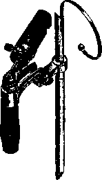
BRONCHOPNEUMONIA, see Pneumonia. BRONCHOSCOPY (from Greek bronchos- bronchus and scopeo- I look), or tracheo-bronchoscopy, is a direct method for examining the deep respiratory pathways. In the 1880s, Mikulicz (Miculicz) developed the method of esophagoscopy (see). Some surgeons, during esophagoscopy, accidentally penetrated with a rigid tube through the vocal cords and saw the trachea. But the founder of the direct method for examining the respiratory pathways is Kirstein, who in 1895 published his method of straightening the oropharyngeal arch with a spatulum to such an extent that it is possible to illuminate and see the entire trachea to the bifurcation. He did not dare to penetrate further with a rigid tube. Only Killian, after thorough study on cadavers, animals, and patients, proved that the bronchial branches are capable not only of moving but also of stretching in the transverse dimension to allow passage of rigid tubes of a larger caliber than the lumen of the bronchus, and with his method made accessible to the eye the entire bronchial tree up to branches of the second and third order. Together with his student Bruinings, Killian invented a simplified, world-renowned bronchoscopic set and necessary instrumentation, created a whole clinic for bronchoscopy, and placed in the hands of physicians a powerful, purely surgical method for examining and treating deep respiratory pathways. - Bronchoscopy is performed in a sitting or lying position, under general anesthesia or local anesthesia (or without any anesthesia), especially in early childhood, where the danger of poisoning from cocaine or chloroform is particularly great. The choice of one method or another depends on the patient, on the structure of his neck and jaw, on the development of reflexes, on the nature of the disease, etc. The bronchoscopic set consists of tubes of various calibers: for men, women, children, and infants; in addition, a tube for esophagoscopy is attached - the widest one. The tubes are double, telescopic, and are inserted into an electroscope (see Figure 1), on which is mounted a Kirstein light bulb with the modification that the filaments are crossed three times and at the point of crossing a strongly glowing point is obtained, the rays of which are collected into a parallel beam by a lens placed on the bulb. The light beam is caught by a flat mirror (with a slit cut in it), rotating on a frontal axis, and is thrown perpendicularly into the tube. Through the slit in the mirror, one can see a strongly illuminated field inside the trachea or bronchus. For manipulation of the instrument, the entire illuminator system is pulled out on a triangular rod from the handle for 10 cm almost without loss of light intensity. The tube is inserted through the mouth into the vocal cords and further into the trachea. In order not to remove the bronchoscope and not to insert a longer tube for penetration into the bronchus and its branches, an inner tube is pushed through the inserted outer tube with the help of a steel spring soldered to its upper end. Insertion of the bronchoscope through the vocal cords is called upper bronchoscopy, in contrast to lower bronchoscopy, in which bronchoscopy is performed through a tracheotomy wound. Lower bronchoscopy according to Bruinings is indicated: 1) in all patients who have previously undergone tracheotomy for one reason or another; 2) in all children under 3 and even 6 years of age; 3) in all severely debilitated patients and persons with pronounced dyspnea; 4) in all persons having foreign bodies in the bronchi, which, according to experience, are difficult or dangerous to remove; 5) in all cases where, despite persistent indications for bronchoscopy, for one reason or another, upper bronchoscopy has failed (inexperience, sharp reflex, etc.); 6) finally, in those cases when repeated examinations of the respiratory pathways are anticipated. In particular, in children, one must take into account when choosing a method that after upper bronchoscopy, edema of the epiglottis (subglottic swelling) occurs, which sometimes forces one to perform tracheotomy on the first or second day.
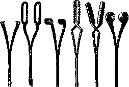
The surgical instrumentation is also constructed on the principle of telescopic tubes: forceps with screw-on tips (see Figures 2 and 3) - claw-shaped for removing solid foreign bodies, pointed with long teeth on the frame - for fruit seeds; with plates perpendicular to the long axis - for pins, needles; cutting - for removing tumors, etc., depending on the application. - Bronchoscopy laid the foundation for a new branch of knowledge, which can be called bronchology, by analogy with laryngology, the foundation of which was the laryngeal mirror (laryngoscopy). A whole clinic of trachea and bronchus pathology was created. In the very recent past, contrast liquids have been introduced through the bronchoscope for bronchography. Bronchoscopy serves for the scientific study of a number of physiological phenomena within the chest cavity. In the clinic of foreign bodies, bronchoscopy has taken first place and is a powerful and sole method for diagnosis and treatment. It is sufficient to mention that in the pre-bronchoscopy period, the mortality rate from aspirated foreign bodies was 40-50, and at present it has fallen to 7-8, with some authors reporting 4-5%.
Figure 2. Telescopic forceps according to Bruinings.
Figure 3. Tips for forceps.

Related articles
Mentioned in
Cite this page
“Bronchopneumonia.” Soviet Medical Encyclopedia. English translation of Bolshaya Meditsinskaya Entsiklopediya, 1st ed. (Moscow, 1928–1936), ed. N. A. Semashko. https://sovietmedicalencyclopedia.pages.dev/article/bronchopneumonia/