Trachea
Historical document, translated for reference. It reflects medical knowledge of the 1920s–30s and is not medical advice.
Summary
The trachea is the initial segment of the lower respiratory tract, a cylindrical tube consisting of 16-20 cartilaginous rings. The article covers its anatomy, developmental anomalies, inflammatory diseases like tuberculosis and syphilis, benign and malignant tumors, and various conditions causing tracheal stenosis.
Encyclopedia article (1928–1936)
Trachea (from Greek trachys-rough), the windpipe. - Anatomy. The trachea, being a direct continuation of the larynx, represents the initial segment of the lower respiratory tract in the form of a cylindrical tube 11-13 cm long from the larynx to the bifurcation; it consists of 16-20 tracheal cartilaginous (hyaline) rings connected by dense fibrous tissue (see figure). Posteriorly, the cartilaginous rings are incompletely closed, and the tracheal wall is formed here by connective tissue and smooth muscle. This part of the trachea throughout its length is adjacent to the esophagus. In the normal state in an adult, the trachea is located from C6 or D1 to D5 vertebra. The connective tissue surrounding the trachea appears loose and allows for large displacements during movements of the larynx and trachea. In its upper part, the trachea lies closer to the surface of the neck, and as it descends downward, it simultaneously deviates backward, being covered by a strongly developed mass of adipose and connective tissue, anterior neck muscles, superficial fascia, and skin. Therefore, incision of the upper and middle tracheal rings during tracheotomy is considered a simpler operation than that of the lower rings. The mucous membrane of the trachea has a similar structure to that of the larynx, differing from the latter by a greater amount of elastic fibers, which form a dense network with a predominance of longitudinally oriented fibers. The cilia of the ciliated epithelium have a direction of movement outwardward. The blood vessels, lymphatic vessels, and nerves of the trachea have the same relationships as in the larynx. Arteries of the trachea originate from a. thyreoidea inf. Developmental anomalies of the trachea may be in the form of fistulous communications with the esophagus (esophago-tracheal fistula), congenital diverticula of the tracheal walls, and more rarely occurring neck-tracheal fistulas, lateral or located along the midline. Lateral fistulas represent remnants of branchial clefts, while fistulas located along the midline are in most cases the result of failure of ductus thyreo-glossi to close. Treatment of tracheal fistulas consists of closing them by freshening the outer edges of the fistula. In more difficult cases, various methods of plastic surgery are resorted to. After operations, frequent recurrences of the disease are noted. - Inflammatory diseases of the trachea see Tracheitis. Tuberculosis of the trachea occurs in the form of superficial or deep ulcers, initially round, later of a potato-like shape with specific features of tuberculous ulcers. Hypertrophic productive forms of tbc and tuberculous tumors, as in the larynx, do not occur here. Due to the appearance of tbc in the trachea most often in the terminal period of the disease, it has no special clinical significance. - Syphilis of the trachea is observed significantly less frequently than laryngeal syphilis and represents phenomena of tertiary syphilis in the form of gummatous nodes and infiltrates, upon the disintegration and healing of which scars form, constricting and reducing the lumen of the trachea. Upon establishing the diagnosis, vigorous specific treatment is necessary to prevent subsequent constrictions of the trachea. Benign tumors of the trachea in many ways resemble similar tumors of the larynx, but occur much less frequently than the latter. Thus, according to the data of Jurasz, out of 42,635 cases of diseases of the upper respiratory tract, there were 2,088 cases of laryngeal tumors and only 3 cases of tracheal tumors. Bru'ns collected from the literature a total of 102 cases of tracheal tumors (from 1767 to 1890): papilloma-33 cases, fibroma-23 cases, chondroma and osteoma-29 cases, intratracheal goiter-7 cases, adenoma-5 cases, lipoma and lymphangioma-5 cases. The most common tumors are papillomas, which are often combined simultaneously with laryngeal papillomas. During life, when of small size, they cause little disturbance to patients and are often discovered only at autopsy. Fibromas of the trachea occur in the form of small tumors on a stalk or on a broader base. According to the same statistics of Jurasz, the literature contained 23 cases of such tracheal tumors. Lipomas and endotheliomas of the trachea are very rare phenomena. Chondromas originating from tracheal cartilages and osteomas of the trachea occur somewhat more frequently. A special type of tumor is the intratracheal goiter - struma intratrachealis, which occurs rarely and owes its origin to the ingrowth of an ordinary goiter into the lumen of the trachea or to a developmental anomaly of the thyroid gland that gives a small process into the trachea. In some cases, islands and bars of cartilaginous and bony tissue develop in the submucosal tissue of the trachea, forming dense protrusions on the mucous membrane. Sometimes these islands are connected to the cartilaginous rings of the trachea, and in other cases they lie freely within the mucous membrane of the trachea. The development of this peculiar disease, which is designated by the term tracheopathia chondro-osteoplastica, is attributed by some authors to congenital detachment of cartilaginous tissue, while others see in this the consequences of inflammatory processes. Similarly to the formation of air tumors of the larynx due to erosion of the thyroid cartilage and protrusion of the soft coverings with the formation of an air sac on the neck, air tumors of the trachea - tracheoceles - are formed. Clinical manifestations of tracheal tumors are insignificant in the initial stage of their development, but as the tumors grow and the lumen of the trachea decreases, the symptoms become more pronounced. In cases of impending unclear stenotic breathing (tracheostenosis), one should think of a tracheal tumor and perform careful laryngological and bronchoscopic examination of the patient. In some cases, the presence of a tumor causes attacks similar to asthma in patients; in such cases, in the differential diagnosis between a tracheal tumor and asthma, a blood test for eosinophilia may help, an increase of which above normal may indicate the presence of asthma, but not a tracheal tumor. - Treatment of all benign tumors of the trachea is surgical. The latter can be performed in two ways: intratracheally - through the natural upper respiratory tract and extratracheally - through incision of the trachea. The advantages of the first method are limited by the necessity of having special instrumentation and special technical training for these operations. This method can be considered particularly suitable for small tumors located on a stalk. On the contrary, large tumors located on a broad base may present significant difficulties when removing them through the natural respiratory pathways. - Of malignant primary tumors in the trachea, sarcoma and carcinoma occur. Sarcoma of the trachea has been described in the form of individual cases, carcinoma somewhat more frequently. In case of early diagnosis, excision of the affected area of the trachea is recommended. The greatest practical significance is attached to stenoses of the trachea (tracheostenoses), which are observed 1) with external compression of the trachea, 2) with diseases of the tracheal walls, and 3) with stenosing foreign bodies (see Foreign bodies). External compression of the trachea is most often caused by the presence of a goiter. The cylindrical lumen of the trachea in many cases is transformed into a slit-like canal. This results in compression of individual cartilaginous rings, their resorption, and sometimes also the ingrowth of the tumor into the trachea, especially in cases of malignant goiter. In some cases of far-advanced goiter, complete resorption of the tracheal rings occurs (tracheomalacia). The latter collapses and threatens the patient with suffocation. In such cases, the trachea is fixed with strong sutures to the underlying muscles, which restores the lumen of the trachea and eliminates the need for wearing a tracheotomy cannula. Treatment of the goiter as a cause of stenosis belongs to surgical measures (see Goiter) - it is only necessary to examine such patients radiologically before the operation, as well as to subject them to direct or indirect tracheoscopy to determine the degree of tracheal stenosis. Due to the close proximity of the aortic arch, its ascending and descending parts to the trachea and the left bronchus, aneurysms in this area can also lead to narrowing of the tracheal lumen. Of neoplasms of neighboring areas, which less frequently cause compression of the trachea and main bronchi, it is necessary to mention tumors of the mediastinum, carcinoma of the esophagus, and tuberculosis of paratracheal lymph nodes.
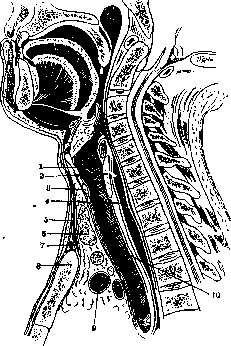
Sagittal section of the neck of an adult: 1- cartilago thyreoidea; 2-lamina cart, cricoideae; 3-arcus cart, cricoideae; 4-oesophagus; 5-m. sterno-hyoideus; 6-glandula thyreoidea; 7-spatium suprasternale; 8-manubrium sterni; 9-v. anonyma sin.; 10-trachea.
Treatment of all benign tumors of the trachea is surgical. The latter can be performed in two ways: intratracheally - through the natural upper respiratory tract and extratracheally - through incision of the trachea. The advantages of the first method are limited by the necessity of having special instrumentation and special technical training for these operations. This method can be considered particularly suitable for small tumors located on a stalk. On the contrary, large tumors located on a broad base may present significant difficulties when removing them through the natural respiratory pathways. - Of malignant primary tumors in the trachea, sarcoma and carcinoma occur. Sarcoma of the trachea has been described in the form of individual cases, carcinoma somewhat more frequently. In case of early diagnosis, excision of the affected area of the trachea is recommended. The greatest practical significance is attached to stenoses of the trachea (tracheostenoses), which are observed 1) with external compression of the trachea, 2) with diseases of the tracheal walls, and 3) with stenosing foreign bodies (see Foreign bodies). External compression of the trachea is most often caused by the presence of a goiter. The cylindrical lumen of the trachea in many cases is transformed into a slit-like canal. This results in compression of individual cartilaginous rings, their resorption, and sometimes also the ingrowth of the tumor into the trachea, especially in cases of malignant goiter. In some cases of far-advanced goiter, complete resorption of the tracheal rings occurs (tracheomalacia). The latter collapses and threatens the patient with suffocation. In such cases, the trachea is fixed with strong sutures to the underlying muscles, which restores the lumen of the trachea and eliminates the need for wearing a tracheotomy cannula. Treatment of the goiter as a cause of stenosis belongs to surgical measures (see Goiter) - it is only necessary to examine such patients radiologically before the operation, as well as to subject them to direct or indirect tracheoscopy to determine the degree of tracheal stenosis. Due to the close proximity of the aortic arch, its ascending and descending parts to the trachea and the left bronchus, aneurysms in this area can also lead to narrowing of the tracheal lumen. Of neoplasms of neighboring areas, which less frequently cause compression of the trachea and main bronchi, it is necessary to mention tumors of the mediastinum, carcinoma of the esophagus, and tuberculosis of paratracheal lymph nodes.
Related articles
Mentioned in
Cite this page
“Trachea.” Soviet Medical Encyclopedia. English translation of Bolshaya Meditsinskaya Entsiklopediya, 1st ed. (Moscow, 1928–1936), ed. N. A. Semashko. https://sovietmedicalencyclopedia.pages.dev/article/trachea/