Connective Tissue
Historical document, translated for reference. It reflects medical knowledge of the 1920s–30s and is not medical advice.
Summary
The article traces the evolving definition of connective tissue in histology from Müller's initial concept to broader interpretations including cartilage, bone, mucous and fatty tissues. It presents various classification systems based on macroscopic appearance, histological structure, and functional characteristics.
Encyclopedia article (1928–1936)
CONNECTIVE TISSUE. The definition of C. t. has repeatedly changed with the development of histology, in the sense of a continuous expansion of this concept, and at present several definitions exist alongside each other, reflecting the views of different periods. The name C. t. (Bindegewebe) as tissue connecting various organs was first introduced by I. Müller (I. Müller, 1835) for tissues consisting of fibers (tela cellulosa and tela fibrosa of older authors), which corresponds in modern terminology to fibrous C. t. In the 1840s and 1850s of the 19th century, with the further study of intercellular substances, as well as the ability of various tissues to replace and transform into each other, the concept of C. t. was expanded to include cartilage, bone, mucous and fatty tissue (Reichert, Kölliker, especially Virchow and Donders). From this time, C. t. began to be defined morphologically as tissue in which intercellular, ground substance predominates over cells; functionally-as primarily mechanical tissue: skeletal, supporting, structural; to this was usually added the indication of a common origin from the embryonic mesoderm. Such definitions are found in most manuals of the period closest to us (Ranvier, Orth, Schiefferdecker, Stöhr, Lavdovsky, Kulchitsky, Ognev), but the histological part of the definition does not withstand criticism due to the existence of supporting tissues of purely cellular character, why some authors consider it possible to characterize C. t. as a whole only from the side of its function (Kölliker, Schaffer). In recent years, the genetic principle-its origin from mesenchyme-has been placed at the basis of the definition of C. t., and on this basis, blood as well as all hematopoietic organs are classified as C. t. (Maximov and his school). In addition, in connection with the clarification of the role of cellular elements of C. t. in immunity and intermediate metabolism, it is necessary to expand its physiological characteristics, adding mechanical, biological and physicochemical functions to it. Thus, the modern concept of C. t. can be united under the name 'support-trophic' tissue (Zavarzin). There is also no generally accepted classification of C. t.: each author groups individual forms in his own way. Leaving aside blood and hematopoietic elements as a completely isolated group, connected with the rest of C. t. mainly topographically, one can (following Kölliker and Schaffer) put the following principles at the basis of a practically applicable classification. The first subdivision is based on the macroscopic appearance and physical properties: from the composition of C. t., solid skeletal tissues-cartilage and bone-are isolated, the remaining are united into a group of soft tissues-C. t. in the proper sense. The latter, taking into account its histological structure and functional features, can be divided into 2 main types: 1) tissue with intercellular substance of a fibrous nature-fibrous C. t. and 2) consisting of rounded elastic cells-cellular tissue. Fibrous tissue is subdivided on the basis of the nature of the predominant fibers and the associated functional features into 3 types: a) with predominance of collagenous fibers, b) elastic and c) reticular. Cellular tissue is divided according to the nature of cells into a) vesicular and b) fatty. An intermediate position between fibrous and cellular tissue is occupied by tissue with predominance of homogeneous intercellular substance: mucous, having an embryonic character, and jelly-like in invertebrates. It is self-evident that in view of the possibility of one type of C. t. transforming into another, any classification is conditional. The classification of C. t. can be presented in the form of a scheme (pp. 79-80). Fibrous C. t. The elements of fibrous C. t. are fibers, cells and amorphous ground substance. According to morphological, physical and chemical properties, fibers are divided into 3 types: collagenous, elastic, or elastic, and fibers of the third type-reticular. Collagenous fibers, swelling from weak acids, occur in the form of so-called collagenous bundles of varying and uniform thickness, composed of thin fibrils by means of a cementing substance. Elastic fibers, not changing from acids, can also have varying thickness, but, unlike collagenous, they branch and form networks. Reticular fibers (see Fibrous reticula) differ from them in not swelling from weak acid and in the ability to be impregnated with silver (argentophilia). They do not branch, but form networks, lying close to each other and then diverging. Between the fibers there is always a greater or lesser amount of amorphous intercellular substance of viscous consistency, sometimes giving a mucus reaction. This substance can in some cases harden, forming a dense basis of connective tissue plates. The cellular elements of fibrous tissue are diverse, they can be divided into permanent cells, wandering cells and variously specialized cells. To permanent cells belong fibrocytes (fibroblasts)-cells of developed tissue and fibroblasts-embryonic elements participating in the formation of fibers and transforming at the end of development into fibrocytes; they can also appear in developed tissue under the influence of various irritations and wounds. The name fibroblasts is often also applied to cells of developed normal tissue, which is facilitated by the custom of studying cellular elements of C. t. on inflamed objects or in life cultures. Fibrocytes (Fig. 1) are thin flat cells, usually lying on the surface of fiber bundles; in sections, only one nucleus is often visible from them, and to detect the body it is necessary to resort to gold plating or staining with iron hematoxylin. Their shape is extremely diverse, often they are provided with processes and plate-like outgrowths extending in different directions; they contain a nucleus with a characteristic fine-grained arrangement of chromatin and 1-2 nucleoli, in the depression of the nucleus the cell center is located. Fibroblasts differ by a large amount of protoplasm, fusiform or triangular body with long processes extending from it; during the functional period they form cellular chains and networks of syncytial character. Such networks are clearly visible in young reticular tissue (cytogenic tissue). Some authors (Moellendorf, Rubashkin) assert that such a syncytial arrangement remains in all types of fibrous tissue, forming a syncytium of fibrocytes. On wandering cells see Wandering cells, Leukocytes, Lymphocytes, Reticulo-endothelial apparatus. Close to wandering cells are also the mast cells of Ehrlich (Mastzellen), although their connection with blood elements is disputed. These cells resemble basophilic leukocytes (especially in rodents), i.e. contain a large number of grains, easily soluble in water and staining with basic dyes, often metachromatically (violet color from methylene blue). Their shape is round or irregular; in amphibians they are provided with long branching processes. Their origin and function are not clarified; it is assumed that they participate in the formation of intercellular mucus substance. To cells in a certain way specialized also belong fat cells (see Fatty tissue) and pigment cells. The latter appear colored in various colors due to the presence of small grains or crystals of pigment, most often yellow-brown, brown or black. Pigment cells of C. t. occur in two types. First, in the form of cells resembling wandering cells, in particular histiocytes, with round or oval body and more or less long, sometimes branching processes; they undoubtedly possess the ability to move and are found in the skin near pigmented areas of epithelium, hair or feathers, serving for the transport of pigment into them (pigmentophages). Whether they are at the same time melanoblasts, i.e. cells producing pigment, remains unclarified. They should be distinguished from macrophages-histiocytes loaded with clumps of blood pigment (near foci of hemorrhage, in the spleen). Second, pigment cells giving color to C. t. itself, i.e. chromatophores; in humans and higher vertebrates they occur only in the vascular coat of the eye, in fish, amphibians and reptiles-in the skin and coats of internal organs. These cells most often have the appearance of plates with outgrowths or cut edges, sometimes star-shaped form, and reach considerable size.
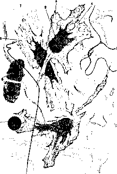
Figure 1. Thin film from subcutaneous connective tissue of a rabbit: 1-collagenous bundles; 2-endoplasm of fibroblast (fibrocyte); 3-ectoplasm; 4-fibroblast; 5-elastic fibers; 6-wandering cell at rest.
By color and nature of pigment, chromatophores are distinguished: black ones—melanophores, yellow ones—xantho- or lipophores, red ones—erythrophores. There are also cells containing guanine crystals, which have a blue-green iridescent color—guanophores (tapetum of the eye). Chromatophores possess the ability, by contracting their processes and moving the pigment to the center, to reduce the colored surface of the cell; mass contraction of cells leads to a change in skin color. It has been proven that the contraction of chromatophores depends on the nervous system, and in fish, nerve endings have been found on them. A. Fibrous connective tissue with predominance of collagen bundles (bundle) is the most common type of soft connective tissue; when simply referring to connective tissue, it is this that is meant. It can be divided into several types, not always sharply demarcated from each other; existing classifications often differ significantly in details, but agree on the main division: namely, fibrous tissue is always divided into a) loose and b) dense. a) Loose fibrous tissue (amorphous tissue, interstitial, interstitial tissue) constitutes what is called cellular tissue (see), filling the spaces between organs and allowing them to move easily during movements; it is located under the skin, between muscles, envelops the internal organs of the neck, chest, retroperitoneal space, pelvis, and accompanies blood vessels to their finest branches. Its main mass consists of collagen bundles of varying thickness, going in all directions and interwoven with each other; they form convolutions, are loosely arranged, and can easily shift; between them go networks of elastic fibers, usually very thin. Elastic fibers can be detected by treating fresh tissue with weak acetic acid, from which the collagen bundles swell and become barely visible (fig. 2). At the beginning of swelling, the bundles acquire a clearly defined character due to the presence of special wrapping fibers that resist the action of the acid, in the form of rings or spirals. The nature of these fibers has not yet been clarified: earlier they were considered elastic, then reticular; at the same time, the opinion is expressed that there are no fibers at all, but the bundles are clothed in a special sheath of amorphous substance, which bursts during swelling and forms constrictions simulating fibers. The collagen bundles are connected by amorphous adhesive substance of a mucous nature, which quickly dries in the air and allows fresh tissue to be stretched on a slide for preparing a specimen. Bundles interwoven in one plane can fuse with amorphous substance into thin plates, which is clearly noticeable in certain types of tissue (e.g., in the subcutaneous tissue of the back in rats); tissue fluid circulates between the plates. The existence of plates is also indicated by the results of interstitial injection of fluid or air insufflation, as a result of which bubbles with thin walls are formed. Therefore, French authors sometimes call loose tissue lamellar tissue (tissu lamellaire). Among the cellular elements in the tissue there are fibrocytes, wandering cells, histiocytes, fat cells individually or in groups; loose tissue is also a repository for adipose tissue, which was formerly classified as fibrous on this basis. The easy stretchability and mobility of the tissue depends exclusively on the straightening of bends and the change in arrangement of collagen bundles, since they can be considered inextensible under normal conditions of the body; conversely, elastic networks easily stretch and, due to their elasticity, return the tissue to its previous position. b) Dense fibrous tissue consists of a dense interweaving of collagen bundles, closely adjacent to each other and connected by a small amount of amorphous adhesive substance. Elastic networks are present almost always, but in varying amounts depending on the organs. The cellular elements are the same as in loose tissue, only the number of wandering cells is less; fat cells are rarely encountered, pigment cells in certain types of tissue—in large quantities (pigment tissue). For the study of dense connective tissue, besides the usual staining methods, gilding and silvering have been used for a long time. The first reveals cellular elements, mainly fibrocytes with all their processes and plate-like outgrowths, and gives especially clear pictures on the cornea. Silver impregnation stains the intercellular fibrous mass yellow-brown, and against this background stands out a negative picture of the cellular elements with the spaces surrounding them, known as the canals of Recklinghausen. These are places through which tissue fluid circulates and which were formerly considered the beginning of the lymphatic system. The function of dense tissue is predominantly mechanical, and in each organ the arrangement of bundles and the amount of elastic tissue determine it. Where the tissue serves to transmit movement (in tendons) and should not stretch, the bundles go in one direction—along the line of tension; where the tissue serves as a covering and should have greater stretchability, the bundles are arranged in a plane, intersecting at various angles, and there is a greater or lesser amount of elastic tissue. Recent data force us to recognize another function for the fibrous mass—physico-chemical. It is determined by the reaction of the tissue (normally weakly alkaline) and the large surface area formed by the bundles. Therefore, an acidic dye introduced into the body (trypan blue) is first adsorbed by the fibrous mass, intensely staining it, and then is absorbed by histiocytes; it also retains water and salts (NaCl) in varying amounts and thus plays a major role in water and salt exchange. According to the thickness of the layers, density, arrangement of bundles, and other features, dense tissue presents a series of modifications characteristic of different organs. 1) The main tissue of the skin (fig. 3)—see Skin. 2) Organ coverings. The bundles of connective tissue are arranged in one plane in different directions, sometimes forming plate-like layers; they can be cut obliquely by bundles serving to connect the plates. This includes first of all the thick and very dense fibrous coverings (t. fibrosae) of the eye: the cornea with very regular arrangement of bundles and the sclera with a dense interweaving of fibers, t. albuginea of the testis, dura mater; further, the capsules of parenchymal organs that allow greater stretching—liver, kidney, spleen; pia mater, outer covering of blood vessels; coverings of parts of the skeleton: perichondrium and periosteum; finally fasciae, aponeuroses, and the proper coverings of muscles (perimysium). Ranvier isolates the coverings of nerve fibers into a special type: plate-like, or sheath tissue, because they can be easily separated into thin plates; they consist of thin fibers, collagen and elastic, fused by dense amorphous substance; such plates are also present in the vascular coat of the eye. 3) Mesenteries, to which belong the mesentery, greater omentum, and ligaments of internal organs. In the mesentery, the bundles interweave more loosely and are connected into a plate by a large amount of ground substance, in which some authors describe a dense network of very thin fibers, probably precollagen. In ligaments (e.g., hepatic, according to Ranvier), the bundles go mainly in one direction, and there are many elastic fibers. 4) Tendons, aponeuroses, and ligaments consist of bundles running parallel to each other in one direction (fig. 4) (see Tendon).
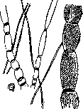
Figure 2. Swelling of bundles in subcutaneous connective tissue.
Development of fibrous connective tissue. The starting point is mesenchyme, which arises at early stages of embryonic development from the sclerotomes of mesodermal somites and consists of rounded or process-bearing cells, freely moving in the fluid surrounding them; filling the spaces between organs, it gives rise to loose interstitial tissue. The first phase of such development is most often described as mucous tissue, and recently as mesenchymal syncytium: cells connect by processes and secrete intermediate mucous substance (Renaut, Laguesse, Maximov) (fig. 5). But there is another view (Karpov), according to which mesenchymal cells, swelling and accumulating mucous substance in themselves, turn into vesicles with a nucleus pushed to the periphery of the cell; such vesicular cells can be isolated, which was done by Schwann, who also described and depicted them. Sections of vesicular tissue give pictures of anastomosing cells. The walls of the vesicular cells then break down, their contents merge into a common mass, and the protoplasmic areas with nuclei initially have the appearance of curved plates, then grow, giving rise to fibroblasts; at this period one can speak of mucous tissue. The development of dense connective tissue occurs later around organs, by
Figure 3. Dense connective tissue of human skin.
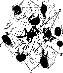
Figure 4. Mesenchymal connective tissue of a human embryo 4½ months: 1-fibroblasts; 2-histioid wandering cells; 3-collagen bundles.
Figure 5. Mesenchyme of a human embryo 15 mm.

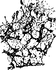
cytium. (After Laguesse.) direct transformation of mesenchymal cells into spindle-shaped fibroblasts arranged in parallel rows. As for the origin of collagen bundles and fibrils, there have been two views on this question since Schwann's time. According to one (Schwann, Boll, Lvov, Flemming), the fibers arise in the peripheral layers of the protoplasm and then detach; according to the other, the fibers develop independently of the cells in the amorphous ground substance (Henle, Kolliker, Ranvier, Ebner). These views are now being brought into unity by proving that the ground substance is nothing other than the ectoplasm of fibroblasts separated from the cells (Mall, Studnicka, Hansen). It is certain, however, that the fibers develop with the closest participation of cells, and newly formed bundles lie next to the cell, repeating all its bends; facts indicating the appearance of fibers in acellular areas (ground substance of the cornea, chorda sheaths) require revision, as obtained from sections. B. Elastic tissue-see Elastic tissue. C. Reticular tissue-see Adenoid tissue, Fiber lattices, Reticulo-endothelial apparatus. Cellular types of connective tissue are formed by the accumulation of cells of considerable size, round in shape, provided with a shell and containing fluid. The nucleus with the surrounding protoplasm usually lies against the wall, and protoplasmic strands and partitions can extend through the cell cavity. In general, the cell represents a bubble possessing a certain degree of turgor. Tissues consisting of such cells have a dual significance: on the one hand, by filling the spaces between organs, they give them a definite shape and contribute to the formation of the body; on the other hand, they counteract pressure by distributing it over a larger surface according to the laws of hydrostatics. In this respect, cellular tissue forms a transition to cartilage, the young forms of which, especially in lower vertebrates, have a cellular character. a) Bubble tissue (blasiges Stiltzgewebe) is widely distributed in invertebrates (hydrozoans, flatworms, crustaceans, mollusks), where it has a supporting function, forming the so-called parenchyma of the body. In vertebrates, bubble tissue represents the embryonic phase of the development of interstitial connective tissue and is found in all classes, but after development is completed, it is preserved in a few places in fish and amphibians (supporting tissue of the sand lance, circular ligament of the eye in fish). The transition to cartilage is represented by the dense sesamoid nodule of the frog's Achilles tendon, in which there are thin layers of fibrous tissue. To bubble tissue should also be attributed the tissue of the notochord, which passes through an epithelial stage in its development and consists of large cells with a dense shell, filled with fluid. b) Fatty tissue (see). Connective tissue with a large amount of homogeneous intercellular substance, mucous and gelatinous, is, like cellular tissue, a filling and supporting tissue. a) Mucous tissue is found in the vertebrate class only in embryos, and therefore is sometimes called embryonic. Loose fibrous tissue passes through the phase of mucous tissue in its development, but it reaches special development in the embryonic organ sheaths and umbilical cord (Wharton's jelly),
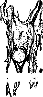
Fig. 6. Gelatinous connective tissue from the human umbilical cord (from Petersen).
where it is usually studied (fig. 6). It is usually described as consisting of spindle-shaped and stellate cells and intercellular substance that gives mucous reactions. Such a picture is obtained only in umbilical cords of a certain age: at the end of pregnancy, a large number of fibers and fiber plates develop; on the other hand, at the initial stages, a large number of bubble cells are found, which are the producers of mucus. b) Gelatinous tissue is found in low-organized invertebrate forms (in the bell of jellyfish, in sponges); it consists of an amorphous gelatinous mass in which cells with mesenchymal-type processes are embedded. The method of origin of the ground substance has not been clarified.
V. Karpov. Pathological anatomy. Connective tissue, together with its vascular apparatus, participates in almost all pathological processes developing in any part of the body, in any of its organs. In addition, independent pathological changes also occur in connective tissue. In particular, it is now established that a number of diseases (scurvy, rheumatism, etc.) have as their basis pathological changes in connective tissue, resp. mesenchyme and its derivatives. At the same time, it must be admitted that far from all violations of the normal physiological state of connective tissue (e.g., violations of salt and other exchange in it) have any definite and clear morphological expression; our methods of studying connective tissue for this are still not sufficiently perfected.
Related articles
Mentioned in
Cite this page
“Connective Tissue.” Soviet Medical Encyclopedia. English translation of Bolshaya Meditsinskaya Entsiklopediya, 1st ed. (Moscow, 1928–1936), ed. N. A. Semashko. https://sovietmedicalencyclopedia.pages.dev/article/connective-tissue/