Bone
Historical document, translated for reference. It reflects medical knowledge of the 1920s–30s and is not medical advice.
Summary
This article from the 1928–1936 Soviet Great Medical Encyclopedia details the histology and embryology of bone, describing its tissue structure, cellular components, Haversian systems, and trabecular architecture.
Encyclopedia article (1928–1936)
BONE. Contents: I. Histology and embryology..........130 II. Pathology of bone...............ш III. Clinic of bone diseases.........153 IV. Operations on bones..............Юб I. Histology and embryology. The B. of higher vertebrates consists of osseous substance or bone tissue, periosteum, bone marrow, blood and lymphatic vessels, and nerves. Bone tissue belongs to the group of fibrous connective tissue and differs from other types by its hardness, which depends on the presence of a large amount of mineral salts (2/3 by weight). The salts include calcium phosphate (85%), calcium carbonate (10%), magnesium phosphate (1.5%), and calcium fluoride (0.3%) (Oppenheimer). Under the microscope, one can distinguish the ground or intermediate substance and bone cells in bone tissue (Fig. 1). The ground substance appears in most cases transparent and homogeneous, but by decalcifying B. in 5% hydrochloric or 5% nitric acid with the addition of 10% NaCl, its composition of fine fibrils assembled into bundles can be revealed (Fig. 2);
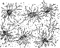
Figure 1. Bone lacunae with canaliculi from the parietal bone (after Kölliker).
they are ordinary collagenous fibers (see Collagenous substances, fibers) and upon boiling yield bone glue (ossein). Mineral salts are deposited in the spaces between the fibers, possibly entering into a compound with the cementing substance, which, according to most researchers, is located between the fibrils (osseomucoid). Upon boiling B. in a sealed tube, it is possible to dissolve the fibers, but B. retains its structure, and in dry ground sections
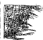
Figure 2. Fibrous bundles
of the bone ground substance (after Ebner), fine tubules filled with air can be seen in place of the fibers (Ebner). Bone cells have the shape of a plum stone or cucumber seed, 22–52 µm in length, 6–14 µm in width, 4–9 µm in thickness (Kölliker) and are provided with a large number of processes extending from both the edges and surfaces—thin (1.1–1.8 µm), long, branching, and anastomosing with the processes of neighboring cells. The cells lie in lacunae hollowed out in the ground substance, which precisely reflect their shape with all processes; in dry ground sections, the cells disappear, while the lacunae become filled with air and appear black (due to total internal reflection); they can also be filled with an alcoholic dye solution (aniline blue, fuchsin). By acting on the bone with strong acids, one can isolate the walls of the lacunae, which differ in composition from the ground substance; they take on the appearance of branched cells, hollow inside, and were considered by old authors to be cells (bone corpuscles; Virchow). By its texture, bone tissue is divided into two types: coarse-fibered and fine-fibered or lamellar (Kölliker). The first is characterized by an irregular interweaving of collagenous bundles, between which spaces remain for cells and blood vessels; it forms the B. of embryos, and in adults is found rarely (at the sites of tendon attachment). In lamellar tissue, on the contrary, fine bundles are arranged very regularly, in parallel rows, and form thin lamellae, 4.5–11 µm in thickness; cells are arranged in rows between the lamellae, and their processes extending from the surfaces penetrate the lamellae throughout their entire thickness. Lamellae and their systems are the main architectural element from which the B. of higher vertebrates is formed in the developed state. On bone cross-sections, one can distinguish with the naked eye osseous substance of two kinds: compact (substantia compacta), having the appearance of a solid homogeneous mass, and spongy (subst. spongiosa), consisting of thin trabeculae intersecting in various directions and delimiting wide cavities in which the bone marrow is housed. Compact substance is located on the surface of all B.: it reaches significant thickness in the diaphyses of long B., is thinner in the lamellae of flat bones, and invests short B. with a very thin layer. Its structure is usually studied on thin transverse (Fig. 3) and longitudinal sections of diaphyses (Fig. 4), where all systems of lamellae are represented (slices of decalcified bones in 10% HCl can also be examined). Each system consists of a greater or smaller number of lamellae closely adjacent to each other; bone cells are located between the lamellae, the processes of which pierce the lamellae and connect with the processes of other cells, as a result of which the entire system is permeated by a connected network.
Figure 3. Cross section of a human metacarpal bone: 1—outer general system of lamellae; 2—Haversian systems; 3—interstitial systems; 4—inner general system (after Kölliker). Bone cavities and channels through which nutrient substances penetrate. Located on the surface of B. is the system of outer general (general) lamellae, encompassing the diaphysis in the form of a tube of large diameter; the same tube of smaller diameter forms the inner surface of the diaphysis, delimiting the bone marrow canal—the system of inner general lamellae. The main mass of B.
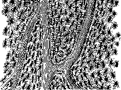
Fig. 4. Longitudinal ground section of the human femoral diaphysis. between these two systems is filled with systems of lamellae rolled into tubes of small diameter that surround cylindrical channels carrying vessels—Haversian canals; these systems are called Haversian, special systems, or the so-called osteons (Biedermann). Haversian systems are located along the length of B.; only at the sites of anastomoses of Haversian canals do they run obliquely or transversely. The gaps remaining in various places between Haversian systems are filled with parallel-running interstitial systems. The boundaries between individual systems appear on ground sections as cementing lines. On transverse sections, all the described systems are clearly visible because the Haversian systems are cut across; on longitudinal sections, a more uniform picture of lamellae running lengthwise is obtained. Examination at high magnifications of ground sections embedded in thick balsam or sections in 10% NaCl reveals the different structure of adjacent lamellae in all systems: some are punctate, others striated, with both types regularly alternating; in polarized light, the punctate ones appear black, while the striated ones are more or less bright [see separate plate (pp. 115–116), Fig. 5]. This phenomenon depends on the different arrangement of fibrous bundles in adjacent lamellae, which in one case are cut transversely, and in another lengthwise or obliquely. The initial assumption that fibers are located in adjacent lamellae at right angles was not confirmed. Studies by Gebhardt showed that in Haversian systems fibers can intersect at various angles and have various inclinations to the axis of the system (Fig. 5). In addition to the listed formations, the compact substance includes Sharpey's fibers and Volkmann's canals. Sharpey's fibers [see separate plate (pp. 115–116), Fig. 6] represent unossified fibrous bundles extending from the periosteum and penetrating into B. perpendicular to its surface; they consist mainly of collagenous fibers, but may also contain elastic ones; they are found only in outer general and interstitial systems. Volkmann's canals also extend from the periosteum and pierce various systems of lamellae without having their own; they contain blood vessels establishing communication between the periosteum and Haversian systems. The compact substance of flat, short, and epiphyseal long B. has varying thickness and consists of outer general and Haversian systems; it passes directly into the spongy substance, which forms the main mass of the listed B. The trabeculae of spongy substance consist of a varying number of bone lamellae layered on top of each other; they are relatively thin and delimit wide cavities containing blood vessels and bone marrow. According to the arrangement of lamellae, three main types of spongy substance can be distinguished (W. Roux): consisting of tubes, plates, and trabeculae. The first type (spongiosa tubulosa completa) is formed of wide tubes, osteons running in one direction (femoral head, sternal end of the clavicle) [see separate plate (pp. 115–116), Fig. 4]. The second (spongiosa lamellosa) represents plates arranged in the direction of trajectories and connected by transverse plates or bridges (femoral neck). The third type (spongiosa trabeculosa) consists of trabeculae, less wide than the plates, forming the fine framework of the vertebral bodies [see separate table (pp. 115–116), Fig. 3].—The entire architectonics of B., their compact and spongy substances, as well as the arrangement of fibers in bone
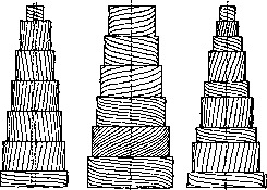
Figure 5. Various types of arrangement of fibrous bundles in bone lamellae (after Gebhardt).
plates, in each given case responds to those mechanical demands that are placed upon the bone during its function (resistance to bending, pressure, tension). The principles of construction of the bone mass correspond fully to mathematical calculations that are placed at the basis of construction structures (e.g., bridges, cranes), and impart to the bone the greatest strength and stability with the least expenditure of material. The periosteum, covering the bone from the outer surface, represents a connective-tissue membrane of varying thickness and density, in which 2 layers are distinguished [see separate plate (pp. 115-116), Fig. 6]: the outer fibrous, consisting of bundles of collagenous fibers and containing a large number of blood vessels, and the inner, in which there are dense networks of elastic fibers and few vessels. In young bones the inner layer contains a large number of osteoblasts, which is why it is sometimes called the cambial layer (blasteme sous-periostale Ollier). The periosteum is connected to the bone by means of Sharpey's fibers, which proceed from the fibrous layer and embed themselves in the bone, and blood vessels. The latter are directed from the periosteum into Volkmann's canals and penetrate from here into the network of Haversian canals, where they break up into capillaries and partly penetrate into the bone marrow; venous trunks also pass through the Haversian canals (in which 2 vessels can often be seen) and exit through Volkmann's canals. Along with this, larger arteries (vasa nutritia) penetrate into the bone through special openings, which are directed into the bone marrow, giving off branches along the way into the bone substance. Lymphatic vessels ramify chiefly in the outer layer of the periosteum; a number of authors described lymphatic perivascular spaces in the Haversian canals, the endothelium of which lies adjacent to the bony wall of the canal. All bones (with the exception of the auditory and sesamoid ones) are abundantly supplied with nerves (Kölliker). In addition to the nerves of the periosteum itself, a large number of myelinated and unmyelinated fibers ramify in the Haversian canals, accompanying the vessel ramifications. Their endings have not been studied; Pacinian corpuscles were found in the periosteum of the vertebrae and epiphyses of long bones. Development and growth of bone. In the embryo, before the appearance of bone tissue, parts of the skeleton are preformed either by connective tissue (bones of the cranial vault, clavicle) or by cartilage (bones of the skull base, spine, limbs). Therefore, a distinction is made between the formation of bone in place of connective-tissue and cartilaginous primordia; essentially it proceeds identically in both cases, but in the cartilaginous primordium the matter is complicated by the simultaneously proceeding process of cartilage destruction. The development of bone in place of connective tissue begins with the fact that at a known place of the primordium an accumulation of special cells--bone-formers, osteoblasts (Gegenbaur)--is formed; they differ from other mesenchymal cells by their size (20-30 µ), angular or cylindrical shape, basophilic protoplasm; mitochondria and various inclusions have been described in their bodies; like other mesenchymal cells, they are provided with thin processes that can connect with the processes of neighboring cells [see separate plate (vol. XIII, pp. 723-724), Fig. 3]. Between the osteoblasts, intermediate substance soon appears, which pushes them apart--the ground substance of the bone (osteoid); part of the osteoblasts turns out to be enclosed in this substance and transforms into process-bearing bone cells, while the remaining

Figure 6. Isocenter of ossification of the parietal bone in a human embryo of 14 weeks (according to Kölliker).
are arranged in rows along its surface. The osteoid substance exhibits a fibrillar structure, stains with acidic stains, and is anisotropic. The very process of its origin cannot be considered fully elucidated. According to the view of some, part of the osteoblasts function as fibroblasts and form collagenous fibers in the usual way, or else fibroblasts are present among the osteoblasts from the very beginning; according to others, the ground substance is formed at the expense of the modified protoplasm of the osteoblasts and is at first uniform or reticular, and then loosens. The second phase of ossification consists in the impregnation of the osteoid substance with lime salts; previously, calcareous salts accumulate in the protoplasm of the osteoblasts in the form of small grains. Ossification proceeds not as a solid mass, but in separate trabeculae that connect with each other like a network (Fig. 6); having begun in one place of the connective-tissue primordium in the form of an islet, it spreads to the periphery. The development of bone in place of cartilage is a more complex process. The cartilaginous primordium of each bone is clothed with embryonic perichondrium, which then transforms into the periosteum; in its deep layers, osteoblasts differentiate, arranged along the surface of the cartilage; they begin to produce bone substance in the form of plates wrapping around the entire surface of the cartilage, and then arrange themselves around the vessels, giving rise to Haversian systems. This is perichondral, periosteal ossification, which forms compact substance [see separate plate (pp. 115-116), Fig. 2]. Along with it proceeds the process of intracartilaginous, enchondral or endochondral ossification, which begins in certain places of the cartilaginous primordium, forming the so-called points of ossification, clearly visible to the naked eye on sections in the form of white opaque islets. In short bones there is usually one point of ossification, in long bones--one in the diaphysis and one in each epiphysis. In the points of ossification, the following changes play out in a regular sequence [see separate plate (vol. XIII, pp. 723-724), Fig. 2]: 1) multiplication of cartilage cells with their subsequent swelling and deposition of lime in the ground substance of the cartilage (stage of calcification); 2) ingrowth of blood vessels from the periphery into the calcified section, as a result of which the cartilage is destroyed and narrow trabeculae of the ground substance remain from it (see Vascularization); 3) formation of young bone substance on the surface of the cartilage trabeculae by osteoblasts that have penetrated along with the vessels. These processes, having begun in the center of the islet, spread wave-like to the periphery, wherein behind the zone of calcification in the growing cartilaginous primordia lies the zone of multiplication of cartilage cells. In tubular bones, ossification, having begun in the middle, proceeds in both directions, reaching the epiphyses, and borders on them with a straight line (line of ossification). This place is especially convenient for studying the details of the process [see separate plate (vol. XIII, pp. 723-724), Fig. 1]. Bordering on the epiphysis is the zone of multiplication of cartilage cells, which, due to lack of space, flatten out and stack into columns; grains of lime already begin to be deposited in them; lower goes the zone of swelling of cartilage cells, their regressive changes and impregnation of the ground substance trabeculae with calcareous salts; still lower--the ingrowth of narrow vascular loops that destroy the cartilage along the line of the columns; the capsules open, the cells perish. The remaining cartilage trabeculae with eroded scalloped edges are covered with osteoid tissue, upon which osteoblasts sit in rows. Endochondral ossification gives rise to spongy substance; the cell mass with vessels remaining between the trabeculae forms the embryonic bone marrow. According to the generally accepted view, the cartilage of the embryonic skeleton only gives the shape of the future bone and is entirely destroyed, but in some places (clavicle, lower jaw) a direct transition of calcified cartilage into bone (metaplasia) can apparently occur with the transformation of cartilage cells into osteoblasts (N. Müller, Streltsov, Kashchenko, Deyneka). During the process of ossification, the forming bone continues to grow in thickness and length; this occurs not only by the imposition of new layers under the periosteum, but also by the further proliferation of cartilage with subsequent endochondral ossification. Between the ossification of the epiphysis and diaphysis remains a strip of unchanged, growing cartilage (epiphyseal cartilage; Fig. 7); along both sides of it go the zones of calcification and vascularization. The bone tube of the diaphysis, formed under the periosteum, with its pointed end extends to the epiphyseal cartilage and ends here, while the periosteum merges with the cartilage; this place, marked on the surface of the bone by a groove (encoche d'ossification of Ranvier), serves for the growth of the periosteum and periosteal ossification. The bone formed in embryonic life bears a coarse-fibered character; Haversian canals have the appearance of wide cavities of irregular shape, and there is also no regularity in the distribution of plates. The final appearance of the bones is acquired after prolonged restructuring, especially intensive when they begin to function after birth (functional adaptation). Restructuring is associated with the destruction of old plates and trabeculae and the formation on
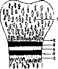
Figure 7. Diagram of the arrangement of ossification lines in the epiphysis of a tubular bone: 1-ossified head of the bone; 4- epiphyseal cartilage, to which the zones of calcification (?) and vascularization adjoin from both sides

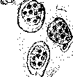
place of their new ones; the latter process occurs in the usual manner with the participation of osteoblasts, while destruction is accomplished by means of "lacunar resorption" with the help of special giant cells—osteoblasts (ostoclasts) (Kölliker) or myeloplaxes (Robin). These are giant cells (43–91 µ in length, 30–40 µ in width, 16–17 µ in thickness) containing 5 to 10, 20, and up to 60 nuclei; mitochondria, vacuoles, and grains of various kinds have been described in their cytoplasm. They appear starting from the earliest stages of ossification and also take part in the destruction of cartilage (chondroclasts). The origin of osteoclasts is not entirely clear; it is assumed that they arise through the fusion of osteoblasts. Osteoclasts adhere to the surface of the bone, which is resorbed at this site in such a way that they appear to lie in pits that become deeper and deeper (Howship's lacunae; Fig. 8). On their surface facing the bone, a border is formed composed of fine rods (like on the epithelium of the small intestine)—an adaptation for absorption; the dissolution of bone tissue occurs, as is assumed, by the secretion of acids or enzymes. During the restructuring of spongy substance, some trabeculae are completely resorbed, while new ones are formed in their place, running along the lines of tension and pressure (trajectories); in the compact substance within wide Haversian spaces, tubular systems are formed that subsequently undergo repeated restructuring [see separate plate (Vol. XIII, pp. 723–724), Fig. 4], sometimes seemingly moving through the thickness of the bone. At the height of restructuring, the compact substance has a mottled appearance: it is composed of remnants of systems running in different directions (fragmentary structure, breccia; Ebner). This character persists in the human femur and humerus throughout life; the bones of the forearm and leg acquire a more regular structure; the systems in the bones of ungulates have the most regular structure.
Comparative histology. Kölliker distinguishes 4 types of bone tissue in vertebrates: 1) true bone with bone cells in mammals, birds, reptiles, amphibians, and some fish; the difference lies in the shape of the cells, which in fish are often spindle-shaped; 2) bone tissue with cells lying on the outside and canaliculi into which processes penetrate—dentin (in acanthopterygian fish); 3) with canaliculi and cells simultaneously—osteodentin (bone and scales of ganoids); 4) osteoid substance without cells and canaliculi (scales, bones of acanthopterygians). The structure of bone in fish is not infrequently complex: among the bone trabeculae there is a homogeneous gelatinous substance, sometimes calcified. In the bones of other lower vertebrates, coarse-fibered bone tissue is found; Haversian canals are often absent. In birds, ossification of tendons occurs not infrequently with the calcification of fibrous bundles and the transition of connective tissue cells into bone cells.
V. Karpov. Microscopic investigation of bone. In microscopic technique, the following are investigated: 1) fresh bone tissue, 2) in ground sections, and 3) in sections of decalcified bone (see Decalcification), with its various components revealed by special staining.—1. Thin trabeculae of spongy bones are broken off with forceps, teased apart with a needle, and thin skull bones (nasal conchae, etc.) of small mammals (e.g., mice, moles, etc.), covering skull bones of fish or amphibians are examined directly under the microscope in indifferent fluids, but not in glycerin.—2. Thin plates in transverse and longitudinal directions are sawn out of compact bones, and ground sections are prepared from dried, fixed, macerated, or untreated bone. Preparation of ground sections: a bone plate 1–2 mm thick is first ground down with a flat file, then glued with melted Canada balsam to a slide, ground on a white grinding stone, and finally polished on frosted glass with provencal oil. Grinding powders (tripoli, emery) are best avoided, as they are difficult to wash out of the preparation. The finished ground section is washed in xylene and then transferred directly to liquid Canada balsam or dried in a thermostat and mounted in melted Canada balsam or directly under a coverslip. In the latter case, the cavities in the bone substance remain filled with air and thus stand out sharply against the rest of the mass. Before polishing, ground sections can be impregnated in the dark with silver nitrate (1%) for 24 hours, after which the silver is reduced in the light, and the section is dried, polished, and mounted in Canada balsam.—3. Sections from decalcified, washed, and celloidin-embedded pieces of bone are subjected to various stains depending on the need. For a general overview of bone structure, preparations are strongly overstained with hematoxylin (preferably Delafield's) and counterstained with eosin. To investigate bone cells and canaliculi, sections are stained according to Schmorl with thionin-picric acid. Frozen or celloidin sections are placed in water for 10 minutes, then transferred to Nicol's carbol-thionin for 10 minutes. Rinsing in water, staining in a saturated aqueous solution of picric acid. Differentiation in 70° alcohol; mounting in Canada balsam. Bone cells are red, the ground substance is light brown, and bone lacunae are dark brown. Alternatively, the so-called "panoptic stain" of bone tissue according to Schmorl is used. Frozen or celloidin sections are stained for 3 minutes with Nicol's thionin with the addition of ammonia, washed in water, and transferred for 1–2 minutes to 70° alcohol, then to distilled water, and rapidly differentiated in a concentrated solution of phosphomolybdic or phosphotungstic acid. The stain is fixed in diluted formalin or ammonia (1:10) for 5 minutes, transferred directly to 96° alcohol, and mounted in Canada balsam. Bone lacunae and their processes are intensely blue-black, the protoplasm and nuclei of bone cells are diffusely blue, the ground substance is light blue (ranging to reddish or purplish-red), and lamellae and interstitial substance stand out clearly.—The fibrillar structure and lamellae according to Weidenreich stand out well when processed by Weigert's method. Sharpey's fibers are also clearly visible when processed by Weigert's method, but can also be detected by the methods of Bělýnovský, Achúcarro-Ranke, and others. The investigation of bone development by vital staining was first proposed by Mizaldus (Mizaud) in 1599 and described as a new method in 1736 (Belchier). If animals (chickens, pigeons, sheep, pigs, rats, rabbits) are fed for a long time with madder root powder (Rubia tinctoria) containing alizarin, certain parts of the bone are in most cases, but not always, stained red. By making ground sections of such vitally stained bone, its development can be traced. Gottlieb (1914) for this purpose injects 12 cm3 of a 1% solution of sodium alizarin sulfonate into the ear vein of a rabbit and after a few minutes achieves a red-violet coloration of the skeleton. The same dye is injected subcutaneously into a rat for 6 weeks, 2 cm3 every week,—2 weeks after the last injection, the skeleton is stained red. Retterer stains bone by feeding animals various dyes, for example: methylene blue, indigocarmine, neutral red, and congo red. By feeding neutral red to guinea pigs, vital staining of the bone can be achieved, and with subcutaneous administration of indigocarmine, the bone ground substance is stained yellow-orange. For further processing, one must use either ground sections or sections without decalcification. To study bone development in mammalian embryos, sections are used after careful decalcification of the object followed by staining with hematoxylin-eosin, picrocarmine, carmine-vesuvin-lyons blue, etc. Among the numerous staining methods, Schaffer's method (1926) deserves attention. Sections from objects fixed in fluids containing chromium salts and decalcified with nitric acid are overstained with Delafield's hematoxylin. They are washed in water and counterstained with an aqueous solution of Congo red (1:300), transferred directly to 95° alcohol, and mounted in Canada balsam. Newly formed bone is brick red, calcified bone is pale red, and cartilage and nuclei are blue.
L. Savateev. P. Pathology of bone. In view of the fact that the constituent parts of the bone (bone tissue, periosteum, endosteum, and bone marrow) are in very close interrelation and dependence, the description of most of their pathological changes must inevitably be general. First of all, this relates to that process of pathological restructuring of the bone which arises in almost all pathological forms associated with changes in bone tissue. This restructuring, which also takes place in normal conditions, can under pathological conditions assume extremely extensive dimensions and unusual courses and forms. The aforementioned restructuring is carried out by the destruction (resorption) of the bone in some places and new formation in others, and under pathological conditions almost always one of these processes proves to be predominant. If new formation proceeds more intensively, the bone mass increases and condenses, which is designated as osteosclerosis; in the opposite case, thinning and rarefaction of the bone sets in—osteoporosis. Pathological resorption (resorption) of the bone can occur in various ways.-1. By means of the usual so-called lacunar resorption, in which pit-shaped depressions (Howship's lacunae) appear on the surface of bone trabeculae with cellular elements located in them—osteoclasts, which evidently also produce the dissolution of the bone (see above). Under pathological conditions, the role of elements resorbing the bone can be assumed by cellular forms of a different origin, such as cells of granulation tissue developing pathologically in the bone, as well as certain neoplasms. In these latter cases, lacunae are not always formed, and sometimes the bone disappears across the entire width of the adjacent cell mass, maintaining smooth contours (so-called smooth resorption—glatte Resorption of German authors); or upon the ingrowth of a neoplasm, the latter can penetrate into the bone substance along cement lines, splitting it into individual plates (lamellar splitting of the bone), which then gradually turn into detritus and disappear.-2. By means of the formation of perforating canals, also called Volkmann's canals. These canals, which do not have a special system of lamellae, are normally always found in small numbers. In various pathological processes, such canals can form in any part of the bone due to the ingrowth of young vessels into it, which as it were bore through the bone tissue in various directions, connecting in this way many bone marrow spaces of spongy substance or neighboring Haversian canals.-3. By means of preliminary extraction of lime—halisteresis (see).-The processes described under headings 2 and 3 are by far not interpreted identically by all authors. In particular, according to some (Axhausen), Volkmann's canals are formed only during the period of bone development, while all those found in the adult organism are pre-existing formations and therefore cannot play a role in bone resorption. As for the formation of bone under pathological conditions, here, besides the appositional growth of bone tissue on pre-existing bone areas from the side of the periosteum or endosteum that is usual for a developed organism, the following processes can also take place. 1. The development of bone from cartilage by the type of normal enchondral ossification, as happens, for example, in certain bone tumors. Sometimes the material for such bone development is not even cartilage, but some calcified pathological product, such as a petrified hyaline area or a cheesy decay impregnated with lime. In these cases, there occurs first, as it were, lacunar resorption of the lime mass with the participation of adjacent connective tissue cells and at the same time the appearance in place of the disappeared lime of young vessels with cells producing bone on the one hand and giving rise to bone marrow elements on the other. 2. Strong proliferation of osteoblasts of the outer and inner periosteum, leading to the formation of more or less large accumulations of this kind of cells with the subsequent appearance among them of numerous islets of osteoid tissue and the gradual transformation of part of them into bone corpuscles. By the further enlargement of these islets, their connection with each other, and impregnation with lime, there is obtained in the end a spongy bone mass, which, for example, must constantly be observed during the formation of the so-called bone callus after fractures (see). 3. Direct metaplasia of fibrous connective and cartilage tissue into bone tissue. In this case, the fibrous substance of the connective tissue condenses, takes on a peculiar hyaline-like appearance, and small angular-shaped cavities become isolated around its cells, which turn into bone corpuscles. Thus, osteoid tissue arises, which subsequently, becoming impregnated with lime, can pass into bone tissue (so-called coarse-fibered bone tissue). In cartilage, the process proceeds by the same path of transformation of its ground substance into osteoid tissue, and of cartilage cavities and cells into bone ones. The bone resulting from all the processes just indicated often turns out to be imperfect in one respect or another. Mostly this concerns the content of lime salts in it, which can either be completely absent (the bone stops in its development at the stage of osteoid tissue) or, conversely, accumulate in an increased amount, depriving the bone of normal elasticity and making it excessively brittle. Finally, simultaneously with all the described pathological deviations in the process of bone restructuring, the state of the bone marrow very often changes, taking on a fibrous or even gelatinous (due to edema or mucous degeneration) character instead of the usual cellular or fatty one. Under pathological conditions, the new formation of bone tissue can occur not only in the bone but also in various other parts of the organism (so-called heteroplastic development of bone). Sporadically, under the influence of occasionally arising special local conditions, bone foci of this kind can appear seemingly decisively everywhere (described in muscles, fasciae, cartilage, skin, mucous and serous membranes, pia mater, vessel walls, lymph nodes, trachea, bronchi, lungs, heart, liver, spleen, kidneys, adrenal glands, penis, testicles, ovaries, tubes, round uterine ligament, even in the sclera and vascular tunic of the eye); but more often they have to be observed 1) in muscles with prolonged repeated traumatic effects on them (e.g., in the adductor muscles of the thigh in horsemen); 2) in various scars; and 3) in the lungs at the site of a calcified primary tuberculous affect. Sometimes these changes turn out to be non-random and bear a systemic character, as in myositis ossificans progressiva and tracheopathia osteoplastica. In all processes of this kind, the element producing the bone is always connective tissue, and the very formation of bone proceeds along one of the aforementioned paths. In this case, the function of osteoblasts is assumed either by perichondrial elements or simply by young connective tissue cells formed as a result of inflammatory irritation, which is stated in the overwhelming majority of observations of this kind. Anomalies of bone development are expressed either in the complete absence of known bones (agenesis) or in the insufficient or excessive growth of either the entire skeleton or its individual parts. The causes of insufficient bone growth can be extremely diverse. These include anomaly of the primary anlage, developmental defects, or embryonic diseases of the nervous system (microcephaly, polioencephalitis, poliomyelitis, etc.), early disorders on the part of the glands of internal secretion (pituitary gland, thyroid gland), and finally disturbances and delays of unexplained etiology in the course of certain processes causing bone growth and development (delay or absence of epiphyseal cartilage growth, insufficiency of osteoblast function). Of the individual pathological forms related hereto, special mention is deserved by 1) dwarfism (see), 2) chondrodystrophia foetalis (see Achondroplasia), 3) osteogenesis imperfecta (see).-Of important significance is also premature ossification of sutures and synchondroses. Too early fusion of any of them entails a cessation of growth in a direction perpendicular to the suture and a compensatory increase in growth in a direction parallel to it. If the skull is concerned, then such an irregularity of course strongly affects its general configuration, which allowed Virchow to divide the pathological forms of the skull into several main types depending on the irregularities in the closure of sutures (see Skull).-Of other synchondroses, synchondrosis sacro-iliaca is of great importance, upon bilateral early ossification of which a transversely narrowed pelvis is obtained, and upon unilateral—an obliquely narrowed pelvis.-Pathologically excessive (gigantic) bone growth can appear in the form of general and partial gigantism (see Giants, gigantism).-Partial giant growth can either be idiopathic, associated with an irregularity of the primary anlage of a given part, or arise—especially in young people with unfinished growth—as a result of inflammatory and other kinds of irritation of individual parts of the skeleton in the order of inflammatory hypertrophy (see below), or finally manifest itself during regenerations after traumatic injuries (see Fractures).
Bone tissue necroses, not excluding so-called phosphorus necrosis frequently observed in the jaw bones of match factory workers, arise in the vast majority of cases under the influence of various kinds of inflammation of both the bone itself and the periosteum covering it (see Osteomyelitis and Periostitis). Much more rarely, they occur as a result of the cessation of nutrition to one or other part of the bone due to disruption of the integrity of the corresponding vessels (anemic bone necroses), as, for example, happens in completely isolated bone fragments in fractures with comminution or in the femoral head in so-called subcapital fractures of the neck (see Fractures). In addition, foci of necrosis of not entirely clear origin (so-called idiopathic) are occasionally encountered in various parts of the skeleton, which by some authors are also mostly linked to various circulatory disorders in the corresponding areas. These include anemic necroses described by Schmorl in the femoral head, as well as necroses of the os lunatum and os navicarue, which constitute the anatomical substrate of Köhler's disease, and finally necrotic foci arising mainly in children in the epiphyses of the long bones of the lower extremities and causing the picture of Perthes' disease.--In bone atrophy, the case is mostly not one of increased destruction of bone tissue, but of the insufficiency of its formation processes during normally continuing resorption. If limited defects are formed in the bone in the process, it is called a bone carious defect (lacunar erosion). The uniform disappearance of bone, proceeding predominantly from the surface and therefore associated with a noticeable decrease in its external dimensions, is designated as concentric atrophy. The same process, proceeding mainly from the inside (from the side of the bone marrow cavity), gives eccentric atrophy. Finally, the gradual expansion of all pre-existing cavities in the bone (bone marrow spaces, Haversian and Volkmann canals) with a corresponding thinning of the bone layers between them leads to an increase in bone porosity and therefore bears the name of osteoporosis (in the proper sense of the word). According to the causes underlying the atrophic process, a distinction is made: 1. Senile or marantic atrophy. It usually covers the entire skeleton, but not all of its parts to an equal degree. In this case, the long bones and vertebrae become extremely porous and brittle, and flat bones (e.g., the bones of the skull vault) can become so thin that through-defects appear in them in places. 2. Atrophy from inactivity, as, for example, is observed on amputation bone stumps, which often acquire a conical shape, on long bones during their forced immobility, e.g., in diseases of the corresponding joints, in paralysis of the corresponding extremities (so-called neuroparalytic atrophy), on the alveolar process of the jaws in the absence of teeth, etc. 3. Pressure atrophy. Here, in contrast to other types of atrophy, the case is mainly one of the intensification of the resorption process—lacunar or so-called smooth—which occurs even in cases where the pressing object is soft. Thus, erosions are formed in the spine, on the ribs, the sternum under the pressure of aortic aneurysms, pits on the inner surface of the skull from the pressure of Pacchonian granulations, thinning of the bones of the skull with severely expressed hydrocephalus, etc. 4. Neurotic atrophy, manifesting most clearly in certain diseases of the central nervous system (tabes dorsalis, syringomyelia), predominantly on the long bones of the extremities and the spine, which in such cases can become extremely brittle due to the gradually developing sharp, mostly eccentric atrophy. The intensification of formative processes in bone tissue leading to its hypertrophy can manifest in various forms. Thus, the thickening of a bone along its entire periphery is designated as hyperostosis, an increase only in length as elongation. The compaction of a bone due to an increase in the volume of the trabeculae of its spongy part and a decrease in the bone marrow spaces is called osteosclerosis. With the complete destruction of intraosseous cavities and the formation of a continuous bone mass, one speaks of eburnation. Limited outgrowths protruding on the surface of the bone bear, depending on their size, the names: larger ones—exostoses, small ones—osteophytes. Finally, the appearance of a limited compact bone area in the spongy substance or in the bone marrow canal is designated by the term enostosis. Regardless of the form, bone hypertrophy can be either primary, idiopathic, as is observed in general and partial gigantism, or secondary, depending on external influences. The latter group includes first of all physiological hypertrophy occurring under the influence of increased functional demands, in other words—as a consequence of an increase in the normal forces of traction and pressure on the bone, e.g., in the case of working hypertrophy of the corresponding skeletal muscles (especially at their attachment sites); further, hypertrophy under the influence of hormonal influences, as in acromegaly (see); finally, hypertrophy arising—more often in young people with unfinished growth—as a result of inflammatory and other kinds of irritations of individual parts of the skeleton (sluggish osteomyelitis, prolonged inflammatory processes in the soft tissues adjacent to the bone, subperiosteal hemorrhages, certain tumors developing in the periosteum and endosteum, traumas, etc.). A peculiar form of hypertrophy is represented by so-called hypertrophic pulmonary osteoarthropathy (see Marie's disease). Hypertrophies in the form of general hyperostosis and general sclerosis also develop with chronic poisonings by phosphorus and arsenic. Finally, general osteosclerosis sometimes accompanies blood diseases, namely—certain forms of leukemias and anemias.—Regarding hypertrophic processes associated with bone regeneration after fractures or bone grafting—see Fractures.—Peculiar diseases of the skeletal system of a dystrophic character, the main feature of which is the depletion of bone tissue in lime salts, accompanied by either atrophic or hyperplastic processes in it, are represented by osteomalacia, rickets, osteitis fibrosa (see) with its subdivisions into ostitis deformans (Paget's disease), ostitis fibrosa generalisata (Recklinghausen's disease) and ostitis fibrosa circumscripta. Into this same group one can also classify those bone changes that are observed in scurvy, Barlow's disease, and Bechterew's disease (see). Nonspecific bone inflammations—see Osteomyelitis, Periostitis. Tuberculosis of bones most often begins with a lesion of the bone marrow, i.e., in the form of tuberculous osteomyelitis, much more rarely—with a lesion of the periosteum (tuberculous periostitis). Favorite sites of localization of the tuberculous process in the skeletal system are those parts of the skeleton that are especially rich in spongy substance, namely—the majority of short bones (vertebrae, bones of the carpus and tarsus, in children also metacarpal, metatarsal, and phalangeal bones), and in long bones—the epiphyses. The tuberculous changes developing here at first almost always bear a predominantly proliferative character, and it is very rarely necessary to observe specific tuberculous foci in unchanged bone tissue of the bone marrow. Usually—tuberculous tubercles are scattered among areas of various sizes made of nonspecific, vessel-rich, fleshy, loose granulation tissue, formed apparently in the order of the so-called perifocal (collateral) reaction. As the granulations develop, whatever character they bear (specific or nonspecific), a gradual disappearance of the bone substance occurs due to its lacunar and smooth resorption, as well as the formation of perforating channels. At first this concerns mainly the trabeculae of the spongy substance, but in the future the compact layer can also be eroded, with the granulations coming out under the periosteum, causing the development of tuberculous periostitis (see below). The entire process is designated as tuberculous caries or caries (see). Granulation tissue over time, as with tuberculosis of other organs, undergoes either fibrous transformation or caseous necrosis, but most often both of these processes proceed simultaneously in different places of the pathological focus. The longer the granulations exist as such in a living state, the greater the amount of bone substance that will manage to disappear; with their transformation into a scar, as well as with the onset of necrosis, bone resorption ceases, but in the latter case, the bone areas enclosed among the dead tissue also become necrotic. Therefore, with early necrosis, caseous decay always contains more or less large pieces of dead bone tissue (sequesters), while with late necrosis—only its crumbly remains (so-called bone sand). Caseous decay, once formed, can either remain for an indefinitely long time without major changes, usually becoming delimited only over time from the surrounding parts by fibrous tissue, or gradually increase in volume as the tuberculous focus grows, or finally—in the case of the dying down of the process—undergo petrification and organization. With this latter outcome, bone metaplasia often subsequently occurs in the formed fibrous areas in a greater or lesser volume. One more outcome is also possible—the melting of caseous masses with the formation of so-called tuberculous pus.
The latter is considerably thinner than real pus and consists of a proteinaceous fluid with suspended granular fatty breakdown, fragments of fibrin, cheesy flakes, bone sand, etc., but with only a small admixture of cells, predominantly of the lymphoid type. Primary tuberculous periostitis is much less frequent than osteomyelitis. It can be observed mainly when the process spreads to a bone from adjacent parts, for example, from the facial skin in lupus to the facial bones, from the parietal pleura to the ribs, etc. Here, too, the matter initially boils down to the development of specific and non-specific granulation tissue, which proliferates especially easily within the inner loose layer of the periosteum, on the one hand destroying this layer, and on the other, eroding the bone surface. Subsequently, this conversion of the tissue may occur (transition to scarring, caseous necrosis with or without melting), and in addition, its ingrowth along the course of blood vessels into the Haversian canals of the compact substance and the nearest bone marrow spaces, as a result of which osteomyelitis with all its inherent pathological picture joins the periostitis in a greater or lesser volume. A feature of bone tuberculosis, in whatever form it manifests itself, is, among other things, the fact that reactive inflammatory proliferations of bone tissue in areas adjacent to the diseased foci (formation of osteophytes in the region of periosteal lesions, osteosclerotic thickenings around subsiding intraosseous foci), so frequent and characteristic of other bone inflammatory processes (suppurative, syphilitic), here appear mostly very weakly and may even be completely absent. A frequent exception to this rule is presented by the small tubular bones (e.g., phalanges of the fingers, especially in children), the explanation for which, according to some authors (Kozlovsky), must be sought in the fact that the lesion of the bone by tuberculosis occurs in these cases secondarily after primary diplococcal osteomyelitis. At the same time, together with the destruction of the bone from the inside due to the development of granulations in the bone marrow, there is also a more or less significant increase in bone tissue from the side of the periosteum with the formation of a new bone layer, due to which the bone, as in osteomyelitis in general, appears inflated (so-called spina ventosa). In the pathoanatomical picture of bone syphilis, in contrast to tuberculosis, both the processes of bone tissue destruction and its reactive new formation take almost equal part. Syphilitic changes in bones, as in other organs, are usually divided into two groups: changes in congenital and acquired syphilis. For congenital syphilis, the most characteristic, frequent, and important lesion in diagnostic terms is the so-called syphilitic osteochondritis (see). A much rarer change in congenital syphilis is periostitis, which has a clearly expressed ossifying character here (periostitis ossificans), and is localized almost exclusively on the diaphyses of tubular bones, sometimes leading to the formation of a voluminous bone capsule around the affected part (Sargbildung of German authors). Changes in so-called lues congenita tarda essentially differ little from the changes characteristic of acquired syphilis. Acquired syphilis causes lesions of the skeleton mainly in its tertiary, gummatous period, and (again in contrast to tuberculosis) the process is localized much more frequently in the periosteum than in the bone marrow, affecting predominantly flat bones (calvaria, sternum) and in general those located most superficially (tibia, clavicles, and phalanges). As for the destruction of the nasal bones and the hard palate characteristic of syphilitic patients, very often it is neither primary nor specific, but develops on the basis of an ulcerous syphilitic lesion of the mucous membranes, leading to the exposure of the bone and its secondary pyogenic infection. Changes both in syphilitic periostitis and in osteomyelitis can have a predominantly gummatous, destructive, or ossifying character, often also giving combinations of these pictures. Periosteal syphilomas form limited or diffuse elastic thickenings of the periosteum consisting of specific granulation tissue. The latter either affects the bone little, causing only pressure atrophy on it, or, proliferating along the course of the blood vessels, penetrates inside the bone, which it gradually and completely destroys in the exact same way as happens with tuberculosis (caries syphilitica). The outcome of a syphilitic granuloma is as a rule caseous breakdown, followed, if the granuloma develops inside the bone, by necrosis of the corresponding bone section. Subsequently, the breakdown either softens into a liquid pus-like mass, which, thanks to the usual proximity of the skin covers, easily paves the way out, giving fistulas, or gradually organizes, turning into a scar. In both cases, if a reactive osteoplastic process does not arise in the surroundings, the bone turns out to be eroded to one degree or another. Even faster and in larger sizes, erosions can occur with less frequently encountered primary gummatous osteomyelitis. When localized in a long bone (clavicle, tibia, femur), the growing granulation focus, destroying first the spongy and then the compact layer, can easily lead to a spontaneous fracture, and in a flat bone (e.g., on the skull) to the formation of a through-defect. Ossifying syphilitic periostitis and ostitis morphologically represent nothing specific to syphilis, due to which their etiology in the absence of other changes is determined only tentatively. The issue here is the appearance of osteophytes, exostoses, hyperostoses, and enostoses of various sizes, shapes, and prevalence, often causing very sharp and ugly thickening of the bone and its significant compaction (osteosclerosis). As already mentioned, the destructive and ossifying processes in bone syphilis are extremely often combined with each other, together giving the picture most characteristic of this disease. In this way, for example, carious shin bones acquire huge peripheral thickenings that increase their volume by half and more and protect them from fracture. Similarly, on the skull, alongside countless diverse defects and even holes, there can be equally numerous sclerotic thickenings and growths that largely cover these defects and give the cranial vault as a whole a very large thickness and an extremely irregular shape. Often these so-called bone scars turn out to be tightly fused with the soft tissues covering them. Actinomycosis of the bone almost never arises hematogenously, but only by the direct transition of the process from adjacent soft tissues. Most often, the thoracic part of the spine and ribs are affected due to the spread of pulmonary or esophageal actinomycosis to one or another part of the chest wall; then the jaw bones (especially the lower jaw) in actinomycosis of the oral cavity; less often the pelvic bones when the process spreads from the intestines or other abdominal viscera. Under these conditions, primary changes naturally always arise in the periosteum, and the specific granulations developing here gradually destroy also the surface layers of the bone substance (caries actinomycotica), however, not penetrating deep into the bone and spreading mainly along its surface (periostally). For actinomycotic granulations here, as everywhere they arise, it remains characteristic, on the one hand, their partial purulent melting, beginning at the sites of the parasite drusen and leading further to the formation of winding fistulous tracts, on the other hand, a sharply expressed fibrous transformation, thanks to which their total mass often acquires the hardness of wood. Perifocal development of osteophytes in actinomycosis is usually insignificant. (For details, see Actinomycosis.) Sporotrichosis can also affect the bone system, arising mostly hematogenously by the introduction of pathogens from places of skin lesions. The favorite site of localization is the tibia and finger phalanges, and in the latter pictures are obtained that very much resemble spina ventosa (see above). Granulations formed under the influence of this fungus often turn out to be extremely similar macro- and microscopically to tuberculous or syphilitic ones with the only difference that the processes of purulent melting here strongly predominate over necrotic ones. (For details, see Sporotrichosis.) Approximately the same pictures are given by the so-called hemisporosis of the bone. In blastomycosis, granulation nests sometimes arising in the bone by a metastatic path initially resemble sarcomatous nodes in appearance, but subsequently often soften and suppurate. Histologically, they are characterized by an abundance of giant cells. Bone changes in leprosy - see Leprosy. Primary bone tumors develop from the elements of the outer and inner periosteum, the bone itself, the bone marrow, as well as epiphyseal or articular cartilage. Less frequently, they originate from some pathological formations, such as from remnants of cartilage tissue inside the bone, from elements of bone callus, etc.
According to their location, bone tumors are classified into peripheral and central. The former originate from the periosteum, and more rarely from the compact layer of the bone; benign ones among them are covered by the periosteum (respectively, its outer layer), while in the underlying bone tissue they cause only pressure atrophy; malignant ones, conversely, destroy the periosteum and invade the bone. Central tumors either gradually cause eccentric atrophy of the bone, and eventually the bone wall may completely disappear and the tumor bulges the periosteum, which often forms a new bone layer over it, gradually and continuously destroyed from the inside and growing from the outside, or they grow through the compact layer along the Haversian and Volkmann canals, for the most part greatly widening them or even completely destroying the bone tissue located between them, and thus grow out under the periosteum, subsequently either destroying or merely lifting it up. - Occasionally, a central tumor causes such a strong production of bone tissue around itself that not only does it not destroy the adjacent bone wall, but, conversely, is surrounded by an especially powerful bony capsule. Among individual forms, osteomas (tumors of bone tissue) deserve mention. In the majority of cases they are indistinguishable from inflammatory and other hypertrophic bone formations, due to which the same terminology is applied to them (hyperostoses, exostoses, enostoses, etc.). A special type is represented by the so-called exostosis cartilaginea, s. ecchondrosis ossificans. These are mostly multiple, often symmetrically located formations of various sizes, sitting near the epiphyseal cartilage or at the edge of the articular surface of the bone and consisting partly of cartilaginous, but mainly of spongy and compact bone mass, covered on the surface by a layer of hyaline cartilage; the latter serves as the source of tumor growth, which occurs via endochondral ossification. From true tumors these formations differ in that their growth ceases with the end of skeletal growth. Closely related to them are cartilaginous tumors of bones, among which ecchondromas and enchondromas are distinguished. The former represent tumor outgrowths of articular or epiphyseal cartilage and are characterized by the absence of a tendency to ossification. However, over time they may acquire this tendency and then finally pass into the previous form. Enchondromas develop, apparently, from stray remnants of cartilage persisting in the bone, appearing as a result of any irregularities of the ossification process in the embryonic or postnatal period. Their favorite site of development is the diaphysis of tubular bones, especially the finger phalanges, where they are often multiple. The peculiarities of enchondromas include their tendency to mucous degeneration and petrification, as well as the ability, despite their apparent benignity, to sometimes give metastases. Sometimes ossification processes develop in enchondromas, or the tumor from the very beginning consists of a mixture of cartilage and bone tissue. In such cases, one speaks of osteochondroma. The so-called osteoid chondroma histologically has nothing in common with chondromas and is called thus only thanks to its cartilaginous consistency and external appearance somewhat similar to cartilage. In reality, this is a tumor of osteoid tissue (therefore the name osteoidoma is more correct for it), the starting point of which is either the cambial layer of the periosteum or the endosteum, depending on which it may have either a peripheral or central position. Rarer benign bone tumors include fibromas, myxomas, lipomas, hemangiomas, and lymphangiomas. Among malignant tumors, the most frequent are sarcomas, which give periosteal (peripheral) and central forms, and both the former and the latter can be extremely diverse in histological structure (round-cell, spindle-cell, giant-cell, alveolar, perivascular, chondrosarcomas, osteosarcomas, osteoid sarcomas, cystosarcomas, etc.). Periosteal sarcomas develop from the periosteum, and although they are not delimited by a bone plate from the surface, nevertheless for quite a long time they often do not invade the surrounding soft tissues. Bone tissue is frequently encountered in them, especially in the central parts, being formed in a metaplastic way from fibrous interlayers and even from the tumor cell mass itself (osteosarcoma). With abundant fatty degeneration in osteosarcomas, so-called osteosteatomas are formed. - Central (myelogenic) sarcomas most often belong to the category of giant-cell sarcomas and primarily develop in the epiphyses. Destroying or growing through the compact layer of the bone and emerging under the periosteum, they can be surrounded by the latter as if by a bone capsule, which, however, in the end they grow through in many places (the so-called Schalensarcome of German authors). Upon sarcomatous transformation of chondromas and osteoids (osteoid chondromas), chondrosarcomas and osteoid sarcomas are obtained. The favorite sites of bone sarcomas in general are the femur and both tibia among long bones, and the jaws among the rest. Practically, it is important to keep in mind the significant histological similarity of giant-cell bone sarcomas with processes of the fibrous osteitis type. There are also indications of the sarcomatous transformation of the latter (see Osteitis fibrosa). A completely special type of primary bone tumors is represented by myelomas (see). - Of secondary tumors, some carcinomas have the ability to give abundant and extensive metastases in the skeletal system. These are precisely carcinomas of the prostate, breast, and thyroid glands, and more rarely carcinomas of the stomach and bronchi. Corresponding to the effect of the tumor on the bone, destructive (osteoclastic) and osteoplastic carcinomas are distinguished. A striking feature of the latter is the abundant formation of bone in the stroma of the tumor, thanks to which the bone is not only not destroyed, but becomes even thicker and denser than normal. Destructive carcinomas frequently lead to bone fractures. In addition to carcinomas, malignant hypernephromas of the kidneys frequently metastasize to the bone. - Cystic cavities in bones (so-called bone cysts) are most frequently formed due to the softening of granulation tissue or fibrous bone marrow in so-called fibrous osteitis, and more rarely as a result of partial or complete softening of certain bone tumors (chondromas, fibromas, sarcomas, carcinomas). Sometimes they may also apparently have a traumatic origin, and their occurrence is here linked to hemorrhage, which gives occasion to the development of a localized chronic rarefying osteomyelitis with subsequent softening of the bone marrow. On parasitic cysts (echinococcus, cysticercus), see below. As regards cysts of jaw bones, besides the categories just indicated, there are often also so-called dental cysts (see) of various origins, as well as cystic adamantinomas (see). - Of animal parasites in bones, cysticercus (extremely rarely) and echinococcus (somewhat more frequently) are encountered. The latter can develop in both long and flat bones and in the spine, sometimes filling the entire bone marrow cavity and causing eccentric atrophy of the bone. Cases of multilocular echinococcus of the bone have also been described. Recently, Rössle and his coworkers have begun to use the measurement of bone hardness, i.e., the resistance that bone tissue (mainly the compact substance) offers to the penetration of a certain hard body into it (a steel ball, a steel cutter). At the same time, it turned out that the degree of "hardness" does not coincide at all with the degree of "strength", i.e., the resistance offered by bone tissue when its particles are separated from each other (e.g., during fracture, rupture, etc.); and also that the value which indicates the degree of "hardness" is extremely invariable both in different parts of the skeleton of the same person and in different persons despite the difference in their age (from the period of puberty to extreme old age), constitution, nutrition, etc. Likewise, all diseases that affect only the quantitative composition of the bone (e.g., various kinds of atrophy, senile osteoporosis, on the other hand hypertrophic processes with or without osteosclerosis) have extremely little effect on its "hardness", despite the fact that "strength" can change significantly in this process. "Hardness" decreases only when the qualitative composition of the bone tissue is disturbed (e.g., in various osteomalacic processes); as for the increase in "hardness", it apparently never exceeds the limits of a known upper normal boundary. In general, this value fluctuates within the same (for the vast majority of cases, very narrow) limits as the specific gravity of the bone tissue and the percentage of ash (respectively, lime) in it, although it goes far from always parallel to the latter. Apparently, the determining factor for "hardness" is not only the amount of mineral salts in the bone, but also the quality of the adsorption bond between its inorganic constituents and the organic colloidal basis. Bone cysts - see Osteitis fibrosa.
M. Skvortsov. III. Clinical picture of bone diseases. Pains in the bone (ostealgia) are observed in various diseases of the bone and the entire organism. They are of various character, intensity, and persistence, depending on the underlying disease; they are common companions of syphilis [see Dolores nocturni (osteocopi)], frequent in various other infectious processes (tuberculosis, typhoid, endocarditis, septicopyemia, gonorrhea, osteomyelitis, influenza, etc.). They are also observed in gout, diabetes, lead poisoning, pernicious anemia (mainly in the sternum and extremities), leukemia, osteomalacia, bone neoplasms, and others. Sometimes it is not possible to establish the anatomical causes of the pains; in other cases, they are associated with small emboli, hemorrhages into the bone marrow, as well as with sluggish inflammatory processes in the bone and periosteum. Pains are observed during the development of genu valgum and coxa vara in the period of puberty. Other bone pains associated with growth (osteomyelite de croissance) in children should rather be attributed to sluggishly proceeding infectious processes in the bone or the consequences of traumas common in childhood. - Professional diseases of the bone are of mechanical or toxic origin. The former manifest either as periostitis in places of constant trauma to bones poorly protected by soft tissues, or as epicondylitis due to frequent tension of the attaching muscles, or as various kinds of deformations and fractures of individual bones in connection with the features of a given profession. The processes in the bone and periosteum present nothing specific in this regard. Toxic occupational diseases occur mainly as a result of chronic poisoning by inorganic poisons when working with them in industries (phosphorus, mercury, arsenic, chromium salts). The consequences of phosphorus poisoning in workers handling white phosphorus (formerly used for the manufacture of matches) have been studied the most. The first description of the consequences of phosphorus poisoning appeared in 1845 (Lorinser). Prolonged inhalation of white phosphorus vapors leads to general poisoning of the organism by phosphoric acid, easy vulnerability of tissues, and excretion with saliva entails inflammation of the gums, especially in the presence of carious teeth and careless oral hygiene. The process easily passes to the alveolar process and the body of the jaw, most often the lower one. The upper jaw can also be affected, down to the base of the skull. The disease sometimes arises 5-8 years after the start, and sometimes even several years after stopping work. Experiments on animals and observations on humans have established the presence of changes in various parts of the skeleton in the form of bone condensations and periosteal deposits. The process, which manifests most strongly in the jaws due to the presence of a ready source of infection, has no characteristic features. One can only note energetic bone regeneration from the surviving periostitis, especially on the lower jaw. Otherwise, the course is like that in any osteomyelitis of the jaw accompanied by necrosis: pains, ulcers, abscesses, fistulas with putrid pus, profuse salivation. Sequestration proceeds slowly. Patients die (comparatively rarely) from general exhaustion, infection, or pneumonia. Recovery is slow. Prophylaxis is important: good ventilation of the workplace, oral cleanliness, treatment or removal of carious teeth, etc. Treatment should be aimed at maintaining strength and oral cleanliness. To prevent complications, it is recommended to perform a subperiosteal resection of the affected bone without waiting for the rejection of the sequestrum. - Mercury intoxications, unlike phosphorus ones, do not produce any specific changes in the bones of the skeleton, but manifest themselves in neglected mercury gingivitis and stomatitis by inflammation and necrosis of the lower (more often) jaw, again due to the penetration of infection from the oral cavity. - Isolated cases of bone lesions have been noted in chronic arsenic poisoning (bone necrosis) and chromic acid. In these cases as well, treatment must begin with the elimination of the causes. - Bone lesions in mother-of-pearl turners, first observed by Englisch in 1867, are rare and insufficiently studied. The cause is seen in the prolonged action of cold water or the inhalation of mother-of-pearl dust, which enters the bloodstream through the lungs and produces emboli mainly in the region of the metaphyses of tubular bones. The disease is encountered almost exclusively in young people (up to 20 years of age), which is why it was more frequently observed in Austria, where almost exclusively youth work with mother-of-pearl. The disease is rarely encountered in other countries where adults are engaged in this work. In the region of some bone, sudden pain appears, gradually intensifying, sometimes accompanied by a high temperature (up to 38.5°), then - a swelling on the bone, passing to the soft tissues, very painful upon pressure, sometimes giving fluctuation. The long bones of the forearm and others are more frequently affected, less often - the face, flat bones. In long bones, the lower metaphyses suffer more often, from which the process passes to the diaphysis. Several bones are affected at once or one after another. Suppuration has never been observed. Pathological anatomy has not been studied in detail; loose bone proliferations were found around similarly loosened bones. The swelling of the soft tissues gradually passes over time; the proliferations on the bone remain. If the patient does not stop their work, frequent relapses occur. Treatment - rest, warmth, potassium iodide internally; a change of work is mandatory. Without knowing the anamnesis, it is easy to confuse it with hereditary syphilis, tuberculosis, or osteomyelitis. Tuberculosis of the bone is clinically characterized by an imperceptible onset, prolonged development and course, and a tendency to recurrences. It arises more often at a young age (50% of all cases up to 15 years) in individuals with burdened heredity. It is often combined with tubercular lesions of other bones and organs. All bones of the skeleton are subject to tubercular lesion, most frequently those rich in spongy substance. The lesion of certain bones is connected with their blood supply: emboli from the primary focus, as well as individual bacilli, are more easily stopped and settle on the one hand in small terminal vessels, with which bones are rich mainly in the epiphyses and metaphyses (Lexer), and on the other hand - in places where the blood flow is slowed - in the expansions of the vascular bed of the spongy part of the bone. In growing bones, this is facilitated by the tortuosity of the expansions of the terminal vascular loops, which explains the high frequency of bone lesions at a young age: according to Tikhov, the first 10 years of life account for 46% and the second decade for 26%. The process, which has not gone beyond the limits of the bone, can for a long time manifest itself only by easier fatigability of the corresponding area, some awkwardness in movements, slowly intensifying soreness that does not cease even at night, is sharper upon pressure, subsides significantly at rest, and upon examination is always localized in one place. Pains are more pronounced in diseases of more accessible bones and in the periosteal form of tuberculosis. Occasionally they may be absent even with extensive bone lesion. The onset of the disease affects the general condition: poor appetite, depressed mood, emaciation, pallor of the skin, slight temperature rises (up to 37.5°). These phenomena are usually accompanied by atrophy and flabbiness of the muscles adjacent to and especially attaching to the sick bone. This early atrophy arises as a result of intoxication from the focus of the muscles themselves and the nerves supplying them. Along the way, a greater thickness of the skin fold grasped by the fingers is noted on the sick limb than on the symmetrical site of the healthy side (L. P. Alexandrov's symptom). Upon lesion of the epiphyseal end of the bone, contractures may appear early, when there are still no other noticeable objective manifestations of the disease. Intensifying soreness and contractions lead to functional impairment, which often happens only a few months after the onset of the disease. As it develops and approaches the surface of the bone, the bone focus causes a fusiform thickening of the affected part of the limb or a indistinctly outlined infiltrate, a swelling in the area of a flat bone, increased muscle atrophy, soreness, and significant functional disorder. Regenerative processes are weak, which is why the destruction of the bone entails fractures and deformations of bones, especially those rich in spongy substance and poor in compact substance (e.g., vertebrae). Swelling is less expressed in dry caries (caries sicca), and stronger in fungous (granulation) form. With caseous breakdown and the formation of an abscess, initially a densely doughy-to-the-touch tumor gradually softens and gives the characteristic sensation of an abscess. The so-called cold abscess develops without the usual inflammatory phenomena. It either stops in development when the process subsides, becomes encapsulated and resolves, or progresses and gradually, without sharp inflammatory phenomena, thinning the tissues, ruptures outward either directly in the area of bone lesion or somewhere at a distance (cold abscess / migrating abscess). Depending on the extent and location of the focus, several fistulous tracts may form. A more destructive tubercular process entails an earlier development of a cold abscess, which is accompanied by a deterioration of the general condition, an increase in temperature, and an intensification of pains if the site of abscess development is surrounded by yielding-resistant tissues. Opening the abscess is unconditionally contraindicated. A fistula can exist indefinitely, especially in the presence of a sequestrum in the affected bone.
The entry of a secondary infection into a fistulous tract and a tuberculous focus causes the development of purulent inflammation, helps to increase the virulence of tubercle bacilli, significantly worsens the general and local condition, and often leads to the death of the patient. Prolonged suppuration without secondary infection in extensive lesions and low body resistance (heredity!) also leads to exhaustion, amyloid degeneration of internal organs, dissemination of tuberculosis, and death. The subsiding of the process and scar formation are observed in favorable cases not only in closed forms, but also in the presence of fistulas. Involvement of the epiphyseal end of the bone near a joint is often accompanied by joint disease, either due to the breakthrough of a focus into it or due to reactive (intoxication) inflammation, often leading to ankylosis. However, epiphyseal tubercular foci can open outward without affecting the joint. The location of a tuberculous focus near the epiphyseal line can cause irritation of the growth cartilage and increased limb growth. This irritation later entails an earlier closure of the epiphyseal line and the cessation of bone growth, which ultimately lags in growth behind the healthy side. From the affected bone, the tubercular process can pass to adjacent tissues and organs: muscles, pleura, dura mater, etc. A reverse transition from soft tissues to bone is also possible. The diagnosis of bone tuberculosis requires careful study of the patient and is not easy at the beginning of the disease: it can be confused with neuralgia, rheumatic pains, the consequences of trauma, etc. The nature of the swelling, abscesses, fistulas, and the course of the disease facilitate recognition in later periods; but even here it is sometimes possible to confuse it with metaphyseal osteomyelitis (acute onset, high purulent temperature), with a malignant tumor (sharper boundaries, development of the venous network), or with syphilis, and the Wassermann reaction does not always help, especially in cases of combined disease (tuberculosis + syphilis). Recognition is facilitated by the presence of tubercular lesions of other organs in the patient, the isolation of a pure culture of Koch's bacillus from the pus of a cold abscess, and animal inoculations. The tuberculin reaction is also of diagnostic value. X-ray images give a lot for the diagnosis of bone tuberculosis. True, at the very beginning of the process, nothing can be caught on the image yet, but after 3-4 weeks, osteoporosis (rarifaction) of the diseased bone is noted, which is stronger in the area of the lesion, manifested by a lower intensity of the shadow (transparency) of the diseased bone, and often of the neighboring ones, compared to the healthy side. Bone osteoporosis consists in a decrease in the amount of bone substance and the mineral composition of the bone (depletion of lime). As the process develops, in the progressive form, the image notes an increase in osteoporosis; in the area of the lesion, where individual tubercles merge into a common focus, the disappearance of the regularity of the bone tissue pattern in the form of a spot of one shape or another is detected. This picture can sometimes be seen only at the 8th-12th week. In rarer cases of a subsiding process, the boundaries of the tubercular focus are sharply outlined, somewhat sclerosed; the phenomena of osteoporosis disappear (see separate plate, Fig. 1). Osteoporosis (bone atrophy) increasing as the disease develops is manifested by thinning of the cortical layer and trabeculae of the spongy part of the bone up to almost complete disappearance of the bone structure pattern. On the x-ray of the bone in the area of the focus, detachment of the periosteum is sometimes noticeable; but almost never are periosteal thickening (except for spina ventosa), osteophytes, or bone sclerosis visible. They occasionally appear only in the healthy parts of the bone adjacent to the focus. It is possible to confuse the x-ray picture of bone tuberculosis with its diplococcal lesion. In the latter, periostitis and bone sclerosis are visible, but the porosity characteristic of tuberculosis is not visible. Unlike tuberculosis, the process is localized in the metaphysis. When the tubercular process in the bone subsides, osteoporosis gradually disappears, and the image shows a bone pattern close to normal. An x-ray image often helps to detect early the presence and location of a deep cold abscess. - The prognosis in bone tuberculosis must always be made with caution, since in general one can speak not of the cessation of the tuberculous process, but only of its subsiding, sometimes for a very long time. Cases of resumption of the process in old foci after several decades under the influence of unfavorable conditions are known. One can sooner expect the process to subside in subjects with an unburdened heredity, showing good protective properties, without fever, and not prone to excessive obesity and the development of anemia. All sorts of concomitant general infections, injuries, etc., worsen the course and prognosis. Comparison of repeated x-ray images at known intervals and comparison of their data with the general condition of the patient can give certain indications of the direction of the course and the possible outcome of the process. The treatment of bone tuberculosis is conservative or surgical; they often complement each other. The conservative method, which requires a long time and is more easily applicable in children, boils down to sanatorium treatment: widespread use of fresh air and sunlight, a quartz lamp, sometimes x-rays, with the use of orthopedic measures that give rest to the affected part. Along the way, various means are used to influence the diseased focus, affecting the composition of the blood or enhancing the reaction of the tissues surrounding the focus. For this purpose, Goetz's iodoform emulsion (9.0 g of 10% iodoform emulsion and 1.0 g of 10% iodine tincture) from 1 to 5 cm3 2 times a week is injected into the muscles or ischio-rectal tissue to enhance lymphocytosis, or injections are replaced by lubrications of iodine tincture on large skin surfaces—usually the entire limb in turn. Calcium is used intravenously and per os in case of violation of calcium metabolism and J and Ca are combined. Injections into the affected area and around it of iodoform emulsion, iodoguaiacol, zinc chloride, creosote, camphor-naphthol, sodium nucleinate or cinnamate and many other preparations, as well as Bier's stagnant hyperemia, are recommended in the hope of causing either isolation of the focus by connective tissue or an increase in local leukocytosis, phagocytosis, and sterilization of the focus. These and other remedies have been successful in a number of cases. Cod liver oil is usually given internally. -- S u r g i c a l treatment is more often used in adults who, due to social conditions, have less opportunity to devote several years to conservative treatment. Surgical intervention boils down either to the removal of the bone focus by thorough scraping or to the resection of the diseased bone within the boundaries of healthy tissues. Surgical treatment is often urgently necessary in case of secondary infection to save the patient. In more severe cases of bone damage, the matter may reach amputation. A necessary condition for the success of any treatment of bone tuberculosis is a nutritious, varied, easily digestible, sufficiently mineralized, vitamin-rich diet that does not burden the intestines and does not disrupt its activity. The most characteristic manifestation of congenital bone syphilis is osteochondritis (see) in premature infants, in newborn syphilitics, and in children in the first months of life. In a more severe degree of its development, osteochondritis manifests itself in the form of the so-called Parrot's disease, in which, due to the separation of the epiphyses from the metaphyses, abnormal limb positions are obtained (see separate plate, Fig. 2). In addition to Figure 1. Large tubercular focus in the upper-inner part of the tibia. Figure 2. Parrot's disease - syphilitic osteochondritis with separation of epiphyses from diaphyses and abnormal limb position. Figure 3. Periostitis of the tibial bones in congenital syphilis. Figure 4. Gummatous periostitis in the upper-inner parts of the tibial bones in congenital syphilis. Figure 5. Gummatous osteoperiostitis. Saber-like curvature of the tibia in late congenital syphilis, resolution of the bone in the area of the gumma and compaction around the focus. Figure 6. Sclerosing osteoperiostitis in late congenital syphilis with the formation of thickening, deformation, and filling of the medullary cavity. Figure 7. Exostosis of the humerus with thickening of the bone at its base and rarifaction at the apex. Figure 8. Multiple enchondromas; rarifactions and cavities in the bones at the site of tumors. Figure 9. Osteochondroma of the ulna growing between the bones of the forearm and bending the radius. Figure 10. Large central sarcoma of the ulna with well-defined bony partitions inside. Figure 11. Periosteal sarcoma of the tibia. Figures 12 and 13. Suture of an unreduceable fracture of the shin bones with Lane's plates. Figure 14. Osteochondrosarcoma of the tibia with a well-defined bony capsule and partitions inside. Figure 15. Complete destruction of the head in coxitis, the remnant of the neck sits deep in the acetabulum. Figure 16. Sharp pathological positioning with adduction of the limb and destruction of the acetabulum with pelvic deformation in coxitis. Figure 17. Coxitis. The acetabulum is flattened. The head and neck of the femur are almost entirely destroyed. The femur is dislocated, rests against the ilium, and is sharply adducted. The shadow of small sequestra and softening in the soft tissues (fistula) are visible.
Figures 1, 5, 6, 7 and 8 are from the Rusakov Children's Hospital; Figures 2, 3 and 4 are from the Institute for the Protection of Motherhood and Infancy; Figure 9 is from the Bauman Hospital; Figures 10, 12 and 13 are from the Junction Hospital of the Moscow-Kursk Railway; Figures 11 and 14 are from the Sklifosovsky Institute. (To the illustrations for the articles Coxitis, Bone.)
To the articles Coxitis, Bone.
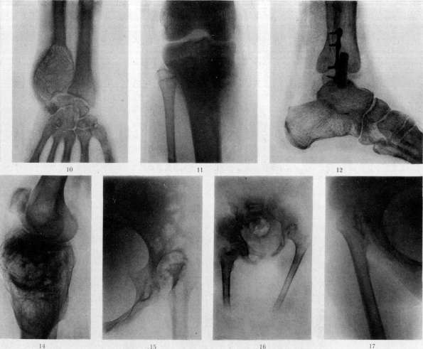
To the articles Coxitis, Bone.
In congenital syphilis at an early age, osteochondritis of the bone is observed, either as independent periostitis or in the vicinity of existing osteochondritis. Here, periosteal thickening is found either in the form of separate bumps or as diffuse, more or less extensive layers on flat and tubular bones. These periostitides may be accompanied by bone deposits. In simple periostitis, an X-ray image reveals thickening of the periosteum; in ossifying periostitis, a dense shadow of the bone capsule layer on the bone beneath the periosteum (see separate plate, figure 3). Aside from these manifestations of syphilis in congenital syphilitics, one can observe osteosclerosis, osteoporosis, and sometimes gummas (see separate table, figure 4)—usually in the second year of life, just as in adults. Likewise, in early age, a characteristic syphilitic dactylitis occurs—a gummatous lesion usually of several finger phalanges or metacarpal bones with the development of periostitis and bone swelling upon them. The process resembles spina ventosa in tuberculosis, but is painless, rarely causes suppuration or fistulae in children, and does not affect the articular ends, which is however observed in adults. Bone manifestations of congenital syphilis are frequently accompanied by anemia and various other signs of syphilis. The clinical course of so-called late congenital bone syphilis is similar to the picture of tertiary bone lesions in acquired syphilis (see below). In late congenital syphilis, one more frequently encounters nutritional decline, emaciation, and cachexia. Simultaneous lesions of several bones, often symmetrical, are not uncommon. The earliest bone lesions in acquired syphilis may be encountered as early as the beginning of the secondary period, sometimes even before the appearance of the first rash and other general phenomena. They occur most frequently in extensive lesions of the skin, mucous membranes, internal organs, and in malignant syphilis. The skull, tibia, ribs, sternum, etc., are most frequently subjected to involvement. In some cases, everything reduces merely to pain felt either deep within the bone or more superficially in areas of the bone rich in fibrous tissue, at the attachment sites of tendons, aponeuroses, on spinous processes, near joints, etc. Deep pains, usually nocturnal, are localized to a definite area of the bone. Pressure can elicit the region of tenderness, but cannot provoke or intensify the pain. Palpation of the pain zone yields no objective data. It is hypothesized that the cause of the pains is syphilitic changes of the roseola type in the bone marrow. Superficial pains may recur from pressure, but otherwise do not differ from deep pains. Pains in the bones are also precursors of periostitis of the secondary period. Their character in this case is acute, sharp, boring, compressive; they intensify at night, interfere with sleep, and are easily elicited and intensified by pressure. In the coming days, edema appears around the bone affected by periostitis; it gradually engulfs the surrounding soft tissues and skin, upon which redness and a local temperature elevation are noticeable. With the intensive development of such periostitis, ulceration may ensue after skin infiltration. The magnitude of lesions varies; small periostitides are barely noticeable and are often found only after the patient precisely indicates the painful area bothering them. Upon superficial examination, they can be confused with neuralgia, rheumatic or muscular pains, etc. Periostitis sometimes resolves spontaneously, disappearing without a trace in 4-6 weeks, but gives frequent relapses. With a long-standing existence of periostitis, slowly developing bone deposits (ossifying periostitis) may appear, which are more characteristic of the tertiary period. Periostitis of the secondary period responds well to specific treatment, resolving without a trace, but bone deposits may persist. In general, it can be noted that bone syphilis in the second period is a non-destructive process, in contrast to the third period of syphilis. Diseases of the bones in the tertiary period are most frequently localized to the tibias, the skeleton of the nasopharynx, and the skull. Gummatous periostitis manifests as a more or less extensive swelling of the periosteum in the presence of several gummas, which is quite dense and causes pain upon pressure and spontaneously (at night). With an increase in swelling and involvement of the integuments in the process, softening of the gumma occurs in connection with its caseous degeneration, followed by ulceration, fistulae, discharge of purulent mass, and sometimes sequestra if the gumma has sufficiently strongly affected the bone. When the process subsides, the swelling gradually diminishes, scars, leaving a scar fused with the bone beneath which a more or less eroded bone can be palpated, usually surrounded by periosteal layers. In gummatous osteomyelitis, which is similar in external manifestations to gummatous periostitis, the bone is affected by the process more strongly than in periostitis, which ultimately leads to extensive necroses, e.g., the rejection of fingers in dactylitis, through-and-through skull defects, etc. Surgical intervention in this case can provoke new bone necrosis. Ossifying periostitis and ostitis in the tertiary period are close in clinical picture to periostitis of the second period, but develop very slowly and produce very extensive bone deposits, differing in persistence of the course and severe nocturnal pains. With it, as with late congenital syphilis, sharp bone deformations are observed (e.g., so-called saber shins with curvature of the lower legs, etc.). Pains are associated with periods of exacerbation of the process after temporary subsidence. Subsequent muscle atrophy is noted with it, but less sharp than in tuberculosis, and appearing later. Most diverse combinations of the more typical forms of bone syphilis listed above are frequently observed, which is why it is sometimes difficult to differentiate them. Secondary infections burden the course of open syphilitic bone lesions and sometimes obscure the picture. Recognition of tertiary forms of bone syphilis is based chiefly on nocturnal pains, significant bone deposits, growths, and at the same time destructions. Unlike infectious osteomyelitis, syphilis predominantly affects the diaphysis, and there is no high suppurative temperature and acute phenomena. Anamnesis and serodiagnosis help distinguish it from typhoid lesions. Suppurated gummatous periostitis is similar to a tubercular process, but the character of the pains, pus (little in syphilis), ulcerous appearance of the fistula, and dense sequestra speak in favor of syphilis. The Wassermann reaction naturally facilitates recognition, but is not absolute. X-ray images in gummatous osteomyelitis usually indicate rarefaction in the area of the gumma, densification of the bone around the focus (see separate table, figure 5); in periostitis, bone deposits encompassing the bone without sharp boundaries, deformations, and densification of the bone up to the destruction of the shadow of the bone marrow cavity in the sclerosing process (see separate table, fig. 6). Diffuse bone rarefaction is encountered significantly less frequently. When all data are insufficient for a diagnosis, one has to resort to trial treatment, which sometimes alone can give a decisive answer. Treatment is specific, according to general rules. In open forms with the presence of ulcers and sequestra, in addition to specific treatment, general surgical measures are necessary with the observance of great conservatism during surgery: remove only the dead tissue, since the rest can be saved by antisyphilitic treatment. Among benign neoplasms during the growth period of bones, so-called cartilaginous exostoses, single or multiple, are encountered more often than others. Developing near joints, they remain hidden for quite a long time by a layer of soft tissues and only upon achieving more significant development do they become noticeable. Attention is more frequently drawn to them thanks to pains in the area of the exostosis, especially upon trauma (bruises, pressure by parts of clothing, shoes, various instruments, a saddle, etc.). Examination establishes the presence of a fixed, dense bone tumor, the base of which often cannot be palpated. The summit of the tumor protruding from the depths is uneven, nodular, often resembling cauliflower. The summit of the exostosis, covered with cartilage, is almost always surrounded by a synovial bursa, which is why these exostoses also bear the name of encysted exostoses (exostosis bursata). The bursa sometimes contains so-called rice bodies. The roentgenogram yields a very typical picture: a more or less long outgrowth on the bone on a broad or rather thin stem, with a nodular summit, a pronounced cortical layer, and spongy substance communicating with the spongy substance and bone marrow cavity of the parent bone (see separate table, figure 7). Usually, the exostosis is located obliquely to the axis of the bone, deviating along the line of action of the surrounding muscles. There are rare indications of the possibility of spontaneous resolution of exostoses, but usually one has to operatively remove those of them that hinder movements. When there are indications for surgery, it is also necessary to keep in mind the possibility of their malignant degeneration. Removal of the synovial bursa is optional. Cartilaginous tumors arising within the bone, as they grow, cause bone swelling, atrophy of the cortical layer to complete disappearance at the site of greatest pressure, but can also not extend beyond the limits of the bone for a long time. An enchondroma that has broken through the cortical layer can gradually reach significant sizes. Enchondromas are more frequently distinguished by a tendency to large growth.
Cartilaginous tumors push aside and spread the soft tissues covering them; to the touch they are painless, sharply contoured, dense and elastic, coarsely nodular, sometimes with areas of softening (mucous degeneration). In multiple enchondromas, severe skeletal deformations leading patients to a state of disability are possible due to the destruction of the bone and the abundance of tumors. On X-ray images, cavities in the bones and lacunae with smooth walls are visible, often with a honeycomb structure due to the fact that individual tumor nodules are separated by bone septa (see separate table, figure 8). The tumor itself usually produces a weak, structureless shadow, the intensity of which depends on the size and density of the tumor; however, lime deposits in the tumor may be visible as formless dense shadows or else (in osteochondromas) bone formations of various shapes and sizes (see separate table, figure 9). The part of the bone adjacent to the tumor either shows no changes, or a coarse-meshed structure of the spongy part and some porosity are noticeable in it. --Treatment is surgical for single tumors, with the removal of the part of the bone adjacent to the tumor to avoid recurrence. Sometimes it is more advantageous to perform a resection of the diseased bone across its entire thickness. In multiple chondromas, radical measures are often impossible in view of the scattered nature of the foci. Various bone sarcomas occur at any age, but more often at a young age (15 - 25 years). First--swelling and pain increasing with rapid growth, minor functional disorders, then--the presence of a more or less dense, depending on the type of sarcoma, sometimes somewhat nodular, not always well-contoured, elastic, immobile tumor in relation to the bone, growing into the soft tissues, skin integument, sometimes joints, the development of superficial veins--all this makes it possible to recognize a sarcoma. The sometimes occurring temperature rises and sharp pains may give reason to think of an inflammatory process, but in sarcoma the phenomena usual for inflammation do not come to the fore. It is sometimes confused with tuberculosis and syphilis. With the development of sarcoma (and other tumors) inside the bone, the cortical layer of the thickened, swollen bone becomes sharply thinned, so that when pressure is applied to a more superficially located diseased bone, one can feel the so-called parchment crackle (false crepitus). Periosteal sarcomas are easier to notice earlier than intraosseous ones. As the tumor develops and the bone is destroyed, spontaneous fractures can occur. Bone sarcomas easily metastasize to the lymph nodes and via blood vessels to various organs, most often to the lungs, as a result of which the prognosis is unfavorable. X-ray examination for sarcoma developing inside the bone indicates a decrease in the density of the bone shadow in the area occupied by the tumor, sometimes to the density of soft tissues. The boundaries of this section are uneven, eroded; in the area of the tumor, it is possible to see shadows of the remnants of the bone structure, individual bone islands, and trabeculae. No changes are noticeable in the surrounding bone. The cortical layer is sometimes sharply thinned, in places defects of its shadow are visible where the tumor grows outward (see separate table, figure 10). In periosteal sarcomas, the cortical layer in the images appears relatively little eroded, the shadow of the tumor has no clear boundaries and is not always distinguishable from soft tissues. In central myeloplastic sarcoma, the shadow of the surrounding bone capsule can be seen even far beyond the borders of the bone. In osteo- and osteoid sarcomas, bone proliferations are visible (see separate table, figures 11 and 14), either radiant, as if emanating from a single center, or in the form of tubercles, and the shadows of such tuberous bone deposits in the images can also be observed in metastatic nodes. --Treatment is recommended to be as early and radical as possible. In early stages, one cannot be limited to the removal of only the tumor: it is necessary to remove it with the adjacent soft tissues and resect the affected bone across its entire thickness within healthy boundaries, replacing the bone defect with a graft. Later, to preserve the limb, in rare cases one can use transverse resection of the limb, leaving only the neurovascular bundle (Bogoraz method). In more neglected cases--high amputation, if it is still possible. Treatment of the tumor with X-rays, according to some authors, gives good results. --Metastases of malignant tumors in the bone, imperceptible in the initial periods of their growth, as they develop, manifest themselves with increasing pain, deformation of the affected bones, spontaneous fractures followed by enhanced tumor growth in the soft tissues. X-rays indicate the presence of foci of rarefaction in the bone without a reaction in the surrounding bone, with irregular boundaries. In osteoplastic cancers, shadows of significant bone deposits are visible. If one can assume the singularity of the metastasis, which also causes severe pain, it is possible in suitable cases to remove the affected part of the bone or amputate the limb, which has to be done mainly in spontaneous fractures to eliminate unnecessary pain. More often, surgical removal of metastases in the bone is impracticable, and it remains to try to stop and limit the growth of tumors with X-rays. Echinococcosis of the bone accounts for about 1.8% of all echinococcal diseases in terms of localization frequency. The pelvic bones, vertebrae, femur, and humerus are most often affected. Developing in the spongy part, small vesicles, growing very slowly, cause bone atrophy from pressure without a reaction from the periosteum. Vesicles that have grown beyond the bone reach significant sizes in the soft tissues. The eroded bone breaks easily, which is often the first sign of the disease, since pain in the area of the affected bone may be absent, and swelling cannot always be noticed in a timely manner. Diagnosis of echinococcosis is not easy. Examination of the fluid when it can be obtained by trial puncture helps somewhat, but hooks are rarely found in bone echinococcosis. On the radiograph, light shadows of round-oval cavities in the bone, separated by dense thin partitions, are visible. Later, the bone is swollen, the cortical layer is thinned. There are no specific features. A similar picture can be given on the image by bone cysts, enchondromas. The prognosis of echinococcosis of the pelvic bones, spine, and skull is unfavorable, since extensive, incurable lesions usually occur before the diagnosis is established; in echinococcosis of tubular bones, the prognosis is more favorable, since with early diagnosis the diseased bone can be resected and the limb preserved; with more significant changes, amputation is necessary. IV. Operations on bones. Operations on bones aimed at restoring the integrity of the bone or correcting its shape require for success the observance of several general rules: 1) carefully stop bleeding and avoid drains, because drains of any kind can be a conductor of infection; 2) spare the periosteum as much as possible; 3) observe the strictest asepsis, which is why it is necessary not to work in the wound with fingers, especially without gloves, but to act only with instruments; 4) do not operate too soon after a past infection in order to avoid a flare-up of latent infection. In such cases, it is useful to prepare the patient with vaccination, protein- or autohemotherapy, or injections of a 10% iodoform solution in almond oil (Wreden). In general, one can operate only with a normal state of white blood. -- Correction of bone curvatures, congenital and acquired, is performed either by osteoclasia or osteotomy. Osteoclasia is the fracture of a bone at a planned site without incision of the soft tissues. First proposed in 1619 (Fabricius ab Aquapendente). In young children in the presence of brittle bones, osteoclasia is easily and gently performed simply by hand: the limb is grasped above and below the intended fracture site and bent until the crunch of the fractured bone is felt. Osteoclasia is easier to perform where the limb bone is less protected by soft tissues. With denser bones, one has to look for a fulcrum in the edge of a table, a placed wooden wedge, one's own knee, etc. In older children (up to 10-12 years old), with denser bones, especially when it is necessary to break a bone closer to the epiphysis, the strength of hands is not enough, which is why a number of osteoclasts have been proposed--devices that transmit pressure to the limb using cuffs, pads, and levers (osteoclasts of Collin, Robin, Lorenz, Heusner, Schultze, Bagrov, etc.). All these devices require careful and skillful handling. In adults, the bones are too dense, and osteoclasia is inapplicable. After osteoclasia, the limb is placed in the correct position, fixed with plaster, and treatment proceeds as with a fracture. Osteoclasia is most often used for genu valgum and varum, for pes equino-varus, etc. In joint ankylosis, osteoclasia is applicable only in the presence of ankylosis at an obtuse angle, otherwise unfavorable conditions are created for the fusion of fragments. Disadvantages of osteoclasia: soft tissue trauma, sometimes significant--sometimes to the point of necrosis--when using an osteoclast. It is far from always possible by hand and by device to obtain a bone fracture precisely in the desired place, especially near the joints. In children, detachment of the epiphysis with subsequent disturbance of bone growth is possible. An exacerbation of the process is also possible if the fracture passes through a focus of infection.
These shortcomings and the development of aseptic technique make the blood method of correcting various bone curvatures—osteotomy, the transection of the bone in the most convenient place by the method most favorable for subsequent fusion in the desired position—preferable. The method is incomparably more precise, less traumatic, and applicable anywhere on the bone. Osteotomy was proposed in 1826 by Rhea Barton, but for a long time did not find wide application due to the infections that were common at the time. Langenbeck in 1854 proposed the so-called subcutaneous osteotomy through a small skin incision with a narrow hacksaw, which reduced the possibility of suppuration. Billroth, to reduce trauma to the soft tissues, replaced the hacksaw with a chisel. According to the method of approach to the bone, open and subcutaneous osteotomies are distinguished. In subcutaneous osteotomy, a single movement of a sharp knife immediately makes an incision down to the bone, sufficient to introduce a wedge-shaped chisel or osteotome along the knife left in the wound (fig.—see art. Surgical Instruments). With a sharp osteotome, one can immediately penetrate to the bone even without a preliminary incision with a knife. Having reached the bone, the osteotome is turned transversely to the axis of the bone (or obliquely) and, striking with a mallet, frequently rocking and without removing the chisel, the bone is gradually transected by at least 3/4 of its thickness, and then the remaining thickness of the bone is fractured, trying not to displace the contacting ends of the bone. For better orientation on some osteotomies (e.g., Macewen's, Mathieu) (fig.—see art. Surgical Instruments), centimeter divisions are marked on the side of the blade. Performing a subcutaneous osteotomy requires knowledge of anatomy, good technique, and the ability to orient oneself by touch and the appearance of the limb. The soft tissues are injured very little during access to the bone, but great caution is necessary so that when breaking the bone, one does not injure the nerve trunks and larger blood vessels passing nearby. It is therefore better to perform subcutaneous osteotomy only where there is no risk of injuring nerves and vessels. Subcutaneous osteotomy is feasible in a transverse or oblique direction to the axis of the bone. More complex types of osteotomy cannot be performed using the subcutaneous method. Open osteotomy is performed while preserving as much as possible the continuity of the greater part of the periosteum. Soft tissue and periosteal incision along the bone. Using periosteal elevators, straight and curved, the periosteum is separated along most of the circumference or around the entire bone. Curved elevators are passed under the bone, pushing aside the periosteum, to protect the periosteum and adjacent soft tissues when working with the osteotome (fig. 9). With a wide, sharp osteotome, the bone is gradually chopped through, frequently rocking the osteotome and freeing it from the bone, and
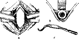
Figure 9. Scheme of the main moments of osteotomy: a—subperiosteal isolation of the bone; b—placement of the elevator behind the bone; c—Lange elevator.
also making sure there are no fragments. With a very dense bone (e.g., the lower jaw), it is recommended to make several holes in advance along the line of bone cleavage with a thin drill or hand drill (fig. 10), which facilitates work with the chisel and helps avoid bone fragmentation. This is especially convenient for complex forms of osteotomy. The bone can also be sawn through with a Gigli saw. With simpler forms of osteotomy, the bone is not chopped through its entire thickness; the remaining layer is fractured toward the direction of the chisel without displacement of the fragments. Bleeding is stopped; sutures are placed on the periosteum and soft tissues. After all kinds of osteotomy, the limb is set in the desired position without displacing the fragments and held until the hardening of a plaster cast that reliably fixes the fragments and adjacent joints. Traction is sometimes used (Steinmann's pin, glue, adhesive plaster), mainly when the position cannot be corrected well immediately due to muscle contractures. In these cases, tenotomy can be performed, and plaster can also be used, changing the bandages every 10–15 days and gradually correcting the position of the limb. The plaster cast remains until the consolidation of the bone callus, the formation of which is sometimes delayed due to a defect between the ends of the fragments of the straightened bone. After
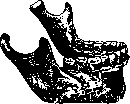
Figure 10. Drilling of the lower jaw during osteotomy.
removal of the cast—general treatment, massage, medical gymnastics; wearing a brace for several months is useful. If for any reason an osteotomy is performed
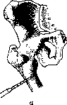
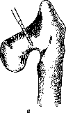
;Гч
Figure 11 a–c. Osteotomy: a—transverse; b—oblique; c—Z-shaped. Ч& not subperiosteally, it is necessary to preserve as much as possible the connection of the periosteum with the muscles so as not to greatly disrupt its nutrition and the nutrition of the bone. Protective elevators are not always convenient to use here, and one must work carefully with the osteotome, protecting the soft tissues. Sometimes an osteotomy is performed under
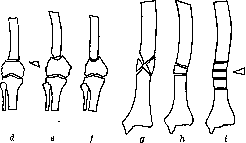
Figure 11 d–z. Scheme of osteotomy: d—wedge-shaped; e—angular; f—hinged; g—cruciform; h—wedge-shaped with 180° rotation; i—segmental.
a bloodless tourniquet. If suppuration appears in the wound in the postoperative period, treatment is conducted according to the general rules of treatment for an open infected fracture, maintaining the plaster cast or traction. A relatively rare complication of osteotomy can be fat embolism. Somewhat more frequently, cases of delayed callus development may be encountered due either to excessive damage to the periosteum during the operation, incorrect alignment of the ends of the fractured bone, or severe exhaustion, diabetes, syphilis, marked bone sclerosis, etc. To prevent the development of a false joint, treatment must be directed at the general disease, applying Bier's congestive hyperemia, massage, percussion, injecting calcium into the vein, injecting the patient's own blood into the callus area, etc. After past bone infections, osteotomy is recommended to be performed away from the former focus (radiograph!). In addition to subcutaneous and open osteotomy, they are also distinguished by location (supra- and supracondylar, diaphyseal, subtrochanteric, etc.) and by the method of bone transection [transverse, oblique, hinged (arched), longitudinal Z-shaped (step-like for limb lengthening), angular, wedge-shaped simple and with a 180° wedge rotation, cruciform, Springer's segmental, etc. (fig. 11)]. A more complex type of osteotomy is the so-called Wreden metaplasia (fig. 12). In accordance with the bone curvature, osteotomy is performed in both the frontal and sagittal planes. It is more advantageous to perform a transverse osteotomy from the concave side of the bone curvature, since upon straightening the fractured bone, the limb lengthens. In wedge-shaped osteotomies, it is important to take a wedge of a definite size, which can be determined in advance from an X-ray photograph and a paper model of the bone contours. Angular and hinged osteotomies do not cause displacement of the fragment ends. The methods are clear from the figures. With complex curvatures, mainly of the lower leg, in two planes, Springer's more complex segmental osteotomy is performed as follows: after careful separation of the periosteum, the curved section of the bone is resected and, outside the wound, sawed perpendicular to the axis into segments with parallel planes of the saw cuts about 1 cm high; the bone is clamped in a vise for this purpose. The remaining small wedge-shaped segment is usually not used, but the rest are laid out in a straight line into the periosteal sleeve, the wound is sewn up, and, having straightened the limb, a plaster cast is applied for about six weeks. Osteoplasty—transplantation of bone to correct a particular skeletal defect. Free transplants and transplants on a soft tissue pedicle are distinguished. For the first time in 1682, Job a Meek'ren successfully transplanted a dog's bone into a skull defect. Bone transplants revived at the beginning of the 19th century (Merrem, 1809; Walther, 1821, etc.). The basis for the wide development of bone transplants was provided by Ollier (1859–67) with his rich experiments and observations.
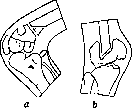
Figure 12. a - 1st stage; b - 2nd stage. 17> times. Many of his propositions retain their full significance to the present time. He proved the possibility of engraftment and production of bone by the periosteum, transplanted on a pedicle and freely, proved the important significance of the inner proliferation layer of the periosteum for bone formation; pointed out that the presence of the periosteum on the transplant during free transplantation of the bone ensures better engraftment of the bone. Later, Barth argued that the transplant's periosteum has no significance; it dies along with the entire transplanted bone, which causes a certain irritation of the damaged bone; the replacement of the defect proceeds gradually through the proliferation of new bone from the periosteum and bone marrow of the defect edges. On this basis, Barth considered the transplantation of living or dead bone a matter of indifference. Subsequent works of a number of authors refuted Barth's opinion on the passive role of the periosteum during transplantation and confirmed its role in bone regeneration, and also clarified: 1) that the bone transplanted with the periosteum dies, but part of its cells survive depending on the presence of favorable conditions; 2) that in bone regeneration, besides the periosteum, the endosteum and bone marrow of the transplant take part, and more strongly the endosteum and bone marrow of the recipient bed; 3) that the connective tissue surrounding the transplant can participate in bone regeneration by metaplasia into osteoblasts and bone cells of connective tissue cells adjacent to the transplant and growing into it. In this case, either lacunar resorption of the transplanted bone by osteoclasts and deposition of new bone in this place is observed, or a barely noticeable direct creeping substitution, when the destruction products of the transplanted bone go directly to the creation of new bone growing from the periosteum and bone marrow of the transplant. Bone transplanted without the periosteum dies entirely, gradually resorbs, and is replaced by new bone growing from the periosteum and bone marrow of the defect edges. The regeneration process here proceeds more slowly than during the transplantation of bone with the periosteum. The presence of bone transplanted without the periosteum enhances regeneration from the edges of the defect, perhaps by virtue of mechanical irritation, perhaps because the salts of the dead transplant go towards the construction of new bone. It should be noted that it is not always easy to strip the entire cambial layer from the bone during transplantation without the periosteum; the remaining part of this layer can contribute to bone regeneration. A fresh autotransplant without the periosteum plays the role of a resorbable prosthesis and can be equated in its participation in regeneration to the transplantation of dead macerated bone of both man and animal (elephant, bull, etc.); but the latter are resorbed and replaced more slowly. Bone material for transplantation is arranged in the following order according to degree of suitability: 1) living bone with periosteum - a) autoplasty, b) homoplasty; 2) living bone without periosteum - a) autoplasty, b) homoplasty, c) heteroplasty; 3) dead bone - a) boiled, b) macerated. Less suitable are decalcified and ossified bones. The success of homoplastic transplantations of living bone makes it possible to use bone from freshly amputated limbs and from fresh corpses no later than 10 hours (Lexer) with the exclusion of any possibility of infection. Kostyukov's studies showed even that under favorable conditions of corpse preservation (+1°-0°), the sterility of the bone and its viability are preserved for over 10 days. Corpse bones fixed in alcohol engrafted in the experiments of Christophe. Lexer used well-boiled or macerated corpse bones freed from the periosteum and bone marrow. Transplanted living bone engrafts and undergoes a series of changes in adaptation to new conditions. It changes its shape: excesses are resorbed, thickness increases to the thickness of the bone where the transplantation was made; the newly developed bone becomes overgrown with periosteum, a bone marrow cavity appears in it, merging with the bone marrow cavity of the main bone. The architectural structure of the transplant changes in accordance with the structure of the main bone and functional requirements of the transplantation area; the new bone participates proportionally in the growth of the skeleton, etc. When transplanting bone on a pedicle, this pedicle can consist of the periosteum alone, of the periosteum and other tissues connected with the transplanted bone (muscles, tendons, aponeuroses, skin). The richer this pedicle is in blood vessels, the more assured is the good engraftment of the transplanted bone, which proceeds by the type of fracture healing if the transplantation is made onto bone. In addition to the above-mentioned general rules for bone operations during bone transplantations, it is necessary to carefully prepare the bed for the transplant: excise scars, which is not always easy and simple, well refresh and smooth the edges of the bone defect, since a tight fit of the transplanted bone to them is necessary for successful fusion. It is important to carefully stop the bleeding, since the poured-out blood disrupts the very necessary close contact of the soft tissues and the transplanted bone and thereby complicates the vascularization of the transplant periosteum and the development of osteogenetic processes in the surrounding connective tissue. For the same purpose, careful suturing of the soft tissues over the transplant is necessary to eliminate voids between them and prevent contact of the transplant with the neighboring bone. A favorite source of borrowing bone for free transplantation is the tibia, which is easily accessible and yields plenty of material. Plates are taken from its median surface, and pins from the crest. In addition, one can of course use any suitable bone, provided that its partial or complete removal does not cause functional disorders (fibula, ribs, plane of the scapula, iliac crest, metatarsal bone, etc.). Usually, the bone is exposed down to the periosteum by a flap incision, the periosteum is incised, slightly extending beyond the boundaries of the future transplant, and peeled off to the desired boundaries; with a sharp, thin osteotome, the boundaries of the transplant are first chopped to the required depth, and then they begin to chop out the outlined piece of bone as parallel as possible to its surface. The chopping of the boundaries and chopping out of the bone plate should be done with not too heavy blows, so as not to split the "donor" bone excessively. The chisel is placed carefully in the line of the cut so that there are no small splinters on the transplant interfering with further work. Raising the chopped-out plate requires caution and gradualness, otherwise it is easy to spoil and even break the plate. Caution in work is also needed when taking a thick plate that also captures the bone marrow cavity, as well as a thick pin, since with careless actions one can split or break the "donor" bone. When borrowing a transplant from other bones, one naturally adjusts to the features of the bone and its topographical relationships. The most convenient chisels for cutting out bone plates and pins are those of Payr, Lexer (Fig. 13), Dyakonov, Bobrov (fig. - see art. Surgical Instruments). Very good transplants can be cut out with a circular saw. Burns of the bone during work with it are prevented by watering the sawing place with a thin stream of physiological solution. Sometimes Bier's arched saw with a very narrow blade is used. At the site from where the bone was taken, bleeding is carefully stopped, the wound is sutured tightly, and a pressure bandage is applied. A carefully fitted transplant is tightly inserted into the bone defect, taking care to ensure a good fit of the edges of the periosteum of the defect and the transplanted bone. On top, the transplant is fixed by suturing the soft tissues. When replacing defects of a tubular bone, the ends of the transplant taken with the periosteum can be inserted into the bone marrow cavities of the ends of the defect (pinning) or the transplant is attached from the side (splinting), tying the ends of the defect and the transplant together (see Bone suture). Examples of more complex methods of fitting and tying the transplant can be seen in Fig. 14. Violating the integrity of the transplant by drilling for fastening with a suture to the ends (edges) of the defect should not be done without the most extreme necessity. When transplanting bone on a soft-tissue pedicle, the flap is fixed by suturing the edges of the periosteum and other soft tissues. It is necessary to ensure that the nourishing vessels are not compressed and the blood supply to the flap and bone is not disrupted. When transplanting on limbs and other places, reliable immobilization after the operation is necessary, ensuring the immovability of the transplant. A good plaster cast is applied to the limb. When possible, this cast in the form of a plaster gutter-shaped splint is prepared before the transplantation so that immediately after the operation the limb can be placed in the ready splint and the risk of bone displacement avoided. In subsequent treatment, it must be remembered that the bone transplanted into the defect does not thicken immediately and long caution in movements is necessary. Cases of fractures of the transplant with subsequent fusion have been observed. Wound infection developed after the operation usually ruins the transplanted bone, but with weak infection there have been cases of engraftment of the transplant even with its partial sequestration. Free bone transplantation has become a routine operation for all kinds of bone defects: congenital, after accidental trauma, resections, various infectious processes, etc.
These include, for example, operations to correct the skeleton of a sunken nose, skull defects, lower jaw, various types of bone transplantation to strengthen the spine in its tuberculosis, arthrodesis of joints using bone pegs, replacement of tubular bone defects, etc. Bone transplantations on
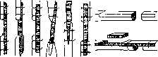
Fig. 14. Various methods of fitting a graft in tubular bones. a pedicle are extensively used in surgery to correct various facial deformations, close skull defects (König-Müller operation, etc.), in all kinds of osteoplastic amputations: Pirogov, Vladimirov, Levshin, Spasokukotsky, Sabaneev, Abrazhanov, Gritti, Kocher, Bier, etc. Here one should also include the replacement of a tibia defect by implanting the end of the fibula (Hahn operation and its modifications), plastics of the upper end of the tibia from the lower end of the femur (Hacker and Klapp operation), transplantation of an embedded bone in a migrating soft tissue flap, etc. To replace a bone defect in the form of a cavity, e.g., after osteomyelitis or fibrous ostitis, one can use the transplantation of a bone piece, filling the cavity with pieces of decalcified or calcined bone, filling with catgut, transplantation of adipose tissue, free or on a pedicle, transplantation of a flap of an adjacent muscle, mobilization of bone walls (Schulten operation), filling the cavity with a blood clot, or plugging with some slowly resorbing mass. Into more superficial cavities, a skin flap is sometimes turned in, achieving epithelialization of the cavity walls. The transplanted soft tissues, filling the cavity, grow together with its walls. In other methods, the cavity slowly grows over with bone, which is intensively produced due to irritation by the graft or plug. Various compositions of the filling mass have been proposed, e.g., Mosetig-Moorhof plug (60.0 iodoform, 40.0 spermaceti, and 40.0 sesame oil) or plaster with 5% carbolic acid (Dreesmann), plaster with rivanol (Oehlecker), etc. The success of plugging depends on the asepsis of the cavity, which must be thoroughly exsanguinated and dried before pouring in the sterilized plug. Transplantations on a pedicle can include so-called temporary resections, when by reflecting a skin-muscle-bone flap
access to deeper-lying parts of the bone is achieved. These include, for example, osteoplastic trepanations of the skull, temporary resection of the nose (Ollier, Bruns, etc.) for access to the vault of the nasopharynx or the pituitary gland, temporary resections of the upper jaw for the same purpose, resections of the lateral wall of the orbit (Krönelein, Czermak), the wall of the frontal sinus (Golovin), the lower jaw (Krasin), the attachment site of muscles to the greater trochanter, etc. These temporary resections are performed with a chisel, saws, and other instruments. Upon completion of the main operation, the temporarily reflected flap is put back into place and secured with sutures. For a more precise fit, it is sometimes good to pre-drill paired holes for sutures on the sides of the future bone cut line. Definite bone resections (osteoectomies) are most often performed for neoplasms. In benign tumors and inflammatory diseases, the bone is usually removed subperiosteally. In malignant tumors, it is necessary to remove the bone together with the periosteum and a greater or lesser layer of adjacent muscles. In subperiosteal resection, it is sometimes possible to leave the defect without replacement by transplantation, especially at a young age, since the bone regenerates well. In large defects in adults during resection with the periosteum, it is necessary to replace the defect with a bone transplant. Sometimes a resection of a part of the bone diaphysis is done in order to make it possible, by shortening the limb, to suture a destroyed nerve or tendons. The bone is connected in these cases by a suture. Resection of articular ends is undertaken for certain joint diseases. - BONE SUTURE - SEE Osteosynthesis. N. Terebinsky.
Related articles
Cite this page
“Bone.” Soviet Medical Encyclopedia. English translation of Bolshaya Meditsinskaya Entsiklopediya, 1st ed. (Moscow, 1928–1936), ed. N. A. Semashko. https://sovietmedicalencyclopedia.pages.dev/article/bone/