Fibrous Osteitis
Historical document, translated for reference. It reflects medical knowledge of the 1920s–30s and is not medical advice.
Summary
Fibrous osteitis is a bone disease first described by Recklinghausen in 1891, characterized by replacement of bone marrow with fibrous tissue, bone destruction, and formation of brown tumors. The condition can occur as a localized or generalized form, affecting either specific bones or the entire skeletal system.
Encyclopedia article (1928–1936)
Fibrous Osteitis (ostitis fibrosa), synonym fibrous osteodystrophy (osteodystrophia fibrosa), a disease of the bones, first precisely outlined by Recklinghausen in 1891; he also gave it the name (ostitis fibrosa von Recklinghausen). It should be noted, however, that the disease was known earlier; undoubtedly, after 1876, when Paget identified the disease of bones under the name 'osteitis deformans', bone lesions belonging to fibrous osteitis were described under the name 'deforming osteitis'; it is very probable that the observation published by Czerny in 1873 under the name 'Eine lokale Malazie des Unterschenkels' also belongs here. Recklinghausen put forward the following five propositions characterizing the essence of fibrous osteitis: 1) replacement of cellular bone marrow by fibrous connective tissue; 2) destruction of bone tissue by lacunar resorption; 3) metaplastic, and partly osteoblastic, formation of new osteoid and bone tissue; 4) tumor-like growths of fibrous as well as giant cell tissue, resembling giant cell sarcomas (of the epulis type), and the presence of hemosiderin in these growths, which gives them a brownish tint, hence the name 'brown tumors' (German 'braune Tumoren'); 5) formation of cysts in the proliferated connective tissue. The listed changes lead to deformities and curvatures of the bones, as well as fractures occurring from trivial causes, and such fractures subsequently usually undergo rapid healing. Furthermore, Recklinghausen established that the mentioned changes can affect locally only one or several bones, which gives local forms of fibrous osteitis (ostitis fibrosa circumscripta), observed most often in childhood and generally young age. On the other hand, the same changes can involve the entire or almost the entire skeletal system, which gives generalized fibrous osteitis (ostitis fibrosa generalisata, s. universalis), occurring more often in middle age. According to Recklinghausen, fibrous osteitis, based on the lesion of bone tissue characterized by him as 'metaplastic Malazie', without any sharp boundaries, adjoins the rel-
FIBROUS OSTEITIS
134 relating to 'hyperostotic-metaplastic malacia' to the deforming osteitis described by Paget and usually observed in advanced age; both diseases, according to Recklinghausen, represent merely variants of the same basic process. Recklinghausen's positions over the subsequent 40 years up to the present time have not undergone any major changes, and in subsequent major works by Frangenheim, Pick, Christeller, Stenholm and many others one can only see a deepening and detailing of these positions. In particular it has been established that fibrous osteitis can give various variants in terms of the spread and direction of the process, changes in bone substance, secondary changes, etc. This served as an impetus for the creation of a rational classification. The school of Pick (Christeller, Stenholm) proposed the following subdivision of forms of fibrous osteitis: a) according to spread in the skeleton—generalized, local, and transitional forms; b) according to spread in individual bones—total (involving the entire bone) and partial forms; c) according to localization in bones—cortical and endosteal (or medullary) forms; d) according to the nature of volumetric changes of the affected bones—hyperostotic forms (with increase in bone volume) and hypostotic forms (with decrease in volume); e) according to qualitative changes in bone substance—porotic forms (with rarefaction of bone substance) and sclerotic forms (with compaction of bone); f) according to secondary changes—forms with tumors, without tumors, with cysts, without cysts; g) according to age—childish, juvenile, adult, and senile forms. In this classification both Recklinghausen's proper fibrous osteitis (porotic, childish, juvenile form) and Paget's deforming osteitis (sclerotic, senile form) find their place. This is due to the fact that both the school of Pick and a number of other leading pathologists, after the works of Recklinghausen, firmly stood on the position that deforming osteitis and fibrous osteitis represent the same disease, which can be called fibrous osteitis, or fibrous osteodystrophy (see manuals on pathological anatomy by Aschoff, Kaufmann and others); its basis is the fibrous transformation of bone marrow, lacunar resorption of bone and at the same time the formation of new bone; the difference between the two variants of the disease lies only in that in the 'Recklinghausen variety' of osteitis (O.f., resp. fibrous osteodystrophy), the processes of bone resorption predominate over the formation of bone substance, and the latter remains in the state of osteoid tissue, whereas in the 'Paget variety' of fibrous osteitis there is a predominance of bone formation, which in this case shows a tendency to quickly acquire the character of calcified mature bone. The identification of both forms was also justified by the existence of transitional and mixed forms (Stenholm, Kaufmann). On the other hand, however, Paget's deforming osteitis has its own, quite special clinical picture (see Deforming osteitis). In recent times it has been further emphasized that in generalized Recklinghausen's fibrous osteitis, tumor-like changes of the parathyroid glands are often found (see below), and surgical removal of the latter results in improvement of the disease; in contrast to this, in Paget's deforming osteitis the parathyroid glands are unchanged, and their removal attempted in several cases gave no results. On the basis of the above clinicians had a tendency to consider Recklinghausen's fibrous osteitis and Paget's deforming osteitis as two completely different diseases. In recent years leading German pathologists, who as early as 1926 at a congress devoted to the program topic on fibrous osteitis, spoke in favor of the identity of these diseases, have changed their opinion on this matter. Indeed at the congress of pathologists in Berlin in April 1930 Schmorl, Sternberg and Pick recognized the error of previous pathological views on the identity of Recklinghausen's and Paget's diseases and definitely spoke in favor of the complete difference between these two diseases of the skeletal system. All these data dictate the necessity of considering deforming osteitis (see) separately from fibrous osteitis. During the period that has passed since the first works of Recklinghausen, considerable attention was also paid to clarifying the relationship of fibrous osteitis to rickets and especially to osteomalacia. Recklinghausen already spoke of the closeness between fibrous osteitis and rickets and of the possibility of transitions between them, and subsequently other researchers insisted that fibrous osteitis represents a metarachitic process (Stumpf) or a continuation of the osteomalacic process (Lang). At the present time it can be recognized that despite the external similarity of some of the forms of widespread fibrous osteitis with osteomalacia and the sometimes observed diagnostic difficulties in this regard, there is nothing in common between rickets and osteomalacia, on the one hand, and fibrous osteitis, on the other. In rickets and osteomalacia the basis of the changes lies in the violation of the process of bone accretion (German Knochenanbau), while bone resorption (German Knochenabbau) proceeds at the usual normal rate; in contrast to this in fibrous osteitis there is observed a combination of sharply expressed disturbances both in the process of accretion and in the process of bone resorption, and the latter in the form of lacunar resorption, which does not occur in rickets and osteomalacia, predominates in the entire patho-anatomical picture. However some admit the possibility of a combination of fibrous osteitis with osteomalacia. Thus at the present time there is no doubt that fibrous osteitis represents an independent disease, a nosological unit, the essence of which is determined by the five positions of Recklinghausen given above. The following data refer precisely to fibrous osteitis in the above-mentioned sense. Pathological anatomy. As was already indicated by Recklinghausen, the basic changes for fibrous osteitis are: 1) replacement of the cellular and fatty bone marrow by fibrous connective tissue (German Fasermark), 2) resorption, resorption of old bone by lacunar resorption, 3) metaplastic formation of new bone; to this must be added 4) dystrophic change of bone substance. 1. The change in bone marrow consists in that in the areas of the pathological process in the bone marrow there occurs proliferation of delicate fibrous connective tissue, which displaces the previously existing fatty and cellular bone marrow. In the initial stages the proliferation of connective tissue appears in direct contact with the bone along the course of bone trabeculae and on the inner surface of cortical bone plates; subsequently all bone marrow spaces in the affected area become filled with this delicate fibrous connective tissue. The latter is sometimes poor and sometimes rich in cells; its collagen fibers are arranged in bundles or in a network; the tissue contains thin-walled, sometimes very wide vessels; phenomena of edema, hemorrhages, grains of hemosiderin, infiltrates of round and plasma cells are not uncommon in it. 2. Bone resorption proceeds by the so-called lacunar resorption. Everywhere in the area of the process it is seen that the bone trabeculae and cortical plates as if gnawed through, contain a large number of depressions—lacunae, in which multinucleated osteoclasts are seated. The bone trabeculae and plates are unevenly thinned, their continuity is disrupted, they disappear [see separate table (art. 239-240), fig. 3]. There are observations in favor of the fact that bone resorption also proceeds by the formation of so-called perforating channels (see Bone). As for the opinion of some authors that the loss of bone substance in fibrous osteitis occurs also by the process of halisteresis (see), at the present time this must be considered rejected. 3. The formation of new bone proceeds parallel to the destruction of old bone and is expressed in that among the proliferated fibrous connective tissue of the bone marrow, new bone trabeculae are formed metaplastically. The first stages of this new bone formation proceed according to the type of direct metaplasia, subsequently however around the formed bone trabeculae osteoblasts may appear and further growth of such trabeculae proceeds osteoblastically. The newly formed bone tissue usually has little tendency to be impregnated with lime salts and to turn into true bone and for a long time remains in the form of soft, lime-free osteoid tissue, often rich in Sharpey's fibers. Often one can also see such newly formed bone trabeculae in which the central parts contain lime, while the periphery represents a zone of osteoid tissue. Besides the formation of new bone trabeculae among the fibrous connective tissue of the bone marrow, appositional formation of layers of new bone on the trabeculae and plates of old bone is often observed; sometimes it happens that on one side of a bone plate or trabeculum its destruction by lacunar resorption is seen, while on the other side new bone is being deposited; this bone can also remain osteoid, without lime, for a long time. Sometimes the newly formed bone is subsequently subjected to destruction. 4.
Degenerative changes in the bony substance have been little studied; in general, they come down to various manifestations of impoverishment of the bony substance with lime, followed by changes in the bone. In the bone tissue, 'lattice figures' appear, the bony substance is sharply oxyphilic (stained with acidic dyes) in places, splitting of the bone into plates is observed, death of some bone corpuscles and their groups. Changes of this kind may not always combine in the same way. In general, in typical cases of F. O., processes of destruction of bone tissue predominate over the formation of new bone, which moreover remains osteoid. However, there are also other ratios of these two opposite processes, which gives various variants of F. O., noted in the above classification by Christeller and Stengholm. Cases have also been described where no formation of osteoid was observed at all (e.g., the case of Askana as a type of progressive bone atrophy). Besides these basic changes for F. O., secondary changes of the fibrous bone marrow are often observed in the affected bones and are in general extremely typical for the disease. These include tumor-like growths and the formation of cysts. Tumor-like growths arise inside the bone and, increasing in volume, destroy the spongy bone and cause atrophy of the cortical plate, sometimes to complete disappearance; the periosteum, however, is usually not destroyed and even with significant growth covers it with a continuous layer. The bone at the site of growth uniformly bulges in the form of a spindle or shows an outgrowth protruding only in one of its parts. On a cross-section, the growth appears as usually soft, rarely dense, pinkish tissue, often with a brownish tint or brown spots, which gave rise to calling these growths 'brown tumors'. Microscopically, the growth rarely consists of dense connective tissue and resembles a fibroma, but usually it is built from a cellular mass, with the main mass formed by small polygonal and spindle-shaped cells, often arranged in bundles; among them in significant numbers are scattered giant cells with numerous nuclei; hence the frequent designation of these growths as giant cell growths. Very often among the cellular mass newly formed beams of osteoid tissue are encountered; a frequent but not obligatory finding is the presence of grains of hemosiderin. The described growths, due to their growth resembling that of a bone-destroying tumor, and due to the microscopic picture, which is very similar to the picture of giant cell sarcoma, are often the basis for diagnostic difficulties in terms of differential diagnosis between sarcoma and F. O., both at the patient's bedside and when examining biopsy material under a microscope. Of the microscopic signs indicating F. O., besides the integrity of the periosteum, one must note the uneven distribution of giant cells, the presence of hemosiderin, and gradual transitions between the sarcoma-like growth and the fibrous bone marrow. Cysts in F. O. are not an obligatory finding, but they are still very common; it is not rare for the formation of a bone cyst to be the first sign of F. O. Cysts form among the fibrous bone marrow and represent either small multiple to larger (up to the size of a child's head) cavities filled with transparent colorless or slightly yellow (sometimes chocolate-colored) fluid, more rarely with a colloidal mass; the inner surface of the cysts is smooth; the wall consists of a thin layer of dense connective tissue. Small cysts do not cause any additional changes in the bone, while larger cysts, as they grow, destroy the bone, cause its bulging and deformation, and are often the cause of a fracture. Another change in the bone deserves mention - this is the special 'mosaic' appearance of the bone beams and plates, found in cases of long-lasting disease and which is an expression of the restructuring of bone tissue, i.e., the alternation of processes of bone resorption and appositional deposition of new bone. Schmorl, who first pointed out this change in 1926, initially considered it specific to F. O.; subsequently, however, it became clear (and in 1930 Schmorl also acknowledged this) that a sharply expressed and irregular mosaic pattern of bone is characteristic only for deforming osteitis, whereas in F. O., as well as in various other bone lesions with restructuring (destruction and formation) of bone, a mosaic pattern can also be observed, but weakly expressed and of a layered regular type (with lines running parallel to the bone surface).-All the above processes lead to disfigurement and softening of the affected bone, to curvatures and fractures. Due to the fact that in F. O. new bone formation easily occurs, fractures often heal easily, but in the same place later, due to degenerative changes of the newly formed bone, progressive tumor-like growth of giant cell tissue, or due to the development of a cyst, a fracture may form again. All this gives the following kind of macroscopic changes in the bones: flat bones of the skull, shoulder blades, sternum, pelvis become thicker and at the same time lighter in weight and softer - easily cut with a knife; on a cross-section a special pale-pink, in places more hyperemic tissue is seen, in which the finger feels like sand grains (remains of bone struts). The thickening of bones is not uniform everywhere, but on one or another surface bulges in the form of nodules are observed, on a cross-section of which soft sarcoma-like tissue is seen, sometimes surrounded as if by a bony shell. In some places on cross-sections cysts are encountered. When ribs and clavicles are affected, they are either diffusely or on limited places soft, deformed, usually in the form of spindle-shaped thickenings, in the area of which later curvature or fracture occurs. Vertebrae in the development of F. O. in them become soft, easily flattened, due to which curvatures occur in the spine; on a cross-section the tissue of the vertebral bodies is gray-pink, poor in bony substance. In the long tubular bones of the limbs F. O. gives rise to either limited spindle-shaped thickenings with parchment-like crepitus or with widespread bone involvement, a uniform thickening, disfigurement and softening; subsequent bulges of giant cell growths, formation of cysts, curvatures and fractures can give enormous disfigurements of the bones. On saws (cross-sections) of affected bones the loss of bony substance with replacement of the entire space by gray-pink tissue is striking, in which cysts, hemorrhages, areas of edema are often visible. Similar changes, but to a lesser extent, are observed when the small bones of the feet and hands are affected. In all these lesions no changes of the periosteum are observed; the latter only passively peels off, is pushed aside, but as a rule never undergoes destruction; in particular, sarcoma-like giant cell growths do not extend beyond the periosteum. Joints also do not participate in the process even with very significant bone involvement; in joints only reflected changes can be observed, which are the result of curvatures and fractures of bones, if they are located close to the joint. An exception to these statements about non-participation of the periosteum and joints in F. O. are those extremely rare cases when the F. O. process is complicated by a true sarcoma; however, few reliable cases of this kind have been observed. As already stated above, F. O. in the form of all the above changes can represent a purely local affliction and in such cases affects only one bone over a small extent, with no pathological changes noted in the rest of the skeleton or in the body in general. Such forms of local F. O. (ostitis fibrosa circumscripta, s. localisata) are observed mainly in childhood and generally in young age, and are not rare. Local F. O. can affect any bone, but most often the process affects the long tubular bones of the limbs (e.g., tibial bones), ribs, as well as jaws; in the latter, starting in the alveolar process, it is expressed in the formation of the usual for F. O. tumor-like giant cell growths, called here epulides (see). A much rarer form is the generalized or widespread F. O. (ostitis fibrosa generalisata, s. universalis), in which the disease affects the entire or almost the entire skeleton. This form of F. O. is observed mainly in adults, and the changes apparently begin simultaneously in different parts of the skeleton. The onset of widespread F. O. in the form of a local F. O., which then passes into widespread F. O., is rarely observed; in general, one must consider doubtful the possibility of transition from local F. O. to general, and on the other hand, incorrect Christeller's opinion that only widespread F. O. exists and that every case of local F. O. is only a local expression of a general lesion of the entire skeleton.
As in both local and diffuse fibrous osteitis, atrophic changes can be found in the muscles related to the affected parts of the skeleton. As for the internal organs, changes in them are naturally observed only in diffuse fibrous osteitis. In severe cases of the disease, deposits of lime in the kidneys, arteries, lungs, etc., are quite common, which are manifestations of the so-called lime metastases (see Lime deposits, metastases); a constant finding is hemosiderosis of the spleen, sometimes of the liver. In severe cases of the disease, there are manifestations of anemia with myeloid hyperplasia of the remaining bone marrow, as well as general cachexia in the form of atrophic changes of internal organs, atrophy of nerve cells with neuronophagia, and degenerative changes of peripheral nerves (Rusakov). Of the endocrine glands, various changes (scleroses, adenomas, etc.) have been described in the pineal gland, epiphysis, thyroid and goiter glands, as well as in the adrenal glands in different cases of fibrous osteitis, but they show no constancy and therefore hardly deserve attention. On the other hand, in a number of cases of diffuse fibrous osteitis, a definite change in the parathyroid glands (epithelial bodies) was observed. This change consists in the fact that one, rarely two, three, or all four parathyroid glands are found to be enlarged to some degree; in many cases this enlargement is colossal (instead of the normal 0.2-0.5 cm up to 5.5 cm in diameter), which leads one to speak of tumors of the parathyroid glands. Microscopically, in such enlarged parathyroid glands, a picture of adenomatous hyperplasia is found, giving a reticular tissue of elements of the type of small chief cells. As a rule, such parathyroid gland tumors in generalized fibrous osteitis are benign proliferations, however, in Rusakov's case there was a true carcinoma of the parathyroid gland with regional metastases. The significance of tumor-like enlargements of the parathyroid glands in diffuse fibrous osteitis was evaluated differently. Until recently, according to the data of Erdheim, the prevailing opinion was that the parathyroid glands in fibrous osteitis increase sequentially as a result of disturbed lime metabolism (release from bone tissue due to destruction of bones of a large amount of lime and hypercalcemia), and such an increase represents a compensatory, as it were, autotherapeutic (Erdheim) process. At present, on the basis of findings of parathyroid gland tumors in the initial stages of diffuse fibrous osteitis, the positive result of surgical removal of parathyroid gland tumors in fibrous osteitis, and the experimental study of this question, such an opinion can hardly be considered correct (see below). The pathogenesis and etiology of fibrous osteitis have not yet been fully elucidated. Recklinghausen considered that the basis of the process in fibrous osteitis is a change in the bone marrow, which in his opinion belongs to productive inflammation; this inflammatory process may be a consequence of bone trauma, overstrain of the bone, vasomotor disorders, etc. Recklinghausen's view of fibrous osteitis as a manifestation of an inflammatory process has gained considerable prevalence and still has supporters: attempts have even been made to find an infectious agent causing this inflammation of the bone marrow; assumptions have also been expressed about the syphilitic nature or the rheumatic character of the inflammatory process underlying fibrous osteitis. Other researchers, considering the change in the bone marrow as primary in fibrous osteitis, regarded it as a manifestation not of inflammation, but of a tumor process (Haeberlin's view of fibrous osteitis as an endosteal fibro-osteoma, Reinhardt's as a myeloma of supporting tissue). Stenholm denies the inflammatory nature of the process in fibrous osteitis, but still thinks that the main changes are in the bone marrow, namely in the latter, as a result of disturbance of nutrition in the broad sense of the word, atrophic changes occur, leading to successive changes in bone tissue. In contrast to the various views assuming the main changes in fibrous osteitis to be localized in the bone marrow, a number of researchers, especially in recent times, shift the center of gravity of the pathogenesis of fibrous osteitis to the change in bone substance. As early as 1916, Kostenko first drew attention to the profound changes in the bone tissue itself in fibrous osteitis. Later, the same was noted by Meyer, Christeller, Abrikosov, and others. The study of these changes in bone substance led researchers to the conclusion that the mentioned changes in the form of impoverishment of bone tissue with lime and other manifestations of dystrophy of bone substance represent the very first and primary change in fibrous osteitis, whereas the proliferation of connective tissue in the bone marrow is a successive process, as it were, a reaction to the change in bone tissue. This view, completely excluding the inflammatory nature of the process in fibrous osteitis, is currently generally accepted among pathologists. On the basis of this, there is an increasing tendency to replace the name fibrous osteitis, implying an inflammatory process, with the term 'fibrous osteodystrophy' (osteo-dystrophia fibrosa), put forward by Mikulicz as early as 1904 and Scheppl in 1908. Despite the current knowledge of the pathological basis of fibrous osteitis in the form of dystrophic change of bone substance, the etiology of this bone dystrophy is not yet quite clear. In this respect, one probably has to approach local fibrous osteitis and diffuse fibrous osteitis separately. In local fibrous osteitis, the matter concerns dystrophy of bone substance over a limited, sometimes very small area; therefore, here one can assume the significance of some purely local and probably not always identical causes. For example, local disturbances of the normal course of bone formation (in childhood), functional damage to not quite correctly formed parts of the bone, improper loading of the bone, trauma, and other factors may be the cause of disturbance of nutrition, dystrophy, degenerative changes of bone substance, which are followed by the mentioned reactive processes in the form of proliferation of connective tissue, resorption of inadequate bone, etc. Such an explanation of local fibrous osteitis may find confirmation also in the fact that bone changes of the fibrous osteitis type (replacement of bone marrow by fibrous tissue and combination of lacunar resorption and bone formation processes) are often encountered as a secondary, reactive change around tuberculous, osteomyelitic, and other destructive lesions of bone. Otherwise one has to look at the etiology of generalized fibrous osteitis. Here the matter concerns dystrophic changes affecting the entire (or almost entire) skeletal system, and therefore one has to think of some general cause causing everywhere a disturbance of the normal state of bone substance with the character of its degeneration. In this sense, views have been expressed about the significance of abnormal nutrition (by analogy with animals, in which diseases close to fibrous osteitis can be observed in connection with irrational feeding, or in wild animals when kept in captivity), changes in the nervous system, and finally lesions of the endocrine glands. The view of generalized fibrous osteitis as a consequence of abnormal function of some of the endocrine organs has become the most widespread in recent years. For a time, among researchers who found changes now in one now in another of the endocrine glands, there was no agreement in deciding which of them is responsible for the dystrophic changes of bone substance; therefore, the assumption of plural glandular insufficiency as the basis of the whole process was often put forward. However, at present one can definitely speak of an etiological connection between the dystrophic changes of the skeletal system in generalized fibrous osteitis and the abnormal function of the parathyroid glands. The following facts speak for this: 1) in most cases of diffuse fibrous osteitis there is a tumor-like proliferation of one or more parathyroid glands; 2) surgical removal of enlarged parathyroid glands in fibrous osteitis results in significant improvement of the disease, not only external but also in the sense of a sharp decrease in lime excretion and progressive increase in the amount of lime in the bones (case of Mashchyan, Gold, etc.); 3) with experimental introduction of parathyroid hormone in animals, a change of the skeletal system is obtained, similar to fibrous osteitis and even accompanied by the formation of lime metastases. These facts can be considered very convincing in the sense of recognizing that in most cases of diffuse fibrous osteitis the cause of the changes in the skeletal system is the adenomatous enlargement and the associated hyperfunction of one or more parathyroid glands. In those rare cases where such enlargement of the parathyroid glands was not observed, one can think of functional changes in them.
At the same time, however, it must be admitted that the mechanism of the influence of hyperfunctioning parathyroid glands on the skeletal system is still not clear; one can only assume that hyperfunction of the parathyroid glands sharply disrupts the balance of lime salts in the body, and in particular in the skeletal system, as a result of which the latter becomes depleted of lime and undergoes dystrophic changes; this is followed by reactive proliferation of connective tissue along the course of dystrophic bone beams and lacunar absorption of them. The formation of giant-cell sarcoma-like proliferations, observed both in local and in general fibrous osteitis, in terms of its assessment, does not cause any disagreements at the present time; these proliferations have no relation to true tumors-blastomas and represent reactive proliferations of endosteal connective tissue, which, due to local conditions, grow more expansively and are therefore richer in cells. Views on the origin of cysts in fibrous osteitis still differ to this day. The majority considers cysts to be the result of edema and loosening of connective tissue by transudate, others attribute the main importance to hemorrhages, possibly of traumatic origin; finally, some see in cysts dilated lymphatic vessels and veins. Diseases similar to human fibrous osteitis are also found in animals (monkeys, goats, pigs, dogs, horses and rabbits.)
A. Abrikosov. Fibrous osteitis, in terms of clinical course, is divided into forms: 1) local (osteodystrophia fibrosa localisata) and 2) generalized (osteodystrophia fibrosa generalisata Recklinghauseni). The local form in turn is subdivided into: 1) osteodystrophy of the skull (osteo-distrophia fibrosa localisata cranii), 2) cystic osteodystrophy, mainly of the tubular bones (osteodystrophia fibrosa localisata cystica), and 3) giant cell tumors. - The limited, or local, forms of fibrous osteodystrophy of the skull are generally observed rarely and almost equally often in men and women aged 20-30 years. The lesion is limited exclusively to the skull. No changes are found either clinically or radiographically in other parts of the skeleton. The onset of the disease is attributed to early childhood. The first changes most often appear in the temporal region, from where the process can spread to the orbit, forehead, zygomatic bone, parietal bone, bones of the facial skull, and to the base of the skull. Multiple isolated foci of the lesion have been observed in the area of the temporal and parietal bones. There are reports of involvement of one occipital bone (Polenov). Usually, in the temporal region on one side or both, a limited tumor or uniform bulging is found. Depending on the absence or presence of large cysts, the tumor is either hard or its apex is soft and elastic. In rare cases, during puberty, the tumor undergoes regression and flattens (Konjetzny). Sometimes the tumor grows more inward into the skull. Among the subjective complaints, headaches in the area of the lesion or of a general nature are foremost. Subsequently, disturbances from the cranial nerves join: paralysis of the n. facialis and acustici, choked disk, visual disturbances, narrowing of the palpebral fissure, diplopia, exophthalmos. Only in a few cases were these disturbances absent. On X-ray films, thickening of the bone is noted, in the area of thickening there is an ill-defined shadow surrounded by dense bone; the pattern of the shadow is uneven - there are lighter and darker areas. When the facial skeleton is affected, the accessory cavities are covered by a dark shadow. - Diagnosis does not present great difficulties. It can be confused with sarcoma, but the signs presented, the data from X-ray examination, and the slow growth facilitate the establishment of the correct diagnosis. - Radical treatment requires extensive resection of the bones, which, in addition to the temporal bone, may involve parts of the orbit, frontal, zygomatic, and parietal bones and extend to the base of the skull. Resections of the upper and lower jaws have been performed. Many times radical excision of the affected parts proved impossible. A conservative operation without opening the skull is possible if there are no symptoms of brain compression and the lamina vitrea is healthy. With extensive resections, it is better to close the bone defects in a second stage, using for this purpose plates preferably from the scapula or from the tibia. Krogius even after an non-radical operation did not see a recurrence after 12 years. Local cystic fibrous osteodystrophy (osteodystrophia fibrosa localisata cystica) is the most common. Bone cyst is a disease of childhood and predominantly juvenile age (8-12-20 years). Cases of intrauterine local fibrous osteodystrophy have been described. In these cases, it is the cause of congenital fractures and pseudoarthroses. In one such case of fracture, Stierling found changes characteristic of osteodystrophy in the tissue connecting both fragments. Beust found these changes at the tops of both fragments of the tibia and in the intermediate tissue between them and in addition a cyst in the distal fragment. Franckenheim observed extensive involvement of almost the entire diaphysis of the tibia and limited involvement of the fibula. It should be noted that in local fibrous osteodystrophy, the cyst is not an expression of the entire process, it is often surrounded by a larger or smaller layer of compact fibrous tissue. This form of osteodystrophy most often nests in the metaphyses of long tubular bones, rarely in their diaphyses. In order of frequency of involvement, first come both metaphyses of the femur, the proximal metaphyses of the humerus and tibia, both metaphyses of the fibula, the metacarpal bones and the proximal phalanges of the fingers, the distal metaphysis of the radius and the metatarsal bones (according to Reenberg); rarely do the spinous processes of the vertebrae (Heller), the scaphoid bone (Cordes) become affected. Local fibrous osteodystrophy has no characteristic subjective symptoms. After a preceding trauma or without it, a uniform thickening of the end of the bone or along its diaphysis appears, painless or slightly painful on pressure. The parchment crack is as a rule absent. Sometimes a thrill can be detected. The skin is unchanged. Adjacent joints are free. The patient begins to be bothered by the thickening of the bone, but most often their attention is drawn to pathological fractures, which occur in almost 60% of all cases of local cystic osteodystrophy (Reenberg). The displacement of fragments and deformation of the limb are insignificant, the function of the limb is often little impaired. Pain is either absent or does not reach the intensity that characterizes a traumatic fracture. In the diagnosis of local cystic fibrous osteodystrophy, X-ray examination is of decisive importance: on the X-ray film, the metaphysis (rarely the diaphysis) occupied by the cyst is swollen, the cortical layer of the bone is thinned, sometimes to the thickness of parchment paper; the cyst never extends beyond the epiphyseal cartilaginous line and the epiphysis is unchanged; the growth of the cyst goes in all directions, but mainly along the medullary canal, which gives it an elongated oval, spindle-shaped or cylindrical form; at the site of the cyst the bone is lighter than normal; cysts are often multilocular, rarely bilocular or unilocular, which is why on X-ray films a series of light chambers separated by bone septa going in different directions are often noted; periosteal reaction is absent and no bone deposits occur, there is neither osteosclerosis nor osteoporosis. In rare cases, foci of local fibrous osteodystrophy are scattered along the length of the bone [see separate table (pp. 103-104), Fig. 3]. The form of pathological fractures is most often a fissure. Pathological fractures heal well, and according to some authors, they lead to self-healing of the cyst. Views on this, however, differ. Fromme asserts that on the site of the fracture no abundant callus is formed, Reenberg, on the contrary, says that the cyst is surrounded by a dense bony callus, the chambers on successive X-ray films become darker, calcify and are filled with bone tissue. The greater the fracture, the more fragmented the bone, the sooner the healing of the cyst occurs and subsequently (after 2-3 years) normal bone is formed. Local fibrous cystic osteodystrophy must be differentiated first from central bone sarcoma, then from the rarely encountered solitary chondroma, from diaphyseal tuberculosis of long tubular bones, and finally from bone echinococcus. It is easiest to confuse a bone cyst with sarcoma, but sarcoma does not have a regular shape, does not lie symmetrically around the long axis of the tubular bone, destroys the cortical layer from within, does not have a lattice-like X-ray pattern, it is accompanied by osteoporosis and periosteal reactive processes. Chondroma grows eccentrically, has a spotted structure and is surrounded by sclerosed bone. The tuberculous focus does not reach the size of a cyst, does not have a regular shape and smooth contours, does not give a lattice-like structure on the film, contains sequestra, periosteal deposits are observed. Bone echinococcus is a rare disease and has no great practical importance in the diagnosis of bone cyst. It should be noted that the X-ray diagnosis does not always settle the question and there are cases when even very experienced radiologists come to false conclusions. Pathological-anatomical examination also sometimes does not give a final answer. In such cases, one has to rely mainly on the clinical course. Among the peculiarities of the course of local fibrous osteodystrophy, the possibility of its transition to sarcoma should be noted. Braytsev observed in one case of osteodystrophy of the femur a rapid growth of fibrous tissue of the type of immature tissue growth. Cases of transition of local fibrous osteodystrophy to generalized form have been described; Henschen and Krogius reported observations in which, with local fibrous osteodystrophy, tumors developed in the muscles, which microscopically represented fibromyxoma. They believe that in the intermuscular tissue a process of the same order as in bone tissue can develop. The prognosis is generally favorable. Treatment of local fibrous osteodystrophy gives good results. Since a fracture in the area of the cyst often leads to healing, for some time it was considered advisable to produce it artificially.
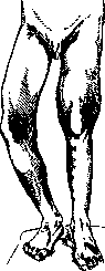
The most commonly used methods are excision of the cyst wall and curettage of its cavity; the latter some fill with fat (Frangenheim), others with plaster of Paris and iodoform or pure plaster of Paris (Oehlecker), still others insert periosteum or a piece of bone into it, and finally some simply suture the wound, leaving the cyst cavity filled with blood, and all these measures lead to bone restoration, since the preserved periosteum has great regenerative capacity. However, not always with these conservative methods does everything go smoothly, sometimes a fracture in the area of the cyst ends in pseudoarthrosis, and in rare cases recurrences of the cyst are observed after apparent recovery. Therefore, in recent years, some recommend more radical treatment: 1) in spontaneous fractures, thorough curettage of the cyst cavity should be performed; 2) in cysts without fracture, the same can be done, and in both cases it is useful to insert bone pins after curettage, both for the purpose of reinforcing the remaining bony shell and for the purpose of stimulating regenerative activity of the bone; 3) in large lesions with significant bone swelling and especially when the fibrous tissue has a tendency to vigorous growth, subperiosteal resection with replacement of the defect with a transplant from the tibia or fibula is indicated. Bone restoration in such cases usually proceeds without complications and radically cures the patients. Giant-cell tumors (the so-called 'Riesenzelltumoren' of German authors) can be more conveniently separated into a separate group based on some features. For a long time they were considered giant-cell sarcomas. Subsequently it became clear that the course of these tumors is not accompanied by any clinical symptoms characteristic of true sarcoma. The tumors grow very slowly (5-8-10 years), do not give metastases either to the lymph glands or to distant organs, and do not recur after their radical removal. Their benign course was already known to Nélaton, Volkmann, and Esmarch. These tumors affect people aged 20-40 years, rarely in younger or older age. Women are affected slightly more often than men. About 1/4 of cases occur in flat bones, 3/4 in tubular bones. Unlike bone cysts, giant-cell tumors are located in the epiphyses of bones. The tumor often occupies the distal end of the radius, the distal end of the femur, and often only one condyle is affected, then the proximal end of the tibia and the trochanters of the femur. Among flat bones, the scapula and pelvic bones are most often affected. The lower jaw is often affected - 10% of all cases (Reinberg). A characteristic feature of these tumors is their singularity and isolation. The affected end of the bone becomes markedly thickened, which interferes with the normal function of the joint (see figure). The skin over the tumor is usually shiny, the subcutaneous veins are somewhat dilated, palpation is painful, and a parchment-like crunch is felt. On the X-ray, the following is observed: the epiphyseal end of the bone is swollen, has the appearance of a hemisphere or candelabrum, the tumor reaches the cartilage and stops here; the tumor grows in all directions, but mainly towards the diaphysis; the structure of the tumor on the X-ray represents irregular honeycombs, giving the appearance of many small cysts separated by septa intertwining in all directions [see separate table (pp. 102-104), figs. 1 and 2]. The cortical substance is thinned, and in large tumors it is completely resorbed and disappears. There is no reaction from the surrounding bone, and no periosteal deposits. The bone marrow canal is separated by a bony septum.
The most commonly used methods are excision of the cyst wall and curettage of its cavity; the latter some fill with fat (Frangenheim), others with plaster of Paris and iodoform or pure plaster of Paris (Oehlecker), still others insert periosteum or a piece of bone into it, and finally some simply suture the wound, leaving the cyst cavity filled with blood, and all these measures lead to bone restoration, since the preserved periosteum has great regenerative capacity. However, not always with these conservative methods does everything go smoothly, sometimes a fracture in the area of the cyst ends in pseudoarthrosis, and in rare cases recurrences of the cyst are observed after apparent recovery. Therefore, in recent years, some recommend more radical treatment: 1) in spontaneous fractures, thorough curettage of the cyst cavity should be performed; 2) in cysts without fracture, the same can be done, and in both cases it is useful to insert bone pins after curettage, both for the purpose of reinforcing the remaining bony shell and for the purpose of stimulating regenerative activity of the bone; 3) in large lesions with significant bone swelling and especially when the fibrous tissue has a tendency to vigorous growth, subperiosteal resection with replacement of the defect with a transplant from the tibia or fibula is indicated. Bone restoration in such cases usually proceeds without complications and radically cures the patients. Giant-cell tumors (the so-called 'Riesenzelltumoren' of German authors) can be more conveniently separated into a separate group based on some features. For a long time they were considered giant-cell sarcomas. Subsequently it became clear that the course of these tumors is not accompanied by any clinical symptoms characteristic of true sarcoma. The tumors grow very slowly (5-8-10 years), do not give metastases either to the lymph glands or to distant organs, and do not recur after their radical removal. Their benign course was already known to Nélaton, Volkmann, and Esmarch. These tumors affect people aged 20-40 years, rarely in younger or older age. Women are affected slightly more often than men. About 1/4 of cases occur in flat bones, 3/4 in tubular bones. Unlike bone cysts, giant-cell tumors are located in the epiphyses of bones. The tumor often occupies the distal end of the radius, the distal end of the femur, and often only one condyle is affected, then the proximal end of the tibia and the trochanters of the femur. Among flat bones, the scapula and pelvic bones are most often affected. The lower jaw is often affected - 10% of all cases (Reinberg). A characteristic feature of these tumors is their singularity and isolation. The affected end of the bone becomes markedly thickened, which interferes with the normal function of the joint (see figure). The skin over the tumor is usually shiny, the subcutaneous veins are somewhat dilated, palpation is painful, and a parchment-like crunch is felt. On the X-ray, the following is observed: the epiphyseal end of the bone is swollen, has the appearance of a hemisphere or candelabrum, the tumor reaches the cartilage and stops here; the tumor grows in all directions, but mainly towards the diaphysis; the structure of the tumor on the X-ray represents irregular honeycombs, giving the appearance of many small cysts separated by septa intertwining in all directions [see separate table (pp. 102-104), figs. 1 and 2]. The cortical substance is thinned, and in large tumors it is completely resorbed and disappears. There is no reaction from the surrounding bone, and no periosteal deposits. The bone marrow canal is separated by a bony septum.
Differential diagnosis must be made with bone cyst, osteochondroma, and mainly with osteogenic sarcoma. These diseases and their characteristic features have been discussed above. The greatest difficulty lies in distinguishing giant cell tumor from sarcoma. To resolve this question in difficult cases, it is necessary to resort to X-ray, biopsy, and clinical course. Treatment is surgical and conservative. It must consist in careful enucleation of the tumor. Recovery after such an operation reaches 90%. In large tumors with extensive destruction, resection with bone graft transplantation is necessary, and in severe cases, amputation is not excluded. X-ray therapy has a certain effect on tumors—it suppresses and stops their growth. The generalized form of fibrous osteodystrophy (osteodystrophia fibrosa generalisata Recklinghanseni) affects many parts of the skeleton. In some cases, individual bones and even entire limbs remain unaffected by the process until death, but cases are observed in which, over time, all bones become affected. More frequently and constantly, the bones of the pelvis and femur, tibia, and humerus, the bones of the cerebral and facial skull, the bones of the forearm, metacarpals, metatarsals, ribs, and vertebrae are changed. The lesions are not symmetrical. In each individual case, there is its own combination of affected bones. On the skull, most often the parietal bones are changed, while the base of the skull is usually not involved in the process. In Germany, generalized O. f. occurs more frequently than in other countries. In France, Italy, and England, there are isolated reports, and in the USSR, cases are known: Abricosov-1, Rusakov-2, Khendrikov-1, Rapoport-1. By 1926, up to 70 observations had been published in world literature (Frangenheim), and in 1929, figures of 80 (Rapoport) and 90 (Greinberg) are cited. The disease is observed in the age range of 10-50 years. Women get sick more often than men, which is associated with pregnancy. The onset of the disease sometimes refers to early childhood. Clinically, it is barely noticeable. Patients begin to complain of small dragging rheumatoid pains in the bones and joints. The first objective signs are often noticed accidentally, for example, while combing the hair, a swelling in the parietal region is suddenly discovered. With the development of the disease, with sharp changes in the bones, sometimes attacks of extremely severe pains occur, so that even simple contact with the bed cannot be tolerated by patients. Patients weaken, cannot get out of bed, move, or eat. Sometimes even in these severe cases, remissions occur. Objective symptoms are manifested as thickenings in various places of the tubular bones, and in severe cases, of flat bones as well. On palpation, a floating sensation and parchment-like crunch are often felt. The surrounding soft tissues are not involved in the process, and there is never any inflammation or redness of the skin. A characteristic feature is that the thickenings lie in the metaphyses or diaphyses, while the epiphyses remain untouched. Due to the impoverishment of lime, the bones become soft and deform under the action of mechanical forces. Spontaneous fractures occur, healing with disfigurement and shortening of the limbs. Flattening of the vertebral bodies occurs, resulting in kyphosis in the thoracic part of the spine and lordosis in the lumbar part. Lateral compression of the chest, deformation of the pelvis—"heart-shaped" and "beak-shaped" pelvis, and flattening of the frontal part of the skull appear. In terms of metabolism, low or normal calcium content in the blood and a sharp increase in calcium excretion in urine and feces are noted. Sometimes stones form in the urinary tract (Abricosov). Diagnosis can be made on the basis of clinical signs, but full certainty is given by X-ray examination of many bones. On the X-ray, it is seen that the thickening of the bone occurs due to the growth of numerous cystic chambers occupying the bone marrow canal and also penetrating the cortical layer. The cysts often lie next to each other or merge with each other. The cortical substance is thinned. There is no reaction from the periosteum. Periosteal deposits are noted only in the area of pathological fractures at the site of bone callus. In flat bones, for example in the bones of the skull, translucent cystic cavities are also outlined on the X-ray, thinning the cortical substance and bulging the bones in the form of large nodular tumors. The affected vertebrae appear translucent, enclosed in thin bone shells. Along with cysts, there are dense fibrous tumors in the bones, which are difficult to distinguish radiologically. In difficult cases, only a trial puncture can resolve this question. Differential diagnosis is not difficult. The question of osteomalacia, multiple cancer metastases, and myelomas may arise. In osteomalacia, there are no cyst-like bone defects, the deformities are symmetrical, and all bones are affected. In cancer metastases and myelomas, small, sharply limited round defects are noted on X-ray, there are no arcuate curvatures of the bones, and there is no thinning of the cortical substance. The prognosis in osteodystrophia fibrosa generalisata is unfavorable. Due to changes in the skeleton, the general condition of patients greatly suffers, breathing is difficult, heart function is impaired, and patients often die from bronchitis and pneumonia. Treatment is unsuccessful and, due to the unclear etiology, is conducted by trial and error. Some authors have seen improvement from the use of strontium lactate and strontium phosphate. Rapoport from the clinic of Prof. Grekov reports significant success from the use of Calc. glycerophosph. (2.0 pro die) and X-ray irradiation of individual parts of the skeleton. A patient who was almost immobile, after half a year, began to get up, after 1 year began to move with the help of a cane, and after 1 year 9 months, could walk freely. Mandl from the Hochenegg clinic performed transplantation of 4 parathyroid glands from an accidentally deceased person to a 38-year-old patient suffering from osteodystrophia fibrosa generalisata. No effect was obtained. Then, examining the patient's own parathyroid glands and finding a tumor in the upper one, he removed it. Under the microscope, the tumor was suspicious for malignancy. Immediate improvement followed. Calcium excretion in urine sharply decreased. After 4 months, lime deposits were seen in the nails on X-ray. The patient, whose lower limbs were as if paralyzed, who could not lift or bend his legs in the knees while lying down, and suffered from severe pain, after the operation could walk on crutches, could sit in bed and on a chair, lift his legs while lying down and bend them at the knees. In the patient Gold with a severe form of osteodystrophia fibrosa generalisata, after removal of the tumor of the upper parathyroid gland, a similar clinical effect occurred, but no radiological improvement followed. Beck after removing a parathyroid gland tumor in a woman observed a rapid decrease in calcium excretion in urine. On the 5th day, the patient developed tetany, and on the 20th day—death. At autopsy, no other parathyroid glands were found. Eggers in a woman with a severe form of osteodystrophia fibrosa generalisata removed the tumor of the lower parathyroid gland and subjected individual bones to X-ray irradiation. Clinical improvement, but after 17 months, the patient died from suffocation (fibromyxoma of the false vocal cord). Microscopic examination of foci of damage did not reveal any improvement. However, these individual reports are exceptions from the general gloomy background of therapy for osteodystrophia fibrosa generalisata. In a number of cases, the disease occurs without enlargement of the parathyroid glands. Some authors do not consider it advisable to either extirpate the parathyroid glands or use X-ray therapy.
V. Braytsev.
Related articles
Mentioned in
Cite this page
“Fibrous Osteitis.” Soviet Medical Encyclopedia. English translation of Bolshaya Meditsinskaya Entsiklopediya, 1st ed. (Moscow, 1928–1936), ed. N. A. Semashko. https://sovietmedicalencyclopedia.pages.dev/article/fibrous-osteitis/