Osteitis Deformans
Historical document, translated for reference. It reflects medical knowledge of the 1920s–30s and is not medical advice.
Summary
This article from the 1928–1936 Great Medical Encyclopedia describes osteitis deformans, or Paget's disease of bone, a condition characterized by bone deformation and structural changes. It details the pathological anatomy, including the characteristic mosaic structure of bone tissue, and discusses the then-current theories regarding its etiology, which remained largely unknown at the time.
Encyclopedia article (1928–1936)
OSTEITIS DEFORMANS (ostitis deformans), Paget's disease, a disease of the skeletal system, first described in detail in 1876 by the English physician James Paget under the name "osteitis deformans." Changes in osteitis deformans are expressed in the deformation of bones and are characterized by a specific clinical and pathological-anatomical picture. Before the widespread use of X-rays, the disease was considered very rare, but at the present time, the number of published and described cases of osteitis deformans is growing quite rapidly. To date, over 600 clinical observations have been published, of which about 30 are in the USSR (including 3 autopsy cases). The disease is obviously encountered more often than it is diagnosed. Pathological anatomy. Most often, changes in osteitis deformans are noted in long tubular bones, especially of the lower extremities, in the bones of the skull, and in the clavicle. According to Schirmer, these changes, in order of frequency of localization, are arranged as follows: skull, tibia, femur, pelvis, vertebrae, clavicle, ribs, radius, etc. The main changes consist of an increase in the length and volume of the affected bones and in their deformation; the affected bones become more porous and acquire a softer consistency; sometimes, as a result of significant new formation of bone tissue, its compaction (eburnation) is noted, especially in the bones of the skull. The most drastic changes are noted in the skull, in particular the cranial vault. The circumference of the skull increases by 6–8–12 cm, sometimes reaching 72 cm (Koch); the bones of the cranial vault thicken sharply (up to 2.5 cm—observation by Shlapobersky and Frank-Kamenetsky) [see separate table (cols. 127–128), fig. 11]. In long tubular bones, the diaphyses are usually affected, with the bone appearing thickened, with an uneven surface, and often curved (e.g., the tibia is curved outward and anteriorly) (fig. 1). A number of new works, in contrast to previous views, state that in osteitis deformans, the epiphyses of tubular bones are also gradually involved in the process. The clavicles are usually increased in volume (thickened), especially in their middle part, and the curves are more sharply pronounced. The bones of the foot and hand are affected significantly less often than others. In the bones of the pelvis, sharp changes are also noted. On a cross-section of the affected bones, uneven porosity of the cortical layer is noted with a significant number of gaps between the bone trabeculae; when studying X-rays of bone sections, a significant change in the "architectonics" of the bones is noted (figs. 7–10): the cortical layer begins to approach the structure of the medullary layer, and small cavities are noted here and there. Microscopic changes in osteitis deformans, according to Schirmer, can be reduced to the following: 1) resorption of compact bone tissue with a subsequent increase in the size of the Haversian canals; 2) new formation of bone tissue in the affected parts and in adjacent healthy areas, with the newly formed bone partly not calcifying and subsequently being resorbed; 3) partial transformation of bone marrow into a vascular-rich connective tissue containing fat, giant cells, leukocytes, etc. It is necessary to emphasize that all these changes are localized predominantly in the cortical layer of the bone. The process of resorption begins in the peripheral layers of the bone (subperiosteally) and gradually develops in the direction of the medullary cavity. Bone resorption occurs predominantly by way of lacunar resorption, i.e., the formation of Howship's lacunae with numerous giant cells (osteoclasts); in the process of resorption, the bone trabeculae decrease in size, take on diverse shapes, and, due to the presence of Howship's lacunae, have a surface that looks as if it has been gnawed. The process of halisteresis (see) is, in the opinion of the vast majority of authors, absent in osteitis deformans, although Kaufmann and Meyer observed halisteresis in small amounts in this disease. In a number of cases, the formation of Volkmann's perforating canals has been noted (see Bone). Due to the processes of bone resorption, the Haversian canals can widen, merge with each other, and form spaces of significant size, filled with fatty or fibrous bone marrow. Simultaneously with the process of bone resorption, there occurs a significant new formation of bone substance, which, according to Recklinghausen and Kaufmann, proceeds from the side of the bone marrow. This new formation occurs either as a result of the appearance of osteoblasts and the formation of osteoid tissue or by direct metaplasia of connective tissue into bone. Regarding the question of the role of the periosteum in the new formation of bone in osteitis deformans, until recently, the generally accepted point of view denied periosteal osteogenesis, although Goodhart (1878), Paget, and Ziegler already admitted the possibility of periosteal bone formation in osteitis deformans. In recent years, a number of authors (Freund, Schmorl), in contrast to their previous point of view, indicate that periosteal osteogenesis in osteitis deformans can take place on a large scale. The newly formed bone remains devoid of lime for a long time and stains light red when stained with hematoxylin-eosin. Of significant interest is the simultaneous existence in the same areas of bone of processes of new formation and resorption of bone. It is often possible to detect pictures of resorption of newly formed bone, and sometimes resorption and new formation of bone take place along the length of the same bone trabecula; in some places, even resorption of immature bone—osteoid tissue—is noted. Due to the repeated alternation of the processes of new formation and resorption, the bone trabeculae consist of a large number of small bone fragments, delimited from each other by connective lines (Kittlinien), which randomly intersect the bone substance in different directions. On cross and longitudinal sections, a picture of a more or less elegant mosaic is obtained, which has received the name "mosaic structure" (fig. 3) (Schmorl; 1926). Regarding the question of changes in the bone marrow in osteitis deformans, at the present time, the point of view that considers changes in the bone marrow to be a secondary, sequential phenomenon is beginning to prevail. This point of view is expressed particularly clearly by Schmorl (1930), who asserts that one can observe cases of osteitis deformans with far-advanced changes in the bones, while the bone marrow remains unchanged. In the majority of cases of osteitis deformans, the bone marrow consists of fibrous connective tissue poor in cellular elements and fatty tissue (fig. 4). A number of authors have noted the development of fibrous tissue directly around the bone trabeculae (Bindegewebsmantel, Askanazy). Thus, it is necessary to come to the conclusion that in osteitis deformans, one is dealing with a process of rapidly proceeding restructuring of bone tissue as a result of alternating processes of new formation and resorption of bone, with the phenomena in the bone marrow being of a secondary, sequential character. The etiology of osteitis deformans has remained unexplained until recently. Czerny and Paget considered osteitis deformans to be a disease of an inflammatory nature; hence the name of the process "ostitis" (Czerny), "osteitis" (Paget). But the clinical and especially the pathological-anatomical pictures of this disease do not provide supporting points in favor of an inflammatory origin of the disease. A number of French authors, basing their conclusions on a positive Wassermann reaction in a certain number of such cases, believed that syphilis lies at the foundation of this disease. But examination of a significant number of patients with osteitis deformans does not provide sufficient grounds to confirm the syphilitic origin of this disease. A lot of misunderstanding is caused by the fact that in a number of reports, authors classify unclear cases as osteitis deformans, including cases that turn out upon further investigation to be atypical forms of lues. There are a number of attempts to link cases of osteitis deformans with trauma, rheumatism, etc. A number of authors, who considered ostitis deformans and ostitis fibrosa to be two forms of the same process, linked the origin of these diseases to changes in the glands of internal secretion, in particular in the parathyroid glands (their adenomas, etc.), although they noted at the same time that lesions of the parathyroid glands are usually found only in cases of ostitis fibrosa. At the present time, when the opinion about the difference between these two diseases is beginning to prevail, the absence of changes in the parathyroid glands in osteitis deformans even serves as one of the distinguishing features of this disease from ostitis fibrosa. Some French authors link this disease with occupational hazards (e.g., poisoning by mineral acids), noting it in painters, tinsmiths, etc. There is a tendency to classify this disease also in the group of vegetative disorders, in the group of dystrophies (osteodystrophia). Of significant interest is the point of view regarding the influence of nutrition on bone changes; thus, some authors in cases of experimental pseudorickets in dogs found a picture in the bones that resembled osteitis deformans; it is possible that further research in this area will bring some clarity to the question of the etiology of osteitis deformans. Clinical picture.
The disease usually develops in the second half of the patients' lives and is observed more often in men than in women. Isolated cases of familial disease have been noted. Based on their distribution in the body, all cases of osteitis deformans can be subdivided into monostotic and generalized forms. In a significant number of observed cases, monostotic forms of the disease prevail; generalized forms are encountered less frequently. The clinical picture in typical cases of the generalized form is very Figure 1. Photograph of the bones of the left lower extremity with osteitis deformans. Sharp change in all bones with thickening and rarefaction. Spontaneous fracture of the femur in the upper third. Figure 2. Radiograph of the skull with osteitis deformans. Spotted structure of the bone. The bone has the appearance of a 'curly head'. Figure 3. Microscopic picture of bone in osteitis deformans. Separate fragments of bone are visible, delimited by connective tissue. Picture of mosaic structures. Figure 4. Microscopic picture of bone marrow in osteitis deformans. The bone marrow consists of fibrous and fatty connective tissue. Figure 5. Radiograph of the bones of the lower leg in osteitis deformans. Sharp changes in the structure of the tibia with the disappearance of the medullary canal. Figure 6. Radiograph of the femur (from a skeleton) with osteitis deformans. A spotted, spongy structure of the bone is clearly visible. Spontaneous fracture. Figure 7. Radiograph of a bone section of the skull in osteitis deformans. Significant thickening of the bone; fine porosity of the cortical layer; smoothing of the difference between the cortical and medullary layers; sharp change in the architectonics of the bones. Figure 8. Radiograph of a bone section of the skull in a normal state. A distinct picture of cortical and medullary substance with a clear bone structure. Figure 9. Radiograph of a bone section of the tibia in a normal state. Compact cortical layer. Figure 10. Radiograph of a bone section of the tibia in osteitis deformans. Significant thickening and porosity of the cortical layer. Figure 11. Cross-section of the cranial vault in a case of Paget's disease. Slight thickening of the bones (~2 cm) with porosity inside. (To the illustration for the article Osteitis Deformans.)
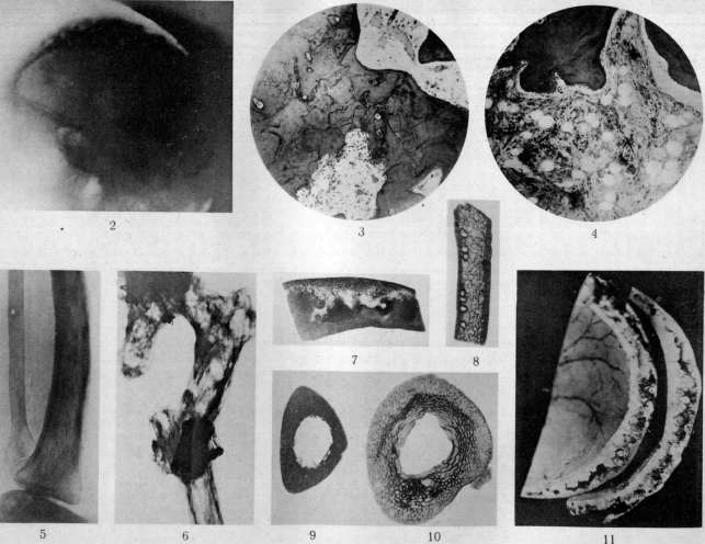
To the article Osteitis Deformans. G O Z N A K. characteristic. Basically, it is the result of processes of deformation of the skull, long tubular bones, and the spine. The circumference of the head begins to increase gradually; patients have to purchase headwear of increasingly larger sizes. In one patient observed by Paget, the circumference of the head increased by 12 cm over 32 years. The bones of the facial skull are usually not affected, as a result of which a small face remains on an excessively large head, usually with a convex forehead. Changes in the bones of the extremities can be symmetrical, asymmetrical, and crossed. In the lower extremities, the changes are usually more sharply pronounced than in other parts. The tibiae, one or both, are sharply thickened throughout their entire length, curved anteriorly and laterally. The thickening usually reaches its greatest degree in the upper part of the tibiae. The fibula is usually not curved. In the femur, the curvature is more noticeable than the thickening. Clinically, lesions of the bones of the foot are usually not noted. In the upper extremities, which are less frequently affected by the process, changes are usually localized in the humerus; in lesions of the forearm, the radius usually suffers. In the bones of the hand, changes are usually not clinically detected. Deformation of the ribs is noted, with the intercostal spaces narrowing. Changes in the spine manifest as a more or less pronounced kyphosis, usually observed in the thoracic region. The curvature of the bones of the lower extremities leads to a significant relative shortening of the latter; as a result of this, and also due to the existing curvature of the spine, the relative length of the torso decreases (up to 30 cm in Wilson's case). The arms of such patients hang down almost to the knees and seem disproportionately long; the head, due to the weight of the cranial vault and the weakness of the musculature, drops onto the chest; in appearance, the patient somewhat resembles an anthropoid ape. In monostotic forms, the tibia is most often affected. Combined lesions of the skull and tibiae are not uncommon. Deformations of the bones, especially of the lower extremities, in far-advanced cases can cause severe movement disorders. There are often neuralgic pains in the affected extremities, sometimes reaching high intensity. In a number of cases, spontaneous fractures are observed. The regular transverse direction of such fractures is very characteristic. Isolated cases of joint involvement in osteitis deformans have been described. However, confusion with arthritis deformans is possible here. Internal organs, including organs of internal secretion, are usually not affected. Many authors note sharply pronounced phenomena of general arteriosclerosis in osteitis deformans. Calcification of vessels can be so sharply pronounced that the vessels are contoured on radiographs of the extremities. However, there are no sufficient grounds to consider arteriosclerosis pathognomonic for osteitis deformans. Here, there is obviously a simultaneous existence of both processes, which is explained only by the advanced age of most such patients. The blood picture usually does not change. Urinalysis does not yield noticeable deviations from the norm. Examination of calcium metabolism also does not yield deviations from the norm: the amount of calcium in the blood, urine, and feces does not change significantly, just as the amount of phosphorus in the blood does not (Snapper). X-ray examination is of decisive importance for diagnosis, since in typical cases the X-ray picture is so characteristic that it cannot be confused with any other disease. Changes in the skull and long tubular bones are especially characteristic. The bones appear thickened, their structure is variegated; along with areas of sclerosis (eburnation), there are small foci of rarefaction. A picture of a spotted, spongy structure is obtained [see separate table (pp. 127-128), Fig. 6]. The areas of rarefaction have a resemblance to cotton flakes. Such a spotted structure on a radiograph is, as it were, a coarse macroscopic substrate of the mosaic structures characteristic of the microscopic picture of the process. Upon X-ray examination, it is discovered that the bones of the cranial vault are thickened, and the normal relationships between the lamina externa and interna and the diploe have disappeared. The surface of the bone has blurred contours, not sharply separated from the soft tissues. The structure of the bone is characterized by a disorderly alternation of lighter and darker areas and has a spongy character. Such X-ray pictures are compared to the curly head of a Negro [see separate table (pp. 127-128), Fig. 2]. Upon more detailed examination, changes are also established in the base of the skull—a decrease in the depth of the cranial fossae, flattening of the sella turcica, thickening of the bones of the base, and blurring of their contours. The tibia appears thickened 2-3-4 times; the medullary cavity is usually narrowed. The boundaries of the bone with soft tissues are not sharp. The substance of the bone appears as a disorderly mixture of denser and loosened areas and has a characteristic spongy appearance [see separate table (pp. 127-128), Fig. 5]. In individual cases, small bone cysts are noted. The fibula is usually not curved and resembles the string of a bow, the arc of which is the curved tibia. Changes in the femur are analogous to changes in the tibia. Not infrequently, changes in the small bones of the hand and foot are discovered on radiographs. Changes in the spine are rarely established radiologically during life. They are usually revealed during the examination of autopsy specimens (Hallermann). The X-ray picture can, however, present significant difficulties in the diagnosis of initial and monostotic forms. One of the manifestations of the initial forms of the disease is obviously Schüller's osteoporosis circumscripta, which consists in the appearance of fairly extensive isolated areas of rarefaction in the bones of the skull, predominantly adjacent to the base, sharply delimited from normal areas by a jagged or scalloped line. The diagnosis is made in some cases for the first time upon the occurrence of a spontaneous fracture, sometimes when the patient seeks medical attention for another disease. Only in far-advanced cases do the deformation of the extremities and pain lead the patient to the doctor. Regarding differen-
OSTEITIS
For differential diagnosis, it is necessary to keep in mind syphilis and fibrous osteitis. In syphilis, the characteristic changes of D. d. in the skull are usually not observed; as for the very frequent changes in the tibiae in D. d., the characteristic feature is their curvature to the lateral side and forward, in contrast to syphilis, where the curvature is usually directed forward. Furthermore, in D. d., periostitis, so characteristic of syphilis, is not observed, and finally, in D. d., there is a typical spotted structure of bone tissue, whereas in syphilis, hyperostosis (eburnation) is usually observed. However, with isolated lesions of the tibiae, differential diagnosis with syphilis can sometimes be difficult. As for ostitis fibrosa, it is necessary first of all to establish that both of these diseases are independent nosological units (Schmorl, Snapper and others). It should be noted that the clinical and X-ray pictures of both of these diseases have always been considered different, whereas the patho-anatomical picture of both processes was identified by many researchers up to the most recent time (see Fibrous osteitis). D. d. usually develops in the second half of life, while fibrous osteitis develops in the first three decades. For D. d., diffuse involvement of the bones of the cranial vault is typical, whereas in fibrous osteitis, the bones of the skull are usually not diffusely affected. Multiple fractures, typical for fibrous osteitis, are not encountered in D. d. While D. d. often proceeds without pain, sharp pain in the affected bones is characteristic of fibrous osteitis. Calcium metabolism, usually unchanged in D. d., is sharply disturbed (increased) in fibrous osteitis. In D. d., internal secretion organs are usually not affected, whereas in fibrous osteitis, enlargement of the parathyroid glands is described. Radiologically, in typical cases, there is also a sharp difference between the two processes: the spongy, spotted structure of bone tissue in D. d., in contrast to the malarial bone tissue with large bone cysts, typical for fibrous osteitis. Outcome. The disease lasts for decades and by itself usually does not lead to a fatal outcome. In individual cases, the development of sarcomas in the affected bones has been noted. Some authors even consider D. d. a presarcomatous condition. However, this view is hardly correct. Most patients die from intercurrent diseases.
Treatment in general is unsuccessful and, due to the unclear etiology of the process, is conducted empirically, by trial and error. There are a number of proposals for treating such patients with preparations of internal secretion iron, such as ovarin, extracts of the thyroid gland, adrenal glands, etc.; in some cases, apparent improvement of general symptoms could be achieved, but no long-term improvement or arrest of the process could be spoken of. Some authors have tried treatment with preparations of iodine, quinine, phosphorus with cod liver oil, etc., but without noticeable results. In view of the connection between D. d. and syphilis assumed by some French authors, attempts at antiluetic treatment were made without particular success. Schlesinger proposed to conduct radiotherapy of the affected bones. To correct deformities in D. d., a number of surgical measures (osteotomies, resections of bone sections, etc.) were proposed and carried out, however, with rather frequent unsuccessful outcomes (slow formation of callus, tendency to form false joints, etc.).
Related articles
Mentioned in
Cite this page
“Osteitis Deformans.” Soviet Medical Encyclopedia. English translation of Bolshaya Meditsinskaya Entsiklopediya, 1st ed. (Moscow, 1928–1936), ed. N. A. Semashko. https://sovietmedicalencyclopedia.pages.dev/article/osteitis-deformans/