Clavicle
Historical document, translated for reference. It reflects medical knowledge of the 1920s–30s and is not medical advice.
Summary
The clavicle is a paired bone of the shoulder girdle that articulates with the sternum and scapula in humans. This article covers its embryological development, anatomical structure, and functional significance in the shoulder complex.
Encyclopedia article (1928–1936)
CLAVICLE (clavicula), a paired bone of the shoulder girdle, articulating with the sternum and scapula in humans. Phylogenetically, the clavicle represents a dermal bone, first appearing in bony fishes in the dermal (connective tissue) part of the skin. Only at the next stage of vertebrate development, in amphibians, does the clavicle lie deeper and cover parts of the skeleton forming the primary shoulder girdle on a cartilaginous basis. In lower mammals (monotremes), the primary shoulder girdle consists of the scapula and a powerful beak-shaped (coracoid) bone extending to the sternum (Figure 1). Subsequently, the coracoid bone gradually reduces and transforms into the coracoid process of the scapula. Only the clavicle remains as the connecting link between the scapula and sternum. The episternum—another, also dermal, i.e., not preformed by cartilage bone, which secondarily enters into the composition of the shoulder girdle similar to the clavicle—initially has a T-shape. In humans, the intra-articular disc of the sternoclavicular joint is a remnant of the horizontal branch, while the suprasternal bones, which are far from constantly found at the upper edge of the sternum, represent remnants of the vertical part of the episternum (Fig. 2). In ungulates and carnivorous animals, the clavicle is underdeveloped or absent, as for example in cattle, horses, dogs. The primary ossification center of the clavicle appears in the human embryo already at the end of the 1st week, consequently at a time when bone tissue is not yet present anywhere else. The middle part of the clavicle forms metaplastically from connective tissue; the terminal sections have a cartilaginous basis (Kulchitsky). A secondary epiphyseal-type ossification center appears on the sternal end of the clavicle approximately at the twentieth year of life and soon (after 10-15 months) fuses with the main mass of the clavicle (Testut).
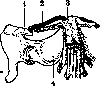
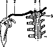
Figure 1.
Figure 1. Coracoid (4); 1-acromion; 2-clavicle; 3-episternum. Figure 2. Suprasternal bone (3); 1- acromion; 2-clavicle; 4-intra-articular disc; 5-manubrium of sternum.
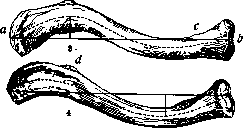

The clavicle (Figs. 3, 4) has the shape of an elongated S, averaging 12-15 cm in length in an adult. The medial convexity faces forward and occupies 2/3 of the length. The lateral third is concave in front and ends in a flattened from top to bottom acromial plate of spongy structure. The middle part of the clavicle is cylindrically narrowed, resembles a tubular bone, and has a bone marrow canal. The sternal spongy end is thickened into a triangular prism with blunted edges and a roughness (tuberositas costalis) on the lower side for the attachment of the costoclavicular ligament. An even greater tuberosity (tuberositas coracoidea) is located on the lower side of the acromial end. It is caused by the attachment of the coracoclavicular ligament, consisting of 2 parts: the trapezoid ligament lying in front and extending in the sagittal direction, and the conoid ligament located behind, in the frontal plane. Both ligaments suspend the entire weight of the upper limb and scapula on the clavicle. Thus, the acromioclavicular joint remains unloaded, and the freedom of its dynamic function is ensured. The mobility in this last joint is considerable due to the thickness of the fibrocartilaginous layer between the bone ends and the weak tension of the strong capsule (Figs. 5 and 6). The layer often forms a more or less isolated disc. On the sternal end of the clavicle, the intra-articular disc is constantly present, has a very irregular shape, is obliquely positioned, with its upper edge closer to the midline of the body, and divides the joint into two chambers. It represents, as mentioned above, a rudimentary bone. The sternoclavicular joint allows active excursions of the acromial end of the clavicle within the dimensions of an oval 10 cm high and 12 cm long (Fig. 7). The remarkable mobility, elasticity, and strength of the joint are due to the powerful development of its ligamentous apparatus.
Fig. 6. fii cartilage tissue. In the posterior-superior part of the joint—cartilage covering of the clavicular end, the transverse diameter of the articular disc and the lining of the sternal fossa taken together form an elastic buffer system up to 13 mm thick. With the first rib, the clavicle is connected by a two-layered, rhomboid costoclavicular ligament. Between the two layers of this ligament, a well-developed serous cavity is sometimes found. In front and behind, the sternoclavicular joint is reinforced by accessory capsular ligaments. Across the jugular notch, the interclavicular ligament is tightly stretched, which, along with the sternoclavicular ligaments, holds the clavicle and the entire shoulder girdle with the upper limb suspended from it in a horizontal position even when all muscles are cut. The ligaments allow excursions of the lateral end of the clavicle only forward and upward (Figs. 5, 7). In rare cases, the lower surface of the clavicle and the apophysis of the coracoid process are articulatively ^rs^m
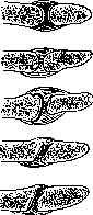
Fig. 5.
On the surface there is a longitudinal groove for the short (10 cm) t. subclavius. Here also is located a small opening for the nourishing artery bone, which goes to the lateral half of the clavicle. At the acromial end of the C., from above in front, the m. deltoideus is attached, from above behind—the m. trapezius. For active movements of the upper limb, the function of both clavicular joints, the sternal and the scapular, is of very great importance. Without the participation of these joints, the shoulder, regardless of the choice of initial position, cannot be raised above the horizon either forward or to the side. To the action of the m. deltoideus on the shoulder must be added the rotation of the inferior angle and the displacement of the entire scapula forward and upward in order to obtain elevation. The mm. serratus ant., trapezius, levator scapulae with their antagonists (rhomboidei, pectoralis major and minor) play the most important role here. Active retroversion of the shoulder is generally not performed in the shoulder joint, but only in the claviculo-acromial joint, mainly thanks to the m. latiss. dorsi. Mechanically playing the role of a hanger for the shoulder and a strut between it and the thoracic cage, the C., while increasing the supporting capacity of the shoulder girdle, regulates the dynamics of the free upper limb. Functionally belonging to the latter, the C. in anatomic-topographic relation represents the boundary between the cervical region and the trunk.—Corresponding to the middle third of the C., between the attachment of the deltoid and the pectoralis major muscles, behind the C., in front of the I rib, is located the subclavian artery with the同名 vein and with the brachial nerve plexus. For supraclavicular anesthesia according to Kulenkampf (Kilenkampff), the needle is inserted in the direction toward the II-III thoracic spinous process in a sitting patient somewhat laterally from the site of pulsation of the artery at the upper edge of the C. and is advanced deeply until clear paresthesia is obtained in the fingers. To avoid damage to the dome of the pleura, it is recommended (Mulley) that the site of insertion be three transverse fingers above the middle third of the C. In fractures of the C., the muscular belly of the m. subclavii and its strong fascia protect the vessels and nerves from injury by bone fragments. By pulling the shoulder backward and downward, the vascular-nerve bundle can be compressed between the C. and the I rib. This maneuver achieves preliminary arrest of arterial hemorrhage. Thus, sometimes due to negligence, when the upper limb hangs over the edge of the operating table, paralysis under anesthesia occurs.—Wide access for the surgical treatment of traumatic aneurysms and nerve injuries in this area, especially in gunshot wounds, is obtained only by temporary resection of the C. The small supraclavicular fossa (Zarig'a) between the sternal and clavicular heads of the m. sternocleidomastoidei occasionally serves as the site for finding and ligating the common carotid artery, more often for auscultation of the jugular vein. The superficial location of the C. and its support-dynamic connection with the upper limb predispose it to injuries. Injuries from direct violence occur less frequently than indirect injuries from falling on the shoulder or on the outstretched hand and from excessive muscle tension when lifting heavy objects. Fractures of the C. are often observed already in newborn children (according to Muns'y, in 1.3% of cases), and not only in connection with difficult artificial delivery, but also after normal births and with origin still in fetal life. In childhood, in addition to the frequency, what is striking is the slightness and short duration of the disorders, the large number of subperiosteal and incomplete fractures. Among adults, due to the peculiarities of domestic and professional traumatism, fractures of the C. occur in men significantly more often than in women. This fracture is a typical injury of cavalrymen, and the mechanism of its origin is uniform: when falling from a horse to the side, the C. is compressed in the direction of its longitudinal axis and breaks from bending at the weakest point, at the boundary of the middle and outer third.
According to Rahman, among men the 'sternal' type of C. with a large index of medial curvature predominates, while the 'acromial' type or transitional forms are more characteristic of the female sex (fig. 3 and 4). Despite the predisposition of the sternal type to fractures in the medial half, in men, violations of integrity occur, just as in women, usually closer to the acromial third of the C., because the sternal part is stronger due to the parallel arrangement of the trabeculae to the long axis of the bone. The typical displacement (fig. 8) of the medial fragment with the broken end upward depends on the traction of the m. sternocleidomastoidei, while the displacement of the lateral fragment downward and toward the sternum is caused by the action of gravity on the suspended limb and the weight of the co-clavicular ligament and the contraction of the major and minor pectoral muscles. Fractures of the acromial third most often occur from a direct blow from above or a thrust from the front. Fractures of the sternal third occur least frequently, and in their mechanism of origin plays a role the disproportion of rapid contractions of the sternoclavicular-pectoral, deltoid and pectoral muscles, for example, throwing and a wide empty swing or a supraclavicular lever resting on the first rib. Recognition of clavicle fractures presents no difficulties if, in the absence of displacement of fragments, one is guided by locally limited sharp pain on palpation. For the treatment of fractures of the C., if one counts the authors, about 100 bandages have been proposed. The most rational are: 1. The ancient figure-of-eight bandage of Petit, bringing both scapulae closer together. Its action is enhanced (according to Burian) by rubber traction downward at the site of the cross on the back. Or the same bandage is applied from plaster bandages with good modeling of the contours of the shoulder (fig. 9); in children—the Pirogov's jacket bandage following the same principle. Here also belong the stick bandage of Wildbolz (fig. 10) and the S-shaped splint of Hartel. 2. Plaster bandages, elevating and abducting the upper end of the shoulder, leaving the fracture site free (fig. 11) and resting on the iliac crests (fig. 12) or encompassing the thoracic cage together with the healthy shoulder. 3. The bandage of Desault, applied over a shirt with starched bandages,
Fig. 10.
or the bandage of Sayre with adhesive tape strips. 4. The method of Cotteaud, i.e., the horizontal position of the patient with the shoulder hanging down over the edge of the bed for self-reduction, with staying in this position until the formation of the initial callus and with subsequent application of a bandage bandage. 5. Bloodless reduction and operative fixation of fragments in cases not amenable to conservative treatment or complicated, like gunshot fractures, by injury to vessels or nerves.—According to Austrian insurance literature, the duration of treatment averages 10th/a weeks, complete restoration of working capacity only in 52% of cases. A significant role in the remaining functional disorders is played by the adduction contracture of the shoulder joint due to
Fig. 11.
prolonged fixation and improper treatment. Traumatic pseudarthroses occur very rarely and restrict movement so little that Kappis proposed to improve function in ankylosis of the shoulder by creating an artificial false joint within the C. Dislocations occur not only of one, more often the sternal end, but of both ends of one C. (Wesk-mann) and simultaneously of both C. (Riedinger). Congenital anomalies of the C. belong to rare but typical systemic malformations of the skeleton. Clinically they manifest as partial or complete defect of the C. (see Diostosis—fig. 2), combined with anomalies of development of the skull
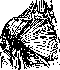

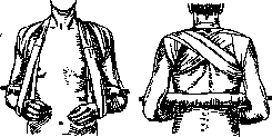
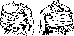
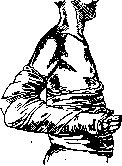
Fig 12 shows a case of hereditary cleido-cranial dysostosis. The genotypic nature of the anomaly is evident from its inheritance according to Mendel's laws: in most cases, it follows a dominant type (in the Carpenter-Langmead case, over four generations); sometimes, it appears to have a recessive mode of inheritance. Parallel to these biological differences between individual cases are clinical differences. For example, there are families in which only a defect of the clavicle is observed, while in others, anomalies of the skull are present. There can be little doubt that under the name hereditary cleido-cranial dysostosis, anomalies are described that differ both biologically and clinically. The opposite anomaly to the hypoplastic cleido-cranial dysostosis was described by Dzerzhinsky under the name familiaris periostal hyperplastic dystrophy. Tower-like (oxycephalic) deformation of the head with a prognathic bird-like facial profile, resulting from premature fusion of the cranial sutures, is combined with thickening of the clavicle and increased curvature of it. Hyperplastic narrowing of the basal foramina, which serve for the passage of blood and lymphatic vessels, leads to increased intracranial pressure and secondarily to atrophy of the optic nerves. Among other anomalies, Bennett described a congenital bifurcation of the clavicle at its acromial end, where one branch articulated normally, while the other was directed backward from the coracoid process and formed a joint with the spine of the scapula. Among acquired diseases of the clavicle, acute and chronic inflammatory processes attract attention early due to the superficial location of the bone. The differential diagnosis of tuberculous and syphilitic lesions, following the general rule, is based on the patient's history, the nature of accompanying symptoms, and serological and other reactions. Arthritides of various (traumatic, infectious, and toxic) etiology are observed predominantly in the sternoclavicular joint and early cause significant pain on movement. Friedrich, in one operated case, found dystrophic changes similar to those in Perthes' disease and Köhler's disease. Among neoplasms with primary localization in the clavicle, sarcomas are most frequently encountered. The prognosis is unfavorable if surgery is not performed early and radically. In relation to tuberculosis and chronic osteomyelitis of the clavicle, total resections should also be more widely applied. Even after complete excision of the clavicle along with the periosteum, the function of the upper limb and the stability of the shoulder girdle are little affected.
Related articles
Mentioned in
Cite this page
“Clavicle.” Soviet Medical Encyclopedia. English translation of Bolshaya Meditsinskaya Entsiklopediya, 1st ed. (Moscow, 1928–1936), ed. N. A. Semashko. https://sovietmedicalencyclopedia.pages.dev/article/clavicle/