Vertebral Column
Historical document, translated for reference. It reflects medical knowledge of the 1920s–30s and is not medical advice.
Summary
The vertebral column is the axial skeleton of vertebrate animals, consisting of a metameric series of more or less movable skeletal elements that protect the spinal cord and serve as attachment points for trunk musculature. This article covers comparative anatomy and ontogeny, describing the development from the notochord in primitive chordates to the complex structure in higher vertebrates.
Encyclopedia article (1928–1936)
156 VI. Professional pathology of the V. . . .......157 Vertebral column, the axial skeleton of vertebrate animals, consisting of a metameric series of more or less movable interconnected skeletal elements that protect the spinal cord and serve as attachment points for trunk musculature. I. Comparative anatomy and ontogeny. The primary axial skeleton of vertebrates is an elastic cylindrical rod - the notochord, forming the sole skeletal axis of lower "chordate" animals. In vertebrates, the notochord is present at least embryonically, and is then supplemented or, more often, completely replaced by cartilaginous or bony elements of vertebrae. In early stages of development, the notochord appears as a cellular cord of entodermal origin, lying directly beneath (i.e., on the ventral side of) the central nervous system. On the outer surface of the notochord, a thin "elastic" sheath develops, and beneath it a thicker "fibrous" sheath as a product of the activity of the notochord's own cells (Figure 1). The cells of the notochord become highly vacuolated. The skeletal role of the notochord is determined by the strength and elasticity of its sheaths under significant internal pressure (turgor). In all vertebrates, massive proliferation of mesenchymal cells occurs from the inner angles (sclerotomes) of the primary mesodermal segments (somites), which then arrange themselves around the notochord and central nervous system. This embryonic connective tissue forms a particularly dense thickening around the notochord and penetrates from here into the intervals between muscle segments, as well as into the partition between the dorsal and ventral parts of the latter. The skeletal axis, the animal's notochord, is thus connected with a complex system of connective tissue partitions, which collectively form a fairly strong framework. This is the stage of the "membranous" axial skeleton. Such is the lifelong axial skeleton of lower chordate animals (lancelet). In lower vertebrates, inside the connective tissue sheaths and partitions, elements of the axial skeleton develop, initially in the form of isolated paired cartilages, adjacent to the notochord on the dorsal and ventral sides. In lampreys, there is only a paired row of small cartilages sitting on the notochord in the connective tissue layer, on the sides of the spinal cord. These are very thin rods, quite regularly alternating with the exits of spinal nerve roots, with two pairs of such "upper", or "neural" arches per segment. In cartilaginous fishes, these cartilages grow stronger and completely surround the spinal canal, with usually one posterior arch predominating in each segment, while the anterior develops more weakly and appears in the form of an "intercalary" cartilage (intercalare). In addition to the upper arches, all true fishes also develop "lower", or "hemal" arches, initially also in two pairs in each segment. The notochord itself remains as a permanent organ not only in cyclostomes (lampreys) but also in chimaeras, sturgeons (Figure 2), and dipnoan fishes. In this case, the V. consists of well-developed paired upper and lower arches, which sometimes even ossify, but sit directly on the notochord ("osetra" of sturgeons); there are no "bodies" of vertebrae in this case. In connection with the primary arches of vertebrae in all true fishes, some further parts of the axial skeleton also develop. By the pairwise connection of neural arches above the spinal canal, unpaired upper spinous processes (proc. spinosus neuralis) are formed (Figure 2). The lower arches, growing sideways along the myosepta, give rise to ribs, which then separate from the main part - the lateral process (parapophysis). In vertebrates, the formation of two kinds of ribs is observed: some are located between the peritoneum and the ventral part of the trunk musculature - these are the lower, or pleural ribs, characteristic of most fishes; others are located in the horizontal connective tissue partition between the dorsal and ventral parts of the musculature - these are the upper ribs, present in some fishes (sharks), sometimes together with the lower ones (in crossopterygians) (Figure 3), but especially characteristic of terrestrial vertebrates. In the caudal region of the V., the lower arches grow not to the sides but downward, enclosing on the sides the continuation of the dorsal aorta - the caudal artery and vein (forming here a special "hemal" canal for them) - and fusing beneath them to form the lower spinous process (proc. spinosus haemalis) (Figure 3). The actual bodies of vertebrae are derivatives of the described "arches", although later they acquire, as it were, an independent significance.
Figure 1.
Figure 2. Figure 1. Sheaths of the notochord in the larva of the midwife toad (Alytes obstetricans): 1-fibrous sheath; 2-perichordal connective tissue; 3-layer of skeletoblasts; 4-elastic sheath; 5-notochordal epithelium. Figure 2. Part of the vertebral column of a sturgeon from the posterior part of the trunk region (cartilage indicated by dots).
№ & 17 <%<? Figure 3. Figure 4. Figure 3. Vertebrae of the crossopterygian Polypterus (A-C): 1 and V-upper and lower spinous processes; 2-upper rib; 3-lower rib; 4-lateral process. Figure 4. Trunk (a and b) and caudal (c-f) vertebrae of the stegocephalus (Archegosaurus).
The bodies of vertebrae, if present, are always solid in modern vertebrates. Their developmental history shows that in fishes, amphibians, and higher terrestrial vertebrates, they have essentially different origins and therefore formed independently in these groups. The bodies of vertebrae of amphibians were formed due to the enlarged bases of the main (posterior) upper and lower arches around the notochord. The bodies of vertebrae of higher terrestrial vertebrates were formed mainly due to intercalary (i.e., anterior, cranial) elements, with which the bases of the upper arches fused. Remnants of the main lower elements are sometimes still observed in the form of special "intercentra", with which the lower arches with lower spinous processes are associated in the caudal region. In any case, such lower (hemal) arches are attached in reptiles and mammals always intervertebrally. The bodies of vertebrae in fishes have a cylindrical shape and are deeply concave in front and behind (cranially and caudally). Thus, between adjacent vertebral bodies, there is a free space in which remnants of the notochord are preserved for life. In lower terrestrial vertebrates, the biconcave (amphicoelous) shape of vertebrae with remnants of the notochord between adjacent bodies is sometimes still preserved, however, in the vast majority of them, the notochord is completely displaced by the ossifying bodies from the outside or by special intervertebral cartilages. However, insignificant remnants of the notochord are preserved even in mammals between the bodies of adjacent vertebrae inside the intervertebral cartilages in the form of a special * gelatinous nucleus (nucleus gelatinosus, s. pulposus). In higher fishes, as well as in terrestrial vertebrates, the vertebrae completely ossify. Adjacent vertebrae are connected to each other more or less movably. These connections are especially mobile in more primitive terrestrial vertebrates (amphibians and reptiles), which move with the participation of lateral bends of the body (especially snakes). In connection with this, the shape of the vertebral bodies is concavo-convex, and a true joint develops between the bodies. The articular processes (zygapophyses) present in fishes on the neural arches develop much more strongly in terrestrial vertebrates vertebral column 11" and are sometimes supplemented by a system of additional unpaired processes (for example in snakes). In animals with a less flexible body, the anterior and posterior surfaces of the vertebral bodies become flatter, and sometimes intervertebral discs or menisci develop between them (in crocodiles, in mammals); in this case, adjacent vertebral bodies are firmly connected by the help of a syndesmosis. Special mobility in terrestrial vertebrates is achieved by the connections of the vertebral column with the skull and between the first vertebrae. On the first vertebra, articular surfaces develop for articulation with the unpaired (in reptiles and birds) or paired
Figure 5.
Figure 6.
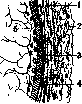
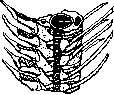
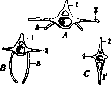


Figure 5. Formation of the atlas and axis in the fossil reptile Ophiacodon: 1-atlas; 2-axis. Figure 6. Anterior thoracic vertebra of an alligator: 1-spinous process; 2-articular process; 3-rib; 4-transverse process. In higher vertebrates (reptiles, birds, and mammals), there is a special composition and relationship between the first two vertebrae (fig. 5). The first vertebra consists only of the upper arch, closed on the ventral side by the intercentrum, and takes the form of a ring. The body of this vertebra connects with the body of the second vertebra (and its intercentrum), forming a protruding 'odontoid' process (processus odontoideus) that fits into the ventral half of the aforementioned ring. Thus, the first vertebra, the 'atlas,' is able to rotate quite freely on the odontoid process of the second vertebra, the 'axis.' The mobility achieved in this way is further enhanced by the development of a generally more mobile cervical section of the vertebral column, in which the ribs are reduced (in some reptiles, there are even movable cervical ribs on the atlas). In the vertebral column of mammals, the strengthening of the pelvic girdle on the vertebral column (and specifically with the help of ribs) causes the isolation of another (sacral) section, inserted between the trunk and tail sections (fish have only these two sections). Finally, the trunk section is divided into the anterior, relatively less mobile, thoracic section, characterized by fully developed ribs that here, together with the sternum, form the thoracic cage, and the posterior, more mobile, lumbar section, in which the ribs are reduced. As mentioned, the ribs of terrestrial vertebrates belong to the upper ribs type. Their connection with the vertebral column is transformed through the division of the proximal part of the rib into two branches, which corresponds to the division of the lateral process supporting the rib into two parts-the dorsal part develops more strongly and shifts (especially in the cervical and thoracic regions) onto the neural arch (fig. 6), while the ventral part remains on the posterior edge of the body (initially the process and rib are attached to the intercentrum) and is reduced. The upper branch of the proximal end of the rib is called the tubercle (tuberculum), and the lower branch is called the head (capitulum). The first is usually reduced, especially in mammals (fig. 7). As a rule, the ribs do not ossify throughout their entire length-their sternal part remains cartilaginous (in crocodiles, birds, and monotreme and edentate mammals, the sternal part of the rib also ossifies). The number of vertebrae in vertebrates is highly variable: in fish, this number ranges from 2-3 dozen to 200-400 vertebrae in forms with long bodies; in terrestrial vertebrates, this number is usually lower, although in snakes it also reaches 200-400. However, the number of presacral vertebrae is much more constant. Although this number also varies considerably, paleontological data suggest that the original number for higher vertebrates (reptiles, birds, and mammals) was probably 26 vertebrae. In amphibians, there is only 1 sacral vertebra, in modern reptiles-2. In birds, there are also 2 primary sacral vertebrae, but they fuse with a series of lumbar vertebrae in front and caudal vertebrae behind, resulting in a complex sacrum of 9-22 vertebrae. In mammals, there are initially 2 sacral vertebrae, but then up to 4 additional vertebrae may join them. These vertebrae fuse with each other and bear on their lateral parts, corresponding to the transverse processes (and partly to the ribs), articular surfaces for the iliac bones of the pelvis. The caudal vertebrae that attach to the sacrum from behind and do not participate in this articulation are called secondary or false sacral vertebrae. The number of vertebrae in the cervical region of mammals is quite constant-7 vertebrae (exceptions: manatee-6 vertebrae, sloths-6-9 vertebrae, pangolin-8). In the thoracic region, there are usually 12-15 vertebrae (most often 13), in the lumbar region usually 6-7. The number of caudal vertebrae varies depending on the length and importance of the tail, usually ranging from 15-30.
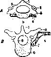
Figure 7. Diagrams of cervical vertebrae of mammals: 1-spinous process; 2-articular process; 3-transverse process; 4-rib; 5-body of vertebra; 6-neural arch; 7-head of rib.
The ribs, as a rule, do not ossify throughout their entire length-their sternal part remains cartilaginous (in crocodiles, birds, and monotreme and edentate mammals, the sternal part of the rib also ossifies). The number of vertebrae in vertebrates is highly variable: in fish, this number ranges from 2-3 dozen to 200-400 vertebrae in forms with long bodies; in terrestrial vertebrates, this number is usually lower, although in snakes it also reaches 200-400. However, the number of presacral vertebrae is much more constant. Although this number also varies considerably, paleontological data suggest that the original number for higher vertebrates (reptiles, birds, and mammals) was probably 26 vertebrae. In amphibians, there is only 1 sacral vertebra, in modern reptiles-2. In birds, there are also 2 primary sacral vertebrae, but they fuse with a series of lumbar vertebrae in front and caudal vertebrae behind, resulting in a complex sacrum of 9-22 vertebrae. In mammals, there are initially 2 sacral vertebrae, but then up to 4 additional vertebrae may join them. These vertebrae fuse with each other and bear on their lateral parts, corresponding to the transverse processes (and partly to the ribs), articular surfaces for the iliac bones of the pelvis. The caudal vertebrae that attach to the sacrum from behind and do not participate in this articulation are called secondary or false sacral vertebrae. The number of vertebrae in the cervical region of mammals is quite constant-7 vertebrae (exceptions: manatee-6 vertebrae, sloths-6-9 vertebrae, pangolin-8). In the thoracic region, there are usually 12-15 vertebrae (most often 13), in the lumbar region usually 6-7. The number of caudal vertebrae varies depending on the length and importance of the tail, usually ranging from 15-30.
I. Shmal'gauzen. Ontogenesis. The human vertebral column also passes through three stages of development: membranous, cartilaginous, and bony; but even earlier appears the notochord. The process of cartilage formation in humans begins at the beginning of the second month. In certain areas of young connective tissue, nests of cells appear, which, secreting cartilaginous ground substance, push away from each other as they grow. Initially, such nests of cartilage cells appear in the bodies and arches of the vertebrae, then the arches grow to the body and only at the 4th month of intrauterine life do the arches (or rather halves of the arches) fuse with each other, closing the vertebral column from the dorsal side. The cartilaginous processes of the vertebrae appear later at separate places, latest of all the spinous process. The entire cartilaginous skeleton of the human embryo has a special appearance, significantly different from the bony skeleton. Even before the complete laying of the cartilaginous vertebral column, from the end of the 2nd month of intrauterine life, in humans, the ossification of the V. begins, proceeding from the head to the tail. Certain areas of the cartilaginous skeleton ossify, others do not, so that the cartilaginous skeleton is not completely replaced by the subsequent bony stage of development. Thus, the bony vertebral column of an adult contains remnants of all stages of development passed through. Each vertebra has 3 main points of ossification: one in the body and 2 in the arch; the points of ossification in the arch fuse with each other in the first year of life and connect with the body in the 3rd year, with the bony substance of the anterior parts of the arch extending into the body of the vertebra. Thus, the posterior lateral part of the future vertebra, with which the future articular process articulates, is formed from the anterior part of the arch. From the points of ossification of the arch, not only the bony arch but also the processes are formed: spinous, transverse, and articular. Additional points appear later—at 15-16 years of age: one at the apex of the spinous process, two at the apex of the transverse processes, and in addition two points on the upper and lower surfaces of the vertebral body, forming two thin, wide epiphyseal plates. In the lumbar vertebrae there are additional points for the mamillary processes. The fusion of the aforementioned additional points of ossification with the rest of the vertebra occurs by the 25th year of life. The rudiments of ribs also participate significantly in the formation of vertebrae, with separate points of ossification in the cervical, lumbar, and upper sacral vertebrae. In the cervical ribs, still in the cartilaginous stage, the costal process connects at one end with the body of the vertebra and at the other with the apex of the transverse process, thereby forming an opening (foramen transversarium), which then remains in the bony state for life. In the thoracic region, the ribs are independent and do not participate in any way in the formation of the vertebrae. In the lumbar region, the rudiments of ribs completely fuse with the transverse processes, forming a complex structure—the costotransverse process. In the upper sacral vertebrae, the rudiments of ribs fuse into the lateral masses, as will be indicated below. In the two lower sacral and all coccygeal vertebrae, the rudiments of ribs are absent. The first and second cervical vertebrae have special features. The atlas has one point of ossification for each lateral mass with the corresponding half of the posterior arch, in addition, in the first year, one or two additional points of ossification appear in the anterior arch. To the body of the axis, still in the cartilaginous stage, the body of the atlas attaches, forming the odontoid process, therefore the axis has an additional point of ossification (sometimes two) for the odontoid process, which fuses with the bony body of the axis at 3-5 years. The sacral vertebrae, like the others, have three main points of ossification, which fuse, starting from the lower vertebrae, at 2-6 years of age; in addition, the three upper vertebrae also have lateral points for the rudiments of ribs (sacral ribs), which participate in the formation of the lateral masses of the sacrum (massa lateralis). The fusion of the sacral vertebrae occurs late—at 14-25 years. The coccygeal vertebrae have one point of ossification (for the bodies of the vertebrae), which appear from 1 to 20 years of age. Fusion of the vertebrae occurs from below; first the three lower vertebrae fuse, and then after 30 years the first vertebra fuses with them.
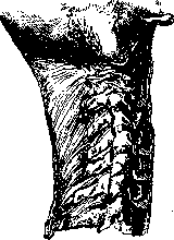
II. Anatomy. The human vertebral column is the main, rather flexible rod to which all other parts of the trunk skeleton are attached; in it are distinguished the cervical region with 7 cervical vertebrae, the thoracic with 12, the lumbar with 5, the sacral with 5, and the coccygeal with 4 or 5 vertebrae. Each vertebra has a massive body (corpus vertebrae) located in front and an arch (arcus vertebrae) located behind; the arch and body limit the opening (foramen vertebrale); in the entire V., these openings, by fusing, form a canal (canalis vertebralis), intended for the spinal cord. The vertebral body is usually oval in shape, its anterior and lateral surfaces are slightly convex from left to right and concave from top to bottom; the posterior surface is slightly concave, and the upper and lower surfaces are flat and also slightly concave. The anterior, posterior, and lateral surfaces bear numerous openings for blood vessels; these openings are especially large on the posterior surface, facing the vertebral canal. The arch consists of two symmetrical halves, extending from the lateral parts of the body and fusing behind along the midline. The initial parts of the arch on both sides are narrowed and are called roots (radices arcus vertebrae); on the upper and lower edges of each root of the arch there are notches (incisura vertebralis superior et inferior), with the lower one being significantly deeper. In the entire V., when one vertebra is placed on another, the notches of two neighboring vertebrae form on each side intervertebral openings (foramina intervertebralia), leading into the vertebral canal. From the arch extend to the sides two paired processes—the transverse processes (processus transversi), further two pairs of articular processes upward and downward (processus articulares superiores et inferiores) and finally one unpaired process extending backward from the middle of the arch—the spinous process (processus spinosus). The size and shape of the vertebrae vary depending on their location. Starting from the cervical vertebrae, the size increases, with the upper sacral vertebrae being the largest, and below them there is a rapid decrease. Each of the individual groups of vertebrae has special features, which are most pronounced in the middle vertebrae of each group; these features make it possible to determine to which group a vertebra belongs even by its individual parts (by the body, arch, or process). They are smoothed out at the ends of each group, where the vertebrae acquire transitional forms to the neighboring group. The human vertebrae can be considered as a complex lattice structure (Bardeleben). They consist mostly of spongy substance (substantia spongiosa), covered on the surface only by a thin layer of dense substance (substantia compacta). The bony trabeculae in the vertebral body are arranged vertically from the upper surface to the lower, horizontally, and obliquely—from the upper articular process to the lower surface and from the lower articular process to the upper surface. In the middle of the body, the bony trabeculae are weakly expressed, and here more or less large cavities are formed. The dense bony substance is more strongly developed in the area of the upper and lower notches, in the roots of the arches, which is why the arches have a stronger structure compared to the bodies and other parts of the vertebra, which explains the rarer fractures of the arch roots. The cervical vertebrae (fig. 8) (vertebrae cervicales), seven in number, are small; a distinctive feature is the opening in the transverse processes (foramen transversarium) for the a. et v. vertebralis; the vertebral artery, emerging from the subclavian, usually enters the opening of C1, and sometimes only C2, C3, and even C4. The upper surface of the transverse process from C4 to C7 is provided with a wide
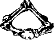
Figure 8. Cervical region of the vertebral column with the nuchal ligament. vertebral column 11 k
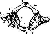
Figure 9. Cervical vertebra of a newborn. Figure 10. Atlas from above: i-tuber-
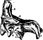
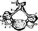
by a groove (sulcus n. spinalis). The bodies of the cervical vertebrae are more elongated in the transverse direction than in the sagittal; the width of the vertebrae, starting from C6, increases downward. The upper and lower surfaces of the body are slightly saddle-shaped. The articular processes are wide and flat, the upper ones look backward and slightly upward, the lower ones forward and slightly downward. The spinous processes (proc. spinosi) in general extend horizontally and only very slightly downward; their apices are usually split into two tubercles except for C7 and C6; short in the upper vertebrae, they gradually increase downward, especially the spinous process of C7, which surpasses all others in length and width. The last, lying almost horizontally, protrudes backward more and is relatively easily palpable through the skin, thanks to which C7 was given the name vertebra prominenens. The vertebral foramen of the cervical vertebrae is of considerable size with rounded angles (fig.9-14). culum post.; 2-sulcus*a. Vertebrali Передняячасть 3 -processus transversus; 4 - for. гтлпрпшцпгппт transversanum; -5-fovea articularis поперечного OT- sup-. 6-massa lateralis; 7-foveaden-pOCTKaCvi(pro- tis; 8-tuberculum ant.; 9-arcus C-eSSUB COStari-
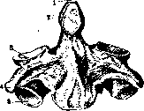
ant.; 10 -arcus post. us) usually protrudes forward more, thanks to which it is more accessible to palpation through the skin coverings. This prominence in topographic anatomy received the name carotid tubercle (tuberculum caroticum), to which the carotid artery can be pressed. The first two cervical vertebrae have different distinctive features, which make understandable the possibility of rotation of the head around the vertical- Fig. 11. The second cervical vertebra- ной оси почему bone (epistropneus): l-dens; 2-
' facies articularis? ant.; 3-faces Двa верхних ШвИ-articular.sup.; 4-processustrans- НЫХ ПОЗВОНКа И versus; 5-facies articularis inf. П0Лучили название вращательных в отличие от всех остальных сгибательных (позвонков). Описание Ci и Си-см. Атлант и Epistropheus. Thoracic vertebrae (vertebrae thoraca-les, s. dorsales), numbering 12, are the simplest typical vertebrae, since in their formation the rudiments of ribs take no part, which in this area are independent and develop separately from the vertebrae. The distinctive feature of these vertebrae^- the presence of articular surfaces on the bodies and on the transverse processes for articulation with the heads of the ribs and their tubercles (fig. 15). These surfaces appear on most
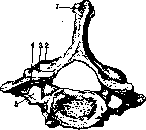
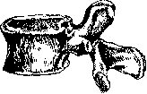
thoracic vertebrae in the form of two half-fossae, lying on the lateral surface of the body, in front of the root of the arch, the upper-at the upper edge, the lower-at the lower (fovea costalis superior et inferior); each of these half-fossae, joining with a similar one of the neighboring vertebra, forms f a fossa for the head of the rib. The upper half-fossa serves for connection . with the corresponding rib and the lower with the underlying one. An exception is D1, which has a whole fossa for the first rib and a half-fossa for the II rib, D10, which has only one upper half-fossa for the X rib and D11, D12, which have whole fossae for the corresponding ribs. Te-Figure 12. second cervical po- la thoracic ver-lateral vertebra: I-dens; 2-faces ков по B0PM TDPM articularis post.; 5-faces arti- нив 1Ю БСЬМ ^ем cuiaris sup.,- 4-processus spi- directions zna-nosus; 5-facies articularis inf.; tELNO more, 6-foramen transversarium. than cervical bodies. Their shape gradually changes from triangular with rounded edges to kidney-shaped. The transverse processes of the thoracic vertebrae are somewhat deflected backward, on their anterior surface at the apex there are rounded, flat-concave articular fossae (foveae costales trans-versales) for the tubercles of the ribs; on D11 and D12 these fossae are absent. The spinous processes are triangular in shape, long and sharply inclined downward, arranged like shingles one Fig 13 Cervical ver-over the other, especially from above: J-processus spino-tebrae; 2-arcus vertebrae; 3- Ппчпптпттттлр nTRpn- facies articularis sup.; 4- vertebral for- tuberculum post.; 5-sulcus СТИе cylindrical nervi spinalis; 6-tubercu-lum ant.; 7-foramen trans- form and smaller than versarmm. cervical and lumbar.
The articular processes stand frontally, with the upper ones looking backward and the lower ones forward. Lumbar vertebrae (vertebrae lumbales), numbering 5, are the largest. Their massive bodies are larger in all directions compared to thoracic ones, the shape is kidney-shaped; the edges of the bodies always protrude sharply, sometimes forming growths, especially at the back (fig. 16). The spinous processes are well developed, have almost a horizontal direction, thanks to which a needle, inserted Fig. 14. Seventh cervical po- when POSSIBLE kyphotic ver-spinosus; 2-processus articu- n MPW7TV ПРТИ_ laris inf.; .?-facies articula- нии u- мeжДУ ОСТИ-ris sup,; 4- processus articula- СТЫМИ отросткамипо-ris sup.;-.5-foramen transver- ЯСНИЧНЫХ ПОЗВОНКОВ sanum-
(between L5 and L4), does not meet resistance and easily reaches the vertebral canal. The transverse processes are long and flat and represent rudiments of fixed ribs; at the base of
VERTEBRAL COLUMN
lie
the transverse processes there is a small sharp spine, an accessory process (processus accessories). The articular processes are thick and stand sagittally, the upper ones are cylindrically concave, and the lower ones correspondingly convex. On the lateral side of the upper articular process there protrudes a small mamillary tubercle (processus mamilla-ris). The vertebral foramen is triangular in shape, of considerable width. -Cervical, thoracic and lumbar vertebrae are called movable, or true (vertebrae verae), and the lower sacral-coccygeal vertebrae, merging in
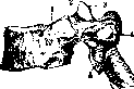
Figure 15. Thoracic vertebra from the side: 1-fovea eostalis sup.; 2-incisura vertebralis sup.; 3- processus arti-eularis sup.; 4-fovea costalis transversal is; .5 - processus articular is inf.; 6-processus spinosus.

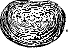
the sacrum and coccyx have received the name of fixed, or false vertebrae (vertebrae spuriae). Description of the lower part of the vertebral column-see Sacrum, Coccyx. Variations and anomalies of the vertebral column are numerous and diverse; some of them are so significant that they disrupt the function of the vertebral column, while others are insignificant, do not manifest during life, and are discovered in the corpse only during special anatomical investigations. Variations concern both the forms of vertebrae and the numerical composition in different parts. Thus, congenital scoliosis, associated with developmental defects, in most cases depends on the presence of additional underdeveloped wedge-shaped vertebrae. Located properly on only one side between normal ones, such wedge-shaped formations influence the development of neighboring vertebrae, which ultimately gives curvature of the vertebral column. The described formations can be observed in all parts of the vertebral column and are sometimes single, sometimes multiple; sometimes they replace normal vertebrae. The formation of the described half-wedge-shaped rudiments can be explained only by improper segmentation of mesenchyme in that initial period of development when the primary vertebra consists of a double paired rudiment. From the diagnostic side, it should be noted that in some cases congenital scoliosis becomes noticeable in older age, and not immediately after birth. Furthermore, the arches of vertebrae may not fuse, the spinous processes then do not form, as a result of which the vertebral canal is more or less open (spina bifida) and the spinal cord along with the membranes may protrude through the resulting slit like a hernia. Such non-fusion of arches is more often observed in the lumbar and sacral, as well as in the cervical part of the vertebral column (see Spina bifida). The most interesting from the morphological point of view are the numerical variations of vertebrae; in many cases they depend on the number of ribs. Thus, with the presence of 13 pairs of ribs, the number of thoracic vertebrae increases, and conversely, with the presence of 11 pairs of ribs, the number of lumbar vertebrae increases; the number of free, precoccygeal vertebrae in these cases remains the same-24 vertebrae. But there are cases, however, when the total number of free vertebrae decreases or increases; this depends on what place the sacrum occupies. It should be noted that the sacrum may consist of 4, 5, or 6 vertebrae and the first sacral vertebra may be XXIV, XXV, XXVI, and XXVII. The last sacral vertebrae may be vertebrae from XXVIII to XXXI, and in general the human vertebral column may consist of 32-35 vertebrae; in the case of extreme elongation of the vertebral column, there are 36 or even 37 vertebrae. Numerical variations of vertebrae cannot be considered as a random phenomenon, depending on various random deviations in development, but as a phylogenetic phenomenon. For a comparatively long time on this question, the coherent hypothesis of Rosenberg (Rosenberg) prevailed, based on the data of comparative anatomy and embryology and easily explaining all such deviations. Rosenberg asserts that in the human embryo, XXX vertebra is usually laid as the sacral vertebra, XXV as the last lumbar, and XX as the thoracic. In further development, the XXX vertebra loses connection with the sacrum, XXV connects with the sacrum, and the ribs of the XX vertebra disappear. Thus, in ontogenesis, the sacrum approaches the head, and the lower end of the thoracic cage shortens. Based on the presented data and facts of comparative anatomy of anthropoid and lower monkeys, Rosenberg asserts that such changes also occurred during phylogenetic development. Therefore he classifies vertebrates, in which the sacrum is located low and the lower boundary of the thoracic cage lies low, as primitive, atavistic forms, and with a high-lying sacrum and a high thoracic cage-as forms of the future, progressive ones. Furthermore, Rosenberg notes that in the human embryo, an independent pair of ribs is laid at C7 and that this pair of ribs loses its independence, and thus in the period of ontogenesis, the upper end of the thoracic cage also shortens. On this basis he asserts that the same reduction also occurred during phylogenetic development, and this reduction did not stop at the ribs of C7, but now begins to capture also the ribs of C8 (D1). Thus, according to Rosenberg, the thoracic cage decreases during ontogenesis from both sides; the same reduction, in his opinion, also occurred during phylogeny; now it begins to spread further to the ribs of the VIII and XIX vertebrae. Rosenberg considers the process under discussion final and does not assume significant shortening of the vertebral column, since in his opinion the loss of individual vertebrae is compensated by the growth in height of the bodies of vertebrae. Rosenberg's hypothesis, which satisfactorily explains all numerical variations of vertebrae, was initially accepted by the majority, being a useful working hypothesis for many researches, which is also acknowledged by its opponents. Over time, the number of voices denying the correctness of this hypothesis increased, and a number of authors (Peterson, Bardeen, Dwight, Fischel) spoke against it, some on the basis of theoretical reasoning, others on the basis of their embryological researches. More than half a century has passed, the hypothesis has undergone the most severe criticism, but even now it cannot be considered refuted, only its application should be somewhat narrowed and limited to primates and its dogmatism should be eliminated. Of course, all observed variations in the vertebral column cannot be explained by phylogenetic processes alone, but one should also take into account the various functions and adaptations that different parts of the vertebral column undergo during ontogenesis. Among other anomalies, fusion of C1 in greater or lesser degree with the skull, as well as fusion of individual vertebrae with each other-congenital synostoses of the vertebral column (see below) should be mentioned. On the lower surface of the occipital bone, in front of the large foramen magnum, bone protrusions of various shapes are often encountered, which are nothing other than various stages of manifestation of the last occipital vertebra (manifestatio vertebrae occipitalis), indicating the origin of the occipital bone from the fusion of vertebrae. In some cases, it goes so far as to form along the median line at the anterior edge of the foramen magnum a more or less sharp protrusion, homologous to the anterior tubercle of the atlas. The manifestation of the occipital vertebra depends on the preservation of sclerogenic tissue in the occipital region, representing remnants of the hypochordal arch and corresponding in shape to C1. The ligamentous apparatus. Connections between bodies, arches, and processes are distinguished. The connection between the bodies of vertebrae occurs by means of intervertebral discs (fibrocartilago intervertebralis), (fig. 17). In the latter, the following are distinguished: 1) the fibrous ring (annulus fibrosus), located along the periphery of the disc and consisting of ring-shaped layers of fibrous cartilage, 2) the gelatinous nucleus (nucleus pulposus), consisting of a gelatinous soft mass of fibrous cartilage and proliferated remnants of the notochord, 3) two thin plates of hyaline cartilage, adjacent to the surfaces of the vertebral bodies. In an adult, the disc contains neither blood vessels nor nerves. The shape of the intervertebral disc corresponds to the bodies of vertebrae. In the cervical and lumbar parts, the cartilage in front is significantly higher than behind; this difference is especially great in the disc between the sacrum and the last lumbar vertebra. In total, there are 23 intervertebral discs in the vertebral column, the last one is absent only between C1 and C2, but there is one between L5 and S1; the total height of the intervertebral discs reaches up to 1/4 of the entire movable part of the vertebral column. With age, the height of the disc decreases, and this circumstance serves as the main reason for the decrease in height in old age. Connection between the bodies of vertebrae by means of intervertebral cartilage fig. 17. intervertebral is extremely strong, cartilage in cross-section: i-nucleus nevertheless mutual pulposus; 2-annulus lib- movements of two neighboring-rosus
vertebrae are possible in all directions due to the fact that the cartilaginous discs possess not only considerable strength but also very high elasticity. The role of the disc here can be compared to a compressed spring or an air cushion. The magnitude of movements depends on the height of the discs-the smaller the surface of the vertebrae and the higher the discs, the greater the mobility. The discs are constantly under pressure of two kinds: internal, coming from the compressed gelatinous nucleus, and external from the pressure of the overlying parts; when the disc is incised, the nucleus expands and protrudes. In cold water it quickly increases to four times its original volume. Congenital anomalies of intervertebral discs consist mainly in improper development of the notochord; they do not give clinical symptoms and can be discovered only on an X-ray.
In addition to the intervertebral discs, the bodies of vertebrae are connected by common ligaments-anterior and posterior. The anterior longitudinal ligament (lig. longitudinale anterius) extends from the base of the occipital bone to the sacral vertebrae (fig. 18). The deep fibers of this ligament are con-
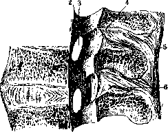
Figure 18 - Connection of vertebrae in the lumbar region, left view in section: 1-lig. longitudinale ant.; 2-lig. longitudinale post.; 3-for. intervertebrale; i-lig. flavum; o-lig. supraspinale; 6-lig. inter-spinale.
The deep fibers connect only adjacent vertebrae, while the superficial fibers pass over 4-5 vertebrae; downward the entire ligament becomes wider and thicker. This ligament is firmly attached to the bodies of the vertebrae, but over the intervertebral discs it merely passes without forming a close connection. In the region of S1 and S2 the ligament thins out and passes into the periosteum.--The posterior longitudinal ligament (lig. longitudinale posterius) is located on the posterior surface of the bodies of the vertebrae, facing the cavity of the vertebral canal. The ligament is less developed compared to the anterior one and is wider at the top than at the bottom, extending from the skull to the sacral bone. Along its length, the ligament is firmly attached to the edges of the vertebral bodies and to the intervertebral discs, while in the middle of the bodies the ligament narrows and recedes, thereby forming a space beneath it occupied by venous plexuses. Due to the bilateral alternation of narrowing and widening, the ligament takes on the appearance of a double-sided saw.--The vertebral arches are connected by strong interarcuate, or yellow ligaments (lig. interarcualia, s. flava). These ligaments consist almost entirely of elastic fibers, giving them a yellowish color; the middle part of the ligament is thickest. When bending the vertebral column, the yellow ligaments tense and stretch, and then return to their previous state, always maintaining their smooth surface.--The transverse processes are connected by relatively weak intertransverse ligaments (lig. intertransversaria); in the lumbar region they are stronger, in the thoracic region they consist of thin plates closely connected with the musculature, on the neck they are formed by thin, few fibers, sometimes they are completely absent here.--The spinous processes are connected by interspinous ligaments (lig. interspinalia), filling the spaces between adjacent spinous processes and consisting mainly of collagen fibers with a small admixture of elastic fibers; in the lumbar region these ligaments are strongly developed. At the tips of the spinous processes, these ligaments pass into a continuous round cord, the supraspinous ligament (lig. supraspinale, s. apiculum) of the vertebral column. On the neck, lig. supraspinale expands into the nuchal ligament (lig. nuchae), which extends from the spinous process of C2 to the external occipital protuberance; from each spinous process, strong bundles depart, weaving into the common mass. In animals, the nuchal ligament reaches powerful development and consists mainly of elastic fibers * in humans, however, it represents a thin fascial plate, forming a partition between the muscles of the occipital region on both sides. The articular processes of two adjacent vertebrae form joints (articulationes intervertebrales); their capsule, attached at the edge of the articular cartilage, is very weak in the cervical vertebrae, while in the lumbar region it is thicker; the joints are slightly movable and should be classified as amphiarthroses.--The lumbosacral junction. The last fifth lumbar vertebra connects with the sacrum according to the type of all free vertebrae: the intervertebral cartilage is significantly higher in front than behind, the amphiarthroses show no special features, all private and common ligaments are present, analogous to the vertebrae above. The sacrococcygeal junction belongs to the type of synchondroses, but with the peculiarity that in the cartilage a cavity is present in most cases. The horns of the sacrum and coccyx are connected by a paired sacrococcygeal ligament (lig. sacrococcygeum articulare), which corresponds to the amphiarthrosis of the vertebrae above. From the lower end of the sacral crest to the spinous process of the first coccygeal vertebra goes a paired ligament (lig. sacrococcygeum lat.). The listed ligaments are often absent. The continuation of the anterior longitudinal ligament is lig. sacrococcygeum anterior, consisting of two crossing bundles extending along the anterior surface of the sacrum and coccyx. The continuation of the posterior longitudinal ligament is lig. sacrococcygeum posterius profundum, extending along the posterior surface of the body of S5 and Co1. In addition, from the edges of the sacral hiatus, closing this opening, goes lig. sacrococcygeum posterius superficiale, corresponding to the yellow ligament.--The craniovertebral ligamentous apparatus. The connection between C1 and C2 and the connection between C1 and the occipital bone are movable connections, forming two joints: the atlanto-occipital joint between the atlas and the occipital bone, and the atlanto-axial joint (Fick) between the atlas and the axis. The atlanto-occipital joint is combined from two paired joints between the articular processes of the occipital bone and the articular fossae of the atlas - the atlanto-occipital joints (articulationes atlanto-occipitales). According to the shape of the articular surfaces, they can be classified as ellipsoid joints with two mutually perpendicular axes of movement. The longitudinal dimensions of the articular processes of the occipital bone and the articular cavities of the atlas converge forward, and the anterior ends of opposite sides are 21-25 mm apart, while the posterior ones are 30-34 mm apart. The joint capsule is loosely stretched, beginning at the edge of the articular surfaces. Various individual differences are observed in the shape of the articular surfaces. Often there is a bifurcation of the articular surfaces into a larger anterior and smaller posterior segments. Their shape can be either wide and low, or narrow and high. Complete symmetry in the shape of the articular surfaces is rarely observed; the right side is mostly flat and low, while the left is high and narrow. The spaces between the occipital bone and the atlas behind and in front are covered by fibrous membranes. Membrana atlanto-occipitalis anterior begins from the upper edge of the anterior arch and goes to the lower surface of the body of the occipital bone; in the middle it is reinforced by the anterior longitudinal ligament. Membrana atlanto-occipitalis posterior begins from the posterior arch of the atlas and goes to the posterior edge of the foramen magnum; this membrane is thinner than the anterior one. The described membranes are rather strong plates containing not only collagen fibers but also a significant amount of elastic fibers. Due to the presence of these membranes, the wide spaces between the atlas and the occipital bone are completely closed, and only an opening remains on the side for the passage of the vertebral artery and nerve trunks.--The atlanto-axial joint is combined from four joints between the first and second vertebrae (articulationes atlanto-epistrophicae). Of these four joints, two paired lateral ones are formed between the lower articular surface of the atlas and the upper articular surface of the axis. The joint capsules are wide and loosely stretched. Two joints are at the odontoid process: one, anterior, between the articular surface of the odontoid process and the anterior arch of the atlas, the second, posterior, between the odontoid process and lig. transversum atlantis. The capsular ligaments of these joints are very thin, giving rise to sacculated bulges on the sides, which can connect the anterior and posterior joints with each other, as well as connect the latter with the lateral joints. The auxiliary ligaments of these joints are: 1) the ligament of the apex of the odontoid process (lig. apicis dentis), stretched between the upper end of the odontoid process and the middle of the anterior edge of the foramen magnum. This ligament corresponds to the nucleus pulposus of the intervertebral disc and initially contained the chorda dorsalis. This ligament is extremely weak and has no mechanical significance. 2) The alar ligaments (lig. alaria), two paired ligaments extending from the apex of the odontoid process sideways and upward to the medial edge of the articular processes of the occipital bone and the lateral edges of the foramen magnum. 3) The transverse ligament of the atlas (lig. transversum atlantis) connects the lateral masses of the atlas behind the odontoid process. This ligament is very strong, in the middle it is wider, on the surface facing the odontoid process it is covered with cartilage. 4) The tectorial membrane (membrana tectoria) is a dense plate covering behind lig. cruciatum and extending from the clivus to the body of the axis; downward it continues into the posterior longitudinal ligament. The upper and lower atlanto-occipital joints are very complex mechanisms facilitating the most diverse movements of the head. The upper one (articulatio atlanto-occipitalis) is a combined ellipsoid joint in which movements occur around two axes. Around the main transverse axis, flexion of the head forward and backward occurs, around the second additional sagittal axis, slight flexion of the head to one side and the other occurs. The lower atlanto-axial joint (articulatio atlanto-epistrophica) represents a combined, rotational joint consisting of 4 joints - two on the sides and two in front and behind the odontoid process. The vertical axis of this joint passes through the odontoid process. Both articular surfaces in the lateral joints are incongruent, convex, therefore they do not contact each other with their entire surface, but only with their middle part. Due to this, during rotational movements of the head, screw-like movements (Henke) also occur simultaneously in them. During rotation of the head, the atlas plays the role of only an interposed meniscus, and the movements occur exclusively in the atlanto-axial joint.
Both head joints—the upper and lower—should be considered in mechanical relation as one combined joint with three axes of movement, i.e., as an arthrodia (H. Meyer). Statics and mechanics of the Vertebral Column. In analyzing the mechanism of the vertebral column, it is necessary to distinguish two main parts, the combined actions of which result in the complex and diverse movements that are possible in the Vertebral Column. The first mechanical part consists of the column formed by the bodies of the vertebrae, connected by intervertebral cartilages and the anterior and posterior longitudinal ligaments; this column, with the laminae cut off, is a very flexible, elastic rod that allows the most extensive and extremely diverse movements in all directions. The strength of this rod is so great that it allows for the harmless removal of 3-7 vertebral laminae, as is done during laminectomy. Even removal of the laminae in the cervical region does not disrupt the function of the Vertebral Column. There are no sufficient grounds to fear that the spinal cord will lose its bony protection and that the mechanism of the Vertebral Column will be disrupted. The second mechanical part, which strengthens and restrains the movements of the rod, consists of the vertebral arches, connected to each other by strong ligaments (lig. flava) and joints. A series of arches or a divided arch plate, as it can be called, not only strengthens the vertebral rod but also limits its movements and directs them along strictly defined lines. The musculature surrounding the arch plate and having the most diverse points of attachment not only strengthens the Vertebral Column but also gives it certain forms and determines the paths along which movements occur within it. Both mechanical parts—the vertebral rod and the arch plate—are extremely firmly connected to each other, forming one whole that has the same strength to break in all its parts. This explains why dislocations of vertebrae in a pure form without bone fractures are observed extremely rarely and, moreover, mainly in the cervical, most mobile section. As a whole, the vertebral column is built on a spiral system having multiple curvatures. In the lumbar region there is a convexity forward, followed by the concavity of the thoracic part, then again the convexity of the cervical section; below is joined the concavity of the sacrum and coccyx. Thus, we have in the Vertebral Column four curvatures: two are convex forward and two backward. These curvatures are clearly visible when examining the Vertebral Column from the side and distinctly protrude in the straightened, so-called military position. In this position, one can spend whole hours with little fatigue, since muscular force is almost not expended; the perpendicular dropped from the anterior tubercle of the atlas in this position passes through the body of Cvi, Dix, Sin and emerges through the apex of the coccyx. With a relaxed posture, the curvature in the thoracic section increases, while in the cervical and lumbar sections it decreases; in old age, the curvature in the thoracic section especially increases. However, these curvatures of the Vertebral Column exist not only in the vertical position but also in the horizontal position, when lying down; the bends in this case decrease but by no means disappear. In the human embryo, the shape of the Vertebral Column has great similarity to the shape of the Vertebral Column of quadrupeds, in which the ventral curvature of the lumbar section and the promontory are absent. In the newborn, the curvatures are barely outlined; only with further growth and under the influence of repeated exercises do the typical curvatures of the adult gradually develop, the degree of which is subject to individual variations. Thus, on the 8th week of pregnancy or so, the cervical curvature clearly appears, in the second half of the first year the thoracic curvature is clearly expressed; the lumbar curvature becomes noticeable later than others (at the beginning of the second year, when the child begins to walk; Gundobin). Two factors undoubtedly play a role in the formation of the Vertebral Column: heredity and mechanical adaptation acquired through experience. In addition to the bends in the sagittal plane, bends are also observed in the frontal plane, mainly the curvature of the thoracic section of the Vertebral Column to the right. But it should be noted that the curvature to the right represents only one link in a whole system of curvatures in the frontal plane, connected together with the sagittal curvatures into one whole (Hasse). Sometimes a curvature of the thoracic section is observed not to the right but to the left. The height of the Vertebral Column as a spiral does not have a constant value but depends on the load. In an adult male, on average, the height of the Vertebral Column is 70-73 cm, in a woman 66-69 cm, with a height of 73 cm, 13 cm accounts for the cervical section, 30 cm for the thoracic, 18 cm for the lumbar, and 12 cm for the sacrococcygeal. In the fetus and infants, the vertebral column is relatively longer, but then it lags in growth compared to the limbs. The final height of the Vertebral Column is reached by different peoples and different sexes at different times: from 23 to 25 years. Women reach maximum growth a year earlier, although this is not observed in all peoples. In old people, the length of the Vertebral Column decreases, and sometimes this decrease reaches 7 cm. The height of the Vertebral Column, in addition to individual differences, depends on its being burdened or freed from weights; in the first case, the curvatures become sharper and increase, in the second they decrease, and the length of the Vertebral Column, on the contrary, increases. If the burden lasted for a long time, the intervertebral cartilages compress, although each of them to a small degree, and with prolonged vertical position of the body, the Vertebral Column becomes shorter. This explains the difference in the height of the Vertebral Column in the morning and evening, which sometimes reaches 2.0-2.5 cm, as well as the increase in height after a prolonged illness, when the patient was in bed for a long time. Movements of the Vertebral Column occur around three axes: frontal, sagittal, and vertical. Around the frontal axis, flexion and extension of the Vertebral Column occur; the former is the most extensive movement; this movement is not uniform in all sections of the Vertebral Column: in the cervical part it is more extensive, in the lumbar somewhat less, and the thoracic section is the least mobile. Around the sagittal axis, lateral bending occurs; these movements are most possible at the boundary between the lumbar and thoracic parts. Around the vertical axis, rotation occurs, most extensive in the cervical section and almost impossible in the lumbar. To these movements is added the so-called spring movement, in which the magnitude of the curvatures of the Vertebral Column changes, for example during jumps. Thus, the cervical section is the most mobile, followed by the lumbar; the thoracic section is less mobile. The magnitude of movements in different directions depends mainly on the thickness of the intervertebral cartilages, as well as on the strength and extensibility of the ligaments and joint capsules; the articular surfaces of the articular processes allow movements only in certain directions. The middle part of the Vertebral Column is connected to the ribs, which connect in front with the sternum and together form the thoracic cage; due to these connections, which significantly restrict movements in the Vertebral Column, the thoracic section is the least mobile. The rib connects with the Vertebral Column at two points: its head connects with the body of the vertebra, and the rib tubercle with the transverse process. The joint between the head of the rib and the body (articulatio capitulae) is formed by the articular surface of the head of the rib and the articular fossae of the bodies of the vertebrae, and from Dn to Dx the head of the rib connects with two adjacent vertebrae. The cavity in these joints is divided by the interarticular ligament (lig. capituli costae inter-articulare), extending from the crest of the head of the rib to the intervertebral cartilage; this ligament divides the joint cavity into two parts. The joint capsule is thin and reinforced in front by rather thick fibrous bundles, radiating from the head of the rib to the bodies of two adjacent vertebrae and to the intervertebral cartilage (lig. capituli costae radiatum). The joints of the first and last two ribs with one cavity and their heads connect only with the corresponding vertebrae. The joint of the rib tubercle with the transverse process (articulatio costo-transversaria) exists only for the 10 upper ribs. The capsule of the joint is thin and reinforced behind by thick bundles of fibers extending from the apex of the transverse process to the rib tubercle (lig. tuberculi costae). In addition, there are short strong fibers extending from the neck of the rib to the corresponding transverse process (lig. colli costae). Two relatively weak ligaments (lig. costo-transversarium anterius et posterius), extending from the upper edge of the neck of the rib to the superior transverse process, complete the ligamentous apparatus. The muscles of the Vertebral Column are located mainly on its posterior surface, while on the anterior surface there are only individual representatives and only in the cervical and lumbar sections, while in the thoracic they are completely absent. On the posterior surface of the Vertebral Column there is a powerful muscular plate consisting of extremely diverse muscle systems, with the broad muscles belonging to the shoulder girdle lying in the superficial layers of this plate, and the long muscles located in the deep layers in such an order—the deeper they lie, the shorter they are. Despite the extreme diversity of the muscles, their arrangement indicates their metameric structure. The arteries of the Vertebral Column, running both along its walls and in adjacent parts, are in general insignificant. In the thoracic section, they originate from the intercostal arteries, and each of them divides into ramus anterior and ramus posterior.
The posterior branch, passing backward through the opening between the vertebra and the anterior costotransverse ligament, divides into two branches: the muscular branch and the spinal branch. The first of them, the muscular branch, supplies the muscles and skin of the back, while the second penetrates through the intervertebral foramen into the vertebral canal and with its twigs forms arterial networks that nourish the bony walls of the vertebral canal, the meninges of the brain, and the spinal cord itself. The same type of branching of the posterior branches occurs in the lumbar region (departing from the lumbar arteries) and in the cervical region (from the vertebral arteries). In the lower part of the neck, branches from the ascending cervical artery (from the thyrocervical trunk) and from the deep cervical artery (from the costocervical trunk) participate in the blood supply of the vertebral column. In the lumbar region, branches of the iliolumbar artery (from the hypogastric artery), the middle sacral artery, and the lateral sacral arteries (the first from the aorta and the second from the hypogastric artery) participate. The veins of the vertebral column represent dense vascular plexuses, located both inside and outside along the entire length of the vertebral column (fig. 19). The external
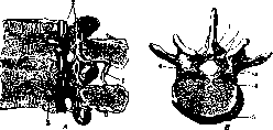
Figure 19. Venous plexus. A-median section on the left: 1- posterior vertebral venous plexus; 2- internal vertebral venous plexus; 3- basivertebral vein; B-horizontal section from above: 1- posterior vertebral venous plexus; 2- vertebral venous rete; 3- internal vertebral venous plexus; 4- basivertebral vein; 5- anterior vertebral venous plexus; 6- intervertebral vein.
venous plexuses (plexus venosi vertebrales externi) are divided into anterior and posterior; they anastomose with each other. In the occipital region, the posterior external venous plexuses (plexus venosi vertebrales externi) reach powerful development and connect with the venous sinuses of the brain and with the external veins of the skull. The internal venous plexuses also divide into anterior and posterior, anastomosing with each other. The anterior internal venous plexuses (plexus venosi vertebrales interni ant.) consist of two venous sinuses (sinus venosi vertebrales longitudinales), which are venous plexuses lying on the posterior surface of the bodies of the vertebrae on both sides of the posterior longitudinal ligament of the vertebral column. Into the anterior venous sinuses of each side flow the veins of the vertebral bodies (vv. basivertebrales), which pass inside the vertebral bodies in the channels of the spongy substance. The posterior internal venous plexuses (plexus venosi vertebrales interni post.) are two venous sinuses (sinus vertebrales longitudinales) descending from top to bottom on both sides of the arches of the vertebrae. Both the anterior plexuses and the posterior plexuses are connected by anastomoses forming venous rings at the level of each vertebra. The entire plexus of the vertebral canal lies between the layers of the dura mater (if we consider the periosteum of the vertebrae as the outer layer of the dura mater). The blood flow from both the internal and external venous plexuses has mainly a horizontal direction and at the level of each vertebra reaches the vertebral veins, azygos, hemiazygos (through the intercostal, lumbar veins), hypogastric veins (through the lateral sacral veins and iliolumbar veins). The lymphatic vessels of the vertebral column go to the intercostal lymph nodes, located in the area of the rib heads, forming plexuses here; the efferent vessels of these plexuses go to the thoracic duct. Cruikshank was the first to see lymphatic vessels emerging from the bodies of the thoracic vertebrae, which was later confirmed by Sommering and Bonomy. The efferent vessels of the upper intercostal nodes on the right side go to the right bronchomediastinal trunk. In the lumbar and sacral regions, the intercostal nodes are replaced by the lumbar and sacral lymph nodes; their efferent vessels go to the sacral lymphatic plexus, and then to the aortic plexus, which pours its lymph into the lumbar lymph trunks, which are on each side the roots of the thoracic duct. In the cervical part, the lymphatic vessels of the vertebral column go to the deep superior and inferior cervical lymph nodes. N. Bushmakin. III. Methods of examination. Anatomical changes associated with a violation of the external form of the vertebral column, as a rule, lead to a limitation of mobility, and often to spinal cord disorders; on the other hand, functional disorders very often make it possible to judge such pathological-anatomical changes of the vertebral column which at a given moment are not reflected in its external form. Therefore, in the clinical examination of the vertebral column, it is necessary to take into account the form, mobility, and spinal cord phenomena. Unlike the normal (fig. 20) form of the vertebral column (see above)
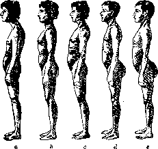
Figure 20. Types of posture according to Haglund: a- round back; b- normal posture; c-e- changes in posture depending on the tilt of the pelvis.
in old people the thoracic kyphosis increases, the lumbar lordosis decreases, and mobility is sharply limited. But also in young people, an increase in the normal thoracic kyphosis sometimes reaches the degree of the so-called "round back" (dorsum rotundum) - a painless, uniform, arcuate kyphosis of the interscapular area without any pathological changes (fig. 21). On the other hand, the opposite variant is also possible - a decrease or flattening of the normal thoracic kyphosis, a flat back (dorsum planum). These variants depend, on the one hand, on certain skeletal features, in particular the tilt of the pelvis and the structure of the sacrolumbar region, and on the other hand, on the degree of muscle tone and determine the so-called posture of a person. The examination of the vertebral column begins with observing the behavior of the patient and his movements. An unnatural position of the trunk and head, unnatural tension, stiffness of movements and disturbance of gait speak of functional disorders, so often associated with anatomical changes of the vertebral column. The general examination allows one to judge the posture, the proportionality of anatomical relationships or disproportions in them. When examining the back in profile, the physiological curves, their greater or lesser expressiveness and uniformity, as well as deformative changes of the vertebral column are established - angular and arcuate kyphoses, intensified or corrected lordoses and deformations of the thoracic cage. From behind, lateral-scoliotic curvatures of the vertebral column, asymmetrical standing of the shoulder blades, r 2 pelvis and deformation of the ribs are determined.
Palpation - feeling the line of the vertebral column - clarifies the data of the external examination and reveals hidden deformities. The determination of the abnormal protrusion of individual spinous processes is best done with the vertebral column bent and the arms crossed on the chest, passing along the line of the spinous processes with the tip of the middle finger (with the index and ring fingers on the sides), going from bottom to top (at this the finger "stumbles" over any protrusion (Boca) or by passing along the vertebral column with the palm of the hand (Turner). Counting of vertebrae is done from the most prominent spinous process of C7, but one must keep in mind Menard's indication that in children D1 protrudes more. One can count from top to bottom from C7 or from bottom to top from L5 (the "tubercle in the hollow"), as well as use reference lines connecting the lower ends of the shoulder blades (on D12) or the upper edges of the crests of the iliac parts (on L5); in lean people D12 can be determined by the last rib. In such an examination, the number of vertebrae involved in the pathological process, the apex of the curvature, as well as its boundaries, defects of the spinous processes and asymmetrical standing of the transverse processes are determined. With the help of palpation, the sensitivity of the vertebral column is examined, which is more pronounced when tapping on the spinous processes with the tip of a finger or a percussion hammer, as well as when displacing-loosening the spinous processes, grasped from the sides with two fingers, or when carefully tapping with a fist. The examination of the mobility of the vertebral column is performed along three axes in relation to flexion-extension, lateral bends and rotations around the axis (torsio). The examination begins with determining the mobility of the head: in normal conditions with maximum flexion the chin rests on the chest, and with extension the head is thrown back, so that the occipito-mental line forms an angle open forward with the vertical; when bending to the side the ear almost touches the corresponding shoulder, and rotations to the right and left are possible almost to the frontal plane. Then the mobility of the trunk is determined: with flexion, which occurs mainly at the bordering regions
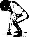
Fig.
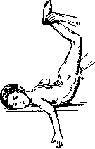
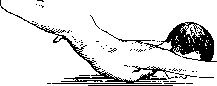
of the cervico-thoracic and thoraco-lumbar regions, the kyphosis sharply increases and the lordoses are corrected. Upon extension, the thoracic portion straightens and the lumbar and cervical lordoses sharply increase; with normal lateral mobility, lateral bending of the torso is possible until the false ribs touch the iliac crest, and rotation around the axis is possible until the arms, extended horizontally to the sides, are moved from the frontal plane to the sagittal plane without pelvic rotation. When picking up an object from the floor, a subject with normal function of the V. bends down uniformly, flexing the spine and extending it freely; in pathological conditions, especially in muscle contractures, patients spare the V., do not bend (Fig. 22), but squat, flexing the knees and hip joints, supporting themselves with their hands on their thighs; also when extending, patients spare the vertebral column and as if climb up the thigh with their hands, raising the unyielding torso; this symptom is particularly important in inflammatory diseases and lumbar pain. Mobility can also be examined in the prone position, both for determining flexion by placing hands under the abdomen and raising the patient, and mainly for extension and determining lateral mobility by placing one hand on the middle of the back and with the other hand raising either the cephalic end of the torso in lesions of the upper portion, or the caudal end (by the legs) in lesions of the lower. In normal conditions, the physiological kyphosis is corrected and the lordoses increase (Fig. 23), in pathological conditions the affected portion remains immobile (Fig. 24). During these examinations, muscle tension, rigidity, and the degree of pain of these movements in the region of a particular portion of the V. are also determined simultaneously. Finally, the degree of endurance of the vertebral column and the tenderness of its individual segments are determined during the so-called dynamic load, which is produced either by pressure on the shoulders and head, or by careful tapping with a fist on the back of the hand of the examiner, placed on the patient's vertex, or by a shock obtained when raising the patient on tiptoes and quickly lowering on the heels.
Measurements of the V. are simplest to perform by Kirchhoff's method, based on measuring in centimeters the levels of curvatures (deformations) and the distances of maximum deviations of various portions of the V. (lumbar and cervical lordoses) from the vertical line passing through the most posterior point of the body (from a centimeter tape with a weight, suspended from a collar and moved backward to the apex of the kyphotic curvature) (Fig. 25). Lateral curvatures, their height and distance from the midline of the body are also determined by the same method. However, this method is not sufficiently accurate. Graphical methods are of greater importance. Tracing of the shape of the V. is performed in the prone position using thick lead wire, a probe, or a lead plate a finger wide, which are applied to the V., well molded to it, and carefully transferred to paper (the patient's history), where the outlines are traced with a pencil. The tracing is made from the occipital protuberance to the coccyx with a mark at the level of L5 and S1. Such tracings are repeated during the illness. To measure mobility of the V., two tracings are made; at maximum flexion, placing a pillow under the abdomen, and at maximum extension-lordosis, placing rolls under the clavicles and thighs (Pitzen). ""0?%?>^ Even greater accuracy can be achieved with plaster casts: longitudinal plates for determining anteroposterior curvatures and transverse ones for lateral ones, or more accurately for changes in the thoracic cage associated with lateral curvatures. For tracing lateral curvatures, a simple window glass in a frame, applied to the back of a sitting patient, can also be used-direct tracing with a wax pencil (Lange). The most important condition for all these measurements and tracings is the constancy of the patient's position, since changes in position make measurements incomparable. During all these examinations, spinal cord disorders should be kept in mind, which are closely related to the condition of the V., often being early, and in some cases the first and only signs of diseases and injuries of the V. This applies to all three functions of the spinal cord-motor, sensory, and trophic in their various combinations. Root phenomena-girdle and shooting pseudoneuralgic pains, increased tendon reflexes, muscle tone reaching the degree of spastic phenomena, increasing weakness of the extremities-pareses and paralyses, changes in sensitivity both in the direction of its increase and decrease, disorders of pelvic organ function-retention and incontinence, as well as trophic disorders-bedsore, trophic ulcers, etc.-this is the circle of spinal cord disorders that must be taken into account when evaluating the condition of the vertebral column.
Figure 25.

X-ray diagnosis is the most important and essential auxiliary method of examination of the V. X-ray photographs determine not only changes in the bones but also processes in soft tissues (e.g. abscesses) complicating bone foci. The radiograph, being the most valuable objective indicator of the anatomical condition of the V., can with improper production technique and incorrect interpretation be a source of gross diagnostic errors obscuring the clarity of the clinical picture. One cannot make a diagnosis and give a conclusion based solely on radiographic data, which should only supplement clinical examination. For a radiograph to really clarify and deepen the data of clinical examination and reveal what is inaccessible to our eye or touch, it must first be technically excellent and clear. One must fundamentally strive for films in two projections, because in many cases only lateral films reveal changes in the bones, especially in the anterior portions of the vertebral bodies. In children, lateral films can be obtained in all portions, in adults it is significantly more difficult in the thoracic portion and relatively easier in the lumbar and cervical. Reading radiographs of the V. should be done in a certain sequence, according to a certain system, going from a general overview of the V. to the relationship and shape of individual vertebrae and intervertebral discs and ending with a detailed evaluation of bone structure. When studying radiographs, both gross changes in the shape of the vertebrae and their integrity, as well as detailed signs should be taken into account: 1) the substance of the bone itself-its density, pattern, and clarity of contours, 2) focal changes in the bone-cavities, sequestra, etc., 3) contact changes between vertebrae-intervals, their shape, erosion of the disks and joints, 4) lateral contours-depositions, proliferations, bone defects. X-ray conclusions should be accompanied by determination of the exact localization of the site of the film and the lesions, as well as correspondence to normal anatomical structure. One must remember that only more significant changes of the V. are found on the radiograph, that in most diseases radiographic changes lag behind clinical symptoms, and that processes of different etiology can give approximately the same picture. Finally, for proper evaluation of pathological conditions, it is also necessary to take into account biological reactions (blood morphology, erythrocyte sedimentation rate, specific reactions, etc.), which have particularly great importance in the absence or insufficiency of radiographic data, especially in inflammatory diseases, for establishing early diagnosis or judging the subsidence of the process. Clinical examinations should be supplemented by "two equivalent auxiliary methods-radiological and laboratory. ЙЙ^"
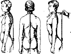
Figure 24. Rigidity of the vertebral column.
Figure 23. Normal mobility.
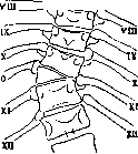
Figure 26. IV. Pathology of the Vertebral Column. Developmental defects and congenital deformities of the Vertebral Column. Congenital developmental defects of the Vertebral Column may manifest as changes in the number, shape, and connection of vertebrae with each other, as well as their connection with other bones—the skull, pelvis, ribs. Changes in the number of vertebrae, as mentioned above, are extremely rare. More commonly encountered are inclusions of incomplete, so-called wedge vertebrae (fig. 26), or more accurately, hemivertebrae, which are the cause of congenital scolioses [see separate table (p.135-136), figs. 1 and 2], differing from static and rachitic scolioses by their relatively small angular flexion without rotation around the axis, which gives them some resemblance to tuberculous scolioses. Complete bony fusions between normal vertebrae—true synostoses—are almost never encountered or occur as exceptions. Asymmetrical synostoses are more commonly observed with wedge and underdeveloped vertebrae—single or multiple. - Disease, or Klippel-Feil syndrome (Klippel, Feil) (fig. 27) is characterized by multiple synostoses of the lower cervical, and sometimes also the upper thoracic, vertebrae, which fuse into one shapeless bony mass and are accompanied by various developmental defects: wedge inclusions, splits of the arches, etc. Clinically, this deformation manifests as a sharp shortening or disappearance of the neck ('frog neck') and is accompanied by kypho-scoliotic curvatures, a sharp limitation of mobility in the upper part of the Vertebral Column, often asymmetrical positioning of the shoulder blades. In rare cases, this deformation may be accompanied by spinal cord and root disorders. In differential diagnosis, this developmental defect is most often confused with cervical spondylitis, rachitic kypho-scolioses, and other deformations, in particular with der 97
formation of Sprengel, which is characterized by a one- or two-sided elevation of the scapula, scoliosis, and is accompanied by a whole series of changes in the ribs and vertebral column, similar to the Klippel-Feil syndrome. Changes in the shape of vertebrae often depend on developmental defects in the posterior parts forming the spinal canal. Either complete splitting of the vertebral canal in the form of an open groove - fissure (rachischisis) or partial non-fusion of the arches - spina bifida posterior is observed. Longitudinal splitting can also occur in the anterior parts of the vertebral column - in the bodies, bearing the name spina bifida anterior (see Spina bifida). To the same category of developmental defects belongs the peculiar separation of the arches in the interarticular part - congenital interarticular spondylolysis (spondylolysis interarticularis congenita Neugebauer), which is based on improper development of the vertebra with the formation instead of one of two non-merging ossification points in the area between the obliquely positioned upper and lower articular processes. Spondylolysis by itself does not cause special disorders, but it has decisive importance as a predisposing cause for forward displacement - slipping of the last vertebra - spondylolisthesis (see). The diagnosis of all these listed changes is complex, as is generally the entire pathology of these so-called transitional parts of the vertebral column, especially the lumbosacral, various developmental defects and deformations of which are distinguished under the general name 'dysplasias' of the lumbosacral region and are characterized by decreased endurance of this part of the skeleton, reduced mobility, lumbosacral pains, lumbalgias, radiculitis, and other so-called myelo-dysplastic changes, which have already been mentioned above. If one takes into account that almost the same symptoms can also accompany inflammatory diseases of the sacro-lumbar region, as well as the very frequent neuro-muscular and rheumatic diseases here (see Lumbago), then the entire complexity of the pathology and diagnosis of these diseases becomes understandable. In this complex of dysplasias, the sacralization mentioned above has the greatest significance in terms of frequency and complexity of the picture. In true sacralization, complete fusion of the body of L5 with the sacrum and enlarged transverse processes with the iliac bones occurs. In the more frequent incomplete sacralization, various degrees of enlargement of the transverse processes of L5 on one or both sides and their fusion with the iliac bones are observed. Painful symptoms are most often observed with incomplete sacralization, usually after 20 years, i.e., after the completion of the ossification process, more often in men. The onset of pain is often associated with heavy physical labor, increased load and mobility in the lumbosacral region. The appearance of pain caused by sacralization always reduces the work capacity of these patients, which, if treatment is unsuccessful, leads to a change of profession or even disability. The diagnosis of sacralization is suspected when the mentioned clinical symptoms appear and is established by X-ray examination, which reveals a decrease in the number of lumbar vertebrae due to an increase in sacral segments. Counting vertebrae is best done from D12 (one must remember the possibility of underdevelopment of the XII rib), for which the thoracolumbar part must also be included in the X-ray. Recognition is facilitated when compared with the healthy side in unilateral sacralization. With complete fusion, the sacrum is correspondingly projected higher and the sacro-lumbar joint is determined on the line connecting the iliac crests, which is usually the boundary between L4 and L5. However, it must be borne in mind that in this very section, the slightest tilting of the pelvis during the X-ray or displacement of the X-ray tube so changes the X-ray image that the same anatomical changes can give rise to various interpretations, which is why special caution is needed in interpreting these pictures. Lumbarization (lumbalisatio) occurs less frequently; also less frequently does this congenital anomaly cause painful symptoms. The latter also occur with increased load and as a manifestation of decreased stability of the lumbosacral region due to its greater mobility. Usually, lumbarization is suspected by radiologists in cases where, below the horizontal line connecting both iliac crests, two free lumbar vertebrae are found, and not one, as under normal conditions, in contrast to sacralization, where the sacrum rises to this line (Reinberg). It remains to mention the frequently observed deformities of the L5 body, which, on the one hand, are a cause for the development of scoliotic changes (in some cases, conversely, scoliosis leads to changes in L5), and on the other hand, to dysplastic symptoms. All the listed bone dysplasias have only relative significance in the origin of the above-mentioned painful sensations, since, on the one hand, not every detected bone change is accompanied by painful phenomena, and on the other hand, similar pain symptoms can occur without any bone changes. Therefore, both the clinical diagnosis and the radiological interpretation of the X-rays present great difficulties. As for the treatment of these dysplasias, it is divided into conservative - providing rest to the affected part with the use of physical therapy procedures and a supporting corset - and operative - bone-plastic fixation of the sacro-iliac part of the vertebral column. Prevention of these diseases comes down to early recognition of congenital anomalies as predisposing causes and elimination of factors that could cause insufficiency of the vertebral column. In particular, the correct choice of profession has enormous significance. Changes in the shape of the spinous processes, their lengthening or shortening, by themselves rarely cause painful phenomena, more often they can give rise to diagnostic errors. Thus, excessive lengthening of C7, T1, D12 and L5 gives button-like protrusions of the corresponding spinous processes and serves as a reason for assuming tuberculous deformations, but preservation of mobility, absence of compensatory lordoses and painlessness, especially with dynamic load, decide the matter. In rare cases, excessive lengthening of the processes serves as a reason for their surgical removal. Acquired deformations on the basis of non-inflammatory changes of the vertebral column. On deformations developing on the basis of uneven load on vertebrae during their growth and formation without disease of the vertebrae themselves - see Scoliosis, Lordosis, Kyphosis. Rickets is one of the frequent causes of this kind of deformations of the vertebral column (fig. 28) (see Rickets). Very rarely are changes in the vertebral column also observed in osteomalacia. Juvenile kyphosis (kyphosis adolescentium), disease of Scheuermann-Mau (Scheuermann, Mai), is based on a peculiar change in the apophyses of the vertebrae, leading to changes in the bodies of the vertebrae. The disease vertebral column is analogous to a whole series of other osteochondropathies of juvenile age, affecting various epi- or apophyseal formations and causing subsequent deformities of developing bones (diseases of Perthes, Schlatter, etc.). In the vertebrae, the role of epiphyseal formations is played by the cartilage rings, in which at the age of 10-12 years, ossification nuclei appear, visible on profile X-ray images as small narrow wedge-shaped shadows at the upper and lower angle of the vertebral body with the base on the anterior longitudinal ligament [see figure 29 and separate table (pp. 135-133), fig. 3]. By 18-20 years (in women somewhat earlier), the process of additional enchondral ossification ends, due to which the shape of the vertebral bodies changes from biconvex to biconcave. In juvenile kyphosis, the ossification nuclei become loosened, fragmented, their contours become indistinct, sinuous, the correctness of the cartilage strip separating them from the surface of the vertebral bodies is disrupted, and then the bodies themselves begin to be involved - first in the form of changes in the contours of the upper and lower surfaces of the bodies, and then there is obtained as if an indentation of the apophyseal shadows into the anterior part of the vertebra, which is compressed here; the structure of the bone becomes indistinct and ultimately wedge-shaped deformities of several vertebrae, usually 3-4, with maximum changes in the middle one, occur. A characteristic feature of these changes is that in the presence of destructive changes,
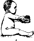
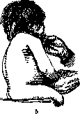
Figure 28. Kyphoses in children: a-normal kyphosis in a 4-month-old child; b-rachitic kyphosis.
and irregularities of the contours of the vertebral bodies, the preservation of the intervertebral discs, which rarely narrow but usually expand (in contrast to tuberculous changes); in addition, the pointed anterior angles of wedge-shaped narrowed vertebral bodies are drawn forward in a lip-like manner, forming an irregular indentation in the middle. The middle thoracic vertebrae are predominantly affected (most often from VII to X). The process usually ends within 1-1½ years and is observed in male adolescents from 15 to 17 years and female adolescents from 12 to 14 years, with the former being affected 4 times more frequently, which is attributed to excessive early physical strain. The process is benign, without any general complications, has a cyclic course, but leaves behind a persistent deformation--a painless, uniform, arcuate kyphosis. This disease is rare and is diagnosed exclusively on the basis of the characteristic radiographic picture of a lateral view. Osteochondropathy of the vertebral body, Calvé's disease, is an even rarer disease and can be considered analogous to the juvenile malacic changes of the tarsal and carpal bones (morbus Köhler I, morbus Kienboeck). In children, a small hump forms with limited mobility of the vertebral column, which is painful on pressure and percussion. On the radiograph, at the beginning of the disease, a compact shadow is determined in the center of the body. Subsequently, the vertebra flattens, narrows to ⅓-½ of its normal size and slightly lengthens, protruding forward beyond the general line of the vertebral column. A characteristic feature is the widening of the intervertebral spaces. The prognosis is favorable, and the vertebra slowly recovers. In diagnosis, it is necessary to consider the no less rare congenital flattening of the vertebral body, 'platispondyly,' and the so-called 'ivory vertebrae' (vertebres d'ivoire of French authors)--a marked densification of the vertebral bodies with preservation of normal shape, sometimes observed in cancerous metastases, primary sarcomas, and in some cases without clear etiology.
Figure 29. Osteochondropathy of the vertebral apophyses: a-initial changes; b-height of the disease-destructive changes of the bodies; c-stage of recovery-wedge-shaped deformation of the bodies with elongated anterior angles.
determined in the center of the body a compact shadow. Subsequently, the vertebra flattens, narrows to ⅓-½ of its normal size and slightly lengthens, protruding forward beyond the general line of the vertebral column. A characteristic feature is the widening of the intervertebral spaces. The prognosis is favorable, and the vertebra slowly recovers. In diagnosis, it is necessary to consider the no less rare congenital flattening of the vertebral body, 'platispondyly,' and the so-called 'ivory vertebrae' (vertebres d'ivoire of French authors)--a marked densification of the vertebral bodies with preservation of normal shape, sometimes observed in cancerous metastases, primary sarcomas, and in some cases without clear etiology. To the same group of rare diseases of the vertebral column, determined mainly radiologically, should be included changes of the nuclei pulposi. The discs may undergo calcification, which occurs quite rarely. Out of 200 films, Geist (E. Geist) found only one case of calcification. Partial prolapse of the disc is fairly frequently observed (according to Geist-in 38%), in which the nucleus plays a predominant role. Prolapse is most often encountered in the lumbar or cervical region, singly or multiple. The cause of prolapse may be damage to the cartilaginous disc or adjacent vertebral bodies from various pathological processes or from direct trauma to the vertebral column in the form of small cracks or even fractures of the cartilaginous disc. Prolapse of the disc varies in size, sometimes the prolapsed part gradually fills the entire vertebral body; in the latter, a reaction may appear, expressed in the new formation of bone substance around the prolapsed part of the disc. Sometimes prolapse occurs into the vertebral canal and leads to various disorders requiring surgical intervention. The prolapsed part of the disc either remains stationary, sometimes becoming vascularized and ossified, or undergoes osseous transformation. Disc prolapse is more frequently observed in strong men of athletic build. The symptoms of prolapse manifest themselves when the vertebral column is loaded with weight. This may also explain the lumbar complaints in adolescence.
VERTEBRAL COLUMN
13th vertebral pains, juvenile kyphosis. A poor posture with sharply expressed poor physique may also depend on multiple disc herniations. Senile changes consist in degeneration of the disc, as a result of which the latter becomes thinner. In various diseases of the V.-in spondylitis, osteomalacia, tbc, osteomyelitis, syphilis, cancer-the intervertebral disc is usually involved in the pathological process. When the cartilaginous membrane of the intervertebral cartilage is damaged, the nucleus is extruded into the spongy part of the vertebra, and depending on this, according to the data of Calve and Galland, the following may develop: 1) herniation of the nucleus pulposus, which in the opinion of these authors is probably the main cause of juvenile kyphosis; 2) impaction of the cartilaginous nucleus into the center of the vertebra body when there is marked osteoporosis of the latter; 3) displacement of the nucleus into the spinal canal with the formation of kyphosis, which is accompanied by compression of the spinal cord and paralysis; 4) deposition of lime in the thickness of the nucleus, detectable on x-ray films. In generalized fibrous osteodystrophy of Recklinghausen (osteodystrophia fibrosa generalisata Recklinghausen), "along with characteristic changes of other bones, mainly long ones, specific changes of the vertebrae are also observed with the formation of cyst-like defects, which on x-ray films have the appearance of soap bubbles against the background of transparent vertebrae, which usually remain intact, do not collapse and do not lead to curvature."-To the group of non-inflammatory diseases of the V., causing persistent rigidity or even complete immobility of the V., belong: 1) so-called deforming spondylitides (spondylitis deformans), which should more correctly be called, by analogy with joint diseases, deforming spondyloses (see), and 2) ankylosing spondyloarthritis (see separate table, fig. 4), or Marie's disease (see Marie's disease). Deforming spondylitides usually involve and fuse two to three vertebrae, whereas spondyloarthritis gradually involves the entire V. and even the hip and shoulder joints. Ultimately, true "woodenness" of the V. in a bent position sets in. Something similar, but to a lesser degree, is also observed in senile changes of the V.-To the category of deforming spondyloses also belongs tabetic spondylopathy (see separate table, fig. 5), observed in the lumbar region in elderly people and leading to various degrees of curvature, completely painless. Radiologically, combinations of severe deformities of the vertebrae with enveloping them huge lime deposits are determined. The diagnosis is facilitated by the presence of other symptoms of tabes. In rare cases, similarly chaotic and atypical changes in the vertebrae may also occur in syringomyelia. Kumme11's disease (Kümmell's disease), traumatic spondylitis (spondylitis traumatica), a deforming process in the vertebra developing after a relatively mild trauma without gross anatomical changes. The basis of the disease is a chronic non-inflammatory aseptic destructive process, and therefore this disease should be classified as a spondylosis, not a spondylitis. This disease is observed in most cases in men engaged in heavy physical labor, and is characterized by three stages. The first stage-immediately after trauma, sharp pains which gradually subside within 2-6 weeks; the second stage-a clear interval, complete absence of any pathological phenomena-painful, functional, anatomical; lasts from 2-6 weeks to 2-3 years, most often 6-8 months, after which the third stage sets in-relapse of independent pains, occurring gradually without a definite cause or after a minor repeated trauma. Painfulness of the vertebra on pressure and load, limitation of mobility, rigidity of muscles appear, and later protrusion of one spinous process and slight curvature of the spinal column develop. Radiologically, in both the first and second stages, no signs of compression fracture or other changes are determined, and only in the third stage typical changes appear-phenomena of softening of the vertebra, its wedge-shaped flattening without any focal changes; the contours remain clear and smooth, sometimes small bone outgrowths are observed, and in some cases in the area of the anterior longitudinal ligament, longitudinal bands of ossification may appear. Most often this disease is treated as tuberculous spondylitis, from which it differs with difficulty mainly on the basis of radiological data: absence of narrowing or disappearance of intervertebral cartilages and contact changes in neighboring vertebrae, good contouring and absence of edge erosion, absence of shadows of cold abscesses. However, the indicated radiological and especially clinical data "are very shaky and far from always convincing to exclude tuberculosis, which is why prolonged observation and biological control are required. Similarly, it is often difficult to distinguish this disease from compression fracture of the V., from which it differs primarily anamnestically-the presence of the second clear stage, although according to the observations of Kornev, a clear interval may also occur with fractures. On profile x-ray films, fractures are determined from the very beginning and often fragments and triangular fragments protruding from the anterior edge of the body and remaining connected with the longitudinal ligament are visible, which is not the case in Kümmell's form. The course is prolonged with exacerbations, but in general relatively favorable: large deformities and spinal cord disorders do not occur, pains gradually begin to subside, but weakness of the back may remain. Treatment-rest, a supporting corset, in stubborn cases-fixation of the spinal column. Injuries of the V. Subcutaneous injuries of the V. are caused either by direct violence-a blow from behind, falling on the back, or by indirect effects-longitudinal compression of the V. and its excessive forced movements during falling or being pressed by a weight. Depending on the degree of injuries and the anatomical changes they cause in the V., bruises, sprains, dislocations and fractures are distinguished, both in isolated form and in various combinations. Bruises and sprains have relatively little significance, rarely causing severe consequences, whereas dislocations and fractures represent a great danger, mainly depending on complications from the spinal cord, which in 2/3 of all cases
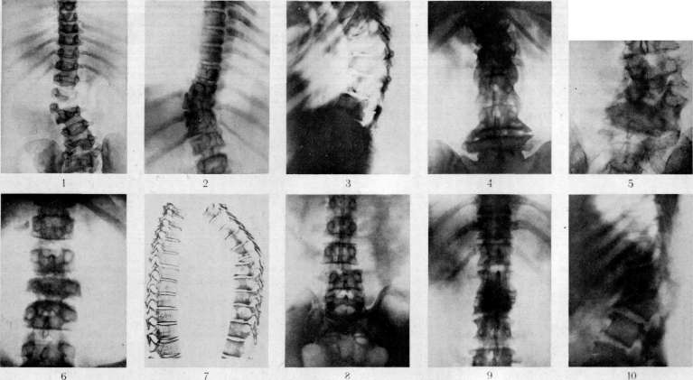
Figure 1 and 2. Congenital scolioses on the basis of additional wedge-shaped vertebrae: fig. 1 between LIV and LV; fig. 2 between LIII and LIV with an additional rib. Figure 3. Osteochondropathy of the apophyses of the vertebrae (Scheuermann-Mau's disease). Figure 4. Spondylosis of the lumbar region spondylosis deformans and spondyloarthritis. Figure 5. Spondylopathy; tabetic changes of LIV and LV. Figure 6. Chronic osteomyelitis of the III lumbar vertebra. Figure 7. Tumor of the spinal column (on the right, cancerous metastases in the vertebrae; on the left, normal spinal column). Figure 8. Dislocation of the III LUMBAR VERTEBRA. Fig. 9 and 10. COMPRESSION fracture of two VERTEBRAE with breaking out of the anterior edge: fig. 9 en face; fig. 10 in profile
Compression or injury of the spinal column occurs. Fractures of the vertebral column occur 10 times more frequently than dislocations, but in relation to fractures of all other bones they constitute only 0.5%, and only depending on professional conditions does this percentage rise to 2% and higher, as is the case, for example, in mining areas and mines, where collapses, falls from height, etc. are more frequently observed (observations of Wagner-Stolper in mining areas of Upper Silesia, observations in the Donbas mines). Bruises (contusions) of the vertebral column are caused by direct impacts and are characterized by damage to soft tissues and the surface of bones without violation of their integrity. The clinical phenomena—bruises, pain, partial limitation of mobility—are usually transient, but in some cases bruises can cause concussion of the spinal cord and even hemorrhage in it, which leads to spinal cord disorders, also mostly transient. More severe bruises can lead to unrecognized surface cracks and apical fractures in the processes, as well as cracks and hemorrhages in the bodies themselves, which may only later manifest as nonspecific inflammatory changes, so-called traumatic spondylitis of Kümmell (see above). Treatment—temporary rest, massage, unloading. Sprains (distorsions) occur under the influence of excessive forced movements (flexion, extension, rotations), causing rupture of the ligamentous apparatus of the vertebral joints, but without persistent displacement, which distinguishes them from dislocations, being as it were a preliminary stage of the latter. These injuries can also affect the intervertebral cartilages. Distorsions are most frequently observed in the same places as dislocations, i.e., in the cervical region, less frequently in the lumbar, and are accompanied by painful limitation of mobility and pain, which are particularly severe in incomplete ruptures (Kocher). It is not always easy to distinguish a distorsion from a fracture without displacement. More severe injuries can be complicated by spinal cord phenomena, as with all other types of injury to the vertebral column. Treatment—rest, in some cases—light traction.
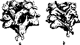
Dislocations of vertebrae may be limited to only the lateral joints or be accompanied by displacement of the bodies themselves in fractures of the latter and damage to the intervertebral cartilaginous discs. Isolated dislocations of lateral joints are observed almost exclusively in cervical vertebrae and depend either on excessive rotation—rotational dislocations, usually unilateral, or on excessive flexion—flexion dislocations, usually bilateral. The upper vertebra is considered dislocated, the articular surfaces of which slide forward and upward along the articular surfaces of the underlying vertebra. If the articular surface of the upper process shifts only to the anterior edge of the articular surface of the lower one, there is an incomplete dislocation, subluxation (subluxatio), but if the upper process shifts beyond the anterior edge of the lower one, jumps over it, catching in the notch in front of the articular process of the lower vertebra, then this is a true dislocation (luxatio). In unilateral (Fig. 30) subluxation due to oblique displacement forward and upward, i.e., elevation of the articular processes, lengthening of the affected side of the vertebral column occurs, depending on which the head tilts to the opposite side, and in dislocation, when the articular processes pass over each other, shortening of this side occurs, and the head tilts to the affected side (Wagner). The spinous process of the upper dislocated vertebra is slightly elevated, shifts somewhat forward, and deviates from the midline, while the transverse process protrudes on the side of the dislocation. At the same time, the head shifts somewhat forward, movements become limited. In bilateral dislocation, the head stands straight, without lateral flexions, but is slightly tilted, as if shifted forward. The spinous process of the upper displaced vertebra also shifts forward, without deviating to the sides, the distance between the spinous processes increases. Bilateral dislocations (Fig. 31) are significantly more severe than unilateral ones and are rarely isolated, more often accompanied by displacement of the bodies themselves (complete dislocation according to Kocher's classification), which as a rule leads to phenomena of compression of the spinal cord depending on the narrowing of the vertebral canal.
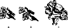
Recognition of dislocations is made on the basis of characteristic anamnesis, typical displacement of the head (Fig. 32), palpation of the spinous and transverse processes, limitation of movements, and phenomena from the spinal cord. Disturbance of swallowing is often observed. Sometimes displacement of the upper cervical vertebrae can be palpated with a finger through the mouth on the posterior wall of the pharynx. Special importance is given to radiodiagnosis (see separate table, Fig. 8). Unilateral dislocations have a better prognosis and in fresh cases can be corrected by constant traction using a Glisson's loop or by reduction of the dislocation under anesthesia, for which, after placing the patient on the abdomen, traction is applied to the head with sequential flexion to the healthy side (a rotation to the healthy side can also be done simultaneously) until the catching articular processes separate; then the head is turned to the affected side and fixed with plaster. Bilateral dislocations (Fig. 33) are also reduced by traction with sequential retraction and extension of the dislocated part of the vertebral column, or an attempt is made to reduce the dislocation one by one from each side using the above method. In general, reduction of bilateral dislocations is rarely successful. Any reduction must be done very cautiously (danger of compression of the spinal cord) and if possible immediately, since after several days and especially weeks reduction becomes impossible.
Particularly dangerous are dislocations of the upper two vertebrae: very rare dislocations of the head, i.e., between the occipital bone and the atlas, and more frequent ones between the atlas and axis. The narrowing of the spinal canal caused by displacement can by itself lead to severe phenomena of compression of the spinal cord, and the frequently observed in this case rupture of the cruciform ligament and fractures of the odontoid process as a rule lead to death from the impaction of the odontoid process into the spinal cord and damage to the latter. Dislocations in other parts of the vertebral column, especially in the lumbar region, more often combine with separation in the area of intervertebral cartilages or with fractures. Fractures of vertebrae Kocher divides into partial (isolated) and complete, combined with dislocations and displacements (dislocation-fractures). Partial (isolated) fractures can be in the processes, arches, and bodies. Fractures of spinous processes occur most often under the influence of direct violence—blow from behind, usually on the most prominent processes (thoracic upper and lower), less frequently from muscle contraction or from the processes pressing against each other during excessive extension. These fractures are recognized by abnormal mobility of the process, its displacement, crepitation, local swelling, and painfulness on pressure. In difficult cases, x-ray decides. These injuries do not require special treatment, in cases of excessive and persistent pain, sometimes the fragment has to be removed.
Violence acting on the spinous processes can be transmitted to the vertebral ring, causing fractures of the arches, often bilateral, with impaction of the fragment into the vertebral canal and compression of the spinal cord (Fig. 34), which is especially dangerous in the cervical region, where fractures of the arches are more frequently observed. Recognition is not always easy. The x-ray decides [see separate table (pp. 135-136), Figs. 9 and 10], and if compression of the spinal cord by the impacted arch fragment is detected, operative removal of the pressure is indicated—elevation and reposition of the fragment or its removal. Fractures of the odontoid process are more frequently observed in pathological conditions (tuberculosis), less frequently in traumatic dislocations and fractures with displacement. The fractured odontoid process can impact into the spinal cord and injure it.
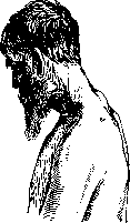
Figure 30. Unilateral rotational dislocations: a - complete dislocation; b - incomplete dislocation.
Figure 31. Dislocations (a - normal position): b - incomplete dislocation; c - rotational bilateral dislocation.

Figure 32. Unilateral dislocation of the cervical vertebrae. Figure 33. Bilateral dislocation of the VI cervical vertebra. Figure 34. Fracture of the vertebral ring with compression of the spinal cord.
to it, which as a rule leads to sudden death. However, in reality fatal outcomes in this condition are observed significantly less frequently than was previously believed (according to Jefferson in about 20%), which depends on the integrity of the ligamentous apparatus, which holds the fragment in place, preventing its impaction. Rupture of the cruciate ligament is a decisive factor not only in fractures of the odontoid process, which as a result can displace and impinge into the spinal cord, but also in anterior displacements-dislocations of the atlas without fracture of the odontoid process, when the latter, not held by the ruptured ligament, remains in place and injures the spinal cord, which, shifting forward together with the upper segment, presses against it. Fractures of the articular processes are comparatively rarely observed in isolation, more often in combination with dislocations and more extensive fractures. Relatively more frequently isolated fractures of the processes of the sacral bone articulating with L5 (Henle) are observed. Finally, among the rare injuries are isolated fractures of the transverse processes, observed almost exclusively in the lumbar region and caused either by direct violence or by excessive contraction of muscles. Usually in this case there is diffuse tenderness in the lumbar region on the affected side, radiating to the legs or abdominal wall. Pain increases with bending and rotation; less painful is bending toward the affected side. Patients spare the lumbar region, hold the trunk bent and inclined to the affected side, where contracture of the m. psoas is also often observed. In diagnosis, one must consider deep contusions, distortions, lumbago, and exacerbations of chronic inflammatory diseases of the vertebral column (tuberculosis), which can give similar symptoms. The matter is decided by a good X-ray, a film on which the avulsion of the outer, narrower part of the transverse process is visible. In rare cases, retroperitoneal hemorrhage is observed, leading to paralytic intestinal obstruction (Volkovitch). Fractures of the bodies of vertebrae most often result from the action of direct violence-a blow from behind, more often from longitudinal compression and bending of the vertebral column forward or backward, falls from a height onto the feet, onto the buttocks or head, falls of heavy objects from above onto the head or shoulders with excessive bending (and simultaneously there may be a fracture of the sternum) or finally from falling on the back onto a hard prominence with excessive backward bending. The degree and form of the fracture are influenced not only by the size and direction of the force of violence, but also by the location, age, and condition of the vertebral column itself. In contrast to dislocations, which occur most frequently in the most mobile segments, fractures of the bodies are observed at the transition from the immobile to the mobile part of the vertebral column, mainly in the thoracolumbar region. According to Menard, almost half of all fractures occur at D11 and L1 and in decreasing progression on the higher and lower vertebrae, with relatively frequent fracture at the apex of the thoracic curvature D5,6. In children with their more elastic ligamentous apparatus and especially with wide, elastic discs, which act as buffers between the vertebrae, fractures occur as a result of relatively greater violence (more often distortions and dislocations) than in the elderly, in whom the buffering capacity and elasticity progressively decrease. Particularly brittle are the fused, ankylosed vertebrae after spondylitis and spondylosis, not to mention active destructive processes (especially in cancerous metastases), in which fracture can occur even with minimal trauma, as if spontaneously. Isolated fractures of the bodies of vertebrae are most often observed in the form of so-called compression fractures (fig. 35-38). Under the influence of longitudinal compression, usually combined with bending, the low-resistance spongy bone tissue of the body is wedged and flattened and spreads sideways. Depending on the elasticity of the bone mass and the force of compression, various degrees and forms of damage are observed-cracks, incomplete fractures, compressions, transverse and oblique breaks, comminuted fractures, and complete comminutions. The injury can involve one or two vertebrae, rarely more.
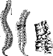
Figure 35.
Figure 36.
Figure 35. Mechanism of vertebral fractures: a-from compression; b-from bending. Figure 36. Compression fracture without narrowing of the canal.
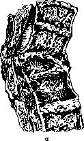
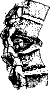
Figure 37. Compression fracture with narrowing of the canal and avulsion of the anterior edge of the vertebral body: a-specimen; b-radiogram of the specimen.
Collapse of the anterior parts of the vertebral column leads to relatively small kyphotic curvatures of the vertebral column. The most dangerous complications are spinal cord disorders caused by displacement of fragments and fragments of bodies into the vertebral canal. Fracture with dislocation, luxation fracture, occurs under the action of more significant violence leading to simultaneous displacement in the lateral joints. Complete displacement of the upper part of the vertebral column, shifting the upper vertebra forward and "downward Kocher calls a complete fracture with dislocation (totale Luxations-fraktur). These injuries are particularly severe and in the vast majority of cases lead to death depending on compression (fig. 39-41) or even ruptures of the spinal cord. A complete transverse fracture of the vertebral body itself can be combined with the most diverse injuries to all other parts of the vertebral column, sprains, dislocations, fractures of processes, and damage to cartilaginous discs, and finally rupture of the ligamentous apparatus; of particular importance is the rupture of the anterior and posterior longitudinal ligaments. The lines of fracture of the vertebral body usually run from above behind forward downward, and in the same direction occurs the displacement of the upper part-slipping forward and downward, so that the upper vertebrae press upon the damaged one ("like a hat on the forehead") or may even stand in front of the last one. In turn, the posterior edge of the lower, undisplaced vertebra may with its corner enter the vertebral canal and impinge into the spinal cord. In some cases lateral displacement or a combination of both occurs.
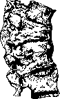
Figure 38. Healed compression fracture.
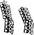
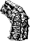
Figure 39.
Figure 40. Figure 39. Mechanism of complete fracture with dislocation. (After Matti). Figure 40. Complete fracture with dislocation with compression of the spinal cord by the posterior protrusion of the vertebra. (After Matti). Symptomatology of fractures. Curvature of the vertebral column in fresh cases is usually slight and in luxation fractures it is naturally expressed much more sharply than in simple compression fracture. In the latter there is usually only some divergence and protrusion (elevation) of the spinous process of the upper vertebra. In luxation fracture, depending on the forward and downward displacement of the upper vertebra, the spinous process of the latter also shifts forward, as if disappearing, and the spinous process of the damaged vertebra protrudes (if several are damaged, the upper of the damaged ones). Curvatures are more expressed in the thoracic region and less in the cervical and lumbar regions, where there is physiological lordosis. Sometimes the protrusion of the displaced vertebra forward can be palpated as a prominence on the posterior wall of the pharynx in fractures of the cervical region
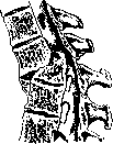
Figure 41. Fracture with dislocation and narrowing of the canal. (After Okinczyc).
or be palpated through the abdominal wall in the lumbar region. In older cases, kyphosis may increase and acquire an angular or arcuate character. Spinal cord disorders in all injuries to the V. are the most important symptoms and the most dangerous complication. Uncomplicated fractures without spinal cord phenomena comparatively quickly lead to consolidation and recovery and by themselves do not represent great danger, in contrast to cases complicated by spinal cord phenomena, which, on one hand, very often lead to death, and on the other hand, leave serious functional disorders for a long time. Diagnosis and course. In fresh cases immediately after injury, it is not always easy to determine the true nature of the injuries to the V. The basic symptoms of fractures-pain, limitation of mobility, curvature of the V. and spinal cord disorders-have only relative significance. On one hand, very severe pain, as well as a sharp limitation of mobility-inability not only to stand, but even to turn in bed-may be observed in distortions and contusions, and on the other hand, even significant fractures without displacement can be comparatively painless and cause moderate difficulty in movement. The size and shape of kyphotic curvatures also do not always decide the question of the nature of the anatomical changes, since deformities of the V. are observed in sprains and displacements without fractures; at the same time, even significant initial curvatures can correct themselves so quickly that they remain unnoticed. Finally, the intensity of spinal cord disorders does not always correspond to the severity of the injury, since even complete paralyses often recover quickly, all the more so that transient spinal cord disorders may depend on simple concussion of the spinal cord. Similarly, there is not always a correspondence between the level of bone injuries and spinal cord disorders, since the latter can spread significantly above the former in hemorrhages and ascending degenerations of the spinal cord. X-ray examination decides the issue, but it must necessarily be compared with the clinical picture, without which one can fall into a gross error in interpreting the films. In compression fractures on frontal films, some densification of the body of the flattened vertebra is found, a decrease in its height and an increase in its transverse diameter (from crushing of the bone mass) with clear outlines and preservation of the intervertebral cartilage; the line of fracture is usually not visible, it is only detected on lateral films; on the latter, wedge-shaped compression of the body and bone fragments are seen with particular clarity, which usually protrude from the anterior edge and are held by the preserved longitudinal ligament in front of the narrowed anterior edge of the body. The clarity of the outlines and the preservation of the intervertebral discs or some narrowing of them is of importance. In the not infrequent injuries of 2-3 vertebrae, the cartilaginous discs usually are destroyed, disappear, the upper vertebra is embedded in the lower one, so that the X-ray picture becomes very close to tuberculous lesions. The diagnosis is made already on the basis of all the data. It is even more difficult radiologically to distinguish a healed fracture of two or more vertebrae with their complete fusion and arcuate sagging from similar pictures of reparative changes after inflammatory diseases (tuberculosis, osteomyelitis). Fatal outcomes occur both directly after the injury from shock (in almost half of all cases) and in the further course from various complications. In the near future, the patient is threatened by hypostatic pneumonia, which so often accompanies severe injuries to the V., especially in elderly people. Then later, with spinal cord disorders, the most dangerous complication is paralysis of the bladder, or rather paralysis of the detrusor, which, with spasm of the sphincter, leads to retention of urine and prolonged catheterization, which always carries the danger of infection of the bladder, suppurative cystitis and ascending pyelonephritis. To prevent this fatal complication, a fistula is sometimes applied to the bladder from the very beginning, which does not always save the patient, but only delays the fatal outcome. When the bladder is overfilled, the sphincter can stretch, and urine begins to be discharged involuntarily drop by drop (ischuria paradoxica), which greatly complicates the care of the patient, especially when bedsores, so often developing after fractures of the V., appear. The latter can lead to deep necrosis involving bone and be accompanied by phlegmonous inflammations leading to sepsis. Paralysis of the sphincter of the rectum leads to incontinence of feces (incontinentia alvi); in those cases where there is simultaneously paralysis of the muscles of the abdominal wall and disturbance of peristalsis in the lower parts of the intestines, retention and accumulation of feces in the ampulla recti and above may occur, which can cause ulceration of the mucous membrane. Sometimes paralysis of the abdominal muscles can cause phenomena of meteorism and intestinal obstruction requiring surgical intervention. The prognosis depends on the level and degree of injury, as well as on the causes causing the wedge-shaped phenomena. The higher the injury, the more serious it is. Injuries in the upper cervical region are especially dangerous, often leading to sudden death; injuries in the lumbar region are much milder-below the end of the medullae spinalis. According to Burrel (Bur rell), in fractures of the upper cervical region mortality reaches 85%, in the upper thoracic-76%, in the lower thoracic-56% and lumbar-50%, with an overall mortality of 64.5%. The decisive significance for the prognosis is the question-whether there was a complete transverse rupture of the spinal cord or only its partial injury and compression. However, this question is not clarified immediately, but after several days after the injury, since even complete paralyses during this period of time can improve or even disappear completely. Preservation of knee reflexes and normal activity of the pelvic organs in spastic and asymmetric paralysis immediately exclude a complete rupture of the spinal cord. In those cases where after 10-15 days there remains a complete flaccid symmetric paralysis with loss of sensitivity, absence of knee reflexes and pelvic disorders, one can confidently speak of a complete transverse rupture of the spinal cord. Such patients sooner or later die. With partial injury or compression of the spinal cord, initially complete paralyses may either pass completely or leave behind some trace in the form of pelvic disorders or disturbances in the motor and sensory sphere. These disturbances can have both symmetric and asymmetric character, with different levels and degrees of motor and sensory disorders. The predominance of sensory disorders over motor ones speaks more for root phenomena. Finally, the cause causing the spinal cord disorders and the degree of its eliminability also influence the prognosis. In some cases, this cause lies only in the mechanical compression of an undamaged spinal cord by bone displacements and fragments, after reduction or removal of which the function of the spinal cord can be completely restored; in other cases, the cause lies in the spinal cord itself, in its injuries and changes, which are in essence not mechanically removable, but undergo successive processes of degeneration or healing. Among these causes, hemorrhages into the spinal cord (see Haematomyelia), which are observed either in the brain tissue itself, more often in the gray matter, or in the central canal, occupy a special place. Such hemorrhages can be insignificant, pinpoint, but can also reach significant size; spreading up and down the spinal cord over a considerable distance, they can cause spinal cord phenomena significantly above the site of injury, which complicates diagnosis. Gradually resolving, the hemorrhage can either be completely eliminated or leave behind changes in the form of delimited cavities in the central canal (syringomyelia traumatica), degenerative changes in the brain itself, adhesions, cicatricial changes with corresponding symptoms of loss in the motor and sensory sphere. Haematomyelia is observed mainly in the cervical region. Extramedullary hemorrhages with intact meninges represent less danger. In individual cases, these accumulations of blood can exert transient pressure on the spinal cord, leaving behind sometimes a trace in the form of cicatricial adhesions and root phenomena. Even more rarely paravertebral hemorrhages are observed, which can sometimes accumulate on the anterior surface of the V., simulating psoas abscesses. As already stated, the prognosis is determined in the first 10-15 days after the injury. If the phenomena from the spinal cord do not improve, then all the mentioned dangers remain in force, but if by this time the phenomena decrease, then gradual improvement and even complete recovery can be expected, despite the severity of the anatomical injuries to the V. However, in a considerable percentage of cases, some disorders remain.
With complications involving spinal cord disorders, partial anesthesias or hyperesthesias, limited paralysis and paresis, contractures, urinary incontinence, impotence, trophic ulcers on the feet, and symptom complexes resembling tabes (see) may remain. But even after uncomplicated fractures, pains, unsteadiness of gait, and reduced endurance may persist for a long time. According to Haumann (1926), restoration of work capacity is observed only in 25% of cases after 2 years, in 61% after 6 years, and in 80% after 9 years. Such outcomes largely depend on the untimeliness and irrationality of treatment, which in turn depends on the difficulty of early recognition of fractures of the Vertebral Column, which in almost half of all cases remain without timely recognition (Volkovich). Therefore, thorough examination of the Vertebral Column after any trauma with the use of radiodiagnosis is the basic condition for improving the outcomes of treatment* of these injuries. Treatment of any injury to the Vertebral Column must first of all begin with protecting the patient-from possible further traumatization during carrying, transportation, moving, and examination, when careless movement can-displace fragments and thereby injure the spinal cord. Therefore, the greatest caution is required in fresh cases immediately after injury. When laying the patient on a flat, unyielding bed, complete rest is ensured for him, and by raising the head end of the bed and fixing the head with Glisson's loop, gradual relaxation of the Vertebral Column and its straightening are achieved, which is especially important in fractures, whereas in contusions and sprains simple rest is sufficient, and in dislocations reduction is required. In fractures of the upper part, traction* by the head is the most convenient method, while in the lower part, reclining position with a pad placed under the affected area is of greater importance, preferably made from flaxseed. Volkovich recommends the prone position for straightening the Vertebral Column in the lower part. To create complete rest, especially with inadequately prepared personnel, it is better to apply a plaster cast to the patient, covering the head, trunk, and thighs, but one must remember about-bedsores. Rest and bed rest-should continue until the moment of fracture consolidation, approximately for 1½-2 months,-The patient should be kept in bed longer rather than put on his feet too soon, as in such cases incomplete restoration of work capacity is more often observed. Absence of pain, symptoms of deficits, stability to load, and the radiographic picture of consolidation allow patients to be put on their feet in a supporting apparatus, plaster cast or removable corset, which the patient must wear for 1/2-1 year. Persisting pains, instability to load and stiffness of movement force either to keep patients in bed or corset longer or to resort to surgical fixation of the spine according to Albee and its modifications (see Spondylitis). With complications involving spinal phenomena, it is necessary to determine the cause of compression and try to eliminate it mechanically. However, solving this problem is very difficult, t. k., even with good radiographs it is not always possible to establish the cause of compression, and in intramedullary hemorrhages and injuries to the spinal cord such an external cause may be absent altogether, therefore the operation proposed for eliminating compression of the spinal cord-laminectomy (see)-in many cases may turn out to be as if trial and not bring the desired results. This operation is particularly useful in fractures of the arches, especially bilateral ones, when by removing or elevating them the spinal cord is freed from compression, just as when removing individual fragments or bone splinters from the spinal canal. This operation also has some significance in displacement of the Vertebral Column, when the arch of the overlying vertebra compresses the spinal cord. There are advocates of early intervention g but most authors adhere to a wait-and-see position, performing the operation-after a certain period, after determining the success of conservative measures and the subsidence of acute phenomena, in particular the reduction of hemorrhages. However, this waiting should not be too long, as scar tissue changes may occur, although, on the other hand, even old cases can be operated on successfully (Polenov). The danger of early operations is great-from 25% (de Quervain) to 72% (Lloyd) mortality and is justified by the exceptional severity of these injuries (in Haumann's 204 cases, 33 patients died soon after admission). Injuries to the Vertebral Column. Injuries to the Vertebral Column with violation of the integrity of the covering are divided into contused-lacerated, punctured-cut and gunshot wounds; they in turn are divided into penetrating into the spinal canal and non-penetrating. These injuries occur even more rarely in peacetime than closed injuries, but they carry greater danger compared to the latter, on the one hand, due to the possibility of infection being carried into the depth, especially into the spinal canal (meningitis), and on the other hand-due to the significantly higher frequency and severity of spinal cord injuries. Contused-lacerated wounds, inflicted by the action of a blunt instrument, occur least often and have relatively little clinical significance, as they are usually more superficial and rarely penetrate into the spinal canal. The danger of these, usually contaminated wounds-is in the infection of tissues and injuries to the spinous processes and arches. Punctured-cut wounds of the Vertebral Column occur somewhat more often. They are inflicted from behind with a sharp instrument, usually in the upper part of the Vertebral Column, at the level of the striking arm. More often these injuries do not penetrate into the spinal canal due to the protection from the vertebral arches. Less often the instrument passes between the arches or violates their integrity and penetrates into the spinal canal. Most often, injuries to the spinal cord are asymmetrical and accompanied by Brown-Séquard type crossed paralysis, but other types of injuries are also possible-from injury to only the membranes to complete transection of the spinal cord. With transection of only the roots, motor and sensory disorders appear on the corresponding side. Injury to the spinal cord membranes can sometimes be recognized by the discharge of cerebrospinal fluid from the wound.-According to Wagner, these injuries give a relatively small percentage of mortality (about 20%) and about the same small percentage of complete recovery, and in 60% of cases some or other disorders remain. Gunshot wounds to the Vertebral Column occur as rarely as punctured wounds, constituting no more than a quarter of a percent of all gunshot wounds. In most cases, penetrating wounds are involved, accompanied by fractures of the bones of the Vertebral Column, and ; in some cases complicated by injuries to internal organs, especially when wounded from the front. Wounds can be through and blind, and the bullet can get stuck either in the body of the vertebra or in the spinal canal. Penetrating through the soft spongy substance of the vertebra body, the bullet forms smooth channels, so-called "perforated" fractures, and hitting the dense processes and arches, it gives comminuted fractures; fragments penetrating into the canal can damage the spinal cord. Injuries to the spinal cord in gunshot wounds occur more often than in closed fractures and are more dangerous, as, on the one hand, the anatomical destruction in them* is more serious, and on the other hand, there is always a threat of infection. Clinical symptoms, depending on the degree of injury to the bones and spinal cord, are similar to those in closed fractures. The prognosis-for gunshot injuries is very serious and depends mainly on the degree of spinal cord disorders. Most patients die in the first hours after injury. In those cases where the wounded person in the Vertebral Column overcomes the initial shock, he faces a second danger-compression and injury to the spinal cord by bone fragments, which can shift during transportation. Therefore, such wounded in a military situation should for the first time most often remain in place or in the nearest rear and be evacuated only after 10-15 days, when acute phenomena subside and the degree of spinal cord injury is determined. Nevertheless, the danger of even such milder injuries is great. Thus, according to Kornev's data from the imperialist war, out of 100,000 wounded evacuated from the front and passing through the city union's hospitals in Leningrad, injuries to the Vertebral Column occupied the last place among other injuries, constituting only 0.3%, while their mortality was at the very first place, constituting 11% with the overall mortality from all injuries at 1.8%. This mortality depends mainly on complications from the pelvic organs and rapidly developing bedsores. According to the same data, complete recovery occurred in 20% of all cases, while the rest of the injuries resulted in varying degrees of disability.-Treatment of gunshot injuries to the spine varies, depending on anatomical conditions and the nature of the injury. For non-penetrating wounds without spinal cord disorders, therapy is conservative: aseptic treatment of the wound, complete rest and traction in case of bone injuries.
For penetrating wounds with spinal cord disorders, surgical treatment may be applied to eliminate the latter. Theoretically, surgical treatment is indicated to eliminate mechanical compression of the spinal cord by a foreign body or bone fragments, as well as to remove them when they have penetrated the spinal cord with partial damage to the latter; the operation is contraindicated in complete ruptures of the spinal cord and extensive hematomyelias. However, accurate recognition of the nature and degree of damage is extremely difficult, especially immediately after injury. X-ray diagnosis also does not provide sufficient data to establish the anatomical causes of spinal cord disorders, except in cases where the presence of a foreign body or bone fragments is found in the spinal canal. Early laminectomy can immediately eliminate the above-mentioned causes of spinal cord disorders, thereby preventing subsequent inflammatory and degenerative phenomena, but at the same time it may prove not only useless in the absence of removable anatomical causes, but even dangerous due to the addition of surgical shock and the possibility of infection. Late surgery has the advantages of greater safety and the possibility of operating under clear indications, since by this time the hemorrhage has resorbed and inflammatory changes have subsided, but waiting can cause persistent degenerative changes in the spinal cord, even with simple compressions without damage to the spinal membranes, whereas timely elimination of compression leads to restoration of spinal cord function. There are advocates of both early and late interventions. Experience from the last war showed the severity and danger of these injuries and strengthened the position of advocates of earlier interventions. However, indications for urgent operations are still very limited, and most adhere to a wait-and-see tactic, depending on the course and changes in spinal phenomena within 10-15 days after injury, when significant improvements may occur. If by this time it is determined that there is a complete rupture of the spinal cord (see fractures of the V. above) or, conversely, rapid recovery of movement and sensitivity occurs, then surgery is not indicated; if, however, signs of definite compression persist but there is no complete rupture, then surgical intervention is fully indicated and often gives a favorable result. Infectious diseases of the V. Inflammatory diseases of the V. can be acute and chronic, localizing either in the joints or in the bones themselves. Acute inflammatory diseases of the intervertebral joints are observed in acute articular rheumatism and as a complication after various acute infections, primarily after influenza and scarlet fever. These synovitis manifest as transient sharp pains and diffuse limitation of mobility of the V. The larger joints of the upper cervical vertebrae are more frequently affected, and unilateral involvement of which leads to painful unilateral muscle contracture (torticollis), pain with certain movements, and radiating neuralgic pains. Acute osteomyelitis of the vertebrae is rare, constituting only about 2% of all osteomyelites (Hahn). According to Volkmann, this disease is more frequently observed in adolescence (50% of all patients from 8 to 17 years of age), predominantly in men (72%) and mainly in the lumbar region; in 58% it affects the arches and processes, in 34% the bodies, and in 8% the entire vertebra. The causative agent is most often a staphylococcus, and the portal of entry for it is paronychia, furuncles, and other purulent diseases. The disease arises acutely, sometimes lightning-fast, proceeds violently and in 2/3 of all cases leads to death, especially when the bodies are affected. Only in 1/3 of cases is the diagnosis made correctly, mainly when the posterior parts are affected; more often, appendicitis, peritonitis, typhoid fever, pneumonia, meningitis is suspected. Usually the temperature rises to 40° and higher, a septic general condition sets in, leukocytosis reaches 40,000, and pains are sometimes very severe, more often radiating to different areas depending on the localization—to the occiput, both scapulae, the iliac regions, the extremities. When the lower thoracic and lumbar vertebrae are affected, meteorism, dilation of the veins of the abdominal wall (Plenz), dysuria (Petersen) are often observed; the patient lies with bent legs. The Kernig symptom is often present. Complications from the spinal cord and membranes are frequently noted (up to %, according to Schonbauer), especially when localized in the cervical region. The resulting abscesses, accumulating on the lateral surfaces of the vertebral bodies, can "descend and rupture into the pleura, bronchi, esophagus, and abdominal organs, sometimes psoas abscesses form. When the posterior parts of the arches are affected, the disease runs much more easily and is more correctly diagnosed by local limited tenderness on pressure, edema, swelling of the outer coverings, and accumulation of pus, the examination of which for pyogenic bacteria in many cases decides the matter. Usually one vertebra is affected, rarely two or more, and in 14% osteomyelitis of other bones is observed. Deformity of the V. is relatively rare, and pathological fractures are even rarer. The prognosis depends to a large extent on the localization (2/3 of deaths fall on body involvement and 1/3 on arch involvement), while the overall mortality according to previous statistics reached 72%, and only recently with improved early diagnosis it has dropped to 41% (Ogloblin). X-ray diagnosis in acute cases is of little help, since more or less clear changes occur only in later stages in prolonged processes. Treatment consists in locating the abscess and opening it, if this is technically possible. This is easiest to accomplish when the posterior parts are affected and much more difficult (and often impossible) when the bodies are affected; in the thoracic region, costotransversectomy (first applied by Heidenhain) can be used, in the lumbar region—opening of the psoas abscess, in the cervical region—opening of the abscess in front of or behind the sternoclavicular muscle. Chronic osteomyelitis of the vertebrae [see separate table (pp. 135-136), Fig. 6] is observed either as an outcome of the acute form with the formation of fistulas and sequestra or as an independent form—primary chronic staphylococcal spondylitis (Oehlecker). This disease is extremely rare (only about 30 cases have been described), and in its course it very closely resembles tuberculous spondylitis—especially the fistulous form, with which it is usually confused. Based on material from about 700 cases of tuberculous spondylitis, osteomyelitis of the V. was established in 4 cases (P. G. Kornev). The lumbar region is predominantly affected, as in the acute forms, but in contrast to the latter, the bodies are more frequently affected than the arches and processes. The disease, sometimes subsiding, can drag on for decades, the general condition suffers little, local and radicular pains are not always constant, spinal cord disorders are less frequent than in acute processes, but fairly frequent are cold abscesses and fistulas, in particular fistulas on the thigh, giving rise to suspicion of tuberculous spondylitis or osteomyelitis of the thigh. Deformities of the V. are relatively rare, mainly in the form of flattening of the lumbar lordosis, sometimes a small gibbus forms with slight limitation of mobility. On X-ray examination, marked sclerosis and bone growth are striking; the affected vertebra or two adjacent vertebrae collapse and flatten, especially at the edges; their lateral and anterior parts protrude beyond the general line of the vertebral bodies, forming periosteal growths, which in the form of bridges can extend to adjacent vertebrae, which prevents more severe collapse. However, it should be noted that these characteristic features do not appear earlier than 3-6 months after the onset of the disease, and before this period usually no changes are found radiologically. Diagnosis is based on a careful history (often acute onset) and course, bacteriological examination of pus, the presence of other osteomyelitic foci, and the characteristic X-ray picture mentioned, which however has conditional value, since sclerosis of the vertebrae and the presence of periosteal growths can also be observed in syphilis and in post-typhoid spondylitis. The presence of staphylococci in the pus in fistulas may be a secondary phenomenon, but at the same time it is not always easy to determine the primary infection in closed old abscesses and in osteomyelitis, therefore many (in particular Eletsky and Demidov) attach great importance to the anti-staphylolysin reaction. Of course, it is also necessary to use other biological reactions: agglutination, RW, Pirquet reaction, focal reaction with Koch's tuberculin, erythrocyte sedimentation rate, examination of blood morphology, etc. With a high degree of probability, tuberculosis of the V. can be excluded if after opening the cold abscess or aspirating pus and applying autovaccine the clinical signs of the disease disappear relatively quickly (Eletsky and Demidov).
In this case, autovaccination is treatment ex juvantibus. The prognosis is significantly better than in acute forms (out of 23 published cases, 4 were fatal). The treatment of recognized osteomyelitis is surgical, where possible, mainly for lesions of the posterior sections: opening of abscesses and removal of sequestra. For lesions of the bodies, only opening of accessible abscesses. Auxiliary treatment-vaccinotherapy-with autovaccine or polyvalent staphylococcal vaccine. Post-typhoid spondylitis is a more benign disease than staphylococcal osteomyelitis of the V. It occurs more often after relapsing fever, less often after typhus and abdominal typhus, and is generally rarely observed. In the USSR, these complications were well studied during former typhus epidemics, when post-typhoid spondylites occurred and were described much more frequently. They arise on average a month after the typhus and manifest as a new rise in temperature, an attack of sudden severe pain in the lumbar region, a sharp limitation of mobility, and sometimes swelling of soft tissues. These acute phenomena gradually begin to subside, and the disease usually ends within several months, leaving behind limited mobility in a small area of the V., sometimes with slight curvature. Neither psoas abscesses nor fistulas are observed. The most characteristic of these diseases is the destruction of the intervertebral cartilage and bony outgrowths, fusing the affected vertebrae. X-ray changes are determined already a few weeks after the onset of the disease and initially manifest as clouding and narrowing of the intervertebral spaces, which may later completely disappear. Changes in the bodies themselves are not always pronounced; sometimes foci of softening are observed, reaching the cortical layer; more often, the body of one or two vertebrae is uniformly compressed. Later, proliferation and thickening of the periosteum occur, changing the lateral contours of the vertebrae, then massive lateral bony spurs appear, which in the form of bridges extend from one vertebra to another at the base of the transverse processes symmetrically on both sides and fuse them. At the same time, the anterior longitudinal ligaments ossify in the area of the lesions, and ultimately complete bony ankylosis of two or three vertebrae occurs. Syphilis of the V. Syphilitic spondylitis occurs very rarely (in Russian literature about 20 cases have been published, in foreign literature up to 150) and is even more rarely diagnosed due to the extraordinary similarity in clinical and X-ray picture with tuberculous spondylitis. Three forms of syphilitic lesions of the V. are distinguished (Synakevich): 1) spondyloarthritis luetica-lesion of the intervertebral joints in the form of acute (in the secondary period) or chronic synovitis, more often occurring in combination with other syphilitic arthritides (arthrolues tardia Schle-singer's), usually passing without a trace under the influence of treatment and leading to ankylosis only in rare cases, especially with simultaneous lesions of the bodies and processes. 2) Spondylitis luetica simplex-simple osteo-periosteal lesion of the bodies and processes without destructions, manifesting as root pains, rigidity of the V., swelling and edema of the outer coverings in the area of the lesion; these phenomena usually easily respond to specific treatment and pass, leaving behind no noticeable traces except slight periosteal thickenings and exostoses. 3) Spondylitis luetica destructiva, s. gummosa, which, in contrast to the first two forms, causes more distinct changes-destructions in the vertebrae, and therefore is more often diagnosed. As in other bones, the process can have either a more diffuse infiltrating character or a more limited one, affecting both the bodies and processes, usually starting from the periosteum and spreading to the bone. Milder cases of bone gummas are characterized by a sharply delimited rarefying process, while in more intense bone formation, a complete narrowing of Haversian channels can occur, leading to impaired circulation, severe necroses and sequestration. The intervertebral cartilages are also involved in the process. Relatively rapidly developing reparative processes and periosteal proliferations prevent too much collapse of the vertebrae, which is why large gibbs are not observed in syphilitic spondylitis. According to Zlesch, spondylitis luetica occurs mainly in adults (in the 3rd-4th decade) and three times more often in men than in women. The favorite localization is the cervical region, the upper four vertebrae, especially the atlas and axis, which many German authors (Neumann, Petren, Schlesinger) explain by the transition of the ulcerative process from the mucous membrane of the pharynx to the vertebrae, as a result of which destruction of the anterior parts and sequestration can occur. French authors (Fournier, Hallopeau) consider the lesion of the vertebrae primary, and the ulceration of the pharyngeal wall secondary. Lesions of the V. often occur in combination with lesions of other bones in particularly malignant forms of syphilis, which gave grounds to speak of a special 'osteotropic' strain of spirochete. The disease usually appears in the tertiary period, several years after infection; acute onset is rarely observed, usually the disease develops slowly and is characterized by intermittent spontaneous pains with nocturnal exacerbations. Sometimes the pains reach an extreme degree, especially with localization in the cervical region, radiating to the occiput, ear and mastoid process; pains on swallowing can occur. The gibbus is usually less pronounced than in tuberculosis, often swelling and infiltration of soft tissues over the affected vertebra, limitation of mobility, muscle rigidity; with cervical lesions-torticollis. In about 1/3 of all cases, spinal cord phenomena are also observed (R. Hunt). The overall mortality according to previous data reaches 40% and depends mainly on the destruction of the upper cervical vertebrae, their displacement and compression of the spinal cord; in rare cases, fatal hemorrhages from the a. carotis ext. were observed. With the refinement of diagnosis, the prognosis has sharply improved. In differential diagnosis, the greatest difficulties are presented by tuberculous lesions, in particular lesions of the two upper vertebrae, proceeding with almost the same clinical phenomena. X-ray changes are also not sufficiently characteristic, in any case they are unreliable (Wimmer). In the initial stages of periosteal changes in syphilis, there may be none. Infiltrates on the posterior wall of the pharynx and swelling on the occiput can also be observed in tuberculosis, but true psoas abscesses do not occur in syphilis (the described cases are doubtful), unless infection and formation of purulent sinuses occur. The diagnosis is based on the general signs of syphilis, especially in the presence of specific lesions of other bones and joints. RW has less importance than specific treatment ex juvantibus, which quickly relieves pains, whereas rest and unloading do not always relieve them, in contrast to tuberculosis. It should also be borne in mind that syphilitic spondylitis can occur in a tuberculous patient, just as tuberculosis of the V. can occur in a syphilitic. In recognition, traumatic spondylitis, tumors of the vertebrae, chronic osteomyelitis and post-typhoid lesions should also be taken into account. Treatment is applied both general anti-luetic-drug, and local-orthopedic. Tuberculosis of the V.-see Spondylitis. Actinomycosis of the V. occurs very rarely and is a secondary disease when the process spreads to the V. from the mucous membranes of the oral cavity, pharynx, esophagus, intestine or from the lungs. One or several vertebrae can be affected; the infiltrating process can involve the entire vertebra and penetrate into the spinal canal, without however causing spinal cord disorders, but more often limited only to root phenomena (Wullstein). The bone appears as if eaten by worms, in rare cases collapse of the destroyed vertebra can occur. Actinomycosis (see) is determined by the presence of a primary progressive focus, the formation of a dense, 'like a board' infiltrate in the area of the affected vertebrae, the appearance of purulent disintegrations with characteristic druses. The prognosis is poor, as the process spreads to the V. in far advanced cases. The iodine treatment used (K J from 3 to 5 g per day) rarely arrests the process. Tumors of the V. occur rarely, constituting about 1.5% of all tumors in general. Benign tumors of the V. have relatively little clinical significance, acquiring it either with particularly strong proliferation or when the tumor narrows the spinal canal and intervertebral foramina, causing compression of the spinal cord and roots. Chondroid exostoses and enchondromas are more common, in most cases multiple. Osteomas, lipomas, as well as angiomas are rarely observed. Malignant neoplasms of the V. occur significantly more often; according to Schlesinger, for one benign there are 10 malignant.
Primary tumors are observed less frequently than secondary ones, among which metastatic carcinomas occupy first place, constituting the vast majority of all tumors of the Vertebral Column. Primary sarcomas of the vertebral column—osteogenic, originating from the bodies and arches—are encountered in younger subjects and on average last 10-12 months, leading to death. Initially developing in the center of the bone, the tumor may for a relatively long time not give visible changes in the shape of the Vertebral Column; its curvature, which occurs after the collapse-compressive fracture of a vertebra, is often sudden and may be accompanied by symptoms of compression of the spinal cord. Radiographically, the development of the tumor manifests as large foci of rarefaction [see separate table (pp. 135-136), Fig. 7], which disappear with compression of the body. Osteosclerosis in sarcoma is the greatest rarity (Reinberg). The intervertebral cartilages remain unchanged. Clinical symptoms depend on the direction and degree of growth of the tumor. Intracostal growth manifests as persistent pain. Soon after, root phenomena join in the form of attacks of severe neuralgic pains. Further growth into the vertebral canal already leads to compression of the spinal cord, after which death usually soon follows. The tumor may grow outward or inward, extending to adjacent tissues and organs. Periosteal sarcomas are observed less frequently, recognition of which is difficult due to the less clear radiographic picture. These tumors are sometimes difficult to distinguish from paravertebral sarcomas originating from surrounding tissues—ribs (chondrosarcomas), pleura, etc., since the former may grow and infiltrate surrounding tissues, while the latter may secondarily involve the Vertebral Column. Significant difficulties for diagnosis are presented by isolated tumors within the spinal canal, which usually originate from the meninges of the spinal cord and are located either extra- or intradurally. For the most part, these are either sarcomas or tumors of mixed type—angio-, myxo-, fibrosarcomas, which early compress the spinal cord, why the differential diagnosis in these cases is made on the basis of the sequence of occurring phenomena—first root, then spinal cord, and only at the end bone; the latter usually do not have time to develop, in contrast to tumors of the vertebrae, which more often first give bone changes (at least radiographic), and then root and spinal cord (see Spinal Cord).-Multiple myelomas are observed in elderly people and when localized in the vertebrae can cause bending of the Vertebral Column and compressive fractures with angular and arcuate curvatures. Radiographically, the bone appears as if porous with numerous round defects having sharp and regular contours. Differentiation must be made from metastatic carcinomas, Recklinghausen's disease, and osteomalacia. Carcinoma of the Vertebral Column is found almost exclusively in the form of metastatic nodules, for the most part multiple, rarely solitary. In percentage terms, most often the transfer to bones is given by carcinomas of the prostate gland, then of the thyroid and mammary glands, less frequently of the uterus and other organs, but in absolute numbers bone metastases are more often observed in the most common of the mentioned diseases—carcinoma of the mammary gland. The Vertebral Column, and especially its lumbar part, along with the pelvic bones, is the most frequent localization of metastases to the bone system. Usually such metastases appear already in far advanced stages of the disease, shortly before death, but they can be detected relatively early. Carcinoma of the Vertebral Column, like other tumors, in most cases, although not necessarily, manifests first with attacks of persistent local and irradiating root pains, passing into constant unbearable suffering, not relieved by rest or by medicinal and physical therapeutic means. Later, phenomena of compression of the spinal cord and bone-destructive changes occur—collapse of the vertebra with formation of deformity of the Vertebral Column. The greatest importance for diagnosis—especially early—is given to radiographic examination, which can detect metastatic carcinomas even before the primary neoplasm manifests clinically, especially in the prostate and thyroid glands (Reinberg). Radiographically, metastatic carcinomas are observed in two forms—osteolytic and osteoplastic. More often osteolytic destructive metastases with resorption of tissue and absence of reactive growth of bone substance are encountered, manifesting as numerous small round homogeneous bone defects with clearly outlined [see separate table (pp. 135-136), Fig. 7], but uneven contours (in myelomas the contours are even); these forms are more often observed with metastases, mainly from the thyroid gland, less frequently from the mammary gland. Differentiation must be made from multiple myelomas, Recklinghausen's disease, primary sarcoma of the Vertebral Column, and in case of compression—from inflammatory destructive diseases (from the latter it is distinguished by the absence of changes in the intervertebral cartilage).-On the contrary, osteoplastic metastases lead to marked densification, sclerosis of the bone, depending on the developing reactive deposition of bone around the smallest carcinoma nodules; on the radiogram the vertebral body, preserving its external form, appears either as a solid dense shadow or as indistinctly outlined merging sclerotic spots. Usually osteoplastic metastases are observed with carcinoma of the prostate gland. These forms must be differentiated from other sclerosing processes—osteomyelitis, syphilis, Calvé's disease, etc. Approximately the same picture can be given by bone metastases in hypernephroma, which can be detected considerably earlier than the primary disease, as well as in multiple metastases of malignant adenomas of the thyroid gland. Echinococcus of the vertebrae is encountered extremely rarely. According to Reinberg, only about 75 cases of echinococcus of bones have been published, of which most frequently the pelvic bones and vertebrae are affected. More often an infiltrating multilocular echinococcus is observed, which penetrates the spongy substance with small vesicles, resorbs it and forms round cyst-like defects. The bone becomes nodular, as if it bulges, the cortical layer thins, necroses, sequestra, and pathological fractures easily occur. Reactive changes in the bones are absent. The growth of echinococcus in the vertebrae is slow—over years, by which it differs from a tumor. The surgical interest is in the unilocular echinococcus. Growing into the vertebral canal, echinococcus vesicles can cause phenomena of compression of the spinal cord, which usually serves as an indication for surgical intervention. Most often echinococcus develops from the posterior mediastinum in the region of Dn-vi and from the retroperitoneal fat tissue adjacent to the lumbo-sacral part of the Vertebral Column. Correct diagnosis is rarely made, more often at autopsy or during operation for compression of the spinal cord. One must keep in mind tumors of the Vertebral Column, cystic osteodystrophies, etc.-Treatment is surgical: removal of large vesicles and especially vesicles pressing on the spinal cord. Infiltrating forms are inoperable. According to Borchard and Rothmann, who collected 48 cases of echinococcus of the Vertebral Column, 17 patients were operated on with 4 cases of recovery. The authors believe that with timely diagnosis most of the 30 deceased patients could have been saved by operation. V. Operations on the Vertebral Column. As already stated above, surgical intervention on the vertebral column has the purpose of eliminating and removing various harmful factors acting mainly on the spinal cord, or strengthening the weakened Vertebral Column. To the first category of operations belong: opening of abscesses and their aspiration in inflammatory diseases, opening of echinococcus vesicles, removal of tumors, bone fragments, and foreign bodies from the vertebral canal, as well as expansion of the latter by removal of the arch in cases of narrowing of the bony canal. To access abscesses forming in the posterior parts of the Vertebral Column, simple incisions of soft tissues serve; to access pathological accumulations located in the anterior parts of the Vertebral Column, the same interventions are used as in cold abscesses in tuberculous spondylitis (see). Accumulations in the spinal canal can also be eliminated by means of punctures or laminectomy (see). Laminectomy is used both for the purpose of examining the spinal cord and for the purpose of removing various harmful factors. By removing the arch and opening the vertebral canal, one can first remove everything that presses on the spinal cord from behind: fragments of the arch, foreign bodies, tumors originating from the walls of the arch and meninges of the spinal cord. By cutting one or two nerve roots, which according to Sherrington's research does not lead to significant disorders, one can retract the spinal cord, examine the anterior part of the bony wall of the vertebral canal and if necessary remove foreign bodies, fragments, as well as remove wedge-shaped protrusions of vertebral bodies in fractures and displacements of the latter, on which Urban particularly insists. At the same time, remnants of hemorrhages can be removed. If the meninges of the spinal cord are damaged, then after examination of the spinal cord and removal of hemorrhages, a suture can be applied, in cases of significant defects of the meninges, free transplantation of fascia can be resorted to.
Finally, for examination of the spinal cord with its membranes intact, it is necessary to open the latter and, depending on the circumstances, remove tumors, hemorrhages, etc. The indication for laminectomy is mainly injuries to the Vertebral Column and tumors within the spinal canal, whereas in inflammatory diseases, especially in tbc, they are very limited (see Spondylitis). lo& Operative osteoplastic fixation of the Vertebral Column—the operation of Albée and its modifications—has recently received very wide application, mainly in tuberculous spondylitis, and less frequently in other diseases and injuries. The indication for fixation is insufficiency of the Vertebral Column and its destructive changes leading to curvature. Thus, operative fixation is applied in juvenile kyphosis, traumatic spondylitis, certain forms of compression fractures, as well as in various dysplasias of the Vertebral Column, especially in the sacro-lumbar region, where it is best to use two plates placed alongside the spinous processes according to Henle. The technique of osteoplastic fixations of the Vertebral Column—see Spondylitis. VI. Professional pathology of the Vertebral Column. In describing individual forms of diseases, the causes influencing the occurrence of various pathological conditions have already been indicated. Among these causes, the professional working conditions and occupational hazards are of great importance, which come down to the following factors: prolonged forced position of the Vertebral Column, its uneven load, excessive strain, trauma, cooling, intoxications, and other working conditions affecting the general condition of the body. In turn, the listed factors can exert varying degrees of influence on the condition and changes of the Vertebral Column depending on whether they affect the normal, anomalous, or already pathologically changed Vertebral Column in various age, constitutional, and conditional peculiarities of the body. The influence of occupational hazards on the normal Vertebral Column is manifested both in the developing and in the formed Vertebral Column. Uneven load on the Vertebral Column, incorrect sitting, too long immobility and weakness of the musculature lead to such frequent 'school' scolioses (see) and kyphoses in children, the rational fight against which lies in conducting physical education, school gymnastics, correct posture and proper alternation of work and rest. Similarly, too early heavy physical labor in an adolescent can lead to incorrect posture, round back, scolioses, etc. Professional influences on posture and the shape of the back, and consequently on the endurance (work capacity) of the Vertebral Column, are also manifested in adults. Thus, a round back bordering on kyphosis is as if an occupational disease of professions associated with prolonged forced bent position, especially in persons engaged in heavy physical labor, e.g., in loaders (up to 30%), then in locksmiths, molders, tailors, office workers, etc. (Golyanitsky). These changes in the Vertebral Column worsen the function of the thoracic cavity and its organs—the lungs, heart. On the other hand, certain professions lead to the formation of a flat back, as for example in tailors (Hoffa). The weaker the musculature and the longer the forced position, the more the Vertebral Column assumes a passive 'fatigue' position already due to the support of bony parts, which ultimately leads to persistent changes in the skeleton, 'uncompensated' kyphoses and scolioses. This loss of elasticity, leading to increased pressure of the vertebrae on each other, affects the condition of the intervertebral joints and discs and is expressed by changes in the cartilage, narrowing of the spaces, limitation of mobility, and appearance of pain. If to this are added excessive strain and especially injuries, even if small but repeated, then conditions are created for a whole series of secondary pathological changes: spondylarthritides, osteoperiostitides, deforming and ankylosing spondyloses, which, on the one hand, lead to limited or diffuse rigidity of the spine, and on the other—to neuralgic and root pains (especially lumboischialgias). Uneven or excessive cooling, past acute infections, as well as intoxications intensify the painful phenomena. To an even greater extent than in the normal Vertebral Column, the influence of professional conditions is manifested on the anomalous, hence inadequate, Vertebral Column. All congenital deformities of the Vertebral Column, clefts, asymmetries, synostoses, inclusions and underdevelopments, and especially lumbosacral dysplasias, are that locus minoris-resistentiae on which the influence of occupational hazards is manifested with particular force, early putting such patients out of commission and making them disabled. The same applies to the influence of occupational hazards on the pathologically changed Vertebral Column, whether it be a latent process in the vertebrae (e.g., tbc) or the consequences of a past disease that caused various deformities, adhesions, etc., not to mention active processes in which any strain is especially unfavorable. Therefore, preliminary thorough examination of the Vertebral Column, especially radiological, and proper selection can, on the one hand, prevent the possibility of severe complications in such persons, and on the other hand, give the key to the correct choice of profession. Finally, it is necessary to emphasize the influence of trauma to the Vertebral Column. In the section on fractures, it has already been mentioned that certain professions, e.g., miners, are particularly often subjected to severe trauma to the Vertebral Column with all its pernicious consequences. However, even less significant injuries in many professions associated with lifting heavy weights and jolts can, without causing clear clinical symptoms at first, subsequently lead to various diseases; these include traumatic spondylitides of Kümmel's type, changes associated with damage to the nuclei pulposi, unrecognized compression fractures, cracks, avulsions, etc. Thus, the influence of professional factors on the Vertebral Column is very diverse, which forces special attention to be paid to studying working conditions to eliminate occupational hazards and prevent their effect on the Vertebral Column. At the same time, it is necessary in persons assigned to work to carefully examine the Vertebral Column to determine: its strong and weak points (musculature, elasticity, posture, anomalies, pathological conditions) for the correct choice of profession corresponding to the revealed peculiarities of this subject and his Vertebral Column.
p- Kornev..
Related articles
Mentioned in
Cite this page
“Vertebral Column.” Soviet Medical Encyclopedia. English translation of Bolshaya Meditsinskaya Entsiklopediya, 1st ed. (Moscow, 1928–1936), ed. N. A. Semashko. https://sovietmedicalencyclopedia.pages.dev/article/vertebral-column/