Spondylolisthesis
Historical document, translated for reference. It reflects medical knowledge of the 1920s–30s and is not medical advice.
Summary
Spondylolisthesis is the gradual forward and downward displacement of one vertebra relative to another below it. This article describes the condition's anatomical features, clinical manifestations, diagnosis, and treatment approaches as understood in the 1930s.
Encyclopedia article (1928–1936)
SPONDYLOLISTHESIS (from Greek spondylos-vertebra and olisthesis-slipping), a gradually developing displacement of the body of one vertebra forward and downward in relation to another, lower-lying vertebra. Most often, due to anatomical-physiological conditions (see Lordosis), S. is observed between the sacrum and L5. The deformation was first described by Herbiniaux in 1782. The name S. was given to it by Killian in 1854. The disease was considered very rare and affecting exclusively women. Until 1892, only about 18 cases were noted in the literature. The question interested almost only obstetricians, since the vertebra displaced into the pelvis served as an insurmountable obstacle for the passage of the child's head through the birth canal. Only since G. I. Turner's presentation at the Leningrad Congress of Russian Surgeons in 1922 did S. attract the general attention of broad medical circles in the USSR and abroad.
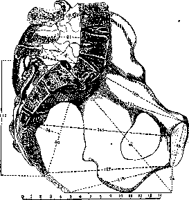
Figure 1. Prague pelvis. Grade I spondylolisthesis.
Lambl, to whom the most extensive monograph on S belongs, distinguished 3 degrees of "self-dislocation of the spine": 1) protrusion of the L5 body over the sacral promontory with partial exposure of the sacral base; 2) strong forward inclination of the displaced L5; 3) descent of L5 into the pelvis and complete exposure of the upper surface of the sacrum (figs. 1-4). The thought suggests itself that
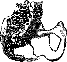
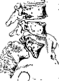
Figure 2. Paderborn pelvis. Grade II spondylolisthesis. (After Lambl,) these 3 degrees are different stages of the same process, however, to the present time the origin and patho-anatomical essence of S. have not been clarified. Lambl's student, Neugebauer, put forward a theory according to which the basis of S. is spondylolysis, i.e., a congenital defect of ossification of the arch of L5 in the area between the upper and lower articular processes (fig. 5). If at this junction between 2 ossification nuclei fibrous tissue remains instead of the development of a bony union, then under the influence of additional mechanical factors, disconnection can occur. Further load in the upright position leads to gradual displacement of the vertebral body with the upper articular processes and the underlying disk forward and downward along the inclined plane of the sacrum, while the posterior part of the arch with the spinous process and the lower joints remains in place. Lambl categorically rejected the causal relationship between incomplete ossification of the arch in the portio interarticularis and the development of S. He pointed out that fibrous unions, bony "sutures" or false joints on the neck of the L5 arches occur, according to the anatomist Chausov, in 5% of people and represent a variant, named by him, Lambl, spondylolysis. If the development of S. depended on the presence of spondylolysis, then the first could not be such a rarity, and the second should always occur together with the first. Meanwhile, among the 15 known specimens of S. at that time, only 3. Prague pelvis A. Grade III spondylolisthesis. (After Lambl.) in 3 the arches had characteristic spondylolysis sutures and that without "dissection" of this area. Lambl's data that extreme degrees of S. are possible without violation of the integrity of the arches are irrefutable, however Neugebauer's theory nevertheless remained dominant and in recent years has found new factual confirmation. First, the observations of G. I. Turner and subsequently the reports of numerous foreign authors proved that S. is by no means a rare disease and does not affect only women. For example, Meyerding from the Mayo clinic, having material on 207 cases, gives the ratio of male to female patients as 71% to 29%. Secondly, Schmorl, the best expert in the patho-anatomy of the spine, substantiated on autopsy material Neugebauer's theory, establishing that spondylolysis between 2 epiphyseal ossification nuclei in each arch cavity can occur simultaneously with signs of S. already in small children. Even reactive processes preventing further slipping can be clearly expressed in early childhood. Furthermore, it was shown that S. is not an anomaly peculiar only to L5. In Schmorl's bone collection
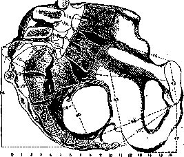
Figure 4.
Moscow pelvis. Spondylolysis and spondylptosis. (After Lambl.) for 23 spondylolistheses of L5 there were 11 spondylolistheses of L4. Finally, a whole series of clinical observations speaks in favor of the significance of repeated mechanical, accidental and professional traumatic injuries that disrupt the established equilibrium and lead to the gradual manifestation of hidden defects in the structure of the spine. The clinical picture of S. is very characteristic (fig. 6). The trunk is shortened due to the lumbar section, which is thrust into the pelvis. The sacrum protrudes backward, clearly outlined under the skin, upward it ends with a step instead of an arched transition into the lumbar region. The pelvis is elevated by the pubis, the iliac crests protrude more and are directed more horizontally than normal. The lumbar lordosis is elongated upward, smoothed in the middle section, deepened below, steeply breaking off above the sacrum. The spinous process of L5 or L4 kyphotically protrudes (Chirkin's symptom). The waist is shortened and widened, bordered by a deep transverse fold, above which another smaller skin groove is noticeable. Functional disorders consist of some limitation of mobility, especially forward bending of the back, and in a less smooth, duck-like gait. Subjective complaints come down to relatively rapid fatigue and the appearance of dull pain in the sacrum and legs when standing for a long time.
The course varies, depending on the frequency and strength of secondary mechanical effects on the primary defect. In the absence of prolonged load or traumatic injuries, S. may not
40
progress, and patients can live to a ripe age, maintaining working capacity. Even with complete displacement of L5 into the pelvis, subjective disorders are moderate and inconstant. Conversely, in initial forms of S., adjacent to spondylolysis, sometimes severe pains and phenomena of acute functional insufficiency of the spine are observed. Recognition is easy in typical cases according to the characteristic deformation of the spine and the absence in the anamnesis of indications of spondylitis or compression fracture in the lumbar-sacral region. Profile X-ray has decisive significance for initial forms. Technical difficulties in clear imaging of L5 in the lateral projection make one appreciate the signs of S. first indicated by Turner on the frontal film. The characteristic changes are considered: 1) superimposition on the sacrum of the shadow of L5, the anterior contours of which
f
shine through with a convex downward line, appearing through the bone like the visor of a cap (chapeau de gendarme according to French authors); 2) the spinous processes of L5 and L4 elevated upward like "sparrow tails"; 3) widening of the space between the sacrum and the posterior part of the L5 arch. Recognition of spondylolysis is also sometimes possible on X-rays in the sagittal projection. Experimental data on this topic are presented by N. S. Markelov. Under the incorrect name of prespondylolisthesis, cases of distortional hyperextension of the lumbar-sacral region are described in the literature. They differ from S. by deepening of the lumbar lordosis throughout and by lowering the pubic arch of the pelvis.
Prevention of S. coincides with treatment, since the dislocation is irreducible and only prevention of further slipping can be discussed. For this purpose, operations connecting the arches of the upper lumbar vertebrae with the sacrum by bone graft and to some degree unloading L5 are used with success (see Spondylitis). Operative treatment is indicated only in cases not yielding to bloodless orthopedic measures. Horizontal position with elevated pelvis, corrective gymnastics, massage and wearing special corsets are widely used. The main and decisive factor is timely recognition of spondylolysis and exclusion of harmful professional-domestic moments.
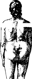
Figure 6.
of the vertebra and service in the Red Army, Voenn.-med. zhurn., 1930, no. 2; Turner G., Spondylolisthesis, its essence, clinical manifestations and significance in changing body statics, Vestn. khir., 1926, book 16; Jungnan n s H., Spondylolisthesen ohne Spalt im Zwischengelenkstiick ("Pseudospondylolisthesen"), Arch. f. Orthop., B. XXIX, 1930; he same, Spondylolisthese, Pseudospondylolisthese und Wirbelverschiebung nach hinten, Beitr. z. klin. Chir., B. CLI, 1931; Meyrding H., Spondylolisthesis, Surg. gynec. a. obstet., v. LIV, 1932; Mouchet A. et Roedeger C., La spondylolisthesis, Revue d'orthop., v. XIV, 1927; Reinbold P., La spondylolisthese acquise et la spondylolisthese traumatique, Schweiz. Z. Unfallmed., B. XXV, 1931; Schmorl G., Beitrag zur Kenntnis der Spondylolisthese, Deutsche Ztschr. f. Chir., B. CCXXXVII, 1932.
Related articles
Mentioned in
Cite this page
“Spondylolisthesis.” Soviet Medical Encyclopedia. English translation of Bolshaya Meditsinskaya Entsiklopediya, 1st ed. (Moscow, 1928–1936), ed. N. A. Semashko. https://sovietmedicalencyclopedia.pages.dev/article/spondylolisthesis/