Spondylodeses
Historical document, translated for reference. It reflects medical knowledge of the 1920s–30s and is not medical advice.
Summary
Spondylodeses refer to bony outgrowths on the vertebrae that can develop as reactive or infectious processes, often associated with spinal static insufficiency or degenerative changes. These formations may appear as spurs, hooks, or bridges connecting adjacent vertebrae, commonly in the lumbar region, and are frequently discovered incidentally on X-rays.
Encyclopedia article (1928–1936)
SPONDYLODESES (from Greek spondylos-vertebra). In the literature there is no unified understanding of the essence of S. and their origin, and consequently there is no unified nomenclature for them. By analogy with bone spurs that develop on the heel with flat feet, on the anterior-lateral edges of the vertebral bodies there first appears a lip-like formation, and then a bony exostosis resembling a spur, hook, beak, or bridge connecting two adjacent bodies (see separate table, fig. 5). These spondylotic formations are sometimes observed singly and on one side, sometimes on both sides and in front of the bodies of several vertebrae, most often in the lumbar region, sometimes they fuse the entire spine into a single bony mass. The origin of S. should be considered in some cases as a reactive and compensatory phenomenon with static insufficiency of the spine, in others-as infectious (toxic). The appearance of S. is associated either with the loss of elasticity of the spine in older age, especially with heavy labor and with load on the back, e.g. in porters, or with various defects of the spine causing its static insufficiency, e.g. with traumatic injuries, as well as with some congenital developmental defects of the lumbar-sacral region. These factors lead to the appearance of static, or deforming, spondylosis. Finally, the appearance of spondylosis is connected with diseases of the joints of the spine, for example with infectious (toxic) spondyl-arthritides or with diseases manifested by chronic progressive ankylosis of the spine--spondylosis or spondylitis ankylotica. Static, or deforming, S. is most often a reaction to insufficient elasticity of the cartilaginous discs and their compression during the period of human decline in the second half of life or with prolonged loading, as well as to congenital defects and trauma to the spine and trauma to the cartilaginous discs. However, not every injury to the spine, not every loss of elasticity of the cartilaginous discs leads to S. The height of the cartilaginous discs decreases either uniformly or asymmetrically, mainly on the right side (in left-handers, according to Schmorl, the opposite arrangement), causing tilting of the spine. Beginning with spurs developing in the anterior or lateral ligaments, the process can end with the fusion of two or more bodies. In this case, this phenomenon of localized static S. is sometimes called deforming S. or even deforming spondylitis. It is characteristic of people of middle and older age engaged in physical labor. When the deforming process or process of adhesion affects not the bodies but the joints of the spine, it is called spondyloarthrosis, or deforming spondyl-arthrosis (see). If both of these forms combine in one clinical picture, they are united under the general term-deforming osteoarthritis of the spine (see Spondylarthroses). In the clinical picture of localized static S., similar to static or deforming spondylarthrosis, there is little that is characteristic. Patients complain little about the back and almost never complain about the legs. Only prolonged forced position and heavy work in a bent position result in dull back pains, which disappear after a short rest or when the patient 'warms up' (hence the indications for mototherapy); 'weather-related' pains as a rule intensify. The appearance of the back differs little from a healthy one, only slight flattening and slight rigidity of the long lumbar muscles are noticeable. More serious 'symptoms' are rare and appear later, with the development of muscle atrophy on the basis of prolonged muscle contracture. On the radiogram (see separate table, fig. 3) there is always a striking discrepancy between the sharp anatomical changes and the insignificant complaints, as well as the minor changes in the function of the spine. This only confirms the idea that the reactive proliferative process by the development of exostoses, spurs and bridges compensated for the static insufficiency of the spine. These phenomena of static (deforming) S. are often discovered accidentally on X-ray films taken for other complaints. The discitides of the lumbo-sacral region deserve great attention, which in most cases lead to primary limited deforming S. and subsequent spondylarthrosis. The question of the mechanism of origin of this deforming S. has been well developed in recent years by the school of Schmorl in Germany (Günz) and the school of Williams in America. The discitides of this region, inflammatory-degenerative, traumatic from overstrain of the spine, with acute and chronic trauma, lead to compression of the intervertebral disc, to crushing of its elements and mainly the pulpy nucleus. The intervertebral space narrows, the contact distance between the vertebrae approaches. Due to the predominance of the ligamentous and muscular apparatus of the posterior part of the spine, there occurs a subluxation of L5 backward. The lateral joint relaxes, becomes loose, the intervertebral lumbo-sacral opening is deformed, narrows, and into its opening projects in the form of a tooth-like thickening the edge of the articular process of the underlying vertebra (S1). The result of sliding and abnormal mobility of the articular facets is periodic compression of the L5 root and lumbo-ischiatic pains with subsequent development of spondylarthrosis and partial deforming S. The latter, according to Schmorl, is possible with rupture of the fibrous ring of the border rim or bead (Randieistenannulus), which is the support for the main ligament of the anterior part of the vertebral bodies. With rupture of this border ring, the free, exposed edges of the vertebrae rub against the anterior longitudinal ligament of the spine, causing reactive inflammation in it, as a result-calcification of the sites of rupture of both the border rim and the disc itself. More severe manifestations of S. develop in the infectious and toxic forms of chronic rheumatic polyarthritis--similar to the spurs that develop on the heels in some forms of rheumatic polyarthritis of the foot. In more severe cases of these forms, following the development of primary phenomena of spondylarthrosis (see), which to one degree or another disturbs statics, sooner or later, phenomena of S. are added--the formation of sharp-pointed osteophytes as marginal exostoses, causing elongation of the upper and lower edges of the vertebral bodies and best detected on lateral and semi-lateral radiograms. In contrast to static deforming S., the intervertebral discs are compressed here not first, but following the spondylarthritic process. In severe cases of the resulting osteoarthritis of the spine (see Spondylarthroses, fig. 5), the vertebrae come into contact with their bony parts. The etiological causes, clinical and radiological phenomena in this case see Spondylarthroses.-In the development of typhoid spondylitis with final fusion of two vertebrae: L4 and L5, 'Fig. 1. Radiogram of spondylosis of the lumbar spine; clinically imperceptible. Fig. 1. Radiogram of the lower part of the lumbar spine (same case). Fig. 3. Deforming osteoarthritis of the lumbar spine in a 53-year-old porter who worked for 30 years without trauma and without infections: cartilaginous discs sharply compressed; lower articular processes sunk into the nests of the underlying arches; osteophytes lie one on another and are soldered along the sides of the bodies from L4 to L5, projecting sharply like hooks, going toward each other along the periphery. Fig. 4. Sharply compressed and flattened intervertebral discs; joints soldered, but roots of the L5 root compressed from above. Fig. 5. Various stages of development of spondylosis from slight lip-like formation at the borders with discs to multiple spurs. Fig. 6. Spondylosis ankylotica; bodies of the ankylosed segment free, 873'
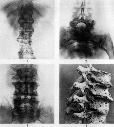
In the dynamics of development of typhoid spondylitis with final fusion of two vertebrae: L4 and L5, 'Fig. 1. Radiogram of spondylosis of the lumbar spine; clinically imperceptible. Fig. 1. Radiogram of the lower part of the lumbar spine (same case). Fig. 3. Deforming osteoarthritis of the lumbar spine in a 53-year-old porter who worked for 30 years without trauma and without infections: cartilaginous discs sharply compressed; lower articular processes sunk into the nests of the underlying arches; osteophytes lie one on another and are soldered along the sides of the bodies from L4 to L5, projecting sharply like hooks, going toward each other along the periphery. Fig. 4. Sharply compressed and flattened intervertebral discs; joints soldered, but roots of the L5 root compressed from above. Fig. 5. Various stages of development of spondylosis from slight lip-like formation at the borders with discs to multiple spurs. Fig. 6. Spondylosis ankylotica; bodies of the ankylosed segment free, 873'
Spondylosis LIII or LIV and LIV has an initial period of hook development, i.e., spondylosis. The cartilaginous disc involved in the process is compressed in front, which results in a slight kyphotic protrusion behind the spinous process of the superior vertebra, while the hooks extend from the lateral edges of the body of one vertebra to another until they fuse together into a common bridge. The joints of these vertebrae, as well as the posterior part of the body near the root foramina, as a rule remain free from adhesions, which is why there are almost no pains in the period of development of typhoid S. or in the period of complete fusion of the bodies, or they are expressed very mildly. The most pronounced picture of S. is observed in the so-called diffuse chronic immobility of the spine. Description of this disease can be traced back to the 18th century. One of the earliest references to it under the name 'spinal gout' is found in Guyot (1736), and in Frank (1792). More detailed patho-anatomical descriptions of this disease are found in Wenzel and Bell (1824), and clinical descriptions in Senator, Braun, and Leyden (1875). Particular interest in this disease began after the works of Bechterev (1893), Strümpell (1897), and Marie (1898) (see Marie's disease). Further research by other authors revealed not only these two forms (Bechterev's and Marie-Strümpell's), but also great variety and the presence of transitional forms (Levi). The presence of kyphosis did not always coincide with the descending development of Bechterev's form of the disease. On the other hand, Schlesinger described immobility of the spine of the Bechterev type, but with complete absence of root phenomena. Therefore, many authors came to the conclusion that there are no data for dividing it into 2 types and that transitional forms exist between both types. However, of all the authors after whom this disease is named, only Bechterev insisted on division. The basis of ankylosing S. is a chronic inflammatory reaction of the bone-ligamentous system of the vertebrae to the action of a poison, possibly through sympathetic or endocrine mediation. Here the cartilaginous discs, if damaged at all, are not severely so, and the spinal joints may remain unaffected by ankylosis. Unlike static S., the process does not begin with compression of the cartilaginous discs, but with marginal growth of the anterior-lateral parts of the vertebral bodies. The ossifications in ankylosing S. are more evenly and symmetrically located. In old cases with complete involvement of the entire spine, a bamboo-stalk appearance is obtained with bone widenings arching around the cartilaginous discs. In the clinical picture of ankylosing S. (see illustration), limitation of spinal mobility is detected very early. The latter is more and earlier expressed either in the cervical part or in the lumbar or thoracic. Very early limitation of thoracic cage mobility is also noticed, manifesting in small lung capacity measured with a spirometer. The possibility of lifting the thoracic cage upward is particularly early limited, which may have as its main cause the involvement of costovertebral joints in the process. However, simple flexion of the thoracic spine and bending of the head toward the chin is sufficient to bring the points of attachment of sternocleidomastoid and scalene muscles closer together, which hinders the upward lifting of the thoracic cage during the act of inspiration. Limitation of thoracic cage mobility leads to poor expansion of the lung apices during breathing, to stasis and poor ventilation. As a consequence of this, such patients often develop tuberculosis of the lungs, from which they frequently die. However, more often they die from deficiency of oxidative processes in conditions of diaphragm paralysis, when the diaphragm has to take on all the work of replacing the functions of the fixed thoracic cage. Abdominal breathing in such cases is extremely pronounced.
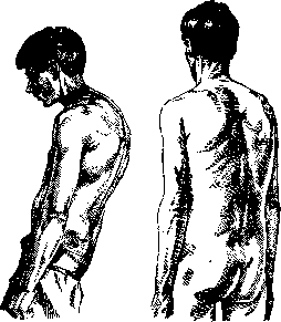
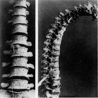
Spondylosis ankylotica.
Nervous phenomena of the root type are very frequent in the course of the disease and initially take the form of neuralgia, and later neuritis with severe pains in the back and legs, first with heightened and then with diminished reflexes and pains with coughing and sneezing. After the onset of ankylosis, the neuritic phenomena gradually subside and may sometimes not manifest themselves at all in the future. The X-ray picture depends on the duration of the disease: ranging from randomly scattered spurs and bone coverings of almost unchanged cartilaginous discs to the 'bamboo stick' inclusive (see separate table, fig. 1). In contrast to static (deforming) S., where the spongy bone of the bodies themselves is hypertrophied in places, in ankylosing S., along with extensive symmetrical lateral growths that quickly fuse the bodies, there is bony atrophy of the spongiosa of the ankylosed vertebrae themselves (see separate table, fig. 1 and 2). Treatment. 1) Static deforming S., due to its benign nature, generally requires no treatment, but where there are pains, massage, baths, diathermy, mototherapy are indicated. For persistent pains, wearing a soft leather belt padded with hair is recommended. Thus, depending on the case, it is necessary to choose between rest and careful non-tiring movement. 2) Treatment of S. on the basis of infectious (toxic) rheumatic spondylarthritis-see Spondylarthritis. 3) Ankylosing S., as a progressive form, is hopeless in terms of results of any treatment. The unilateral extirpation of the parathyroid glands propagated by Oppel gives neither resolution of ankyloses nor loosening of the 'woodenness' of unfused vertebrae. Therefore, in the treatment of ankylosing S., one has to resort to those physical therapeutic measures that only soften what is not yet fused. These include primarily heat in all forms, baths, diathermy and especially the mud of our resorts, but all this is palliative: pains are somewhat alleviated, stiffness is decreased, but despite repeated seasonal mud therapy, the process continues its progressive course. To alleviate the bent position and prevent further bending of the spine, such patients have to be given a supporting leather-shin corset with crutches that recline the spine from the armpits and sternum backward.
Related articles
Mentioned in
Cite this page
“Spondylodeses.” Soviet Medical Encyclopedia. English translation of Bolshaya Meditsinskaya Entsiklopediya, 1st ed. (Moscow, 1928–1936), ed. N. A. Semashko. https://sovietmedicalencyclopedia.pages.dev/article/spondylodeses/