Ankylosis
Historical document, translated for reference. It reflects medical knowledge of the 1920s–30s and is not medical advice.
Summary
This article from the 1928–1936 Great Medical Encyclopedia discusses the classification, causes, and treatment of joint ankylosis, distinguishing between fibrous and bony, congenital and acquired forms.
Encyclopedia article (1928–1936)
ANKYLOSIS (from Greek ankylos - crooked), immobility of a joint due to the fusion of articular surfaces by intermediate tissue. Depending on the type of connecting tissue, fibrous and bony ankylosis are distinguished. Furthermore, the fusion of joint ends can be primary-congenital or secondary-acquired. In the embryo, the articular ends of the cartilaginous skeleton are initially embedded in undifferentiated intermediate tissue. Only gradually, under the influence of functional irritation, do clefts form within the solid mass. However, almost until the 5th month of embryonic life, the articular cavity
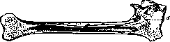
Figure 1. Section of a hip ankylosis; d - cortical bone at the site of articulation.
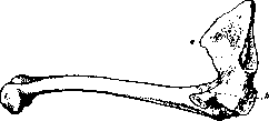
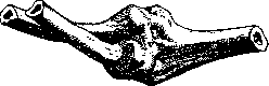
is still permeated by septa and cords that limit mobility. If, due to defective muscle development or other causes, such differentiation of joints does not follow in the fetal period, children are born with more or less immobile, as it were, fibro-ankylosed joints (arthrogryposis). In another form of congenital ankylosis, only individual joints are affected, but they are fused by bone. Such congenital synostoses are generally rare, most frequently occurring at the proximal end of the forearm, between the radius and ulna, and at the interphalangeal joints of the fingers and toes. Congenital ankylosis of the fingers is often combined with brachydactyly; in most cases, they are of hereditary, endogenous origin. Acquired ankylosis is the consequence of trauma or a pathological process (see figures 1–2). Traumatic ankylosis is inevitable if comminuted fractures of the articular ends heal with a common continuous callus. Even in mild cases, for example, in contusions of the articular cartilage with hemorrhage into the joint, an outcome in ankylosis is possible. Only two conditions are necessary: first, the development of granulation tissue that erodes the cartilage cover and organizes blood clots; second, local rest, sufficiently prolonged so that the connective tissue replacing the granulation, whether ossifying later or not, can fuse the opposing ends of the joint into a single whole. A variety of traumatic ankylosis occurs when the articular cleft is preserved, but the joint ends are fastened by parts of the detached periosteum and torn capsule that have ossified in the form of brackets or crossbars. Pathological ankylosis arising on the basis of arthritis occurs most frequently. Among them, three main types can be distinguished and characterized by the names of destructive, adhesive, and periarticular forms. Purulent osteoarthritides usually heal with such destruction of the articular apparatus that mobility is lost (see figure 3). The degree of deformity and shortening accompanying ankylosis depends on the magnitude of destruction, the extent of the process, and, in children, the participation of the epiphyseal cartilage. Besides purulent inflammation, tuberculosis (although rarely on its own without mixed infection) is a cause of destructive ankylosis. Many of the ankyloses formed in early childhood as a result of acutely onset and chronically proceeding "bone caries" are caused by pneumococcal infection. Among specific infections widespread among adults, gonococcal poison possesses a special ability to ankylose joints. Entering the joint via the hematogenous route, the gonococcus causes changes that even in non-purulent cases threaten ankylosis. The plastic exudate binds the articular
Figure 2. Bony angular ankylosis of the right hip joint. View from the medial side: a - spina iliaca anterior superior; b - pecten ossis pubis; c - secondary curvature of the pelvis (from the collection of G. I. Turner).
surfaces from the inside, while the phlegmonous infiltrate of the soft parts, gradually shrinking, produces fixation from the outside. Added to this is the sharp pain during movements characteristic of gonococcal processes. Among the large group of rheumatic, infectious, and toxic polyarthritides, adhesive forms are sometimes encountered, which are accompanied by the destruction of mobility. This is facilitated by the participation in the pathogenesis of certain endocrine glands (thyrotoxic forms) and the presence of peripheral innervation disorders (primary chronic articular rheumatism). A special place is occupied by hemophilic arthritis due to the combination of toxic and traumatic, degenerative and inflammatory moments. Neurogenic arthropathies of central origin never lead to ankyloses. In the spine, ankylosing processes occur frequently and in great variety (fusion of vertebral bodies, ankylosis of apophyseal joints, bony brackets). Diagnostic tasks in ankylosis boil down to clarifying the nature of the underlying disease, the degree of immobility, and the character of the fusions (fibrous or bony, intra- or periarticular). The prophylaxis of traumatic ankylosis consists in the rational treatment of fractures, in the use of such immobilization methods that do not disturb muscle tone, do not hinder blood circulation, and allow early active movements. In case of unsuccessful closed reduction of joint fractures, one should not delay operative reposition and fixation of fragments. The prophylaxis of pathological ankylosis, on the one hand, is a question of the prompt and complete cure of the underlying disease, and on the other hand and mainly depends on nursing care. The treatment of ankylosis should in principle strive to restore normal mobility. This is impossible without active separation and plastic shaping of the joint ends, with the interposition of living or dead tissue to prevent new fusion. Such an operation of arthrolysis is contraindicated in the event of the danger of a flare-up or relapse of the underlying disease. It is useless and even harmful (flail joint) if, due to complete muscle atrophy and extensive scars, material is lacking for the active stabilization of the joint. Finally, for the success of the operation, normal regenerative capacity of the tissues and the conscious participation of the patient in subsequent treatment are necessary. Therefore, old people and children are unsuitable subjects for the operation. An exception is ankylosis of the lower jaw in view of the severe micrognathic facial disfigurement of patients not operated on in childhood. In ankylosis in a position absolutely unsuitable for function, for example, in a strongly flexed and adducted hip, palliative operations such as osteotomy are indicated.
Figure 3. Bony ankylosis after purulent synovitis of the left elbow joint (from the collection of G. I. Turner).
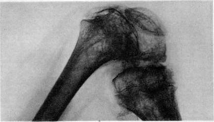
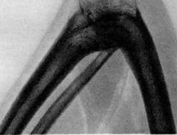
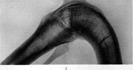
1 - angular ankylosis in a 13-year-old patient after purulent synovitis of the knee; 2 and 3 - secondary arcuate ankyloses after resection of tuberculous knee joints.
E. Osten-Sacken. Ankylosis of the temporomandibular joint. Joint immobility and bone deformation, the two main signs of ankylosis in general, are also typical for lesions of the temporomandibular joint. Pathological changes in the joint can be of various characters. Sometimes there is only a fibrous adhesion between uneven joint surfaces devoid of cartilage; more often, a true synostosis is observed between the articular process and the temporal bone. Bone thickening in severe cases reaches large dimensions, exceeding several times the normal width and thickness of the articular process. The deformation of the head and neck of the articular process is clearly visible on the radiograph and serves as a good sign for determining the side of the joint lesion. Ankylosis of the temporomandibular joint is rarely congenital, and usually occurs as the outcome of various joint diseases. Injuries to the articular process and the articular fossa (fractures along the shaft, birth traumas) occur in approximately 30%. The most frequent cause of ankylosis is purulent inflammation of the joint complicating infectious diseases of childhood (scarlet fever): primary—about 22%, secondary, accompanying inflammation of the middle ear, also about 22%. Much less frequently, osteomyelitis of the lower jaw and gonorrheal or rheumatic diseases of its joint are found in the anamnesis. Accordingly, the disease and ankylosis of the lower jaw, as a consequence thereof, in the majority of cases (about 75%) are unilateral. The opposite joint, even with long-standing ankylosis of the lower jaw, can remain healthy and capable of restoring normal movements (slight movements of the head are palpated through the anterior wall of the external auditory meatus). The inevitable consequence of ankylosis of the lower jaw formed during the growth period (80% occurs at the age under 15 years) is microgenia, underdevelopment of the lower jaw of the corresponding side, and facial asymmetry. The chin and the healthy side are displaced toward the underdeveloped half, which consequently appears more convex. External measurements, displacement of the midline of the dental arch and the chin clearly indicate which side is shortened and where, therefore, the joint lesion should be sought. Ankylosis of the lower jaw is a serious condition that severely disrupts the functions of nutrition and speech. The teeth are tightly clenched, coated with tartar, food is passed through the interdental slits or through the gaps formed by the separation of the teeth.—Preventive and therapeutic measures are reduced to the application of unloading and rest during the period of acute inflammation of the joint. For this purpose, a piece of cork or rubber drainage tube from 1.5 to 1 cm in thickness is inserted between the posterior teeth of the upper and lower jaw in the form of a spacer. Rough mechanical impacts (mouth gags) are useless both in the initial period of jaw trismus and in established synostosis.—Treatment of ankylosis of the lower jaw can be rationally carried out only by surgical means and is reduced to the formation of a new artificial false joint either in the place of the former normal joint (operation of resection of the articular head according to König, 1878) or somewhat lower, in the region of the ascending ramus, by wedge-shaped excision of a piece of bone about 1 cm wide (Rochet, 1894, Schmidt, 1899). In order to ensure the formation of a false joint, into the bone defect resulting after resection, a flap of muscles is inserted by means of transplantation on a pedicle (Verneuil, 1860, Helferich, 1895, Rochet and others) or (by free transplantation) a flap of fascia and adipose tissue (Korneyev, 1914 and others). This interposition prevents the approximation of the ends of the fragments and the formation of a bone callus. It is necessary during the first week to provide the wound with complete rest and to fix the fragments in the position of the greatest possible divergence for them. Thus, new, artificially created anatomical conditions are secured, the graft takes root over a wide area, the correct position of the horizontal ramus is maintained, and an easily movable false joint is formed. Movements of the lower jaw can be restored to normal. In some cases, the use of special mechanotherapy (see) of the lower jaw is indicated.
Related articles
Mentioned in
Cite this page
“Ankylosis.” Soviet Medical Encyclopedia. English translation of Bolshaya Meditsinskaya Entsiklopediya, 1st ed. (Moscow, 1928–1936), ed. N. A. Semashko. https://sovietmedicalencyclopedia.pages.dev/article/ankylosis/