Ankle Joint
Historical document, translated for reference. It reflects medical knowledge of the 1920s–30s and is not medical advice.
Summary
An anatomical and functional overview of the ankle joint (articulatio talocruralis) from the 1930s Soviet Medical Encyclopedia. It describes the skeletal structure, the critical role of the malleolar fork and ligaments, and the surrounding musculature and tendons.
Encyclopedia article (1928–1936)
ANKLE JOINT represents a highly stable connection of the bones of the lower leg with the foot. The talus, articulating with the inferior articular surface of the tibia and the lateral surface of the lateral malleolus (malleolus lateralis) above and the tarsal bones below, divides this region into an upper joint—the ankle joint proper (articulatio talocruralis)—and a lower subtalar joint (articulatio talotarsalis). The latter joint, consisting of a posterior subdivision (articulatio talocalcanea) and an anterior one (articulatio talocalcaneonavicularis), plays a significant role in the mechanism of gait and, in the event of destruction of the articulatio talocruralis (ankylosis), compensates to a certain extent for the loss of movement in this upper, ankle joint, preserving the elasticity of the gait. The tibia and fibula participate in the formation of the ankle joint proper, creating, by means of the medial and lateral malleoli, a fork that very tightly grips the lateral surfaces of the talus (see Figures 1 and 2). The weight of the body is transmitted from the inferior articular surface of the tibia almost exclusively to the trochlea of the talus, which must be held very tightly by the malleolar fork. Due to the fact that the center of pressure on the trochlea is located closer to the outer edge than to the inner edge, the talus, in order to maintain its balance, must have a solid support in the lateral malleolus. The force with which the talus presses on the lateral malleolus is a direct force from within outward and slightly from front to back. Under the influence of this force, the talus would shift outward and backward if it were not held by the lateral malleolus. Such a displacement indeed often occurs in fractures of the malleolus (see Figure 3). The malleolus lateralis is located somewhat more posterior to the malleolus medialis in relation to the frontal plane, which is advantageous from the standpoint of the mechanics of the ankle joint. The strength of the joint, in addition to the skeleton, is provided by a strong ligamentous apparatus (see Figures 4–6). The following ligaments possess the
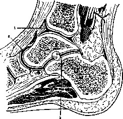
Figure 1. Ankle joint (sagittal section): 1—articulatio talocruralis; 2—articulatio talocalcanea; 3—articulatio talocalcaneonavicularis.
greatest strength and have great practical importance: lig. deltoideum, lig. talofibulare anterius and posterius, as well as calcaneofibulare. The first of these—the deltoid ligament, which is very strong—is located on the medial (inner) side and, spreading out like a fan into superficial and deep layers, plays an extremely important role in the stability of the ankle joint. Its strength is so significant that during sudden abduction of the foot, it often tears off the tip of the medial (inner) malleolus, but does not rupture itself (see Figure 3, I E). Laterally, the ankle joint is strengthened by 3 ligaments: lig. talofibulare anterius, posterius, and lig. calcaneofibulare. The lateral ligamentous apparatus is weaker than the medial one and more frequently ruptures during traumatic injuries of the ankle joint. The connection of the tibiofibular bones below (syndesmosis tibiofibularis) is slightly movable and is strengthened by the anterior and posterior ligaments of the lateral malleolus, as well as the interosseous ligament. The very minor mobility in this joint is important in the mechanism of movement of the trochlea of the talus in the malleolar fork. During dorsal flexion, the trochlea of the talus, which is wider in front than in back, wedging between the two malleoli, widens the distance between them. The physiological separation barely exceeds 1–2 mm. In traumatic injury of the joint with rupture
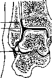
Figure 2. Ankle joint (frontal section): 1—articulatio talocruralis; 2—articulatio talocalcanea; 3—recess of the synovial membrane.
of the syndesmosis tibiofibularis, the malleolar fork widens, creating conditions for the displacement of the talus, usually outward or backward. The strong ligaments of the ankle joint significantly strengthen the joint capsule, mainly on the lateral sides. Posteriorly and anteriorly, the capsule is thin and less protected. When fluid accumulates in the joint, effusion is easily detected anteriorly under the extensor tendons and posteriorly on both sides of the Achilles tendon. In these areas, it is convenient to perform joint puncture. The ankle joint is constructed as a hinge joint, and movements in it occur almost exclusively in the direction of plantar and dorsal flexion. Purely rotational movements of the foot around the longitudinal axis in this joint are as insignificant as in the wrist joint. But in terms of
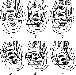
Figure 3. Diagram of injuries in the region of the ankle joint during forced rotation of the foot: A, B, C—inward; D, E, F—outward. A—rupture of the lateral ligament; B—fracture of the lateral malleolus; C—supramalleolar fracture of both leg bones; D—rupture of the medial ligament; E—fracture of the medial malleolus; F—Dupuytren's fracture with a fissure of the tibia. 1—medial malleolus; 2—medial ligaments; 3—talus; 4—interosseous ligament; 5—calcaneus; 6—lateral ligaments; 7—lateral malleolus; 8—lig. peroneotibiale; 9—fissure of the tibia. (After Testut-Jacob.)
abduction and adduction in combination with flexion, they are possible. Normally, the range of motion in the ankle joint (dorsal + plantar flexion) in adults reaches 70°. Excessive dorsal flexion is limited on one side by the malleolar fork, and on the other by the Achilles tendon and the tension of the posterior muscle group (m. triceps surae). Excessive plantar flexion is limited by the extensor tendons along with the m. tibialis anterior and the tension of the lateral ligaments. The stability of the ankle joint is largely explained by the large number of muscles and tendons surrounding the joint on all sides. It should be noted that not a single tendon attaches directly to the talus; this is of significance both from the standpoint of mechanics and from the standpoint of surgical approaches to this joint. In the anterior compartment of the ankle joint, the muscle tendons run in the following order: medially—the tendon of m. tibialis anterior, which has its own sheath; next to it lies the tendon of m. extensoris hallucis longi, which also has its own sheath; more laterally pass the tendons of m. extensoris digitorum
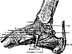
Figure 4. Ligamentous apparatus of the ankle joint from the medial side: 1—lig. deltoideum; 2—lig. talotibiale ant.; 3—lig. tibionaviculare; 4—lig. calcaneotibiale; 5—articular margin of os talus; 6—tuberosity of os naviculare; 7—tendon of m. tibialis post.; 8—sheath of m. flexor digitorum longus; 9—sheath of m. flexor hallucis; 10—lig. talotibiale post.; 11—os trigonum; 12—m. abductor hallucis. (After Dujarier.) longi and finally the last—the tendon of m. peronaeus tertii. All these tendons are tightly pressed in front by the transverse and cruciate ligaments of the leg, which enhances their action. Behind the medial malleolus run the tendons of m. tibialis posterior and flexor digitorum longus; somewhat more posteriorly, between the medial malleolus and the Achilles tendon, passes the tendon of m. flexor hallucis longus.
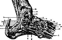
Figure 5. Ligamentous apparatus of the ankle joint from the lateral side: 1—lig. talofibulare ant.; 2—lateral articular surface of os talus; 3—lig. calcaneofibulare; 4 and 5—lig. cruciatum cruris; 6—capsula artic. talocalcanei; 7—retinaculum mm. peronaeorum; 8—tendo m. peronaei brevis; 9—tendo m. peronaei longi; 10—lig. malleoli lat. ant.; 11—membrana synovialis; 12—lig. talotibiale ant.; 13—collum tali; 14—lig. talonaviculare dorsale; 15—lig. interosseum; 16—caput tali; 17—lig. bifurcatum; 18—m. extensor digitorum brevis (cut); 19—lig. calcaneocuboideum dorsale; 20—tendo m. peronaei tertii; 21—tendo m. peronaei brevis. (After Dujarier.)
Behind the lateral malleolus pass the tendons of mm. peronaei longi and brevis, the former being thicker than the latter and located more superficially (see Figure 7).
In pathological conditions accompanied by partial or complete loss of function of the muscles surrounding the ankle joint (infantile paralysis), the ligamentous apparatus also loses its normal tone. In such cases, lateral movements in this joint are possible, as well as excessive plantar and dorsal flexion. An unstable joint results, and various types of foot deformities such as varus, valgus, and calcaneus arise. The mutual arrangement of the tendons and the preservation of some muscle groups when others are lost makes it possible to use active tendons for plastic surgery. Thus, in paralysis of the peroneal group, the m. tibialis anterior can be transplanted to the peronaeus tertii. In paralysis of the m. triceps surae, a satisfactory effect is obtained by transplanting the tendons of m. flexor hallucis longus and m. peronaei longi to the calcaneus, etc. Of the vessels that should be kept in mind during interventions on the ankle joint, the a. tibialis anterior and a. tibialis posterior deserve attention. The former, together with the n. peronaeus profundus, lies in front of the ankle joint between the tendons of m. extensoris hallucis longi and m. extensoris digitorum communis. The a. tibialis posterior is located behind the medial malleolus and runs accompanied by two veins and the n. tibialis. The ankle joint is innervated by branches from the n. saphenus and n. peronaeus profundus.
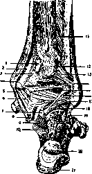
The exposed position of the ankle joint and the fact that it bears the entire weight of the body explain the relatively frequent injuries and diseases of this joint. Among traumatic injuries in the region of the ankle joint, ruptures of the collateral ligaments and fractures of the malleoli with simultaneous subluxation of the foot are most common. In so-called distortions (sprains) in the region of the ankle joint, a rupture of one of the lateral collateral ligaments occurs more frequently. The stronger medial collateral ligament tears off the tip of the medial malleolus, but itself ruptures only in exceptional cases. It should be noted that ligament ruptures in the region of the ankle joint are significantly less common than fractures. Depending on the mechanism of injury, Figure 6. Ligamentous apparatus of the ankle joint from behind; 1—tuberc. tib. post.; 2—groove for m. tib. post. (sulcus malleol.); 3—groove for m. flex. digit. longi; 4—os tali; 5—lig. calcaneo-tibiale; 6—lig. talo-tibiale post.; 7—os trigonum; 8—groove for m. flex. hallucis longi; 9—lig. talo-calcaneum post.; 10—part of lig. talo-calc. post., enclosing the tendon of m. flex. hallucis longi from behind; 11—lig. interosseum; 12—groove for m. flex. halluc. longi; 13—lig. malleoli lat. post.; 14—groove for m. peronei longi; 15 and 16—lig. poster.; 17—lig. talo-fibulare post.; 18—talo-calcaneal articulation; 19—lig. calcaneo-fibulare; 20—bursa tend. Achill.; 21—tendo Achillis. (After Dujarier.)
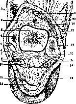
Figure 7.
the following fractures are possible: single-malleolar, mainly fract. malleoli lateralis, often combined with lateral subluxation of the foot; bi-malleolar fractures [see separate plate (cols. 675–676), Fig. 4]; and Dupuytren's fracture. The latter type of fracture is characterized by an avulsion of the medial malleolus and a fracture of the fibula above the lig. malleoli lateralis ant. Most so-called "malleolar" fractures are intra-articular. Treatment consists of the earliest possible reduction, which can be achieved by a system of corrective traction or manually. In the latter case, the achieved result should be maintained with a plaster cast or splint, placing the foot in a varus position. Dislocations and subluxations in the ankle joint are almost always combined with one of the specified types of fracture, requiring immediate reduction and subsequent functional treatment.—Among chronic inflammatory diseases of the ankle joint, tuberculosis occupies a prominent place, and the tuberculous focus can arise both in the talus and in the lower metaphysis of the tibia and fibula. Due to the anatomical structure of the ankle joint, a whole series of bones is often involved in the tuberculous process, making conservative therapy difficult. In isolated focal lesions, surgical intervention is indicated in individual cases. Syphilitic arthritis of the ankle joint is less common and requires specific therapy.—So-called "rheumatic" arthritis of the ankle joint is often bilateral, frequently in combination with lesions of other joints.—It is also necessary to note the possibility of typhoid and paratyphoid inflammation of the ankle joint, which, starting acutely or subacutely, runs a chronic course thereafter.—Among acute arthritides of the ankle joint, gonorrheal lesions occupy a definite place.—Furthermore, this joint, like others, can be the site of purulent synovitis of staphylococcal or streptococcal origin. In such forms, surgical intervention in the form of arthrotomy or resection is often required. Transverse section of the leg at the level of the malleoli: 1—tendon of m. tibialis ant.; 2—a. dorsalis pedis; 3—talus; 4—medial malleolus; 5—tendon of m. tibialis post.; 6—n. saphenus; 7—tendon of m. flex. digit. communis longi; 8—tendon of m. flexoris hallucis longi; 9—a. and vv. tibiales post.; 10—n. tibialis; 11—tendo m. plantaris and lateral to it bursa tend. Achill.; 12—Achilles tendon; 13—n. cutaneus surae lateralis; 14—m. peronaeus brevis; 15—tendon of m. peronaei longi; 16 and 18—terminal branches of a. peronaea in the region of the lateral malleolus; 17—lateral malleolus; 19—tendon of m. peronaei tertii; 20—tendon of m. extensoris digit. longi; 21—tendon of m. extensoris hallucis longi. (After Testut-Jacob.)
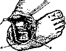
Surgical approaches to the ankle joint depend largely on the purpose of the operation. Kocher's typical lateral incision (see Figure 8) is convenient for joint resection and extirpation of the talus (astragalectomy) according to Whitman. For the purpose of ankle joint arthrodesis, most orthopedists use an anterior or posterior incision. In this case, one can limit the manipulations to the bony skeleton alone, or combine the arthrodesis proper with tenodesis (intraosseous tendon fixation according to Biesalski). The latter type of arthrodesis for the ankle joint, with simultaneous ankylosing of Chopart's and Lisfranc's joints, gives satisfactory results in severe cases of pes paralyticus. In milder cases, to prevent foot drop, good results are achieved by Campbell's operation, known among American orthopedists as "Campbell's bone block." "Blocking" of the ankle joint is performed from behind with a graft of cancellous bone. There are also methods of arthrodesing the ankle joint with a bone plate inserted through the joint (see Arthrodesis).—For the purpose of arthroplasty on the ankle joint, an anterior longitudinal or lateral Kocher incision is usually used. The diastasis between the separated bones must
be at least 0.5–1 cm; it is more convenient to shape the joint as a saddle joint. Fascia, fat, and muscle (from m. triceps) are suitable for interposition. Reconstructive operations on the ankle joint are also used for malunited "malleolar" fractures, when static conditions are severely disrupted. Restoring the normal ankle mortise and correcting the subluxation of the talus is particularly important in these cases. In all surgical interventions on the ankle joint, as well as in the treatment of inflammatory diseases, one should strive to preserve at least a small amount of mobility, which is extremely important for walking. If the joint is threatened with ankylosis, it is necessary to position the foot at a right angle to the axis of the leg. An equinus position and lateral deformity of the foot make walking extremely difficult and require subsequent orthopedic intervention.
Related articles
Mentioned in
Cite this page
“Ankle Joint.” Soviet Medical Encyclopedia. English translation of Bolshaya Meditsinskaya Entsiklopediya, 1st ed. (Moscow, 1928–1936), ed. N. A. Semashko. https://sovietmedicalencyclopedia.pages.dev/article/ankle-joint/