Radiocarpal Joint
Historical document, translated for reference. It reflects medical knowledge of the 1920s–30s and is not medical advice.
Summary
This article from the 1928–1936 Soviet Great Medical Encyclopedia provides a detailed anatomical description of the radiocarpal joint, including its bones, ligaments, and movements. It discusses the triangular cartilage and the intercarpal joints.
Encyclopedia article (1928–1936)
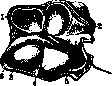
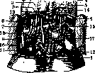
RADIOCARPAL JOINT (articulatio radio-carpea) connects the hand with the forearm. The name art. radio-carpea indicates that of the two forearm bones, only the radius articulates directly with the hand. The ulna, however, does not reach the hand; its lower end is separated from the wrist by a triangular cartilage (cartilago triangularis, syn.: lig. triangulare, discus articularis). The mentioned cartilage has the shape of a triangular plate; one of its sides is attached to the articular notch of the lower end of the radius, while its apex is connected to the styloid process of the ulna by a thick and short ligament (lig. subcruentum). The morphological significance of the triangular cartilage is not completely studied. In the embryo, a bone is found on the lower surface of the cartilage, which, due to its position between the radius, ulna, and the first row of wrist bones, received the name intermedium bone of the forearm (os intermedium antebrachii). The older the embryos, the less frequently this bone is encountered; in adults it is almost never observed; so far only one case of such an anomaly is known; this bone must be considered morphologically vanished and phylogenetically extremely ancient. Thilenius points out its presence in marsupials (Phaseolomys) and in some anthropoids (in gibbons) (Testut). The R. joint belongs to arthrodiases, so-called head-shaped or elliptical joints (condyloarthrosis according to Cruveilhier) (Lesgaft). Of the two articular surfaces of the joint, one belongs to the forearm, the other to the wrist. The articular surface of the forearm is concave (Fig. 1), has the shape of an ellipsoid, the long axis of which runs from one styloid process to the other. This articular surface is formed by the expanded end of the radius with a cartilaginous continuation (discus articularis) towards the elbow. The articular surface of the radius is triangular in shape, with the apex turned outwards, on the surface of which light indentations, the impressions of the adjacent scaphoid and lunate bones, are noticeable. The articular surface of the wrist is formed by three bones: the scaphoid, lunate, and triquetral; the pisiform bone belongs to the sesamoid bones and has no direct relation to the formation of the joint; the spaces between the bones of the first row of the wrist up to the level of the articular surfaces are filled with ligaments, as a result of which a nearly smooth head of the joint is obtained, elongated in the transverse direction of the hand. The number of bones of the first row of the wrist is sometimes increased due to the doubling of some of them; for example, a double scaphoid bone [os naviculare bipartitum, syn.: os naviculare radiale, os naviculare ulnare: ossa navicularia secundaria radiale et ulnare (Gruber)]; a double triquetral bone (os triquetrum bipartitum, syn.: os triquetrum radiale, os triquetrum ulnare). The line of the R. joint runs obliquely from outside to inside and from below upwards. The concavity of the articular surface of the radius exactly corresponds to the convexity of the scaphoid and lunate bones; the participation of the triquetral bone in the joint is negligible, which can be judged by its very small articular surface; the surfaces corresponding to the most lateral part of the triangular cartilage. The articular line on X-ray images is thin and clearly visible; the posterior edge of the epiphysis of the radius gives a faint shadow on the lunate bone; in the ulnar part of the articular line it is wider.
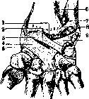
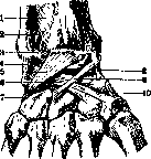
Ligaments of the joint. The bones of the joint are held by the fibrous capsule and a reinforcing ligamentous apparatus. The upper edge of the capsule is attached along the edge of the articular surface of the radius and along the edge of the triangular cartilage (discus articularis); the lower edge of the capsule is attached to the edge of the articular surface of the first row of wrist bones. The capsule is reinforced on the outside by ligaments. Although the ligamentous apparatus forms almost a continuous connective tissue layer around the joint, nevertheless it is customary to distinguish and describe individual bundles of ligaments going in various directions: on the palmar side there is a wide radiocarpal ligament (lig. radio-carpeum volare) (Fig. 2), going from the styloid process and the palmar edge of the articular surface of the radius by several bundles to the lunate, triquetral, and capitate bones; the second ligament of the same surface goes in a fan shape from the base of the styloid process of the ulna to the triquetral and capitate bones (Testut). These ligaments are related to supination of the hand; when the hand passes from pronation to supination, the radius, with their help, pulls the hand along with it. The ligaments of the dorsal side of the joint are weaker, covering only about one-third of the posterior surface of the joint and stretching from the radius to the triquetral bone, sometimes to the lunate and capitate (Fig. 3).
Fig. 2. Radiocarpal joint from the palmar surface: 1 and 2 - interligamentous protrusions of the synovial membrane of the joint; 3 and 4 - lig. radiocarpeum volare; 5 - lig. collaterale carpi radiale; 6 - lig. interosseum antebraehii; 7 - interligamentous protrusion of the synovial membrane between the radius and ulna; 8 - lig. radio-ulnare distale; 9 - the same in front of the styloid process; 10 - lig. carpo-ulnare; 11 - lig. collaterale carpi ulnare; 12 - the same between the pisiform and triquetral bones. The dorsal ligaments are closely connected with the fibrous sheaths of the extensors. These ligaments are tense during pronation of the hand. From the styloid process of the radius to the scaphoid bone runs the lateral collateral ligament (lig. collaterale carpi radiale); from the styloid process of the ulna to the triquetral extends the medial collateral ligament (lig. collaterale carpi ulnare). The entire inner surface of the articular capsule is covered with a synovial membrane, ending exactly at the edges of the articular cartilages. The R. joint communicates with the lower radioulnar joint (40%) by a slit-like opening at the base of the triangular cartilage. In half of the cases the cavity of the R. joint communicates with the joint between the triquetral bone and the pisiform, and only in exceptional cases does the R. joint communicate with the joints of the wrist (Testut). In addition to this, the articular cavity in some cases gives off, as it were, processes, bays, cavities between bundles of ligaments (Fig. 2 and 3); these bulgings are true synovial hernias penetrating through the fibrous capsule of the joint and are the beginning of the cysts (hygromas) of the joint. The boundaries of the R. joint are established differently by various authors and conditionally. The upper boundary is formed by a horizontal plane passing immediately above the head (capitulum ulnae) of the ulna; the lower one by a plane drawn parallel to the previous one immediately below the pisiform bone. Movements in the R. joint can be considered only in conjunction with movements in the joints between the first and second rows of wrist bones, i.e., here movement occurs as in a complex joint with a bony meniscus, formed by three bones of the first row of the wrist. In this complex joint, movements are possible around the transverse and anteroposterior axes: flexion, extension, adduction, and abduction, and the combination of movements around these axes is circular rotation. During flexion the palm inclines towards the anterior surface of the forearm, during extension the opposite movement occurs. The participation of both parts of the complex joint in these movements is well visible on X-ray images (Fig. 4). The bony meniscus (first row of wrist bones) inclines towards the forearm during flexion; in the same direction the inclination of the second row of wrist bones also occurs (Fig. 4); this double movement occurs around the transverse axis passing through the highest point of the head of the capitate bone. By adduction or ulnar flexion is understood the movement in which the ulnar edge of the hand inclines towards the ulnar edge of the forearm; abduction or radial flexion is called the opposite movement. This movement occurs around the anteroposterior axis passing through the center of the head of the capitate bone; in this case, when the base of the capitate bone tilts outwards (abduction), the lunate bone, sitting on top of the first,
Fig. 4. X-ray image (schematic) in profile of the radiocarpal joint in the position: A - average between flexion and extension; B - in flexion; C - in extension; 1 - radius; 2 - os lunatum; 3 - os capitatum; 4 - third metacarpal bone.
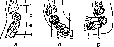
it deviates inward and vice versa; therefore the articular surface of the first row of the carpus slides along the articular end of the forearm in a transverse and opposite-to-the-hand direction, i.e., upon ulnar flexion the wrist moves toward the radius, and upon radial flexion toward the ulna (Fig. 4); circular rotation is obtained as a result of the successive transition from flexion to abduction, extension, and adduction. The range of motion in this case is greater around the transverse axis of the joint, i.e., during flexion and extension, than around the anteroposterior axis-abduction and adduction (Sapreyu); therefore the base of the cone described by the hand is not a circle but an ellipse (Testut). Normally the range of motion from full extension to full flexion is slightly less than 180°, with more than 90° falling on the motion in the R. joint and less on the joint between the two rows of carpal bones (Testut); Lesgaft considers the arc of this motion to be 135-140° and around the anteroposterior axis up to 75°. Of the listed movements, flexion in the R. joint is performed either only by the muscles acting directly on it, such as: the radial flexor of the wrist (m. flexor carpi radialis), the long palmar muscle (m. palmaris longus) and the ulnar flexor of the wrist (m. flexor carpi ulnaris), or these muscles together with the other flexors. Extension involves all the extensors. Abduction is obtained as a result of the combined action of m. flexor carpi radialis, both radial wrist extensors (mm. extensores carpi radialis brevis et longus), the long abductor muscle of the thumb (m. abductor pollicis longus), and both extensors of the thumb (mm. extensores pollicis brevis et longus). Adduction is performed by m. flexor carpi ulnaris and the wrist extensor (m. extensor carpi ulnaris).
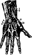
The flexor muscles lie under the aponeurosis of the flexor surface of the joint and are arranged in 4 layers (Fig. 5); in the first layer (counting from the radial edge to the ulnar) go the tendons of the brachioradialis muscle (m. brachioradialis), the radial flexor of the wrist (m. flexor carpi radialis), the long palmar muscle (m. palmaris longus) (Testut), more precisely: the tendon of this muscle passes in the bifurcation of the volar carpal ligament and the m. flexor carpi ulnaris; in the second layer - four tendons of the superficial flexor of the fingers (m. flexor digitorum sublimis); in the third layer - the long flexor of the thumb (m. flexor pollicis longus); four tendons of the deep flexor of the fingers (m. flexor digitorum profundus); in the fourth layer - the quadratus pronator (m. pronator quadratus). In the canal for tendons from the palm to the forearm pass two synovial sheaths: one for fl. pollicis longus and the second for fl. digitorum communis (see Hand); in the fifth layer - skeleton. Between the tendons of m. brachioradialis (laterally) and m. flexor carpi radialis (medially) runs the radial artery (a. radialis) with two of its accompanying veins; the radial nerve (n. radialis) has already passed higher to the dorsal surface of the forearm; the artery lies on the m. pronator quadratus and can be easily pressed against the radius; covered only by skin and aponeurosis the artery is easily accessible for the examination of the pulse; approaching the base of the styloid process of the radius, the artery changes its direction and goes to the back of the joint; on the flexor side the radial artery gives a superficial palmar branch (ramus volaris superfic.) and a carpal palmar branch (ramus carpeus volaris), going in a transverse direction along the lower edge of the m. pronator quadratus and anastomosing with the同名 branch of the a. ulnaris. Between the tendons of fl. carpi ulnaris (medially) and fl. digitorum sublimis (laterally) go the ulnar artery with two accompanying veins and the同名 nerve, lying nearby and medially to the artery. Under the m. pronator quadratus passes the volar interosseous artery (a. interossea volaris), which passes through the interosseous ligament to the dorsal surface of the forearm. Of the nerves here there is also the median nerve (n. medianus); with the nerve goes the同名 artery (a. mediana), lying on its anterior surface; the nerve passes under the m. palmaris longus at the radial edge of fl. digitorum sublimis, dividing the sheath of fl. pollicis longus from the sheath of fl. digitorum sublimis. All these muscles, vessels and nerves are directly covered by aponeurosis, which in this place is thicker than on the forearm and forms a strong ligament with transverse fibers (lig. carpi volare), 3-4 cm wide. Above it passes into the aponeurosis of the forearm, below - into the palmar aponeurosis, from it also originate some of the muscles of the thenar and fifth finger eminences; one end of the ligament is fixed to the pisiform and hook-shaped bones, the other - to the scaphoid and trapezium bones; from the deep layer of the ligament bundles go to the anterior surface of the carpal bones. Under the ligament there are channels for the passage of flexor tendons; through these same channels pus can spread from the hand to the forearm. The subcutaneous tissue here is never rich in fat, even in obese people; in this layer pass superficial veins, which are the beginning of the system of venae basilicae, and nerves, which are the continuation of the forearm nerves: along the radial edge goes the cutaneous lateral nerve of the forearm (n. cutaneus antebrachii lateralis), in the middle - the palmar branch of the median nerve (ramus palmaris n. mediani), along the ulnar edge - branches of the cutaneous medial nerve of the forearm and the palmar cutaneous branch of the ulnar nerve (rami n. cutanei antebrachii medialis et ramus cutaneus palmaris n. ulnaris). The skin in this place is thin, devoid of hair, has transverse folds, is fused with the aponeurosis, therefore little movable. The extensor muscles lie under the aponeurosis of the dorsal surface of the R. joint; here the aponeurosis forms the dorsal carpal ligament (lig. carpi dorsale), the fibers of which go in a transverse direction, being reinforced by the ulnar end to the scaphoid and pisiform bones, the radial end - to the lower end of the radius. The ligament above passes into the aponeurosis of the forearm, below - into the aponeurosis of the hand. From the deep layer of the ligament septa go to the forearm bones and form 6 channels for 12 extensor tendons, arranged in the following order, starting from the radial edge (Fig. 6): the long abductor muscle of the thumb (m. abductor pollicis longus); the short extensor of the thumb (m. extensor pollicis brevis); both muscles pass through the first channel, having a common synovial sheath, rising 2-3 cm above the upper edge of the ligament; the radial wrist extensors (mm. extensores carpi radialis longus et brevis) pass through the second channel
Fig. 6. Synovial sheaths of the tendons of the back of the hand: 1-m. abductor et extensor pollicis brevis; 2-mm. extensor carpi radialis longus et brevis; 3-m. extensor pollicis longus; 4-m. extensor digitorum communis; 5-m. extensor digiti quinti proprius; 6-m. extensor carpi ulnaris.
Fig. 7. The anatomical snuffbox area (right hand): 1-skin and subcutaneous tissue with superficial veins and nerves; 2-superficial aponeurosis; 3-mm. extensor carpi radialis longus et brevis; 4-m. abductor pollicis longus; 5-m. extensor pollicis brevis; 6-m. extensor pollicis longus; 7-a. radialis.
in the following order, starting from the radial edge (Fig. 6): the long abductor muscle of the thumb (m. abductor pollicis longus); the short extensor of the thumb (m. extensor pollicis brevis); both muscles pass through the first channel, having a common synovial sheath, rising 2-3 cm above the upper edge of the ligament; the radial wrist extensors (mm. extensores carpi radialis longus et brevis) pass through the second channel
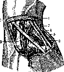
also in the general synovial sheath, which is divided at the top and bottom into two separate sheaths; above them, in the third canal, the m. extensor pollicis longus runs obliquely from the ulnar edge to the radial side; its sheath extends 1-2 cm above and below the ligament. The interval between the long and short abductor of the thumb is called the anatomical snuffbox (la tabatiere anatomique); on its floor, under the tendons, the a. radialis runs obliquely to the first interdigital interval (Fig. 7). The next in order are: the muscle extending the index finger (m. extensor indicis proprius), the common extensor of the fingers (m. extensor digitorum communis); their tendons pass through the fourth canal, covered by one sheath rising 1 cm above the ligament and descending to halfway and below the metacarpal bones; the extensor of the little finger (m. extensor digiti quinti proprius) in the fifth canal; its sheath begins at the level of the head of the ulna and reaches halfway of the fifth metacarpal bone; the last is the extensor carpi ulnaris (m. extensor carpi ulnaris) in the sixth canal; its sheath runs from the head of the ulna to the upper end of the fifth metacarpal bone. - The synovial sheaths of the tendons, cylindrical in shape, consist of two layers: the parietal, lining the bony-fibrous canal, and the visceral, enveloping the tendon itself; these two layers are connected to each other in places by synovial folds extending from one layer to the other and called "mesotendo"; through them pass the vessels and nerves nourishing the tendons; therefore their destruction can lead to tendon necrosis. The vessels and nerves of the back of this area are of less importance than on the flexor surface. The superficial arteries are all small. The main artery is the radial, giving off the dorsal carpal branch (ramus carpeus dorsalis), anastomosing with the corresponding branch of the ulnar artery; here also pass the terminal branches of the palmar and dorsal interosseous arteries. - The arteries nourishing the joint arise from several sources: from the front - from the transverse arch of the wrist passing along the lower edge of the m. pronator quadratus and the ascending branches of the volar arcus prof.; from the back - from the a. interossea and from the rete carpi dorsale; on the radial edge - from the a. radialis, on the ulnar edge - from the a. ulnaris. - The joint receives nerves from the front from the nn. medianus and ulnaris, from the back from the n. radialis and the dorsal branch of the n. ulnaris. - Lymphatic vessels here, as on the anterior surface, divide into superficial and deep; they usually run with the veins and partly empty into the ulnar lymphatic glands, and partly pass directly to the axillary glands. - The superficial veins of the subcutaneous layer are well developed, have no constant location, and are the beginning of the venae cephalicae. The subcutaneous tissue is richer in fat; often in this layer above the styloid processes of both forearm bones there exist serous bursae. The skin here is thicker than in front and more mobile, covered with hair, and has many small transverse folds. A. Sirorkin. Pathology of the R. joint. In view of the special significance of the hand and fingers as the main working organ of man, injuries and diseases of the R. joint region acquire great importance. This region is especially frequently subjected to injuries in the very process of work. These include injuries by hand tools, the material being processed, glass, machine injuries (getting into shafts, drums, etc.), and finally burns, both thermal and chemical. Injuries can be: 1) closed - contusions, sprains, strains, fractures, and (extremely rarely) dislocations (Fig. 8) and 2) open - wounds (incised, contused, lacerated and gunshot wounds) and burns. As regards open injuries, they are dangerous first of all due to the anatomical peculiarities of this region, abundant in tendons, nerves and vessels, secondly due to the possible entry of infection, which is especially dangerous precisely in the R. joint region, where infection easily and quickly spreads along the course of the tendons, often causing their necrosis, thirdly these injuries, giving rise to extensive and deep scars upon healing, disrupt the function of the hand and fingers. Even superficial skin injuries, giving rise to scar contractures after healing per granulationem, strongly limit the normal function of the hand and fingers. In cases where the tendons of the flexors or extensors lying directly under the skin are compressed by a scar, limitation of movement of the hand and fingers occurs. Knife wounds and glass wounds of the R. joint region are very often complicated by the cutting of the flexor tendons. Besides professional factors, domestic ones also play a role here: wound
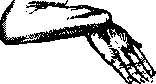
Fig. 8.
Spontaneous dislocation of the wrist with a knife in a fight, injury with a broken bottle while intoxicated, etc. In this case, the ends of the severed tendons (especially the central ones) move far to the side due to muscle contraction, and finding them often presents great difficulties. Along with injury to the flexors, damage to the vessels and nerves passing through here is always possible. While the vessels (a. and v. radiales and a. and v. ulnares) give themselves away by strong bleeding, forcing the victim to seek a doctor immediately, damage to the nerves and tendons remains unnoticed. Hence it is clear that in any injury in the area of the R. J. it is necessary to establish the diagnosis accurately, i.e., to orient oneself regarding accompanying damage to deeper organs. Damage to tendons can be judged by the loss of function of the corresponding fingers, and severance of nerves by loss of sensitivity. It must be decided once and for all that probing of such a wound is inexcusable. It gives the investigator nothing and causes suppuration in the patient. Possible damage to the joint capsule with penetration into the joint and damage to bones during deep injuries (especially on the dorsal side) will be discovered during the wound treatment itself, which must be undertaken upon delivery of the patient to an institution where the wound can be treated in a corresponding, sufficiently aseptic environment (outpatient clinic, emergency ward, hospital). If the wounded person reaches the doctor no later than the first day after the injury, primary treatment of the wound by excision and freshening of its edges should be performed immediately. A subsequent thorough examination of the wound in connection with the preceding examination of function (movement and sensitivity of the fingers and hand) will give an accurate idea of which organs (tendons, nerves) are damaged. The widening of the wound necessary for finding the ends of the tendons will make it possible to verify damage to the joint itself and the corresponding bones. After excision of the edges, thorough examination, and stopping of the bleeding, one should proceed to finding and suturing the tendons and nerves, and then to the layered suturing of the joint capsule. When finding and suturing the ends of the tendons of the long flexors, one must remember the position of the median nerve, which differs from the neighboring shiny tendons by a matte color and special structure on cross-section (cases of erroneous suturing of the median nerve with a tendon are described; Hess, etc.). Finding the ends of severed flexor tendons sometimes presents great difficulties, and therefore this task should not be undertaken either in an insufficiently aseptic environment or by a doctor lacking the appropriate technique and knowledge of anatomy. Primary suture of tendons and nerves in the area of the R. J. with correctly conducted primary treatment of the wound (within the first day) is the method of choice. In incised wounds and mechanical injuries, all bruised, lacerated areas of tissue should be excised as thoroughly, completely, and carefully as possible, and in intra-articular bone injuries one should not stop before partial resection (see Tendon suture, Wounds, Injuries). Firearm injuries of the R. J. represent a great danger 1) in connection with anatomical destruction of functionally important organs, often disproportionate to the small size of the external wound, and 2) due to favorable conditions for the development and spread of infection in the area of the R. J. The narrow and long course of a wound in a firearm injury of the area of the R. J. usually does not allow, due to anatomical conditions, sufficiently radical excision of the edges and bottom of the wound and proper treatment of it, without which the application of a primary suture is inadmissible. Therefore, in firearm injuries of the area of the R. J., it is recommended to adopt a waiting treatment without applying a primary suture. In case of injury to vessels—widening of the wound and ligation of vessels. In cases of developing inflammatory or purulent process—timely incisions, and in subsequent treatment—baths, stasis hyperemia. As for closed injuries, the R. J. is especially frequently subjected to contusions—upon impact or push, which is accompanied by hemorrhages, intra-articular or only in the soft tissues surrounding the joint. In both cases, clinically there is swelling, edema, and temporary limitation of mobility. Unsuccessful turns of the hand, lifting an impossible weight, and pulling the hand in children cause stretching (distorsio) of the R. J. Such stretching of the joint capsule is accompanied by rupture and tearing of the auxiliary ligaments of the R. J. Clinically, distorsio of the R. J. is expressed as a slight swelling and sharp pain upon pressure and upon movements. A less sharp twitching, push, or turn usually causes a dislocation in other joints, but the R. J. (as well as the ankle joint) does not give a dislocation thanks to the extraordinary strength of the lateral ligaments. In these cases, for example, upon falling on an outstretched arm, a fracture of the epiphysis of the radius and the styloid process of the ulna occurs. As a rule, a dislocation of the hand in the R. J. is accompanied by a fracture of the radius in its classic location. These fractures of the radius can be considered as intra-articular fractures, since they occur below the attachment of the capsule, and the interarticular space of the R. J. communicates with the corresponding eversion of the capsule located between the radius and the ulna bone. As for intra-articular fractures of the small bones of the R. J., before the existence of X-rays they were diagnosed extremely rarely. At present, X-ray images [see separate table (st. 371-372), figure 8] make it possible to detect fractures of these bones where before only distorsio was thought of. Most often occur typical fractures of the navicular bone and dislocation of the lunate bone. According to data from large statistical summaries, fractures of the navicular bone constitute from 1 to 2% of all fractures. Clinical signs of fractures of the navicular bone are swelling and smoothing of the folds in the area of the anatomical snuffbox and sharp pain upon pressure in the same area; upon pressure on the heads of the I, II, and III metacarpal bones, pain is transmitted along the axis of these bones to the area of the broken navicular bone. All these signs should force the attending physician to subject the patient to X-ray examination, which can finally establish the differential diagnosis between distorsio and fracture of one of the small bones of the R. J. A transverse fracture of all small bones of the hand can occur upon the hand and forearm getting caught between rollers. (For more on fractures of the wrist bones—see Hand.) Acute inflammatory processes arise in the R. J. 1) as a result of the introduction of infection upon direct damage to the R. J. (penetrating wounds, stab and firearm wounds); 2) upon the transition of inflammatory and purulent processes from surrounding tissues (from the hand or from the forearm); 3) as metastatic foci in a generalized process—during pylemia, general sepsis, scarlet fever, and other infectious diseases; phlegmon of the hand and forearm, tenovaginitis, osteomyelitis of the epiphysis of the radius, etc., take on a particularly threatening character upon transition to the R. J. Purulent processes, which are a frequent complication of injuries to the fingers and hand, spreading through the subcutaneous tissue, infect the entire area of the R. J., causing a sympathetic inflammatory process within the joint itself. Purulent inflammation of the tendon sheaths, especially of the I and V fingers, which pass under the volar carpal ligament and are also in contact with the joint capsule, represent a particular danger; such purulent processes can easily spread to the joint. The capsule is destroyed, cartilages necrotize, and a severe purulent arthritis of the R. J. develops. In cases where the etiological factor of purulent arthritis of the R. J. is osteomyelitis of the epiphysis of the radius, the process initially proceeds sluggishly, slowly capturing the spongy part of the bone and causing a sympathetic serous effusion in the R. J. When the entire epiphysis of the radius is soaked with pus, the pus easily penetrates into the joint, and a flare-up of purulent arthritis occurs. Purulent arthritis of the R. J. clinically proceeds violently at a high temperature. The process involves the cartilaginous surfaces of the bones, and necrosis of the adjacent small bones of the hand occurs. The process is serious, requiring wide arthrotomy, and sometimes even resection of the joint. Arthritis of the R. J. connected with osteomyelitis of the radius or with multiple flexor tenovaginitis proceeds especially violently; they often end with amputation of the forearm. In those cases where timely incisions (and sometimes with the help of resection) manage to save the limb, its function remains severely impaired. Destruction of the cartilaginous surfaces of the joint gives a persistent ankylosis of it, to which is added complete limitation of movement of the fingers as a result of the death of the tendon (mainly of the flexors) of the fingers, which are usually involved in the process to a greater or lesser extent. Thanks to timely arthrotomy and a whole series of additional incisions, it is sometimes possible to save the tendon, but even in the best cases mobility is strongly limited due to abundant adhesions, adhesions, and scars, which are the final result of inflammatory processes in the vicinity of the joints.
In cases where ankylosis of the wrist joint and stiffness of the fingers are present, it is necessary immediately to apply appropriate splints to give the wrist the correct functional position: dorsal flexion of the wrist at an angle of 60° and volar flexion of the fingers at an angle of 60°. Only in this position, even with ankylosis of the wrist and limited movement, can the hand be used as a working organ. Physiotherapy and mechanotherapy after the process has ended are of great importance in the treatment of stiffness of the R. joint. One must remember that prolonged inflammatory processes in the R. joint lead to changes not only in the tendons but in all the bones of the wrist and fingers, which is confirmed by X-ray examination: atrophy (spongiosis) of the bones of the wrist and fingers after purulent arthritis. Among acute inflammatory diseases of the R. joint, gonorrheal inflammation of the R. joint occurs quite frequently. Gonorrheal arthritis of the R. joint occupies the 3rd place among other gonarthritises according to Mokler (Maiclair), and the 2nd place according to Velyaminov, who considers this localization very characteristic of the gonorrheal process. Gonorrheal arthritis of the R. joint occurs more often in women than in men, most often in the form of serofibrinous synovitis with characteristic periarticular infiltration and involvement of the tendon sheaths. Along with the serofibrinous form of gonorrheal disease of the R. joint, there is a form of arthritis gonorrhoica phlegmonosa with slight effusion and concentration of the process around the joint, in the ligamentous apparatus and periarticular connective tissue. The process is accompanied by softening of the ligaments, laxity of the joint with subsequent subluxation. Among chronic inflammations of the R. joint, one should mention specific diseases: tuberculosis, syphilis, rheumatism. Tuberculous arthritis of the R. joint occurs relatively rarely (according to Tikhov, it constitutes 6% of all tuberculous arthritises); it is more common in men than in women. It most often occurs in the form of synovitis; however, it should be noted that the focal form is also encountered frequently, localized in individual bones, with a tendency to fungous growths, caseous decay, suppuration, and the formation of abscesses and sinuses; the anatomical close connection between the tendons and the articular bag determines the frequent transfer of the tuberculous process from the R. joint to the tendon sheaths. The clinical picture corresponds to that of fungous tuberculous arthritises: swelling, thickening of the joint, and pain. Early disturbance of the function of the fingers and atrophy of the muscles of the forearm and wrist are characteristic. The anamnesis and course facilitate the differential diagnosis. The prognosis in tuberculosis of the R. joint is quite favorable, however in old age the outcome is significantly worse (dissemination phenomena, e.g., miliary tuberculosis, are possible). In terms of restoring working capacity, the results are extremely unfavorable because the tendons of the finger flexors are involved in the process. Treatment. Along with conservative methods of treatment of tuberculosis of the R. joint (immobilization in a functionally convenient position, stagnant hyperemia, treatment with a quartz lamp, heliotherapy, etc., medicinal therapy - iodine, iodoform emulsion, creosote, etc.), some surgeons also apply methods of operative intervention, in particular resection. Along with supporters of resection in tuberculosis of the R. joint (Konig, Oilier, Kocher, etc.), insisting on complete resection of the radiocarpal joint as an operation that gives not only a cure but also the restoration of working capacity, Garre, Velyaminov, Krasnobaev, etc., consider this operation contraindicated precisely in tuberculosis of the R. joint. Some surgeons apply partial resection, curettage of sinuses, combining these methods with medicinal therapy, heliotherapy, or treatment with immobile dressings. Amputation of the forearm in tuberculosis of the R. joint is permitted only in cases of severe generalized process where there are life indications; in all other cases one tries to achieve, if not the restoration of function (which is far from always possible), then ankylosis in a functionally advantageous position, i.e., maximum restoration of working capacity. Syphilis of the R. joint occurs extremely rarely. Velyaminov describes hydrops fibrinosus villousus, a disease easily distinguishable from other diseases of the R. joint by the fact that in no other disease of the R. joint is usually observed swelling of this joint, bulging of the bag, laxity, and partial painlessness of the joint. Deformities and disfigurements of the R. joint - see Hand. Among other diseases of the R. joint, ganglia and Kienböck's disease (see) are also encountered. Operative access to the R. joint. Typical operations on the radiocarpal joint are performed: 1) For inflammatory (purulent) processes: a) puncture, b) arthrotomy, c) resection of the R. joint. 2) For ankylosis: a) resection, b) arthroplasty. 3) For laxity of the R. joint (in infantile paralysis and paralysis of the radial nerve): arthrodesis and plastic operations on the tendons. 4) Atypical operations - for intra-articular fractures, for fractures and dislocations of the os. naviculare and os. lunatum, for tumors, and for other diseases. Due to the large number of tendons adjacent directly to the R. joint, access to it, especially from the palmar side, is extremely difficult. The most convenient place for penetrating into the cavity of the R. joint is its dorsal surface. For puncture of the R. joint, the favored place is the space located at the intersection of the line connecting the styloid processes of the radius and ulna and the line passing between the long extensors of the thumb and index finger (from the ulnar side of the II metacarpal bone) (Fig. 9 and 10). Arthrotomy of the R. joint is performed most often through a cut passing on the dorsal surface of the joint; however, in cases where the purulent process associated with purulent tenovaginitis of the flexors captures primarily the palmar surface of the forearm, it is necessary to make incisions located on the volar surface of the joint. As a general rule, in incisions in this tendon-rich area, after the skin incision one should proceed layer by layer anatomically, exposing and carefully spreading the corresponding tendons and vessels to the sides. For better orientation, it is recommended to perform the operation under a tourniquet. The skin incision is most expediently made along the resection incision line of Langenbeck (see below) on the dorsal surface of the hand and forearm (Fig. 11, 12 and 13). After the skin incision and division of the lig. carpi dorsale, the tendons m. extensor pollicis longus (to the radial side) and m. extensor indicis proprius (to the ulnar side) are bluntly separated, and between them, with slight palmar flexion of the hand, the joint capsule is incised; a counterincision is made in case of necessity between the tendon flexor carpi radialis and the radius (careful of the radial artery!). On the ulnar side on the dorsal surface, the incision is made along the line of the fourth finger between the tendon flexor carpi radialis and the radius. On the palmar surface, in cases accompanied by severe phlegmon, access to the R. joint is made from the ulnar side through the tendons m. palmaris longus. Resection of the R. joint is performed in far-advanced acute purulent arthritises of the R. joint.
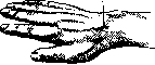
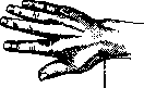
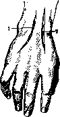
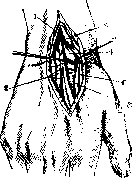
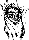
In cases of tuberculosis processes and finally in ankyloses, arthroplasty is performed. Classical incisions. 1. Dorsoradial Langenbeck incision; it begins at the middle of the II metacarpal bone and proceeds along the axis of the limb for 10 cm upward, ending at the middle of the radius epiphysis. 2. Dorsoulnar Kocher incision; it is carried out slightly arched from the middle of the V metacarpal bone along its axis through the middle of the width of the joint and upward for 4–5 cm. After the fascia and lig. carpi dors. are incised, the joint is opened. The edges of the joint capsule are seized with a hook, the ligaments, periosteum, and the tendons lying on it are separated in the ulnar direction. The operation is performed subperiosteally. The soft tissues are retracted in the ulnar direction, after which the ligaments and tendons are separated from the ulnar and palmar surfaces of the joint. The hamulus of the hamate bone is driven in with a hammer, and after the tendons are separated, the small bones of the wrist are skeletonized. The ligaments and tendons of the radial part of the incision are also separated subperiosteally. Kocher pays special attention to preserving the attachment site of the m. extensor carpi radialis on the II metacarpal bone, considering that only this muscle, which supports the bone in the extended state, cannot be replaced in case of its loss. Having thus separated the joint ends subperiosteally from the tendons and ligaments, the joint can be freely dislocated and the joint ends sawed off. 3. Maisonneuve and Titze perform a resection incision not on the side but in the middle along the axis of the limb. 4. Other authors (Lister, Bourgery) open the joint with two incisions from opposite ends of the joint. The advantage of these incisions is that they are technically simple and lie far from the aforementioned dangerous sites. The Bourgery incision, starting from the styloid process of the ulna, is carried upward for 5–7 cm, passing layer by layer between the flexor and extensor muscles of the hand. The incision reaches the bone. After the periosteum is removed all around, the epiphyseal end of the ulna is sawed off with a Gigli saw and removed. The same is done on the radial side, the second incision passing between the m. extensor carpi radialis and the m. supinator longus. After partial removal of the radius and ulna, access to other parts of the joint is easily obtained, and by dislocating the articular surface of the carpal bones into the wound, it can be scraped, trimmed, or sawed off. The simple Bourgery technique provides good access to the joint and spares the tendons. In cases where resection was performed for ankylosis of the radiocarpal joint and the goal of the operation is to restore mobility of the radiocarpal joint, arthroplasty is added to the resection. After resection, the bone ends are covered with a freely transplanted strip of broad fascia of the thigh or a piece of fat is placed between the newly formed joint ends. The success of the operation depends: 1) on asepsis, 2) on whether the newly formed joint space is wide enough, and 3) on whether movements in the radiocarpal joint were started in time. After arthroplasty of the radiocarpal joint, some authors apply traction, others place a fixing bandage for the first 1–11/3 weeks after the operation; but the whole point is to start movements as early as possible, systematically continuing them for several months. Subsequent movements, gymnastics, physio- and mechanotherapy are indispensable conditions for the success of radiocarpal joint arthroplasty. Arthrodesis-fixation of the radiocarpal joint is performed for laxity of the radiocarpal joint, for a hanging hand, which is most often a consequence of infantile paralysis. Since the most favorable position of the hand in terms of function is dorsal flexion, it is usually desired to give the hand a position of dorsal flexion and, having achieved ankylosis, to keep the hand in that position. To achieve ankylosis of the radiocarpal joint, there are several methods: open the joint with one of the resection incisions, dislocate it, and, removing the joint ends with a knife and a sharp spoon, expose the bone surfaces of the wrist and forearm, after which the hand is fixed in the desired position for a long time. However, it is not always possible to obtain a stable ankylosis in this way. In these cases Spitzy applies osteoplastic operations, driving a transplant from the tibia through the styloid process of the ulna and through all the carpal bones. Wittek inserts a bone transplant into a bony groove which he makes on the dorsal surface of the radius, wrist, and at the base of the first metacarpal bone. However, experience shows that these bone transplants also often break or are resorbed. Other authors try to achieve stable fixation of the hand in dorsal flexion through plastic operations on the tendons: shortening the extensors and simultaneously transplanting healthy flexors to the place of the paralyzed extensors. However, these operations do not always achieve the goal. Then the help of a prosthesis is resorted to, which, embracing the hand like a cuff, holds it in the required position without interfering with work, by V. Gorinievsky.
Related articles
Mentioned in
Cite this page
“Radiocarpal Joint.” Soviet Medical Encyclopedia. English translation of Bolshaya Meditsinskaya Entsiklopediya, 1st ed. (Moscow, 1928–1936), ed. N. A. Semashko. https://sovietmedicalencyclopedia.pages.dev/article/radiocarpal-joint/