Urachoplasty
Historical document, translated for reference. It reflects medical knowledge of the 1920s–30s and is not medical advice.
Summary
Urachoplasty refers to surgical repair of the urachus, a fetal structure connecting the bladder to the umbilicus. This article describes the embryonic development, anatomy, and pathological conditions associated with the urachus, including persistent urachus and related complications.
Encyclopedia article (1928–1936)
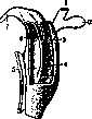
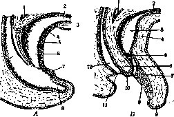
URACHOPLASTY, see Cleft Palate. URACHUS (from Greek uron-urine and icho-I pour), the urinary duct (canal) extending from the apex of the fetal bladder to the umbilicus (fig. 1) and being a derivative of the upper part of the allantois (see) (fig. 2). By the time of birth, the U. becomes obliterated and transforms into a connective tissue cord with an admixture of longitudinally running smooth muscle fibers (continuation of m. detrusoris) (see Human Muscles), called in anatomy ligamentum umbilicale (s. vesico-umbilicale) medium. This cord determines the presence on the inner surface of the anterior abdominal wall of a fold of parietal peritoneum—plica vesico-umbilicalis medialis, s. media. The connective tissue fibers of lig. umbilicalis medii in the region of the umbilicus intertwine with those of lig. umbilicalia later. In individual cases, non-closure of the urinary duct is observed in the postnatal period, with preservation either of the canal or of a narrow fistulous tract between the bladder and the umbilicus—U. persistens. The urinary duct in such cases is lined with an epithelial covering similar to the internal covering of the bladder (so-called transitional epithelium). Over this layer is located a layer of connective tissue and smooth muscle fibers. As it approaches the umbilicus, the epithelium is first represented by two layers, and higher up by one. The degree of non-closure of U. may vary: complete non-closure (along its entire length), closure of the segment nearest to the bladder or nearest to the umbilicus, or both, or finally preservation of the lumen only in certain segments of the duct. Within the substance of lig. umbilicalis med., the urinary duct passes either in the central part of the ligament or eccentrically; it has either a straight direction or forms individual protrusions, dilatations. At the transition of the bladder mucosa into the U. mucosa, a fold is observed that prevents urine from entering the urinary duct. Non-closure of the urinary duct leads to the appearance of a number of pathological formations, such as: fistulas, cysts, etc. The most frequent manifestation of non-closure of U. as a developmental anomaly is the presence of a congenital umbilical (urinary) fistula (fistula umbilicalis congenitalis). The latter is more frequently observed in males. The lumen of the fistulous opening may vary (e.g. in the case of Stuckey it was 3 mm). The presence of an umbilical urinary fistula leads to urine being excreted to a greater or lesser degree through the umbilicus. Simultaneous severe disorders in the development of the bladder or urethra create conditions for the excretion of urine only through the fistula. Besides fistulas, in more rare cases, cysts are observed along the course of the urinary tract, one or several, ranging from microscopically small to the size of a bean and larger (fig. 3). In the case of Walter (Waither), the cyst was 2.5 cm in length, 1.6 cm in width. The origin of the cyst is explained by partial obliteration of segments of the canal with the preservation between them of a closed cavity, sometimes communicating with the bladder and filling with urine. Cases of stones in the urinary duct or its cyst have also been observed. In individual cases (already from the period of development), a connection is established between the bile ducts and the urinary duct, so that there are even observations of the excretion of gallstones through the urachus. (See also Umbilicus, Meckel's diverticulum.)
URACHUS (from Greek uron-urine and icho-I pour), the urinary duct (canal) extending from the apex of the fetal bladder to the umbilicus (fig. 1) and being a derivative of the upper part of the allantois (see) (fig. 2). By the time of birth, the U. becomes obliterated and transforms into a connective tissue cord with an admixture of longitudinally running smooth muscle fibers (continuation of m. detrusoris) (see Human Muscles), called in anatomy ligamentum umbilicale (s. vesico-umbilicale) medium. This cord determines the presence on the inner surface of the anterior abdominal wall of a fold of parietal peritoneum—plica vesico-umbilicalis medialis, s. media. The connective tissue fibers of lig. umbilicalis medii in the region of the umbilicus intertwine with those of lig. umbilicalia later. In individual cases, non-closure of the urinary duct is observed in the postnatal period, with preservation either of the canal or of a narrow fistulous tract between the bladder and the umbilicus—U. persistens. The urinary duct in such cases is lined with an epithelial covering similar to the internal covering of the bladder (so-called transitional epithelium). Over this layer is located a layer of connective tissue and smooth muscle fibers. As it approaches the umbilicus, the epithelium is first represented by two layers, and higher up by one. The degree of non-closure of U. may vary: complete non-closure (along its entire length), closure of the segment nearest to the bladder or nearest to the umbilicus, or both, or finally preservation of the lumen only in certain segments of the duct. Within the substance of lig. umbilicalis med., the urinary duct passes either in the central part of the ligament or eccentrically; it has either a straight direction or forms individual protrusions, dilatations. At the transition of the bladder mucosa into the U. mucosa, a fold is observed that prevents urine from entering the urinary duct. Non-closure of the urinary duct leads to the appearance of a number of pathological formations, such as: fistulas, cysts, etc. The most frequent manifestation of non-closure of U. as a developmental anomaly is the presence of a congenital umbilical (urinary) fistula (fistula umbilicalis congenitalis). The latter is more frequently observed in males. The lumen of the fistulous opening may vary (e.g. in the case of Stuckey it was 3 mm). The presence of an umbilical urinary fistula leads to urine being excreted to a greater or lesser degree through the umbilicus. Simultaneous severe disorders in the development of the bladder or urethra create conditions for the excretion of urine only through the fistula. Besides fistulas, in more rare cases, cysts are observed along the course of the urinary tract, one or several, ranging from microscopically small to the size of a bean and larger (fig. 3). In the case of Walter (Waither), the cyst was 2.5 cm in length, 1.6 cm in width. The origin of the cyst is explained by partial obliteration of segments of the canal with the preservation between them of a closed cavity, sometimes communicating with the bladder and filling with urine. Cases of stones in the urinary duct or its cyst have also been observed. In individual cases (already from the period of development), a connection is established between the bile ducts and the urinary duct, so that there are even observations of the excretion of gallstones through the urachus. (See also Umbilicus, Meckel's diverticulum.)
URACHUS (from Greek uron-urine and icho-I pour), the urinary duct (canal) extending from the apex of the fetal bladder to the umbilicus (fig. 1) and being a derivative of the upper part of the allantois (see) (fig. 2). By the time of birth, the U. becomes obliterated and transforms into a connective tissue cord with an admixture of longitudinally running smooth muscle fibers (continuation of m. detrusoris) (see Human Muscles), called in anatomy ligamentum umbilicale (s. vesico-umbilicale) medium. This cord determines the presence on the inner surface of the anterior abdominal wall of a fold of parietal peritoneum—plica vesico-umbilicalis medialis, s. media. The connective tissue fibers of lig. umbilicalis medii in the region of the umbilicus intertwine with those of lig. umbilicalia later. In individual cases, non-closure of the urinary duct is observed in the postnatal period, with preservation either of the canal or of a narrow fistulous tract between the bladder and the umbilicus—U. persistens. The urinary duct in such cases is lined with an epithelial covering similar to the internal covering of the bladder (so-called transitional epithelium). Over this layer is located a layer of connective tissue and smooth muscle fibers. As it approaches the umbilicus, the epithelium is first represented by two layers, and higher up by one. The degree of non-closure of U. may vary: complete non-closure (along its entire length), closure of the segment nearest to the bladder or nearest to the umbilicus, or both, or finally preservation of the lumen only in certain segments of the duct. Within the substance of lig. umbilicalis med., the urinary duct passes either in the central part of the ligament or eccentrically; it has either a straight direction or forms individual protrusions, dilatations. At the transition of the bladder mucosa into the U. mucosa, a fold is observed that prevents urine from entering the urinary duct. Non-closure of the urinary duct leads to the appearance of a number of pathological formations, such as: fistulas, cysts, etc. The most frequent manifestation of non-closure of U. as a developmental anomaly is the presence of a congenital umbilical (urinary) fistula (fistula umbilicalis congenitalis). The latter is more frequently observed in males. The lumen of the fistulous opening may vary (e.g. in the case of Stuckey it was 3 mm). The presence of an umbilical urinary fistula leads to urine being excreted to a greater or lesser degree through the umbilicus. Simultaneous severe disorders in the development of the bladder or urethra create conditions for the excretion of urine only through the fistula. Besides fistulas, in more rare cases, cysts are observed along the course of the urinary tract, one or several, ranging from microscopically small to the size of a bean and larger (fig. 3). In the case of Walter (Waither), the cyst was 2.5 cm in length, 1.6 cm in width. The origin of the cyst is explained by partial obliteration of segments of the canal with the preservation between them of a closed cavity, sometimes communicating with the bladder and filling with urine. Cases of stones in the urinary duct or its cyst have also been observed. In individual cases (already from the period of development), a connection is established between the bile ducts and the urinary duct, so that there are even observations of the excretion of gallstones through the urachus. (See also Umbilicus, Meckel's diverticulum.)
Related articles
Cite this page
“Urachoplasty.” Soviet Medical Encyclopedia. English translation of Bolshaya Meditsinskaya Entsiklopediya, 1st ed. (Moscow, 1928–1936), ed. N. A. Semashko. https://sovietmedicalencyclopedia.pages.dev/article/urachoplasty/