Uterine Artery
Historical document, translated for reference. It reflects medical knowledge of the 1920s–30s and is not medical advice.
Summary
The uterine artery is a major blood vessel supplying the uterus, originating from the internal iliac artery. It undergoes significant changes during pregnancy and is important in surgical procedures involving the uterus.
Encyclopedia article (1928–1936)
UTERINE ARTERY, uterine artery [corresponds to the artery of the vas deferens in men]. The uterine artery arises from the hypogastric artery (a. hypogastrica, fig. IV).
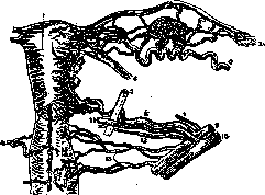
Figure 1. Diagram of the vessels of the uterus, its appendages, and vagina. Posterior surface of the uterus and right broad ligament: /-uterus; г-tuba Fallopii; 3-fimbriae; ^-lig. uteri rotundum; s-ureter; 6-a. ovarica; 7-obliterated umbilical artery-lig. vesico-umbilicale laterale; «-a. uterina; 9-a. hypogastrica; 10-v. hypogastrica; 11-v. uterina; 12-a. vesicalis superior; 13-a. vaginalis.
to the wall of the uterus. The uterine artery is spiral-shaped, especially in its peripheral portion—a circumstance having physiological significance during pregnancy: the vessels must stretch in length and dilate considerably to supply the fetus and hypertrophied walls of the uterus. In this process, the walls of the uterine arteries and veins not only stretch but also undergo marked hypertrophy. A similar phenomenon is observed in pathological conditions, in neoplasms, etc. In the postpartum period, undergoing regressive metamorphosis, the vessels do not return to their original size and remain somewhat dilated. Embryologically, the aa. uterinae, corresponding to the right and left sides of the uterus, originate from arteries supplying the Müllerian ducts or cords (see Müllerian duct). In the absence of fusion of the latter, each embryonic primordium corresponds to one uterine artery. The uterine artery arises from the anterior surface of the hypogastric artery (a. hypogastrica, s. a. iliaca interna), directly opposite or slightly below the origin of the superior gluteal artery (a. glutaea superior). Often the uterine artery arises with a common trunk with the umbilical artery (a. umbilicalis), sometimes above, sometimes below the latter. These three sites of origin, close to each other, can be considered typical. Initially, the uterine artery, covered by parietal peritoneum, runs along the wall of the pelvis forward, then turns medially and through the parametrial tissue of the broad ligament runs obliquely forward to the cervix of the uterus. From here the artery runs, winding, along the lateral wall of the uterus from below upward (fig. 1). The terminal part of the uterine artery anastomoses widely with the ovarian artery (a. ovarica). "The anastomosis is so large that it is impossible to determine where one of these arteries ends and the other begins: the amount of blood brought by each of them should be considered completely equal" (Gubarev). From the point where the uterine artery approaches the cervix downward, vaginal arteries (aa. vaginae, fig. 1) branch off. After giving branches to the vaults, they descend along the lateral wall of the vagina. The vaginal arteries may arise independently from the hypogastric artery. Regardless of their site of origin, the terminal parts of these arteries anastomose richly with branches of the middle hemorrhoidal, inferior vesical, etc. arteries. In the peripheral parts of the vagina, anastomoses of the perineal arteries and external pudendal arteries from the internal pudendal artery system (a. pudenda interna) flow into this arterial network. Some authors (Hyrtl) distinguish special unpaired arteries of the vagina—anterior and posterior (aa. azygos vaginae anterior et posterior, fig. 1). Gubarev considers that the unpaired arteries are a large anastomosis between branches of the vaginal arteries and the uterine artery. From the main trunk of the uterine artery, numerous anterior and posterior branches arise, supplying the tissue of the cervix and uterus. They run in the superficial layers of muscular tissue and anastomose with each other; in this connection, an anastomotic ring at the level of the middle third of the cervix is particularly prominent, consisting of two branches on each side, which can serve as a source of severe bleeding, especially with low-lying uterine tumors. Mention should be made of the small anastomotic arteries running in the uterosacral ligaments, which empty into the aforementioned ring and bleed when cut during operations for removal of the uterus. In the region of the angle of origin from the uterus of the round ligament, there is a small anastomosis with the artery of this ligament (a. ligamenti rotundi). The latter is a branch of the inferior epigastric artery (a. epigastrica superficialis); in its peripheral part, at the exit of the round ligament of the uterus from the inguinal canal, the artery in question anastomoses with the external pudendal artery (a. pudenda externa), a branch of the femoral artery. When removing the uterus, the artery of the round ligament of the uterus must be ligated separately. The most powerful anastomosis, already mentioned above, is formed by the connection of the uterine artery with both the branches of the ovarian artery (a. ovarica) itself and its tubal branch (ramus tubarius, fig. 1). All these anastomoses make the uterus an organ abundantly supplied with blood from the most diverse sources: the uterine artery—from the hypogastric, ovarian artery, from the abdominal aorta; by the vaginal arteries—from the internal pudendal, from the vesical and hemorrhoidal; by the artery of the round ligament of the uterus—from the inferior epigastric and external pudendal, by the latter's anastomosis—from the femoral artery. The most powerful trunks are the uterine and ovarian arteries. Topographically, the uterine artery is divided into three segments: in the first, together with its vein, it lies on the lateral wall of the pelvis, lateral to the ureter, covered by parietal peritoneum (fig. 2). Enveloped in connective tissue, the uterine artery enters the base of the broad ligament of the uterus (ligamentum latum uteri, also lig. cardinale uteri). The second practically important site is the crossing of the ureter with the uterine artery, occurring in the connective tissue of the broad ligament of the uterus. The trunk of the uterine artery crosses the ureter in front (fig. 1 and 2), one loop of the artery passes behind the ureter. The uterine vein runs with its main trunk most often along the posterior wall of the ureter; around the latter an arteriovenous ring is formed (Gubarev, fig. 1). Knowledge of the relationship of the uterine vessels to the ureter is very important in the extended operation for removal of the uterus for malignant tumors. The third segment of the uterine artery, about 11/2-2 cm long, from the point of crossing with the ureter to the cervix, is characterized by the beginning of the branching of the uterine artery into ascending and descending branches. The ureter, in relation
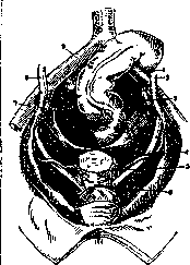
Figure 2. Position of the ureter in the female pelvis and its relationship to the uterine artery, hypogastric artery, and vagina. 1 and 9-a. iliaca communis; 2 and «-ureter; Зж 7-a. hypogastrica; if-a. uterina; 5-cervix uteri; 6-vagina.
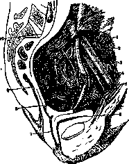
Figure 3. Ureter and uterine artery and their mutual relationship to the pregnant uterus: 1-plexus pampiniformis; 2-a. hypogastrica et ureter; 3-a. et v. iliaca ext.; 4-a. uterina; 5-a. vesicalis sup.; 6-vesica; 7-vagina; S-orificium int. uteri; 9-rectum; 10-promontorium.
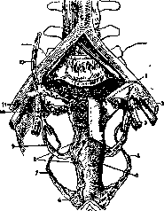
Figure 4. Blood supply of the uterus and vagina; 1-uterus; 2-a. et v. hypogastrica; S-a. et v. uterina; i-v. pudenda int.; 5-vagina; 6-bulbus vestibuli; 7-a. et v. pudenda int.; «-plexus utero-vaginalis; 9-v. uterina; 10-plexus ovaricus; 11-ovarium; 12-a. et v. ovarica. *4»> to this segment lies lateral and downward on the lateral wall of the vagina (fig. 3). On this path the artery gives small branches to the broad ligament, to the ureter, and to the posterior wall of the bladder. The vein accompanying the artery here becomes double, and along the lateral wall of the uterus and vagina the uterine artery in essence runs through the uterovaginal venous plexus (plexus utero-vaginalis venosus) (fig. 4). The innervation of the uterine vessels comes from the uterovaginal nerve plexus (plexus utero-vaginalis). The influence of the autonomic nervous system, subordinate to the endocrine glands, is monthly reflected in the vascularization of the uterus in the direction of its increase, and consequently also in the temporary change of the uterine artery. Pregnancy, uterine tumors, especially fibroids, lead to sharp, often persistent hypertrophy of the uterine artery. In subinvolution of the uterus and senile atrophy, the uterine artery sometimes turns into a thin cord. Anomalies of the uterine artery are extremely diverse; it may arise from any branch of the anterior trunk of the hypogastric artery: from the umbilical (a. umbilicalis), superior vesical (a. vesicalis sup.), obturator (a. obturatoria), inferior vesical (a. vesicalis inf.), common pudendal (a. pudenda communis), from the middle artery of the rectum (a. haemorrhoidalis media) and finally from the sciatic artery (a. ischiadica). "From a practical point of view, it is important that the uterine artery never arises below the upper edge of the greater sciatic foramen, consequently it can be ligated only in the cavity of the small pelvis" (Gubarev). The opinion of authors about the possibility of a double (Haller) or triple (Weber) uterine artery. Gubarev denies this, considering that in all these cases the additional uterine arteries were actually additional vesical arteries, but practically it is important to remember the possibility of their existence, as they give branches to the lower parts of the cervix of the uterus.
Related articles
Mentioned in
Cite this page
“Uterine Artery.” Soviet Medical Encyclopedia. English translation of Bolshaya Meditsinskaya Entsiklopediya, 1st ed. (Moscow, 1928–1936), ed. N. A. Semashko. https://sovietmedicalencyclopedia.pages.dev/article/uterine-artery/