Ureter
Historical document, translated for reference. It reflects medical knowledge of the 1920s–30s and is not medical advice.
Summary
The ureter is the excretory duct of the kidney. This article describes its embryological development, anatomy, histology, topography, and variations in structure and position.
Encyclopedia article (1928–1936)
URETER, the excretory duct of the kidney. From the perspective of the embryological development of the urinary system in humans, one can speak of primary and secondary ureters. With the two primary, successive renal formations (pronephros and mesonephros), the role of the ureter is performed by the Wolffian duct (see), which can be considered as the primary ureter. The ureter of the definitive, permanent kidney (metanephros), i.e., the organ present in humans in extrauterine life, is a secondary formation. It develops in the embryo at 5 mm in length from the lower part of the Wolffian duct by the evagination of its posterior medial wall (fig. 1). Extending in the cranial direction, this primordium reaches the nephrogenic tissue of the caudal segment of the Wolffian body (see), grows into it, and together with it, its terminal upper part participates in the formation of the permanent kidney.
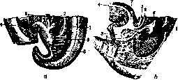
Fig. 1. Development of sinus urogenitalis (according to Keibel). a: 1-ureter; 2-Wolffian duct; 3-cloaca; 4-urachus; 5-rectum; b-chorda; b: 1-ureter; 2-Wolffian duct; 3-cloaca; 4-urachus; 5-rectum; c-renal pelvis; 7-urinary bladder.
Anatomy and histology. In the developed state, the ureter has the appearance of a cylindrical, slightly flattened tube. The beginning of the ureter is considered the narrowed end of the renal pelvis, or calyx (isthmus ureteris). The end of the ureter obliquely pierces the wall of the urinary bladder and opens on its mucous side with a slit-like opening at the plica ureterica, in the upper corner of the trigone of Lietoaudi [see separate table for article Neuroglia (fig. 6) and separate table-article 143-144 (fig. 1)]. The wall of the ureter is provided with smooth muscle, which divides into 2 layers: the outer, circular, and the inner, more powerful, longitudinal. Upon entering the bladder, both layers maintain their independence and are not connected with the muscular activity of the bladder, so that when it is stretched by fluid, the lumen of the ureter does not expand, and the periodic emptying of urine into the bladder does not depend on the degree of tension of the walls of the urinary bladder. Not far from the bladder opening, the ureter acquires a third muscle layer located along its periphery-longitudinal, so-called ureteral sheath, which, conversely, is in close dependence on the bladder muscle (fig. 2). The mucous membrane lining the ureter consists of a transitional epithelium of Henle and a base rich in fibrous connective tissue and elastic fibers. Throughout the entire length of the ureter, the mucous membrane forms longitudinal folds, due to which the ureter in cross-section has a star-shaped shape (fig. 3). Entering the bladder obliquely at an acute angle, the upper wall of the ureter forms a fold lined with mucous membrane on both sides-valvula ureteris (fig. 4). Due to the presence of muscle fibers in its thickness, it is capable of contracting and, by closing the lumen of the ureter, plays the role of a valve, which under normal conditions prevents the backflow of urine from the bladder into the ureter. Outside, the ureter is covered by loose connective tissue, which includes its vessels and nerves and is surrounded by a thin fascia-a direct continuation of the renal fascia. The ureter is located in the retroperitoneal connective tissue of the posterior abdominal wall and, bending over the linea innominata, s. terminalis, passes to the lateral wall of the pelvis. Corresponding to this location,
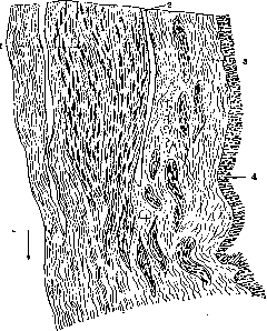
Fig. 2. Longitudinal section of the wall of the lower part of the ureter: 1-ureteral sheath; 2-slit separating the sheath from the wall; 3-wall; 4-mucous membrane.
in the description of the ureter, it is divided into 2 parts: pars abdominalis and pars, s. curvatura, pelvina. Its total length in an adult is calculated as 28-34 cm and is subject to large variations depending on the individual's height and the level of position (development) of the kidney; in connection with this, in the same subject, the ureter is shorter on the right than on the left, by approximately 1 cm. About 2 cm of its length falls on the intravesical part, while the remaining extent is divided almost equally between the abdominal and pelvic parts. The width of the ureter is uneven. Narrowings alternate with spindle-shaped expansions (fig. 5). The narrowest lumen is in the region of the neck of the ureter (isthmus), reaching 2-4 mm here, and at the point of bending over the terminal line-up to 4-6 mm in diameter. The expansion located above it, clearly expressed even in children (fig. 6), is called ampulla terminalis. The greatest width of the abdominal segment in an adult is 8-15 mm. The pelvic part of the ureter represents a more uniformly expanded tube with a lumen up to 6 mm, while in the part located in the thickness of the bladder wall (pars intramuralis), the lumen narrows to 4 mm. However, the given dimensions refer to the empty ureter; with excessive filling with urine, they can vary significantly; since the ureter has very loosely arranged connective tissue between muscle bundles (fig. 3), it possesses great elasticity and in case of difficulties in urine outflow can expand to significant sizes (up to 8 cm in diameter).
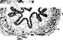
Fig. 3. Cross-section of the ureter: 1-longitudinal muscle fibers; 2-circular muscle bundles.
Fig. 4. Longitudinal section of the ureter in the area of its entrance into the urinary bladder: 1-valvula ureteris; 2-ureter; 3-ureteral orifice; 4-wall of the urinary bladder; 5-upper wall of the ureter; 6-lower wall of the ureter.
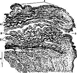
Fig. 5. Course of the ureter (diagram): 1-isthmus; 2-spermatic vessels; 3-iliac vessels; a-pars abdominalis ureteris; b-pars iliaca; c-pars pelvina; d-pars intramuralis.
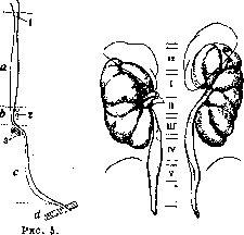
Fig. 6. Kidneys and ureters of a newborn (49 cm). (According to K. Peter.)
Topography of the ureter. Abdominal part. Depending on the shape and position of the axis of the kidney, the ureter at its point of origin can already give significant variations in its relationship to the kidney. With a well-developed pelvis, the ureter originates below the crossing of its
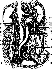
Fig. 7. Course of the ureters in the retroperitoneal connective tissue: 1-right ureter; 2-iliohypogastricus and ilioinguinalis; 3-cutaneous femoris lateralis; 4-spermatic vein; 5-psoas muscle; 6-genitofemoral nerve; 7-vas deferens; 8-urinary bladder; 9-rectum; 10-severed mesentery of S-Romanum; 11-permanent nerve; 12-inferior mesenteric artery; 13-psoas muscle; 14-spermatic artery; 15-spermatic vein; 16-left ureter; 17-duodenum.
renal vessels, while with a short renal pelvis, it is usually located behind most of their branches and, descending, crosses them; in this case, the lower pole of the kidney may lie laterally to the ureter or to a greater or lesser degree overlap and cover it. In the presence of additional vessels (see Kidney), some of them may pass behind the ureter and in front of it, far below its exit from the kidney. Along the abdominal part, the ureter lies on the anterolateral surface of the m. psoas, in the vertical projection-at the level of the transverse processes of the vertebrae [see separate table (article 135-136), fig. 8]. Descending, it obliquely crosses the m. psoas along a gentle spiral, so that when bending over the iliac vessels, it is found on its medial surface (see vol. IV, article 148-149, separate table, fig. 3). On its path along the anterior surface of the muscle, it ventrally crosses the n. genitofemoralis (fig. 7). The crossing of the ureter over the iliac vessels is not always symmetrical. On the right side, the ureter bends over the a. iliaca ext., while on the left it does not reach it and, descending more medially, crosses the a. iliaca communis. At the vessels at the medial edge of the m. psoas, the ureter crosses the a. and v. spermatica, s. ovarica (which usually lie closer to the peritoneum and depart together with it when it is detached). The segment of the ureter between the spermatic vessels and the bend over the iliac vessels is distinguished by some authors as portio iliaca. In front of the ureter, besides the spermatic vessels on both sides, are visceral organs, i.e., various parts of the intestine and associated vessels and mesenteries or parietal areas of the peritoneum. Thus, on the right side, the ureter is covered: in the initial part-by the descending portion of the duodenum (with its low position, a larger part of the abdominal part of the ureter may be hidden behind its descending part); in the middle of its length-by the parietal peritoneum with the vessels passing under it-a. and v. colica dextra and ileocolica and the lymph nodes lying on them; in the lower part, the ureter is crossed by the terminal portion of the ileum with its shortening mesentery not far (3-4 cm) from the place of entry into the cecum. Usually, the cecum and ascending colon are located lateral to the ureter and do not come into contact with it (fig. 8). However, with an excessively developed mesentery (normally absent), these parts may adhere to the retroperitoneally located ureter. The vermiform appendix may come into close proximity to it with medial displacement and with a mobile cecum. On the left side, the abdominal part of the ureter lies more openly from the intestinal elements, but from top to bottom it is transversely crossed by retroperitoneal blood vessels (fig. 9) with adjacent lymph nodes.
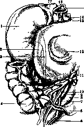
Figure 8. Topography of the right ureter: 1- right kidney; 2- duodenum; 3- ureter; 4- cecum; 5- v. iliaca interna; 6- ileum (its free edge is cut); 7- ureter (pars iliaca); 8- v. iliaca com.; 9- a. iliaca com.; 10- vasa spermatica; 11- a. mesenterica superior with adjacent lgl. ileo-caecales; 12- pancreas; 13- a. hepatica; 14- v. portae; 15- v. cava inf.; 16- ductus choledochus; 17- suprarenal gland. (Original drawing from the author's specimen.)
mi. Of the parts of the intestine, only its lower part is covered by the root of the mesentery and the flexura sigmoidea itself. On the rest of its course, it is covered by the parietal peritoneum with its vessels: branches of a. and v. colica sinistra superior et inf., which go to the vascular arcades (arcuate anastomoses) of the descending colon and sigmoid colon. The pelvic part of the Ureter. In the small pelvis, the Ureter appears at the level of the sacroiliac joint (sometimes somewhat lateral to it), directly behind the peritoneum (easily separated from it), in front of a. and v. hypogastrica in the closest neighborhood with the adjacent lymphatic l nodes for pelvic organs. Below the terminal part of the Ureter, it runs along the lateral wall of the pelvis in a curve forward, crossing on its way from the medial side a. umbilicalis lateralis and below it a., v. and n. obturatorii (fig. 10). After this last crossing, the ureter bends inward and approaches the bladder, entering the venous bladder plexus, here meeting the horizontal portion of the vas deferens crossing at a right angle and at its end touching behind the upper pole of the seminal vesicle. In the entire lower segment, starting from the crossing with vas deferens, the Ureter is displaced from the peritoneum by the vas deferens and the surrounding cellular tissue with the venous plexus. In women, in the area of the terminal part of the Ureter, it crosses the vessels of the ovary located in lig. suspensorium ovarii. Somewhat below the border line, the Ureter enters the area of fossae ovaricae and comes into proximity with the ovary, being separated from it only by a leaf of peritoneum (fig. 11). After crossing with aa. umbilicalis and obturatoria, descending Fig. 10. relation of the
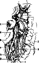
KNIzhu, the Ureter ENTERS the cellular tissue of the BROAD ligaments at the level of the upper edge of the uterus *Tvas deferens; 5- a. and V. ц
p+ou-pg, t-1 iliaca externa; 6- ureter; 7- bladder; 8- rectum. here makes a sharp turn from above outward inward and among b. or m. abundant venous parametral plexus, it goes around the Ureter, curving around it in front (fig. 12). From the cervix, the Ureter in this area is at a distance of 0.8-2.5 cm (fig. 13) (with a pregnant uterus, it is in direct contact with it). At the level of the cervical os, being outside the parametral cellular tissue, the Ureter lies against the anterior wall of the vagina, after which it empties into the bladder 2-3 cm below the level of the lower edge of the cervix.
Vascular supply of the Ureter. The vessels serving the Ureter are located in the surrounding cellular tissue (adventitia) in the form of long thin descending and ascending loops. Arterial branches depart from the corresponding arteries, mostly in the upper part from the renal arteries, in the middle part from a. spermatica interna or a. iliaca (rarely), in the lower part from any branches of a. hypogastricae (haemorrboida
Figure 12. Relation of the ureter to a. uterina: 1- ureter; 2- a. uterina; 3- a., v. and n. obturatorii; 4- bladder; 5- vagina; 6- uterus.
lis media, vesicalis, uterina). Venous blood drains accordingly to the same areas in vv. renales, spermaticae, branches of v. hypogastricae and plexus vesicalis. Regional lymph nodes for the lower part are lymph nodes on vasa hypogastrica, for the middle part- lymph nodes along the course of v. cavae (on the left- directly the nodes of the renal vessels), for the upper part- lymphatic nodes of the renal hilus. Innervation- see Vegetative nervous system.
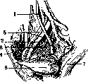
Figure 11. Topography of the left ureter in cross section: 1- ureter; 2- ovary; 3- rectum; 4- v. iliaca ext.; 5- uterus; 6- sacrum. (According to Tandler.)
Figure 13. Diagram of the relation of the ureter to the uterus in cross section: a- ureter; b- cervix of uterus; c- bladder. (According to Nagel.)
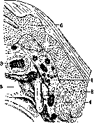
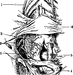
B. M. E. t. XIX.
K. Yesipov. Pathology. Diseases of the ureter have two features: 1) primary forms of diseases are rare, usually ureteral lesions develop secondarily following diseases of the kidneys or bladder; 2) as soon as the ureter becomes involved in the process, the course of the disease changes sharply for the worse, as a new factor appears—obstructed outflow of urine from the kidney. According to autopsy material (Poirier), anomalies of the ureter occur in 3% of all autopsies, but in clinical practice, calculated per number of kidney patients, they are significantly more frequent, as they themselves serve as a cause of kidney diseases. The very large variety of forms of ureteral anomalies can be reduced to several main groups: anomalies in the number of ureters, changes in their course, length, lumen, method of departure from the renal pelvis, or termination in the bladder. The number of ureters can be increased, duplicated, and even tripled on one or both sides, so that sometimes 3 to 6 openings are found in the bladder; but duplication may not concern the entire length of the ureter, but only part of it. The ureter—one or both—may open into the bladder not on its own side, but on the opposite side (crossed). In length, the ureter can be very shortened, as is observed with dystopic kidneys. The vesical opening of the ureter can be very narrowed (hypoplasia) or even completely closed (atresia). Sometimes it opens not at the usual place, but closer to the midline or closer to the neck of the bladder, even into the urethra, into the seminal vesicles, in women—into the vagina or next to the external opening of the urethra. The upper end of the ureter may depart from the renal pelvis abnormally at an acute angle, making a sharp bend, or it begins very high. Changes in the lumen of the ureter are expressed by the formation of kinking, twisting, narrowing up to complete closure, or the formation of valves similar to venous ones, consisting of duplication of the mucous membrane, diverticula, or diffuse dilatations. Thus, with an anomaly of the ureter, difficult urination is often associated, which leads to dilatation of the renal pelvis, development of hydronephrosis or sclerosis and atrophy of the kidney; therefore, recognition of anomalies has great practical significance. Modern clinical practice has chromocystoscopy, ureteral catheterization, radiography with an X-ray opaque bougie introduced into the ureter, or with filling it with contrast solutions for this purpose. A purely functional disease is atony of the ureter, which can be secondary and primary. Any atony due to changes in hydrodynamic conditions leads to the development of hydronephrosis. The basis of primary atonies is either congenital insufficiency of the ureteral sphincter and generally weak development of the musculature of the entire urinary system, or lesion (as a result of injuries) of the nerve nodes and branches supplying the ureter. Secondary atonies are observed with the most diverse processes affecting the muscular layer of the ureter. The lesion can be unilateral and bilateral. The atonic ureter is stretched, has thinned walls, its musculature gradually disappears, being replaced by scar tissue. Atony is recognized by the presence of a wide, gaping opening, weakly contracting or completely immobile, and by the sluggish discharge of urine from it. But in some cases, the vesical openings remain normal, and to confirm the presence of atony, one must observe the nature of the urine discharge or introduce a catheter into the ureter and fill it with a colored solution, whereby one can see that the liquid flows back out of the ureter past the catheter in a constant stream, and not in an intermittent jet, as in normal conditions. Finally, by means of pyeloureterography, definite indications regarding the dilatation of the ureter can be obtained. The term ureterocele denotes a cystic dilatation of the lower end of the ureter, connected with prolapse into the bladder of either only the mucous membrane of the ureter or all its layers; sometimes the bulging is only lateral within the intramural part. The cause of cystic dilatations of the ureter is narrowing of its vesical opening, atresia due to congenital anomaly or inflammatory origin. The upper part of the ureter, gradually stretching with urine and turning into a cyst, bulges into the bladder cavity as a large tumor, and in women even protrudes outward through the urethra. In some cases, the presence of ureterocele is not without significance, but causes pain, frequent urination, and successive dilatation of the renal pelvis and kidney. Recognition of ureterocele is quite easily established by means of cystoscopy, but to determine the extent of the lesion, it is necessary to resort to ureterography. Treatment is possible only by operative endovesical route or through high incision of the bladder. The goal of the operation is to create a wide anastomosis between the cyst and the cavity of the urinary bladder. With large cysts, especially those protruding outward, it is better to go through the opened bladder and resect part of the cyst wall or to amputate it entirely at the base. On a par with ureterocele, one can place diffuse dilatation of all or a considerable part of the ureter, known under the name of hydroureter. The basis of this disease is constant stasis in the ureter, caused by one or another organic obstacle to the outflow of urine or atony of the ureter. From organic causes, hydroureter can be caused by stones, strictures of congenital or inflammatory origin, new growths, compression by tumors of neighboring organs, the pregnant uterus, inflammatory processes in the perirenal tissue. Finally, obstacles to the outflow of urine in the urinary bladder and in the urinary passages can also be accompanied by dilatation of the ureter. The dilatation affects either the entire ureter throughout its entire length or only part of it, depending on the localization of the obstacle. Almost always the effect of urine retention is then transmitted to the renal pelvis and kidney. Thus, hydroureter is always a secondary disease and moreover its significance recedes to the background before the usually simultaneously present hydronephrosis (see). In the presence of infection of the urinary passages, hydroureter turns into hydropioureter and then 229
URETER
230 into a clean pyoureter. In pyonephroses of various origins, the development of the pyoureter proceeds in a descending order from the kidney, with the walls of the U. undergoing changes in the sense of sclerosis, ulcer formation, and narrowing.-Special attention should be paid to those cases of pyoureter that develop after nephrectomies in the remaining, unremoved peripheral end of the U. Due to obstruction by a stone or narrowing at the exit and atony, the remaining U. can be stretched by pus to the size of an intestine, which is associated with a number of further complications-pains, elevated temperature, pyuria, and rupture of pus into the surgical scar or cellular tissue. Recognition of this complication is easily achieved by cystoscopy, catheterization, and based on anamnestic data. As for treatment, preventive measures should be kept in mind first, i.e., to determine before nephrectomies or during the operation itself whether there is narrowing in the lower part of the U., and to excise the U. as low as possible during nephrectomies. In already developed complications, either the opening of the U. should be dilated to allow pus to drain into the bladder, and then attempts should be made to achieve scarring of the U. through cauterizations, or a secondary operation-ureterectomy-should be performed through an extraperitoneal incision. Inflammatory processes in the ureter-ureterites as a primary disease are extremely rare and were first described by Israel under the name ureteritis membranacea bact.; similar observations are reported individually by other authors; the process occurs at high temperature, is accompanied by colic and the excretion of pus, fibrin, and films impregnated with microbes and lime salts. The disease can sometimes be cured only by removal of the U. and kidney. In the vast majority of cases, ureteritis is a secondary disease that develops as a complication of purulent diseases of the kidney or bladder, thus proceeding in a descending or ascending manner. The nature of the infection in ureterites is the most diverse, including specific (tuberculous). Changes in the ureter itself, starting with hyperemia of the mucosa and its catarrhal swelling, can lead to the formation of erosions and ulcers, infiltration of the entire thickness of its wall with subsequent spread to the surrounding cellular tissue of the U. (periureteritis). The inflamed ureter becomes dense to the touch, thickened sometimes to the size of a finger, its lumen is narrowed by inflammatory infiltrate and scars or dilated due to extensive ulcers, loss of muscle tone, and stretching by urine. If there is narrowing of the U., sequential dilation of the renal pelvis and kidney is inevitable. Clinically, as a secondary disease, ureterite naturally recedes into the background, and similarly therapy should be directed at the diseases of the bladder and kidney.-A special form of chronic inflammation of the U. is cystic ureteritis, which occurs as a rare disease (30 cases described); it is manifested by the formation of cysts with transparent content on the mucosa of the U., scattered throughout its entire length. The walls of the ureter are thickened, and its bladder opening (during cystoscopy) gapes. The origin of this disease is attributed by some authors to inflammatory processes caused by parasites (myxosporidia, psorosporidia), while others, which is more likely, associate the disease with congenital local accumulations of flat epithelial cells (see Bruni's epithelial nests). The clinical importance of the process lies in the fact that it secondarily affects the condition of the kidneys due to narrowing of the ureter or spread of the inflammatory process to the renal pelvis. The diagnosis can be tentatively made if there are uniform changes in the bladder or at the bladder opening of the U.; the latter is usually dilated in such cases. Conservative treatment in the form of cauterizations is unlikely to be successful; therefore, in complicated severe cases, only nephrectomy + ureterectomy remains. Tuberculous U. (tuberculous ureteritis)-is always a secondary disease, spreading to the U. usually from the kidney and more rarely developing in an ascending manner from the bladder. The lesion begins with the appearance of tubercles, then ulcers, caseous necrosis, and a reactive sclerosing process in the perireteric cellular tissue. In far-advanced stages, the U. has the appearance of a cord thickened to the size of a finger, firm to the touch, palpable through the abdominal walls, especially at the place where it passes into the small pelvis, and its lower end can be palpated through the rectum or vagina. The lumen of the U., despite its great thickness, is usually narrowed, the bladder opening during cystoscopy appears eaten away, uneven, sometimes gaping and immobile due to infiltration and atony, and sometimes drawn inward due to shortening of the entire U. Lesion of the U. complicates the clinical picture of the disease, as well as the technique of nephrectomies: the affected U. should be excised along its entire length, but since this is technically always difficult, usually the U. is removed as much as the operative wound allows; the peripheral end is then ligated after smearing its lumen with carbolic acid or injecting an iodoform emulsion into its lumen. Most often, the peripheral end subsequently undergoes scarring and atrophy, but sometimes it can serve as a soil on which a pyoureter develops, abscesses and fistulas form even several years after the operation. In such cases, it is necessary to resort to introducing an iodoform emulsion into the U. by catheterization from the bladder or to perform a secondary operation to excise the remaining U. down to the bladder. At the border between inflammatory processes and neoplastic processes in the urinary tract, there are two processes that are to some extent similar to each other-leukoplakia and malakoplakia, consisting in changes in the properties of the epithelium (metaplasia, keratinization). Both processes predominantly affect the urinary bladder and renal pelvis; the U. is apparently also secondarily affected in these cases, and its role in the development of the clinical picture is only related to narrowing of the U.'s lumen and obstruction to urine outflow, and especially to the movement along the U. of desquamating epithelium, shedding in the form of whole films. Both processes are considered as precancerous stages. Among true tumors, both malignant and benign tumors are observed in the U. Primary benign tumors are generally observed in the U. very rarely: only individual cases of lipomas, fibromas are described; papillomas are encountered relatively more often, usually of small size and only in exceptional cases reaching the size of an orange; their favorite site of development is the lower third of the U., and often they prolapse into the bladder. Sometimes papillomas of the U. occur in the form of multiple tumors scattered throughout its entire length. On microscopic examination of such papillomas, atypical epithelial proliferations or even a clear picture of villous carcinoma resembling similar forms in the urinary bladder are often found. Of malignant tumors that are malignant from the start, primarily developing in the ureter, solid and squamous cell carcinomas, sarcomas, myxosarcomas are relatively rare; Kümmel counts 53 cases of primary malignant tumors from the literature. Another part of malignant tumors are of secondary origin, developing by seeding from tumors of the kidney (Dobrotvorsky). All kinds of neoplasms of the U., by narrowing its lumen, lead to dilation of the renal pelvis and the development of hydro- or hematonephrosis, and in the case of infection joining, to pyonephrosis. Early and constant symptoms of tumors of the U. are bleedings; later pains join depending on the difficult outflow of urine and the passage of blood clots or the obstruction of the U.'s lumen by a villus of the tumor. Recognition of ureteral tumors is easily established if during cystoscopy proliferations are visible around the opening of the U. and especially if a tumor villus protrudes from the opening. Sometimes by examination through the rectum or vagina, the lower part of the U., affected by a tumor, can be palpated; in this case, a characteristic sign can be considered the appearance of blood in the urine after palpation, which was previously free of blood. In the absence of the noted signs, it is very difficult to make a differential diagnosis with kidney tumors. The excretion with urine of villi or individual cells of the neoplasm is conclusive, but it can also be observed with other localizations. Characteristic are the weak contractions of the U.; when a catheter is introduced, an obstacle is felt; after catheterization, bleeding appears; it is conclusive if during catheterization of a bleeding U., by passing the catheter higher, clear urine is obtained. Ureterography can also give a positive answer. Any tumor of the U. should be removed; usually the kidney is removed along with the U. Stones of the U. are almost always of secondary origin and form from kidney stones or fragments that have become stuck in the U. and here begin to grow by the deposition of new layers of salts. Most often (90%) stones are solitary, but multiple stones are not rare. The size of the calculi is extremely diverse-from a small sand grain to stones of considerable size, round, rod-shaped, or ovoid in shape.
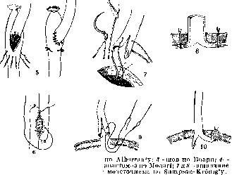
Sometimes, despite their large size, stones in the Ureter nevertheless do not completely close its lumen, and urine from the kidney continues to flow into the bladder, while the stone grows, taking the form of a mold of the Ureter and reaching a considerable length. In the clinic of Fedorov there are specimens of two stones that occupied the entire length of the Ureter from the renal pelvis to the bladder, 16-20 cm long and weighing 52 and 68 g. The majority of stones in the Ureter are located in its lower part, in the cavity of the small pelvis; of these, 3/4 have already penetrated into the intramural part of the Ureter [see separate table (pp. 135-136), figures 6 and 7]. Stones are located in the lumbar section [see separate table (pp. 135-136), figure 5] or in the iliac part of the Ureter much less frequently. Bilateral stones are observed comparatively rarely. In composition, the stones are in most cases mixed and consist of urates and oxalates or urates and phosphates. In relation to the lumen of the Ureter, stones are divided into movable ones, capable of displacement, especially upward, where the Ureter is mostly dilated, and into completely immobile, wedged stones, especially if they are located in a diverticulum or in strictures. The symptoms of stones in the Ureter vary depending on their position and the degree of closure of the Ureter's lumen, as well as on whether the case is complicated by infection of the urinary tract or remains aseptic. Stones located at the exit from the renal pelvis or in the lumbar section give symptoms similar to kidney stones, stones in the pelvic section cause pain in the iliac fossa or in the pelvis, sacrum, and perineum, and often give rise to erroneous diagnoses and confusion with diseases of the vermiform appendix, female genital organs, or rectum. If a stone does not cause any significant closure of the ureteral lumen, it may for a long time not manifest itself in any way and is discovered on an accidental X-ray. In case of obstruction of the Ureter, even if incomplete, a series of secondary changes gradually develop in the kidney—hydronephrosis or secondary atrophy of the kidney as a result of sclerosing nephritis. The farther from the kidney the site of obstruction, the slower the kidney dies, and sometimes the kidney retains its functionality for several years. If the obstruction is complete, the kidney can completely lose its function within a few days. It is precisely this possibility of complete obstruction and the always present slowly increasing pressure in the urinary organs that make the disease always serious and requiring even more attention than kidney stones. In the presence of stones in the kidneys, it is always necessary to ascertain whether there are simultaneously stones stuck in the Ureter. The diagnosis of stones in the Ureter is based on clinical manifestations, on the data of cystoscopy [see separate table (pp. 143-144), figure 1] and catheterization of the Ureter, but mainly on X-ray examination [see separate table (pp. 135-136), figures 5, 6 and 7]. Clinical symptoms are more clearly defined only in cases where the stone has caused obstruction of the Ureter, and especially when it is located in its lower part; in such cases, frequent painful urination appears, and the stone itself can sometimes be palpated through the rectum or vagina. For a long stay of a stone in the Ureter, manifesting with indefinite symptoms, the constant presence of erythrocytes in the urine is characteristic. If a stone is stuck in the intramural part of the Ureter, it can sometimes be seen during cystoscopy protruding into the bladder, or the bladder opening becomes gaping, atonic with signs of edema in the surrounding area. By means of catheterization of the Ureter, one can easily confirm the presence of an obstacle and determine its level, but all other types of obstruction of the Ureter must be taken into account. The most reliable method of detection is X-ray examination, which determines all the details of interest to the surgeon—size, number, shape, position of the stone; however, even with this, the possibility of errors should not be overlooked, as other formations can give shadows simulating stones, especially when located in the pelvis: calcified glands, ossified ligaments, arthritic deposits, foreign bodies of the intestine, etc. To avoid errors, X-ray examination should be performed with an opaque bougie introduced beyond the obstruction, or ureterography should be performed with the filling of the Ureter with bromide or iodide solution. Treatment. As soon as a stone is identified in the Ureter, it is necessary to take all measures to remove it. Spontaneously or under the influence of conservative measures, stones up to 1.5 and even 2 cm in diameter can pass naturally or at least into the bladder. Of conservative measures, drinking large amounts of fluid and vertical position of the body are recommended. The passage of a stone can be facilitated by catheterization of the Ureter with several catheters simultaneously in order to dilate its lumen; for the same purpose, special dilators are also used, introduced with the help of a cystoscope. Often successful is the simple procedure of injecting into the Ureter through a catheter brought to the stone or inserted above it, Provence oil or glycerin, which cause increased peristalsis. Among other endovesical methods, the incision of the bladder orifice with scissors or electrocoagulator is used; finally, with a stone protruding from the Ureter or even located higher, one can attempt to grasp it with special forceps and extract it into the bladder. Conservative measures manage to remove up to 75% of all stones. If conservative and endovesical methods do not lead to the desired result or for some reason are impossible, then surgical removal is resorted to. Access to the Ureter, depending on the position of the stone, is opened either through an oblique lumbar incision, continued downward, or with a low position of the stone through an incision along Key's (Key) line along the midline above the pubis. When removing stones located in the juxta-vesical part, it is more advantageous, if possible, to push them upward with a finger and remove them from here. Low-lying stones in the Ureter (juxta-vesical) are the most difficult to remove surgically, and in surgery the most diverse approaches to them have been tried. At present, almost exclusively the extraperitoneal route through the anterior abdominal wall is used—lateral, according to Israel-Fedorov, or median, proposed by Key in 1925. The operation according to Key is performed as follows: a midline incision 10 cm long from the symphysis upward; after incising the transverse fascia, the finger penetrates behind the symphysis and bluntly frees the lateral wall of the bladder, pushing the latter together with the covering peritoneum to the opposite side; upward, the peritoneum is dissected to the site of division of the common iliac artery, where the ureter is most easily found; from here it is easy to isolate the Ureter to its entrance into the bladder. Having located the stone, a longitudinal incision is made along the Ureter, the stone is extracted, and the incision in the Ureter is sutured with catgut. Key's method is especially suitable for a narrow (high) pelvis, i.e., in men. Isolated injuries and damage to the Ureter, not associated with damage to other organs, are very rare due to the deep position and mobility of the ureter. Combined injuries (pelvis, spine, intestine) are more frequently observed. Isolated gunshot wounds during the last world war were observed only in isolated cases (Kümmell, Stutzin, Voskresensky). Puncture wounds are observed comparatively more often and usually from the lumbar side. In open wounds, the outward flow of urine is usually observed, which determines the diagnosis, but at the same time the cellular tissue is also impregnated with urine. In closed injuries with complete or partial rupture of the Ureter, initially there is slight hematuria, then urine partially or completely flows into the cellular tissue, and with simultaneous rupture of the peritoneum—also into the abdominal cavity. In fresh cases of injury to the Ureter, urgent surgery is necessary; the site of injury is exposed by an extraperitoneal incision, and after determining the nature of the injury, a suture is applied, paying special attention not to cause narrowing of the Ureter; in case of complete rupture of the Ureter and the need for a circular suture, the ends of the Ureter are additionally incised longitudinally (figure 14). In injuries to the lower end of the ureter, instead of a circular suture, which is very difficult to perform in depth, the central end of the Ureter can be directly sutured into the apex of the bladder. If primary suture is impossible, then for the first time urine is diverted outward, the wound is drained, and only later is one of the plastic operations or nephrectomy undertaken. If the wounded person is admitted a considerable time after the injury with a urinary fistula, an expectant method should be applied, as sometimes such fistulas close on their own. Special mention should be made of injuries to the Ureter inflicted by the surgeon's hand during various operations, mainly gynecological in the pelvis; in this case, the Ureter is either consciously resected, or accidentally injured, or it gets into clamps and ligatures, which later gives rise to a fistula or stricture. The first task in such cases is to restore the continuity of the Ureter during the operation itself by one or another method of applying a suture or to suture the ureter into the bladder (figure 14). The consequences of strictures are always the same—dilation of the Ureter above, pyelectasis and hydronephrosis, and subsequently pyonephrosis or atrophy of the kidney.
Diagnosis, concerning the location and presence of stricture, is established by means of catheterization of the U. and ureterography, but the cause and nature of the narrowing can be determined only presumptively in many cases. -Treatment- depends on the nature of the narrowing. For scarred strictures, one can begin by using bougienage or dilators. In case of failure, it is necessary to perform surgical intervention to save the kidney, in scarred


^1
Figure 1 h. 1-suture of a transverse wound of the ureter according to Hook; 2-suture of an oblique suture according to Bove; 5-lateral anastomosis according to Emmel'y; 0-lateral ureter into the bladder according to Sampson'y; 9 and 10-suturing
If these methods are not applicable, but preservation of the kidney is necessary, then recourse is had to one of the numerous methods of plastic reconstruction of the U. (insertion between the ends of the U. of an artery, vein, isolated loop of small intestine) or simply the central end of the U. is sewn into the large intestine. If for some reason these methods cannot be applied and the second kidney is fully functional, the kidney is removed or the central end of the U. is ligated with the aim of causing atrophy of the kidney. Kawazoe proposes for this purpose to tie the ureter with a tight double knot, and in addition to ligate the end.
Narrowings (strictures) of the U., besides compression from the outside and obstruction by stones or tumors, are often observed depending on other causes as well. Part of these strictures is associated with congenital developmental defects of the U. and anomalies of the kidneys, leading to bends at the site of departure from the renal pelvis or further along the course when crossing with abnormal vessels or to twisting of the U. The second group consists of traumatic cases, including those associated with the spontaneous passage of stones or with surgical procedures, and finally the third group of narrowings is the result of inflammatory processes in the U. itself or in the para-ureteral cellular tissue. In all types, the degree of stricture can reach complete obstruction. Narrowings are observed throughout the course of the U., but predominantly in the area of its physiologically narrow places. For narrowings - in the form of ureteroplasty or resection.
Related articles
Mentioned in
Cite this page
“Ureter.” Soviet Medical Encyclopedia. English translation of Bolshaya Meditsinskaya Entsiklopediya, 1st ed. (Moscow, 1928–1936), ed. N. A. Semashko. https://sovietmedicalencyclopedia.pages.dev/article/ureter/