Abdominal Wall
Historical document, translated for reference. It reflects medical knowledge of the 1920s–30s and is not medical advice.
Summary
This article from the 1928–1936 Great Medical Encyclopedia describes the anatomy and structural divisions of the abdominal wall, including its musculature, fascial layers, and regional variations across different constitutional types and ages.
Encyclopedia article (1928–1936)
ABDOMINAL WALL limits the contents of the abdominal cavity and is divided into anteromedial, posterior, upper, and lower sections. The description of the upper wall of the abdominal cavity (diaphragm) and the lower one (regio diaphragmatis pelvis) is presented separately.-The anteromedial section represents a muscular
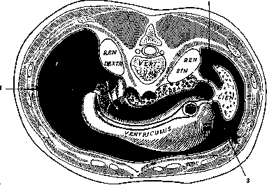
Figure 3. Course of the visceral layer of the peritoneum: 1 - lig. hepato-duodenale; 2 - lig. phrenico-lienale; 3 - lig. gastro-lienale; the arrow indicates the entry through the foramen of Winslow into the lesser sac (bursa omentalis) (diagram according to Corning). aponeurotic plate stretched between the lower edge of the chest and the upper pelvic bones and lined externally by a layer of fat and skin, and internally by the preperitoneal tissue and peritoneum. Depending on the ratio of the dimensions of the chest and pelvis, the shape of the space formed by the abdominal wall is very inconstant: with a wide chest and a narrow pelvis, the abdomen resembles a cone with its base facing upward, and with a narrow chest and a wide pelvis, the reverse (see Figure 1). Each
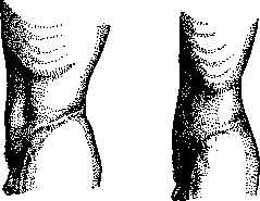
Figure 1. Two profile shapes of the abdomen in a man.
of the forms is characteristic of specific constitutional types: the first to the hypersthenic, or muscular, the second to the asthenic. Depending on age, the profile relief of the anterior abdominal wall undergoes strong changes, which boil down mainly to the retraction of the supraumbilical region and the uniform protrusion of the infraumbilical region toward old age (see Figure 2).-In the lower medial sections of the anterior abdominal wall, on each side, there is an opening through which the spermatic cord passes in men, and the round ligament of the uterus in women (inguinal
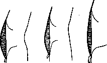
8 years
11 years
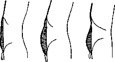
25 years
38 years
Figure 2. Profile of the anterior abdominal wall at different ages. canal), and along the midline, approximately at the middle of the distance between the navel and the xiphoid process, a third opening, which overgrows at birth into a scar—the navel (umbilicus). These areas, in view of their complexity and important practical significance, require special description.-The muscles of the anterolateral section of the abdominal wall include the external and internal oblique abdominal muscles, transversus abdominis, rectus abdominis, and pyramidalis; the last two muscles are located along the midline along the long axis of the body, while the first three have an oblique, mutually intersecting direction. By virtue of this, the nature and composition of the layers of the abdominal wall differ in different sections. Near the midline, the layers are as follows: 1. Skin, covered with hair below the navel. 2. Subcutaneous adipose tissue containing many veins that hypertrophy during congestion in the abdominal cavity. 3. Superficial fascia, which gives off from itself, together with the linea alba, a ligament to the root of the penis (superficial suspensory ligament of the penis or clitoris). 4. Anterior wall of the rectus sheath, formed by the tendinous aponeurosis of the external oblique abdominal muscle and the anterior lamina of the internal oblique abdominal muscle.
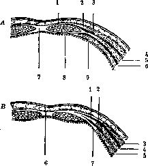
Figure 3. Sheath of the rectus abdominis muscle above (A) and below (B) the umbilicus. A: 1 - vagina of rectus muscle (anterior lamina); 2 - tendon of external oblique abdominal muscle; 3 - tendon of internal oblique abdominal muscle; 4 - external oblique abdominal muscle; 5 - internal oblique abdominal muscle; 6 - transverse abdominal muscle; 7 - linea alba; 8 - vagina of rectus muscle (posterior lamina); 9 - transversalis fascia. B: 1 - tendon of internal oblique abdominal muscle; 2 - tendon of external oblique abdominal muscle; 3 - external oblique abdominal muscle; 4 - internal oblique abdominal muscle; 5 - transverse abdominal muscle; 6 - linea alba; 7 - tendon of transversus abdominis. At the medial edge of the muscle, it fuses with the posterior lamina, and both of them, together with the sheath of the rectus abdominis muscle of the other side, form the so-called linea alba; right at the symphysis, within the thickness of the anterior wall, or rather in its duplication, lies the pyramidalis muscle, which originates from the pubic symphysis and attaches to the linea alba; sometimes this muscle is absent. 5. The rectus abdominis muscle is a long, ribbon-like muscle, four finger-breadths wide; it originates from the anterior surface of the cartilages of the 5th, 6th, and 7th ribs and the xiphoid process of the sternum and inserts into the pubic symphysis. It has several transversely disposed tendinous intersections (inscriptiones tendineae) embedded within it, which allow it to contract in parts and represent a trace of rudimentary ribs; there are usually four intersections: two above the umbilicus, one at the level of the umbilicus, and the fourth below it; the latter is frequently absent. The tendinous intersection serves as the site of entry of blood vessels into the muscle and is fused with the anterior lamina of the sheath; throughout the rest of its extent, the sheath is freely separated from the muscle. 6. The posterior wall of the rectus sheath is formed differently above and below the umbilicus (see Figure 3). Above the umbilicus, its composition includes the posterior lamina of the aponeurosis of the internal oblique abdominal muscle and the transverse abdominal muscle; below the umbilicus, or rather below the so-called linea semicircularis of Douglas, the tendons of the oblique muscles do not take part in the formation of the posterior wall of the rectus sheath; they pass anteriorly, and the sheath is formed by the fascial lamina of the following layer. 7. The transversalis fascia lines the deep internal surface of all the abdominal muscles, passing posteriorly to the renal region and superiorly to the diaphragm; behind the rectus muscle, it is somewhat thickened. Blood vessels run along the posterior wall of the rectus sheath, represented by the inferior epigastric artery (from the external iliac artery) and the superior epigastric artery (the terminal branch of the internal mammary artery), which anastomose with each other at the level of the umbilicus; they serve to restore collateral circulation when the common iliac artery is occluded; in the scattered type, their trunk is replaced by several finer branches; the terminal branches of the intercostal nerves (7th–12th) also run within the rectus sheath; having an almost horizontal course until then, they abruptly change their direction here and run almost vertically; therefore, incisions along the fibers of the rectus muscle damage them only to an insignificant degree. 8. The preperitoneal adipose tissue, loosely connected with the transversalis fascia and more closely with the peritoneum, is quite abundant in the region of the muscles, especially in the epigastrium; lower down it thins out, but above the symphysis, where the peritoneum of the anterior abdominal wall passes onto the urinary bladder, its amount increases. In the posterior parts of the abdomen, the preperitoneal tissue turns into thick layers enveloping the kidney and descending along the colon (paranephron, paracolon). 9. Peritoneum (see). The anterolateral region of the abdominal wall has the following differences compared to that described. The superficial fascia consists of two leaves: a superficial one, passing uninterruptedly from the abdomen to the thigh, and a deep one, attaching to the so-called Poupart's ligament. By virtue of this, accumulations of blood, gases (emphysema), as well as lipomas do not spread to the thigh if they have developed beneath the deep lamina, and pass lower if they are located between its two layers. The superficial vessels of the abdominal wall also run here: the superficial epigastric artery, superficial circumflex iliac artery (from the femoral artery), and Haller's abdominal vein (emptying into the saphenous vein). The muscular layer consists of three muscles clothed with a perimysium, which sometimes assumes the character of a fascia. The external oblique abdominal muscle originates by digitations, interleaving with the origin of the serratus anterior muscle, from the last eight ribs and inserts partly into the external lip of the iliac crest, while its main mass passes into a broad flat tendon approaching the edge of the rectus abdominis muscle and participating in the formation of the anterior wall of its sheath. The lower part of the aponeurosis, attaching to the anterior superior iliac spine, is thrown over the anterior notch of the pelvic bone in the form of the so-called Poupart's ligament (or Fallopius ligament, or crural arch) and attaches to the pubic bone by two bundles: lateral (inferior crus) to the pubic tubercle, and medial (superior crus) to the pubic symphysis; a narrow cleft remains between the bundles, reinforced from above by transversely running fibers of the so-called intercrural fascia. Through this cleft passes the spermatic cord in men, and the round ligament of the uterus in women. The cleft, bearing the name of the external inguinal ring, is usually closed in newborns, but later, especially in individuals constitutionally predisposed to hernia (asthenic habitus), the inguinal ring opens. Similarly, the bundles of the aponeurosis of the external oblique abdominal muscle, which in newborns and children are closely adjacent to each other and do not yield to splitting, become split, clefts and even apparent ruptures arise, closed by a thin leaf of fascia; sometimes the cleft between the superior and inferior crura extends almost to the anterior superior iliac spine, and the intercrural fascia covering it forms a thin transparent lamina. The direction of the fibers of the external oblique abdominal muscle corresponds to the course of the ribs. The internal oblique abdominal muscle originates from the intermediate line of the iliac crest, the two outer thirds of Poupart's ligament, and attaches partly to the last three ribs, partly passing into an aponeurosis. The latter, upon approaching the edge of the rectus muscle, divides above the umbilicus into two laminae forming the anterior and posterior walls of the sheath of this muscle; below the umbilicus, the aponeurosis of the internal oblique abdominal muscle passes into the anterior wall of the rectus sheath. The direction of the fibers of the internal oblique abdominal muscle corresponds to the course of the internal intercostal muscles, and in the region of Poupart's ligament it is almost horizontal. The transversalis abdominis muscle originates from the six lower ribs, the deep leaf of the lumbodorsal fascia, the inner lip of the iliac crest, and the outer third of Poupart's ligament and, having a horizontal course of fibers, passes along a particular line convex in the lateral direction (Spigelian semilunar line) into a broad tendon directed toward the outer edge of the rectus muscle. It participates in the formation of the posterior wall of the rectus sheath above the umbilicus and the anterior wall below it. In the layer between it and the internal oblique abdominal muscle run the vessels and nerves of the region. These include the last six intercostal arteries and five lumbar arteries, which give off lateral branches and, within the rectus sheath, anastomose with the superior and inferior epigastric arteries and the circumflex iliac artery. The nerves—the last 5 intercostals, iliohypogastric, and ilioinguinal—also run segmentally; along the axillary line they give off the lateral perforating branch, and within the rectus sheath the anterior perforating branch, innervating the skin near the midline. According to Lavochkin's studies, the course and branching of the nerves can be extremely diverse. In the so-called main type, the peripheral branches arise late, and the nerves do not anastomose with each other; in the scattered type, peripheral branches arise at the site of origin of the nerves and connect with each other (the same applies to the iliohypogastric and ilioinguinal nerves) by numerous anastomoses, so that a kind of extensive peripheral plexus is formed; its anastomoses capture the branches of three adjacent segments. If one examines the abdominal wall from the side of the peritoneum, then here
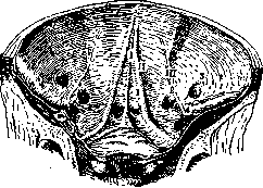
Figure 4. Internal surface of the anterior abdominal wall: I - middle vesico-umbilical fold; II - lateral vesico-umbilical fold; III - epigastric fold; 1 - supravesical fossa; 2 - medial inguinal fossa; 3 - lateral inguinal fossa; 4 - femoral fossa.
three abdominal folds located below the umbilicus catch the eye; the middle one - plica vesico-umbilicalis medialis - covers the ligament - ligam. vesico-umb. med. (remainder of the urachus) and extends from the umbilicus to the apex of the bladder, the lateral ones - plicae vesico-umbil. laterales - cover another ligament (from the lateral walls of the bladder to the umbilicus) - the obliterated a. umbilicalis (branch of a. iliacae int.). Between these two folds and the bladder, there is a fossa on each side (fovea supravesicalis). More laterally - approximately above the middle of Poupart's ligament - there is a depression into which the components of the spermatic cord enter from various sides (annulus inguinalis abdominalis); the peritoneum slightly enters the fossa [see figure 4 and separate table (vol. III, art. 99-100, fig. 1)]. More medially to the inguinal ring, the plica epigastrica rises from Poupart's ligament to the umbilicus, under which the a. epigastrica passes. It serves as the boundary between two fossae: the one just described and the fovea inguinalis medialis (the site of entry of the so-called direct hernia), located between it and the plica vesico-umb. lat.; fovea ing. lateralis is the site of entry of the so-called oblique hernia. Above the umbilicus, folds of the peritoneum (processus falciformis) also run towards the left longitudinal sulcus of the liver, and along its lower edge is the ligament (lig. teres hepatis), which represents the obliterated vena umbilicalis. The posterior wall of the abdomen, usually described as the lumbar region, is bounded by the XII rib, the spine, and the crista iliaca. Behind the arches and transverse processes of the vertebrae lies a powerful muscle, 4 fingerbreadths wide - m. erector trunci, which constitutes a complex of muscles that extend and rotate the spine. Outwardly are located the muscles of the lumbar region proper. In addition to the skin with subcutaneous tissue and superficial fascia, there is m. latissimus dorsi, which has an extensive origin from the spinous processes of the lower three thoracic, all lumbar, and all sacral vertebrae, the lower four ribs, and the posterior half of the cristae iliacae; the lumbar part begins from the posterior leaf of the so-called fascia lumbo-dorsalis, which covers m. erector trunci and divides into two leaves at the outer edge of the muscle. The muscle is covered by its own fascia. Deeper lie m. obl. abd. int. and m. serratus post. inf., beginning from the superficial leaf of the fascia lumbo-dorsalis in the region of the XI and XII thoracic and I and II lumbar vertebrae; it attaches to the lower four ribs. Deeper lies the origin of m. transversus abdominis, covered by a leaf of fascia transversae, and, finally, m. quadratus lumborum. This latter muscle has a complex origin: on the iliac crest, on the lig. ilio-lumbale, and on the transverse processes of the III, IV, and V lumbar vertebrae, and insertion on the XII rib and the transverse processes of the I and II lumbar vertebrae; it is covered by its own fascia. Deeper lie the perirenal fat (capsula adiposa renis), the kidney, and the peritoneum. Between the lower-outer edge of m. latissimus dorsi, on the one hand, the edge of Mm. Sk
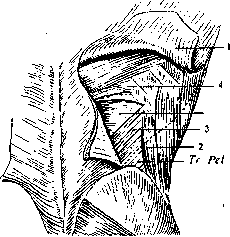
Figure 5. Tr. Pet. - Petit's triangle; Tr. Gr. - Grynfeltt's triangle; 1 - cut across and reflected m. latissimus dorsi; 2 - m. obliquus abd. ext.; 3 - m. oblq. abd. int.; 4 - m. serratus post. inf.; the dashed line indicates the outer edge of m. latissimus dorsi (from a specimen of the Inst. of Oper. Surg. of the 2nd Moscow State Univ.). m. obl. abd. int. - on the other hand, and the iliac crest in 77% of cases, a special triangle described by Petit in 1738 is expressed, which serves as the site of penetration of lumbar hernias. The base of the triangle can reach 2-4 cm, and the height - 3-7 cm. At the bottom of the triangle lies a thin layer: m. obl. abd. int. and fascia lumbo-dorsalis (see figure 5). The second weak spot of the lumbar region is Grynfeltt's triangle (Grynfeltt). It is formed by the edge of m. obl. abd. int. from below, the longitudinal muscles of the spine and m. quadratus lumborum from the inside, and m. serratus post. inf. and the XII rib from above. The shape and size of the triangle depend (according to Barbe) on the length of the XII rib: with a long XII rib, the triangle assumes quadrangular outlines. It is found in more than half of cases. In Grange's statistics (45 cases), lumbar hernia most often appears through Petit's triangle, less often through Grynfeltt's triangle, and even less often through openings in the aponeuroses and muscles that serve as the passage for vessels and nerves. Pathology of the abdominal wall, excluding the linea alba, umbilicus, and inguinal region, reduces to developmental defects, traumatic injuries, inflammatory diseases, and tumors. Among developmental defects of practical significance, underdevelopment of the musculature should be noted. According to Stumme, underdevelopment of m. obl. int. and transversi is most common, and less common - m. recti, m. latissimi dorsi, and quadrati lumborum. At the site of the missing muscles, a protrusion of the viscera of the hernia ventralis congenita type is formed. If the defect is not too large, attempts are made to close it first by mechanical devices (bandages, straps) or operatively - by transplantation of aponeuroses and muscles. Anomalies of the opposite character, with excessive tissue growth of the foetus in foetu type, are not very rare. They are found most often in the lumbar region in the form of appendages having the character of an extra limb; they are subject to surgical removal. Traumatic injuries of the abdominal wall are closed and open. The former arise most often as a result of blunt force and may, depending on the force of the impact, be accompanied by damage to internal organs (ruptures of the liver, spleen, intestines). But even in the absence of visible damage to internal organs, they are dangerous due to the possibility of shock in the event that the point of application of force is such that a contusion of the large nerve plexuses of the abdominal cavity occurs. There may be no visible changes in this case. A special variety of closed injuries is represented by muscle ruptures that occur during an impact at the moment of muscle contraction. The defect formed as a result of the rupture leads to the formation of a hernia. According to Thiem, out of 25 cases of ruptures, m. rectus suffered in 16, the oblique muscles in 4, and the lumbar muscles in 3. Open injuries or wounds of the abdominal wall are distinguished, by the nature of the wounding instrument, as stab, incised, bitten, gunshot, etc. (see Wounds). Incisions of the abdominal wall. The abdominal wall serves as the most frequent object of surgical intervention, since access to various organs of the abdominal cavity is opened through it. Since ancient times, the question of the technique of abdominal wall incisions has aroused much controversy and doubt, since the surgeon has to simultaneously comply with two requirements: to obtain sufficient access to the operated organ and to observe all conditions preventing the occurrence of a postoperative hernia. Over the past two decades, the viewpoint of relative conservatism has taken hold, taking into account the role and physiology of the abdominal press and, especially, the significance of nerves. Its best exponent is Sprengel, who proposed a number of incisions for various regions that are sufficient in terms of access size and cause little trauma. A mandatory condition for Sprengel's so-called "physiological" incisions is: 1) preservation of the nerves, 2) separation of the muscles along the fibers, thereby achieving the creation of a complex scar crossing in different directions, and 3) careful adaptation of the edges of the peritoneal incision, pieces of which, falling into the suture line, can contribute to the occurrence of a hernia.
A. Sozon-Yaroshevich. Abdominal muscles. Pathology. With bilateral paralysis of the abdominal muscles, difficulties are detected in bending the trunk forward (e.g., when getting up from a horizontal position), difficulty in urination and defecation, since these acts require the action of the abdominal press, and difficulty in forced expiration, necessary for a loud cry, cough, singing, etc. When standing, lordosis is noted with a displacement of the pelvis backwards - the plumb line dropped from the upper part of the thoracic vertebrae falls on the middle part of the sacrum. The abdomen protrudes forward, bulges, and this bulging increases when straining. Attempts to tense the abdominal muscles do not lead to success: the muscles remain flaccid, which can be verified both by palpation and simple inspection - their relief does not change, their contours do not stand out more clearly, as happens normally under tension. With unilateral paralysis of the abdominal muscles, displacement of the umbilicus to the healthy side and a decrease in muscle elasticity compared to the healthy side (palpation) are characteristic. When the lower parts of the abdominal muscles are damaged, the umbilicus is displaced upwards. If this damage is unilateral, the umbilicus is displaced upwards and to the healthy side. The study of abdominal reflexes, as well as the study of electrical excitability (spasms of the abdominal muscles, see Hyperkinesia, as well as Hysteria), significantly helps in clarifying the localization of partial paralyses. The abdominal muscles, especially the rectus muscles, are often the site of so-called waxy degeneration (for example, in typhus), as well as hematomas.
Related articles
Mentioned in
- Abdomen (a concept encompassing the representation of)
- Abdominal Cavity
- Abdominal Press
- Abdominal Reflexes
- Abdominoscopy
- Aeroperitoneum
- Asynclitic Presentation
- Ballottement
- Caput Medusae
- Cecum
- Cesarean Section
- Clitoris
- Colica Mucosa
- Colpopexy
- Decompression Sickness
- Dockers
- Douglas Space
- Duodenum
- Embalming of Corpses
- Embryotomy
Cite this page
“Abdominal Wall.” Soviet Medical Encyclopedia. English translation of Bolshaya Meditsinskaya Entsiklopediya, 1st ed. (Moscow, 1928–1936), ed. N. A. Semashko. https://sovietmedicalencyclopedia.pages.dev/article/abdominal-wall/