Clitoris
Historical document, translated for reference. It reflects medical knowledge of the 1920s–30s and is not medical advice.
Summary
This article provides a detailed anatomical and physiological description of the clitoris, highlighting its embryonic development and homology to the male penis. It also covers the organ's erectile function, its vascular and muscular structure, and mentions historical cultural practices of clitoral excision.
Encyclopedia article (1928–1936)
CLITORIS, pokhotnik (clitoris), is an organ of the female external genitalia. The clitoris is located in the upper angle of the pudendal cleft, directly under the pubis, protruding here with its glans in the form of a small, blunt papilla (Fig. 1). The clitoris begins with two
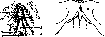
Figure 1.
Figure 2.
Figure 1. 1-clitoris; 2-orif. ext. ureth.; 3-labia minora; 4-vagina; 5-praep. clitorid. Figure 2. 1-cavernous bodies of the clitoris; 2-glans clitorid.; 3-lig. suspensor. clitorid. [The clitoris begins with] cylindrical crura, about 1 cm thick, extending from the ascending ramus of the ischium and the descending ramus of the pubis on both sides (Fig. 2 and Vol. III, p. 42). Both crura, covered by the fascia of the perineum, before reaching the level of the pubic arch, merge to form the body of the clitoris, which, under the pubic arch, bends downward and ends in the glans. Between the pubic arch and the point of the bend of the clitoris pass its nerves and vessels. From its convex part, a fibrous cord—the suspensory ligament of the clitoris—extends to the pubis. Under the concave part of the clitoris is located the venous plexus (Kobelt's), which communicates with the veins of the bulbs and the vestibule and with the cavernous tissue of the body and glans. From above and below, the crura of the labia minora approach the glans of the clitoris, forming its prepuce and frenulum (Fig. 1). Developing primarily from the genital tubercle, the clitoris is embryologically and by the nature of the structure of its tissues an organ analogous to the male penis, but without a urethra. The clitoris consists of cavernous tissue, which, with the exception of the glans, is enclosed in a connective tissue sheath. This sheath forms a septum along the midline, which is interrupted in places. The free surface of the glans is covered with thin, mobile skin, resembling the mucous membrane in color. At the site of the fold of the prepuce, where it transitions to the glans of the clitoris, convoluted paired glands are observed, reaching the cavernous bodies on both sides of the septum. The skin of the glans has a strongly developed papillary layer, rich in small vascular branches and heterogeneous nerve endings (Pacinian and Meissner corpuscles, Krause end bulbs, genital corpuscles). Genital corpuscles consist of clusters of small cells to which the finest nerve branches approach; specific sexual sensations are attributed to them. The fibrous sheath enveloping the crura and body consists of a connective tissue stroma with elastic fibers. The superficial layer of the sheath contains a large number of nerves running in regular rows and giving off numerous branches to the skin of the clitoris. The prepuce of the clitoris, being part of the labia minora, is constructed according to their pattern and contains sebaceous and sweat glands on its inner surface and behind the glans. The cavernous tissue of the clitoris is formed by the insertion of numerous trabeculae originating from the fibrous sheaths of the crura and the median septum (Fig. 3). The trabeculae consist of elastic and muscle fibers, which, by intertwining, form small, interconnected caverns washed by blood entering them directly from the afferent arteries (deep arteries of the clitoris). Blood returns from the caverns of the body via veins (vv. emissariae), which form plexuses and drain into the vein running along the dorsal surface of the clitoris (v. dorsalis). Veins encircling the clitoris and communicating with the veins of the bulb of the vestibule of the vagina also drain here. The venous blood of the cavernous bodies of the crura of the clitoris joins into its deep veins, which go into the deep perineal plexus, and from there into the pudendal vein (v. pudenda communis). The clitoris receives arterial blood from the pudendal artery (art. puden
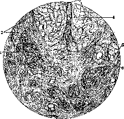
Figure 3. 1-nerve bundles; 2-fibrous tissue; 3-cavernous bodies of the clitoris; 4-septum between the cavernous bodies; 5-genital corpuscles; 6-blood vessel. da communis), which gives off two terminal branches to it—the dorsal and deep arteries of the clitoris (aa. dorsalis et profunda clitoridis). The muscular apparatus of the clitoris consists of three pairs of muscles, extending predominantly along the course of its crura. The ischiocavernosus muscle (m. ischiocavernosus) runs along the lower surface of the crus and, gradually moving outward, partially transitions to the dorsal side of the body, where it merges with the muscle of the same name on the other side. By contracting, they compress the dorsal vein of the clitoris and also press the crura of the clitoris against the ischial tuberosity, thereby hindering the outflow of venous blood and promoting erection of the organ. The deep transverse perineal muscles (mm. transversi perinaei prof.) embrace the vagina with their fibers on both sides and join along the midline of the perineum into a tendinous junction. Upon contraction of this pair, the deep perineal venous plexus, which drains blood from the crura of the clitoris into the pudendal veins, is compressed. The muscle compressing the pudendal cleft (m. constrictor cunni) is a crossed continuation of the anal sphincter, running anteriorly along both sides of the vestibule of the vagina. Located outside the bulbs and Bartholin's glands, it gives off fibers to them along the way and attaches to the posterior wall of the urethra and the cavernous bodies of the clitoris. Contraction of these muscles delays the outflow of venous blood, pulls the glans of the clitoris somewhat closer to the entrance to the vagina, reduces the cavity of the latter, and squeezes the contents out of Bartholin's gland. Depending on the degree of filling of the cavernous bodies and vessels with blood, the volume and consistency of the clitoris change. An increase in the influx of arterial blood and a weakening of the venous outflow during sexual arousal in a woman leads to swelling of the clitoris and, by causing contraction of the muscle and elastic fibers, makes its consistency firm (erection). With the subsidence of sexual arousal, muscle contraction ceases, blood outflow increases, and the clitoris becomes soft and flaccid. It should be noted that during the period of clitoral erection, the amount of venous blood flowing out of it, despite the difficulty of outflow, is several times (8) greater than at rest. This circumstance shows that during erection there is not only an overfilling with blood but also an acceleration of blood circulation. The clitoris moderately swells during pregnancy, primarily due to stasis in the lymphatic vessels. In the climacteric period, its tissues undergo atrophy, similar to other parts of the external genitalia. Observations (far from accurate, however) by some anatomists give reason to assume that the size of the clitoris depends not only on the constitution and other individual characteristics of the woman but also on race. According to these observations, the clitoris reaches larger sizes in southern women than in representatives of the temperate zone, and it is very weakly developed in northern women. In the relative hypertrophy of the clitoris in southern women, they try to find the reason for the custom of bloody excision of the clitoris, which took place in ancient Egypt and has been preserved to the present time among Abyssinians and some other southern tribes. Among some peoples, this kind of peculiar circumcision is performed only by excision of the glans of the clitoris with its prepuce; among others, the labia minora are also excised together with it. Followers of the Skoptsy sect are subjected to identical operations. The sectarians give the name "first purity" or "great seal" to the operations of excising the clitoris, labia minora, and the upper part of the labia majora.

clitoris;
Figure 4. 1, 2 - urethra; 3 - vaginal entrance; 4 - ovary. Developmental defects. Rudimentary development and even complete aplasia of the clitoris, both as an independent defect and in connection with developmental defects of other parts of the external genital apparatus, is observed not infrequently. No less frequently observed is the opposite defect - congenital hypertrophy of the clitoris, whereby its size sometimes reaches significant dimensions and it protrudes from the pudendal cleft. In connection with other developmental defects of the vulva (hypospadias, descent of the gonads into the labia majora, etc.), hypertrophy of the clitoris can present difficulties in determining the sex of an individual (Fig. 4). Hypertrophic phenomena of the clitoris are noted both in girls and in adult women with certain tumors of the endocrine glands, especially the adrenal glands (see Hirsutism). See also Hermaphroditism. Significantly rarer is the duplication of the clitoris, or rather, incomplete fusion of its cavernous bodies. Usually, this defect is combined with so-called female epispadias, i.e., splitting of the urethra. - D i s e a s e s. Inflammatory processes limited only to the region of the clitoris are observed as an exception. Usually, acute inflammations of the clitoris represent a partial manifestation of general vulvitis, most often of infectious origin. The edema of the tissues observed in such cases, spreading to the prepuce of the clitoris, sometimes causes a picture similar to phimosis. Chronic inflammatory processes of the vulva on the basis of irritation during masturbation, lupus, pruritus, ulcus rodens, etc., give corresponding changes in the clitoris insofar as it is affected by the disease. Sometimes such processes lead to elephantiasis. More often, the clitoris serves as the starting point for the formation of tumors, both benign (fibroma, elephantiasis, angioma, etc.) and malignant (cancer, sarcoma). The most frequent and practically important of the tumors of the clitoris is cancer, both primary and metastatic. Primary cancer is usually observed at a later age (55-70 years) and initially has the appearance of a small nodular tumor; subsequently, it grows rapidly, and ulceration and decay appear. Upon contact of the clitoris tumor with the labia, new nodules may form on the latter (autoinoculation). In addition to the sequential involvement of the superficial and deep inguinal glands, characteristic of cancer of the clitoris is the development of metastases in the pubic lymph glands, lying in the amount of 2-3 near the pubic symphysis. The infiltration gradually spreads to the paraurethral and paravaginal tissue; in this case, the pelvic glands are often already affected. The prognosis is always doubtful, since recurrences, even after radical surgery, are sometimes observed after five years or more. Treatment is exclusively operative, with subsequent application of radiant energy. In inoperable cases, X-rays, radium, and mesothorium are the only appropriate measures. In
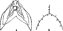
Figure 5. Scheme of the incision (dotted line) for extirpation of the clitoris: a - incision for extirpation; b - wound sutured with interrupted sutures. (According to Gubarev.)
In the operation for cancer of the clitoris, the inguinal and suprapubic glands are first enucleated, then the entire clitoris with the surrounding parts is excised by means of two semilunar incisions (Figure 5).
I. Sudakov. CLOACA of vertebrates represents the posterior section of the intestine into which the urogenital ducts empty. A cloaca is present in cartilaginous and lungfish, as well as in all terrestrial vertebrates with the exception of viviparous mammals. In amphibians, in connection with the abdominal wall of the cloaca, a urinary bladder develops, which in higher terrestrial vertebrates acquires the significance of an extremely important embryonic organ and is known as the allantois. Partly at the expense of the stalk of the allantois, the definitive urinary bladder of reptiles, birds, and mammals subsequently develops. The trunk musculature on the sides of the cloacal opening forms a ring-shaped sphincter muscle (m. sphincter cloacae), and the urinary bladder in amphibians and reptiles retains its direct connection with the cloaca, opening into it from the abdominal side, so that waste products also enter the urinary bladder only through the mediation of the cloaca. In snakes, crocodiles, and birds, the urinary bladder is reduced. In connection with the wall of the cloaca in reptiles and some birds, various kinds of copulatory organs develop, reaching particularly significant development in mammals. With the formation of these organs, it is subdivided from front to back into an abdominal section - the urogenital sinus - and a dorsal section, which serves as a continuation of the rectum. A short cloaca is still present in lower, monotreme mammals
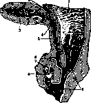
Figure 1. Right half of the cloaca and adjacent parts of a young echidna: 1 - rectum; 2 - coelom (body cavity); 3 - urinary bladder; 4 - papilla; 5 - canalis urogenitalis; 6 - vas deferens; 7 - preputial sac; 8 - phallus; 9 - ectodermal cloaca. (Fig. 1). In marsupials, some rodents, and insectivores, the openings of the urogenital sinus and the anus still lie very close to each other, surrounded by a common cloacal sphincter. In higher mammals, with the progressive development of the perineum, the distance between these parts increases. In viviparous mammals in the adult state, therefore, there is no cloaca, however, it always develops in the embryo. In embryos of higher vertebrates, the cloaca represents a significantly expanded endodermal space, in which the hindgut and allantois connect in front and the tailgut behind. From the dorsal side, the ducts of the primary kidney empty into the cloaca. The abdominal wall of the cloaca lies closely against the integuments, forming together with the latter the so-called "cloacal membrane," lying in front of the base of the tail. In front and from the sides, as the embryo develops, the "urorectal" fold increasingly cuts into the cloaca, separating the abdominal, urogenital section of the cloaca, leading further into the allantois, from the dorsal section, which constitutes the continuation of the rectum (Fig. 2). The abdominal section gives rise to the urinary bladder and the urogenital sinus. The ducts of the primary kidney, which initially empty into the cloaca, subsequently turn out to be connected with its abdominal section. The ureters, arising on these ducts, gradually shift from them and, due to the uneven growth of the cloacal walls, move further forward onto the walls of the urinary bladder itself. The mesenchyme, condensing around the cloacal membrane, raises the latter in the form of a tubercle ("cloacal tubercle"), in the middle of which the membrane itself takes on the appearance of a longitudinal cloacal groove. The anterior part of the cloacal tubercle then protrudes more strongly and gives rise to the copulatory organ (penis, clitoris), along the lower surface of which runs a groove, passing into the cloacal groove. A pair of small ridges on the sides of the latter subdivides it into an anterior longitudinal urogenital slit and a posterior transverse anal slit. The perineum developing in this way separates these openings more and more from each other. Around the anal pit, a special anal tubercle develops, and the pit breaks through into the rectum. The urogenital sinus stretches in the male into a long urogenital canal, and in the female gives a small section, the so-called vestibule of the vagina. The copulatory organ is initially located in the cloaca itself, surrounded by a fold of mucous membrane forming its vagina, and can be extended outward and retracted back into the cloaca (Fig. 1). In viviparous mammals, with the disappearance of the cloaca, a continuous communication is established between the urogenital sinus and the canal of the copulatory organ, so that the latter now also serves for the excretion of urine. Thus, the complete separation of the urogenital tracts from the posterior section of the intestine is completed. The above-mentioned normal development of the cloaca in mammals, ending in humans with the complete separation of the urogenital tracts from the posterior section of the intestine, called the rectum, is sometimes subject to a disturbance, which has the character of a developmental delay. Such a developmental delay of the cloaca in humans leads to various deformities of the perineal region, the lower section of the urogenital tracts, and the rectum; common to all these deformities is some degree of atresia of the rectum. This primarily includes pure cases of rectal atresia, when the developmental defect is expressed only in some degree of underdevelopment of the rectum with the absence of communication of its lumen with the perineum (atresia ani, atresia recti); furthermore, there are cases of rectal atresia with the preservation of communication between it and the urinary or genital tracts. The preservation in a full-term fetus of a true cloaca, into which the urogenital tracts and the intestine open, belongs to very great rarities; more often it is a matter of the presence of communication of the atretic rectum with a correctly formed vagina (atresia ani vaginalis), urinary bladder (atresia ani vesicalis), or urethra (atresia ani urethralis) (see Rectum).
Related articles
Mentioned in
Cite this page
“Clitoris.” Soviet Medical Encyclopedia. English translation of Bolshaya Meditsinskaya Entsiklopediya, 1st ed. (Moscow, 1928–1936), ed. N. A. Semashko. https://sovietmedicalencyclopedia.pages.dev/article/clitoris/