Vulva
Historical document, translated for reference. It reflects medical knowledge of the 1920s–30s and is not medical advice.
Summary
This article from the 1928–1936 Great Medical Encyclopedia details the anatomy, development, vascular supply, innervation, and various pathological conditions of the vulva, including congenital malformations, circulatory disorders, inflammation, and dystrophies.
Encyclopedia article (1928–1936)
VULVA consists of the mons pubis, labia majora and minora, clitoris, and vestibule of the vagina (see Fig. 1). It is separated from the vagina by the hymen (hymen), and from the anus externally by the so-called perineum. Shortly before puberty, light, delicate hairs appear in certain areas of the external genitalia. Vegetation on the genital organs usually begins to appear in the middle part of the mons pubis and along the edges of the labia majora, and the earlier puberty occurs, the earlier hairiness develops. In some peoples of East Asia, the "childish" type of hair growth remains for life (lymphatic type of East Asia). In a sexually mature woman, the hairy part of the genital organs has the shape of a triangle, with an even horizontal border at the top. The male type of hair growth—a triangle pointing upward toward the navel—is found in our latitudes much less frequently (in 5%). Among southern women, especially brunettes, the male type of hair growth on the genital organs is apparently more common. The color of the hair on the pubis is usually darker than on other parts of the body, and graying occurs much later. Senile thinning of the hair on the genital organs occurs only in advanced age. At first glance at the external genitalia of a woman lying on her back with her legs apart and bent, the pubis (mons Veneris), covered with hair and rich in subcutaneous adipose tissue, is seen, from which two pillow-like folds, the so-called labia pudenda majora, extend downwards. These two skin folds are usually strongly pigmented and covered with hair on the outer surface. Posteriorly, the labia majora merge with each other by means of the posterior commissure (commissura labiorum posterior). When parting the labia majora, on their inner side, above the commissure, a thin transverse fold appears, the so-called frenulum (frenulum). The cleft formed between the labia majora is called the pudendal cleft (rima pudendi). In nulliparous women, the labia majora are so close to each other that only the crests of the labia minora are visible between them in front. In parous women, due to the rupture of the posterior commissure, as well as due to the loss of elasticity of the vaginal walls, the vulva gapes to such an extent that the introitus vaginae remains uncovered. Medially from the labia majora lie the labia minora (labia pudenda minora), thin skin folds, tender pink in virgins, devoid of hair and fat. The size of the labia minora varies depending on age, race, constitution, and the intensity of sexual life. Sometimes the labia minora, like the pudendum (clitoris), reach gigantic proportions ("Hottentot apron"). The anterior edge of each labium minus splits into two leaflets. The two lower leaflets, connecting with each other and with the lower side of the glans clitoridis, form the frenulum of the clitoris (frenulum clitoridis), and the two upper folds, connecting above the glans of the clitoris, form its prepuce (praeputium clitoridis). The sulcus between the praeputium and the glans of the clitoris is called the sulcus clitoridis. The labia minora, gradually decreasing posteriorly, merge with the inner surface of the latter approximately at the middle of the labia majora. Upon stretching of the labia and tension of the frenulum, near the posterior commissure, in front of it, one can see a boat-shaped depression, the so-called fossa navicularis. The external genitalia in various periods of a woman's life, depending on their functional activity, i.e., depending on the function of the ovary, represent a certain typical picture both in relation to the vegetation covering them, the development of adipose tissue, and in relation to pigmentation, size, and so on. If you part the labia minora, the so-called vestibule of the vagina, vestibulum vaginae, is revealed—an area bounded in front by the clitoris, behind by the frenulum of the posterior commissure, and on the sides by the labia minora. Into the vestibule open their external openings: the urethra (located on a special elevation of the anterior wall of the vagina—carina urethrae) and paraurethral ducts, the vagina bordered by the hymen, Bartholin's glands, and small vestibular glands. The vestibule is divided into two parts: the anterior urethral (introitus urethrae) and the posterior hymenal (introitus vaginae). The sulcus between the labia minora and the hymen is called the sulcus nympho-hymenalis. On both sides of the external opening of the urethra, there are two small depressions, the so-called paraurethral crypts, rudiments of Wolffian ducts. Between the external opening of the urethra, as well as in the region of the sulcus nympho-hymenalis and fossae navicularis, more often in children than in adults, there are the so-called glandulae vestibulares minores, lined with cylindrical epithelium and producing mucus; they, just like the vagina and Bartholin's glands, open into the hymenal part of the vestibule. In the vicinity of the urethral opening, in addition, there is a number of blind tracts, so-called lacunae, lined with cylindrical or transitional epithelium (on Skene's lacunae—see Urethra). The external genitalia are supplied with arterial blood through the art. pudenda communis s. interna (a branch of a. hypogastricae), through a. pudenda externa (a branch of a. femoralis), and, finally, through a. spermatica externa (a branch of a. epigastricae int.). Venous blood is removed from the external genitalia in three directions: 1) through v. dorsalis clitoridis into the plexus vesicalis behind the pubic symphysis and then into v. pudenda interna; here too, into the internal pudendal vein, through v. haemorrhoidalis, venous blood pours out from the bulbus vestibuli, from the labia, and from the perineum; 2) through v. pudenda ext. into the vena saphena magna and 3) through v. obturatoria. Lymphatic vessels of the external genitalia gather into lymphoglandulae inguinales superficiales, lying partly above, partly below Poupart's ligament. The nerves of the external genitalia are branches of the nervi pudendi communis (from the sacral plexus) and nervi genito-cruralis. Diseases of the vulva. A. Developmental defects. The most important developmental defects of the external genitalia (according to Klebs' scheme) are false male and false female hermaphroditism (see), as well as atresias (atresia ani vulvaris s. vestibularis). B. Circulatory disorders. 1. Edema of the vulva. The loose tissue of the external genitalia and the abundance of cavernous tissue contribute to the fact that stagnation phenomena of a general or local nature very quickly lead to edema of the vulva. Pregnancy and the postpartum period easily lead to stagnation in the venous vessels and edema of the vulva. The formation of vulvar edema in toxicosis of pregnancy is well known. Treatment of vulvar edema comes down to therapy of the underlying disease. Scarifications used in these cases can lead to infection and gangrene. 2. Varicose veins of the external genitalia—see Varicose veins in pregnancy. 3. Vulvar hematoma (see Figure 2) is most often observed intra et post partum both after instrumental delivery and after spontaneous labor. Much less frequently, injuries serve as the cause of vulvar hematoma. Hemorrhage can take on fairly large dimensions: sometimes the blood tumor reaches the size of a child's head and spreads from the pubis to the perineum and even to the anus. Vulvar hematomas usually do not spread deep down because there is a barrier in the form of the diaphragma urogenitale. Patients usually complain of severe pain and shooting in the region of the bladder and rectum. Treatment comes down to rest and compresses. Incision should be resorted to only in extreme cases, keeping in mind subsequent hemorrhage. C. Inflammatory processes, see Vulvitis. D. Hyperkeratoses, see Ichthyosis vulvae and Leukoplakia vulvae. E. Neuro-trophic diseases of the vulva. 1. Pruritus vulvae (vulvitis pruriginosus Sanger's), see Pruritus. 2. Kraurosis vulvae (see Figure 3), kraurosis, a rare disease of the external genitalia, is the final outcome of vulvitis; it consists in the shriveling and atrophy of the labia minora and majora; it is more often observed in menopause and old age; it is characterized by kraurosis with sharply expressed itching.
Author signatures: M. Kushnir
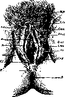

Figure 2. Hematoma of the vulva (after Veit). Tuberculosis, syphilis, and soft chancre of the vulva—see the respective entries. J. Fusion of the labia and cicatricial changes in the region of the external genitalia. 1. Fusion of the labia majora or minora (conglutinatio labiorum; see Figure 4) is encountered almost exclusively in children; some authors (Rausch, Ring) regard fusion as a developmental defect or the result of intrauterine vulvitis (Bokai), while others consider the main etiological factor to be extrauterine vulvitis in children, predominantly of gonorrheal origin. It should be noted that childhood infections (diphtheria, scarlet fever, etc.) may play no less a role in fusion; Kushnir in rural practice even had to observe an entire "epidemic" of vulvar fusions on this basis. Fusion of the labia is usually incomplete, and urination occurs without difficulty. Treatment must be operative: the adhesions are easily severed using a scalpel along a grooved probe inserted. The application of several

Figure 3. Kraurosis vulvae (after Meyer-Rüegg).
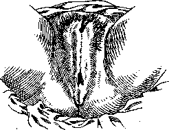
Figure 4. Fusion of the labia (after Veit).
catgut sutures is not always necessary. 2. Cicatricial changes. Scars both in the perineal region and in the region of the labia minora, clitoris, or urethra are treated surgically if necessary (plastic operations). 3. Hypertrophic processes of the external genitalia include elephantiasis (see Elephantiasis) of the vulva (see Figure 5)—a disease of the connective tissue elements of the skin of the genital organs, labia majora and minora, or clitoris; it can be unilateral or bilateral; tumor-like growths can reach very large sizes (weighing sometimes up to 10 kg) and can vary in their external appearance and consistency. Cracks and ulcerations frequently form on their surface. The etiology of the condition appears to be heterogeneous. Therapy is exclusively surgical. 5. Elephantiasis vulvae (after Meyer-Rüegg). II. Among benign tumors of connective tissue origin, fibromas (see Figure 6) of the external genitalia are encountered, originating from the cellular tissue of the labia majora, more rarely from the fasciae and connective tissue of the pelvis. The size of the tumor ranges from a pea to the head of an adult. These tumors usually sit either on a broad base, if they originate from deeply lying cellular tissue, or on a thin stalk (fibroma pendulum molluscum), if they originate from superficially lying cellular tissue. Diagnosis is easy. Hernia should be excluded. Therapy is surgical. Lipomas and myomas are less common. Myomas originate from the outer end of the round ligament of the uterus and very frequently contain cystic and adenomyomatous inclusions. Vascular tumors—angiomas and telangiectasias—are relatively rare tumors on the vulva. Benign epithelial tumors—papillomas, pointed condylomas, condylomata acuminata (see Vegetations, fig.)—warty weeping formations, are usually localized in groups on all parts of the vulva and vagina up to the vaginal portion. They are of two types: papillary growths on a thin stalk, having the appearance of cauliflower, reddish in color, and on a broad stalk, dry, grayish or yellowish in color. The cause of the appearance of condylomas is currently considered to be not only gonorrheal disease of the genital organs and rectum, but also other infections. Pregnancy may serve as a predisposing factor. Patients complain of burning, itching, and severe leukorrhea with a very foul odor. Although condylomas do not cause special pain, they can nevertheless hinder patients during walking, urination, and also during coitus. Sometimes patients complain of "bleeding," which most frequently occurs with the first form of condylomas. The best therapy is removal of the growths with scissors or a sharp spoon, as well as the application of X-rays. Shaving or cauterization is performed, in view of the painfulness of surgical removal of pointed condylomas, under local anesthesia or, in cases of large growths, under general anesthesia. For small growths, medical treatment with caustic powders is sometimes recommended (Pulv. frondosi sabinae, Aluminis usti aa 10.0, Cupri sulfurici 1.0. S. dusting powder),

Figure 6. Fibroma vulvae (Winter-Ruge).
ointments (Summitat. sabinae pulv., Vaselini aa 7.5, Ol. terebinthinae rectif. 6.0. S. ointment), lubrications with a 10% solution of salicylic acid, trichloroacetic acid, etc. Sweat gland adenomas of the labia majora have been described in only 17 cases. They are formations the size of a pea and are sometimes encountered on both sides. The tumor is benign, but in view of the possibility of malignant degeneration, its surgical removal is indicated. Of malignant tumors, sarcoma is a rather rare disease; it originates from the labia majora or minora, as well as from the clitoris. The diagnosis is usually established only with the aid of a microscope. Therapy must be surgical and as radical as possible. Melanoma is a relatively frequent tumor of the external genitalia (40%); therefore, a heavily pigmented tumor of the external genitalia should arouse the physician's suspicion. Diagnosis and therapy are the same as for sarcomas. Cancer of the external genitalia (see Figure 7) is encountered predominantly in elderly women and, according to Schottmüller's statistics, constitutes about 30% of all cancers of the female genital organs in general. According to the statistics of Rothschild and Labhardt, of all parts of the external genitalia, the labia majora are affected by cancer most frequently, namely in 30%. According to its histological structure, cancer of the external genitalia, with the exception of cancer of Bartholin's glands, is squamous cell carcinoma. Cancer of Bartholin's glands is usually of a glandular character (adenocarcinoma). As for the spread of vulvar cancer, Labhardt distinguishes three pathways. The first—per continuitatem—a) to the skin and mucous membranes, b) to neighboring hollow organs (vagina, rectum, bladder, urethra), c) to the connective tissue cellular tissue (fossa ischiorectalis, septum rectovaginale, paravaginal cellular tissue), d) to the pelvic bones. The second—spread via lymphatic pathways. 1st stage—superficial inguinal lymph glands; 2nd stage—deep inguinal lymph glands; 3rd stage—external iliac, hypogastric, and obturator lymph glands. The third pathway—metastases of vulvar cancer (to the lungs, heart, spleen, liver, and spine). Small tumors initially cause no complaints. Subsequently, itching, burning, pain, leukorrhea, and difficulty in urination may appear. Diagnosis is usually easy to make at first glance. In doubtful cases (ulcus vulvae chronicum, lues), microscopic examination decides the question. Advanced age, metastases, complications (cystitis, pyelitis, etc.), as well as recurrences make the prognosis doubtful. Therapy can be twofold: operative and radiant. According to Ederle's statistics, after the operation for the removal of vulvar cancer, the absence of recurrence for 5 years is observed in only 4.17% of all cases. In view of this, it is recommended, in addition to the cancer focus, to remove at least the inguinal glands on both sides. Stoeckel proposes removing not only the inguinal, but also the retroperitoneal glands. Seitz and Wintz developed a special method of roentgentherapy for vulvar cancer. Other authors obtained good results from the use of radium (Kehrer) or radium and X-rays (Warnekros). Baisch reports a good result from a combination of operative removal of the tumor and the use of radium and X-rays. Mixed tumors (teratomas) are encountered on the vulva extremely rarely. Atheromas are encountered on the vulva relatively rarely. Hydrocele muliebris—a cystic fluctuating swelling of the upper part of the labium majus—represents a protrusion into the tissue of the labium majus of the lower part of the vaginal process of the peritoneum, which has become segregated in the form of a cyst and contains a small amount of fluid. The possibility of the presence of a true hernia should be kept in mind. Therapy must be surgical with observance of all rules of herniotomy. Hernias of the vulva, see Hernia.
Related articles
Mentioned in
Cite this page
“Vulva.” Soviet Medical Encyclopedia. English translation of Bolshaya Meditsinskaya Entsiklopediya, 1st ed. (Moscow, 1928–1936), ed. N. A. Semashko. https://sovietmedicalencyclopedia.pages.dev/article/vulva/