Perineum
Historical document, translated for reference. It reflects medical knowledge of the 1920s–30s and is not medical advice.
Summary
This article from the 1928–1936 Great Medical Encyclopedia describes the anatomy, fascial layers, and musculature of the perineum in humans. It covers both the urogenital and recto-anal regions, detailing structures such as the superficial transverse perineal muscle, bulbocavernosus muscle, and ischiocavernosus muscle.
Encyclopedia article (1928–1936)
PERINEUM (perineum, s. perinaeum, s. regio perinealis), the space formed by the soft parts between the anus and the external genitalia; this space closes the pelvic cavity from below and constitutes the pelvic floor, diaphragma pelvis. In women, the perineum extends forward to the posterior commissure of the labia majora, while in men it extends further: to the scrotum and the root of the penis inclusive. The boundary of the perineum in a broader sense runs conventionally from the lower point of the symphysis in front to the tip of the coccyx behind, along the lower rami of the ischial and pubic bones and the sacrotuberous ligaments on the sides. Thus, in general, a rhombus figure is obtained. These soft parts consist of alternating muscular and fascial plates through which pass the rectum and urethra, and in women also the vagina. The human pelvic muscular floor, especially in connection with its upright position, plays an important role in retaining the viscera, which exert greater pressure on it than on the anterior abdominal wall. Since the perineum is intimately connected both genetically and topographically with the urogenital system, important sexual differences manifest in its structure. For practical purposes, the perineum is divided into two triangular regions: the anterior, or urogenital (regio urogenitalis), and the posterior, or rectal region (regio recto-analis). These two regions are conventionally separated by the so-called bi-ischial line (linea biischiadica). In the rectal region lies the anal part of the rectum and, on its sides, the ischiorectal fossae (fossae ischio-rectales); in the urogenital region in men is lodged the initial part of the urethra, and in women, in addition, the vagina. The male perineum is relatively longer and more complex than the female and is composed of the following layers: 1) skin with subcutaneous adipose tissue; 2) superficial, or subcutaneous fascia (fascia perinaei superficialis); 3) superficial aponeurosis of the perineum, or its proper fascia (fascia perinaei propria, s. aponeurosis ano-perinaealis); 4) the first muscular layer of the perineum consisting of the superficial transverse perineal muscle (musculus transversus perinaei superficialis), bulbocavernosus muscle (musculus bulbo-cavernosus), and ischiocavernosus muscle (musculus ischio-cavernosus); 5) the middle aponeurosis (in the anterior section of the perineum, the ligament of Carcassonne), or deep fascia of the perineum (fascia perinaei profunda); 6) the second (deep) muscular layer — the deep transverse perineal muscle (musculus transversus perinaei profundus, or Guthrie's muscle), the levator ani muscle (musculus levator ani, s. diaphragma pelvis propria, s. recta), the iliococcygeus or ischiococcygeus muscle (musculus ischio-coccygeus, s. musculus coccygeus), Wilson's muscle (musculus Wilson!); 7) the upper aponeurosis, or fascia of the perineum, otherwise the pelvic fascia (fascia endopelvina, s. fascia pelvis), which is also partially the obturator fascia; 8) the subperitoneal layer of cellular tissue; 9) the peritoneum. As a result of the development of the perineum from two later-fusing halves, a trace remains on the skin of the perineum, which has the appearance of a longitudinal, more or less pigmented line, the so-called perineal raphe (raphe perinaei). The latter extends from the scrotum and stretches all the way to the anus. The skin of the perineum is generally thin, especially along the edge of the anus, motionless, and difficult to separate from the underlying parts; it is marked by radially diverging folds, in the thickness of which a particularly large number of sebaceous glands are embedded. The superficial, or subcutaneous, fascia of the perineum can be more or less artificially divided into two plates: the outer one, which directly passes into the corresponding subcutaneous-fascial layer of neighboring regions, and the deep one, passing anteriorly into the dartos tunic of the scrotum and posteriorly into the fascia of the external sphincter of the rectum. Between these fascial leaves is a layer of adipose tissue, particularly powerful in the so-called ischiorectal fossae. The superficial aponeurosis, or proper fascia of the perineum, varies greatly in thickness and density, which has given rise to a different understanding of its individual leaves, its position and distribution, and a different order of division of this intermuscular connective tissue formation of the fascia propria type, i.e., it is broken down into as many leaves (coverings) as the muscles it entirely covers. Often this fascia is so thin, "velum-like," that it cannot be naturally separated from the proper subcutaneous fascia. It has the shape of a triangle corresponding to the contours of the urogenital region of the perineum; the wide base of this fascia coincides with the posterior edge of the superficial transverse perineal muscle (see below) and the anus, and its anterior narrow section is located at the root of the penis; on both sides of the pelvis, the fascia is fixed on the anterior lip of the ascending ramus of the ischium and the ascending ramus of the pubis. Thus, on both lateral sides, it does not enter the ischiorectal fossae, but, having covered from above and below the underlying superficial transverse perineal muscle from the side of its free posterior edge, it already terminates as a separate leaf and is fixed anteriorly on the urogenital diaphragm, i.e., on the next, deeper layer of fascias and muscles with which it fuses. Thus, a strong fascial demarcation from the ischiorectal fossa is ensured. Posteriorly, at the level of the posterior edge of the superficial transverse perineal muscle, the fascia, bending upward, passes directly into the underlying middle aponeurosis, or deep fascia of the perineum (see below); anteriorly and superiorly onto the scrotum and penis (fixed along the midline of the perineum on the raphe of the bulb of the penis and continuing into the scrotal septum), forming around it the fascia of the penis (fascia penis), where it ends at the head of the latter. In the anal region, the fascia of the external anal sphincter can also be attributed to it, into which it directly continues here. Thus, the superficial perineal fascia, merging in certain places with the deep fascia of the perineum following it, constitutes a peculiar sheath in which the superficial muscle of the perineum and the penis are enclosed; from below this sheath, as indicated, is closed at the level of the posterior edge of the superficial transverse perineal muscle, and from above and in front at the head of the penis. The first muscular layer of the perineum following the superficial perineal fascia and covered by it has the shape of a triangle with its base facing backward and its apex forward and upward (the ischio-bulbar triangle — trigonum ischio-bulbosum). This layer includes three muscles: the already mentioned superficial transverse perineal muscle, the bulbocavernosus and ischiocavernosus muscles (Figure 1). The superficial transverse perineal muscle lies at the base of the specified triangle; along its outer edge is located the ischiocavernosus muscle, and along its inner edge the bulbocavernosus muscle. The free middle section of the triangle is filled with adipose tissue. The upper (outer) fascial wall of the named muscles is formed by the deep perineal fascia. The superficial transverse perineal muscle (see Human Muscles) usually runs from
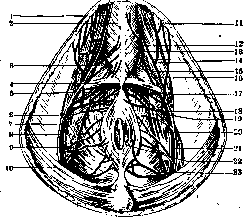
Figure 1. Male perineum: 1-posterior scrotal artery and nerve; 2-bulbocavernosus muscle; 3 and 16-urogenital triangle; 4-transverse perineal muscle; 5-levator ani muscle; 6 and 20-internal obturator muscle; 7 and 15-dorsal nerve of the penis; 8-perineal nerve; 9 and 23-inferior hemorrhoidal nerve and artery; 10-gluteus maximus muscle; 11-ischiocavernosus muscle; 12-dorsal artery of the penis; 13-deep artery of the penis; 14-posterior scrotal arteries; 17-urethral artery; 18-artery of the penis; 19-perineal artery; 21-internal pudendal artery; 22-external anal sphincter.
the ischial tuberosities (inferior ramus of the ischial bone) to the perineal raphe in the sagittal plane (central tendinous point of the perineum), where it limits the base of the specified triangle. Often the muscle is buried in subcutaneous fat, where it is not easily discovered. In its place may be the separated fibers of the neighboring levator ani muscle. Since this muscle rests on the ischial tuberosities and is directed toward the middle of the perineum, its action boils down to tensing the perineum and, in men, supporting the cavernous bodies from below. It is innervated by the perineal branches of the pudendal nerve (Figure 2). The bulbocavernosus muscle originates from the central tendinous point of the perineum, from the external anal sphincter, whence its fibers go to the perineal raphe, on the middle of the lower surface of the bulb of the corpus cavernosum of the urethra, enclosing the bulb; this connection is particularly strong due to the participation of fascias and muscle fibers that circularly enclose the bulb and connect behind it; therefore they, especially with their deep parts, act directly on the initial part of the urethra. The superficial layer of the muscle loop-like encloses the root of the penis, as it is fixed on the fascia of the latter. Therefore, this muscle is able to compress the root of the penis to a certain extent voluntarily and thus contribute to its erection. Despite the fact that this muscle belongs to striated, voluntary muscles, it is believed that during orgasm it is excited reflexively and contributes to the expulsion of seminal fluid from the urethra to the outside. Sometimes there are in addition separate rare fibers that enclose the paired cavernous bodies of the penis (the so-called compressor hemisphaeriorum bulbi muscle). It is innervated by the deep branches of the pudendal nerve. The ischiocavernosus muscle (see Human Muscles) is
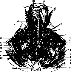
Figure 1. Nerves of the male perineum: 1 - m. bulbocavernosus; 2 - m. ischiocavernosus; 3 - trigonum urogenitale; 4 and 16 - ramus perinealis n. cutanei femoris post.; 5 - n. dorsalis penis; 6 and 14 - n. perinei; 7 and 9 - lig. sacrotuberosum; 8 and 13 - nn. haemorrhoidales inf.; 10 - m. glutaeus maximus; 11 - n. coccygeus; 12 - fossa ischiorectalis; 15 - m. sphincter ani ext.; 17 - m. transversus perinaei superficialis; 18 - n. cutaneus femoris post.; 19 - nn. scrotales post.; 20 - scrotum; 21 - testis. (After Spalteholz.)
synergist of the preceding. Acting on the right and left simultaneously, the muscle can compress both cavernous bodies of the penis and thus express blood in the direction of the anterior cavernae; hence its old name m. erector penis. The middle aponeurosis, deep fascia of the perineum (f. perinaei profunda, s. aponeurosis ano-perinaealis), occupies the entire urogenital region of the perineum; it has been described many times and moreover differently, therefore several different names corresponding to this formation have been preserved: Carcassonne's ligament (in the anterior section of the muscle), subpubic ligament (lig. subpubicum), ligament supporting the urethra (lig. suspensorium urethrae), triangular ligament of the urethra (lig. triangulare urethrae), puborectal aponeurosis (aponeurosis pubo-rectalis), and others; already the very list of names of this fascial-muscular plate indicates the complexity and variability of this formation, studied from various points of view. The middle aponeurosis represents a connective-tissue, fibrous triangular plate, which entirely occupies the subpubic space and directly passes on both sides into f. glutaea propria, and in the depth into cavum ischio-rectale. The apex of this plate constitutes a direct continuation of the subpubic ligament (lig. subpubicum), and the transverse base falls at the level of the bi-ischiadic line, i.e., it is located almost at the anus, where it passes to m. sphincter ani ext., getting lost in its wall. Here this thinned lower edge is concave and merges from below with the superficial aponeurosis of the perineum, above with the prostatoperitoneal aponeurosis; the lateral edges of the plate are fixed on the inner lips of the ischiopubic rami and from here rise upward, lining the outer walls of fossa ischio-rectalis along m. obturator int.; then they merge with the fascia of the latter and at the place of its connection with m. levator ani descend under the name fasc. diaphragmatis pelvis inf. At the level of the posterior edge of the underlying deep transverse muscle of the perineum, the middle aponeurosis of the perineum splits into two whitish dense (fibrous) leaves, or plates—upper and lower (lamina sup. et inf. fasciae perinaei prof., s. f. trigoni urogenitalis sup. et inf.) in such a way that both leaves cover from above and below this muscle, forming a closed fascial receptacle both for the specified transverse deep muscle and simultaneously for the internal pudendal arteries, veins and their branches, as well as for the internal pudendal nerve. Usually the free, posterior edges of m. transversi perinaei prof. et superf. coincide here, therefore f. perinaei superf. merges precisely here with f. perinaei profunda. Both leaves of f. perinaei prof. connect again at the upper edge of the urogenital diaphragm, forming a dense transverse cord here (lig. transversum pelvis). Thus, the very upper section (apex) of the pubic angle remains free and here in a small gap between this transverse ligament and lig. arcuatum pubis passes v. dorsalis penis profunda. The arteries and veins of the cavernous bodies, although they pass through diaphragma urogenitale, are embedded not in its muscular part, but in the connective-tissue plate located between the crura of the cavernous bodies of the penis (lamina intercruralis); therefore, the veins of the penis cannot be compressed by the musculature of the diaphragm, as was erroneously assumed earlier by some researchers. According to a number of authors, the superficial plate of this fascia (and not f. superf. perinaei) covers mm. transversus perinaei superf., bulbo-cavernosus, and m. ischio-cavernosus (along their free surface) and anteriorly continues into f. penis; but in essence the difference here is conventional, since the leaves of the superficial and deep fascia, overlaying each other and merging in some places, diverge again in others, so that in essence it does not matter whether to consider the new secondary fascial formations as a continuation of certain old fasciae or to give them new designations in connection with these or those muscles. Lamina superf. f. perinaei prof. (lamina media f. perinaei, s. f. diaphragmatis urogenitalis inf., s. f. trigoni inf.) is located between mm. transv. perinaei superf., bulbo- et ischio-cavernosus on one side and m. transversus perinaei prof. on the other. Another plate of this fascia (lamina prof. fasciae profundae perinaei, s. f. diaphragmatis urogenitalis sup.) covers the upper (inner) surface of m. trigoni urogenitalis, which faces the cavity of the lesser pelvis. The complex muscular-fibrous septum, or diaphragm, thus constituted, i.e., as a whole diaphragma urogenitale, s. diaphragma pelvis accessorium, closes the outlet from the urogenital region of the pelvis and encloses in itself, besides the indicated muscular and neurovascular formations, also Cowper's glands and the bulb of the urethra together with a part of the latter. However, at the top of the pubic angle, the diaphragm does not fill the latter, and here both leaves of the fascia merge into one strong ligament of triangular shape (lig. triangulare urethrae, s. Carcassonne). Around the urethra, in its pars membranacea, there is a closed ring of striated musculature in the form of a special external sphincter of the urinary bladder [rhabdosphincter, s. m. sphincter vesicae (urethrae) ext.], passing into circular fibers under the mucous membrane of the urethra (m. sphincter urethrae membranaceae, also striated). The fibers of the striated musculature from here partly continue further onto the outer surface of the prostate gland. M. sphincter urethrae membranaceae and m. transversus perinaei profundus during their development merge together along the midline (raphe) of the perineum, so that in small children the right and left parts of the latter are not yet entirely connected directly. Both muscles are abundantly (sometimes up to 50%) permeated with additional smooth muscle fibers. The fibers of m. sphincteris urethrae near the urethra run circularly, the more distant ones—radially; part of the latter are fixed on the pubic bones, therefore they not so much compress the membranous part of the urethra as fix its position, providing, together with its other components (raphe, fascial and numerous elastic layers thereof), support for the fixed part of the latter (pars fixa urethrae). In addition to the indicated muscles in the second muscle layer, in the posterior section of the perineum, there are two more muscles: a small part of the levator ani muscle (m. levator ani), and the ischiococcygeus muscle (m. ischio-coccygeus). In addition, it should be noted here the presence in this same layer anteriorly of two small, but practically important muscles—Wilson's muscle and Guthrie's muscle. M. levator ani, thanks to its funnel-shaped arrangement in the pelvis, constitutes as it were a valve closing the pelvic cavity from below; at the same time, this muscle fills the space between the rectum posteriorly and the prostate gland anteriorly. Firmly connected along the midline, both halves of m. levat. ani represent the most important soft part, highly mobile and providing support to the pelvic floor. Both plates of diaphragma pelvis posteriorly and laterally and diaphragma urogenitale anteriorly complement one another. This general muscular-fascial plate of the pelvic floor closes the entire pelvic outlet and with its musculature encompasses not only the anal section of the rectum, but also the urethra, and in women, in addition, also the vagina. Diaphragma urogenitale represents a special muscular-fascial layer that strengthens only the single weakest anteroinferior region of the pelvic outlet. Another part of the pelvic floor—diaphragma pelvis—serves simultaneously for the act of defecation and for closing the outlet opening of the rectum at the end of this act. The anterior fibers of m. levatoris ani form well-defined inner margins that surround the prostate gland and proceed to the lateral surfaces of the rectum; the posterior fibers of this muscle are fixed in addition on the anococcygeal raphe (raphe anococcygeum), part of them merges with the longitudinal fibers of the rectum and ends in the deep layer of the skin, in the vicinity of the anus. M. levator ani originates on the inner surface of the pelvis at the level of for. obturati; its fibers depart from the ligamentous thickening of the proper fascia of m. obturatorius internus, which is called the tendinous arch (arcus tendineus levatoris ani); along both edges of this tendinous arch, the muscle origins continue further into the periosteum, where they are fixed: anteriorly—on the descending ramus of the pubic bone (up to the symphysis), posteriorly—on the iliac bone, below linea terminalis; therefore, in m. levator ani, the pubic and iliac parts are distinguished (pars pubica et pars iliaca). The attachment site (punctum mobile) of the fibers of both indicated parts is different: pars pubica loop-like embraces canalis urogenitalis and the rectum, where it connects along the midline with its small component—lig. ano-coccygeum (approx. 3 cm in length and width), which proceeds from the apex of the coccyx to the anus.
This loop is located on the corresponding notch of the rectum, i.e., on the flexura perinaealis, fills it, and tightly embraces the intestine here like a sphincter; thus, when the muscle contracts in its pars pubica, the posterior wall of the rectum is inevitably pressed against the anterior wall, as a result of which the anus is closed. This loop can easily be felt with the fingers on the posterior wall of the intestine as a dense ridge during the active elevation of the anus. During the passage of fecal masses, no significant elevation of the anus occurs; it only bends slightly: while its anterior wall lowers, the posterior, conversely, rises somewhat. The fibers of the pars iliacae are anchored on one side to the coccyx and on the other to the lig. sacro-iliacum; in addition, by a single tendinous plate they pass to the anterior surface of the sacrum; they only pass alongside the described loop-shaped embrace of the rectum from the side of the pars pubicae, but are not included in the latter. Due to the oblique course of this part of the muscle from top to bottom and from front to back, it acts on both sides on the almost immobile coccyx in such a way that when its fibers are stretched between its two indicated supports on the coccyx, a support and then an elevation (up to 2 cm) of the entire pelvic floor is created; this elevating action of the muscle is largely facilitated by the low mobility of its support on the coccyx. Thus, the m. levator ani belongs an important static role in creating an elastic support for the pelvic floor and no less important dynamic role as one of the factors in the act of defecation and the closure of the anal opening. The m. levator ani may be partially absent. The fibers of its pars pubicae may enter into the composition of the m. sphincteris ani ext. The innervation of this muscle is carried out by branches of the III-IV spinal sacral nerves (from the plexus coccygeus). If there is a connection of the m. levatoris ani with the m. sphincter ani ext., branches from the n. pudendus also approach it. The m. coccygeus (s. ischio-coccygeus) - see Human muscles. Directly related to the perineum are also the external sphincter of the rectum (m. sphincter ani ext.). It represents a flat striated muscle, developed from the embryonic cloacal musculature, which annularly embraces the end of the rectum. The width of the muscle is approximately 3 cm. That layer of the muscle which lies directly against the intestinal wall has no bony points of support. The fibers of the muscle from both sides cross each other and terminate in the midline in front of and behind the anal opening, in the aponeurotically thickened subcutaneous tissue here. In the front, some of their fibers attach to the superficial aponeurosis of the perineum, while others cross like the figure 8—in men with the m. bulbo-cavernosus, in women with the m. constrictor cunni; therefore, contractions of these muscles occur simultaneously. The middle main layer of the muscle is divided into two halves: the fibers of each of them depart from the end of the coccyx (lig. ano-coccygeum) and attach between the anal opening and the scrotum (in women - the vagina) to a tendinous plate located in the midline of the perineum (centrum tendineum perinaei), at the border between the regio uro-genitalis and regio analis. The upper and deepest layer of the muscle consists only of whole circular fibers, not fixed to the bones; this layer is located 3-4 cm away from the anal opening, borders on the muscular loop of the m. levatoris ani (its pars pubica), partially merges with it, as well as with the smooth muscle fibers of the longitudinal layer of the intestine. Upon tension of those fibers of this muscle which are fixed to the skeleton (coccyx), the anal opening and the rectum (its pars perinaealis, s. analis) close, forming longitudinal folds; by the action of the loop of the m. levatoris ani (its pars pubica), the flexura perinaealis of the rectum is compressed into a transverse fold. Such a muscular mechanism of the end of the recti ensures the hermetic closure of it. This mechanism of the rectum, together with the subsidiary action of its smooth circular musculature, the gluteus maximus, and other muscles of the perineum, ensures sufficient reliability and strength of both the static and dynamic functions of the axes of the rectum and perineum. This muscle is innervated from the rr. haemorrhoidales inf., departing from the n. pudendus. Wilson's muscle is composed of a small bundle of longitudinal deep fibers of the m. transversi perinaei prof.; it has the appearance of a small triangle, the base of which faces upward and is attached to the lig. subpubicum, while the apex faces downward and is fixed on the membranous part of the urethra. It is separated from the underlying m. transversus perinaei prof. by means of a fascial process (lamina sup. fasc. perinaei profundae). Therefore, it is easier to see from the inside, from the side of the pelvis, upon removal of the peritoneum, pelvic fascia, and puboprostatic ligament, behind the latter, in which it is enclosed. Spasmodic contraction of this muscle, as well as other muscles related to the urethra, can lead to a spasm of the latter and present an obstacle to urination or catheterization. Following further upwards behind the second muscular layer is the fascial layer—lamina profunda fasciae perinaei profundae—the upper, relatively thin aponeurosis of the pelvic floor, known as the pelvic fascia (fascia pelvis, s. endopelvina); this fascia represents proper the upper wall of the fascial sheath of the m. levatoris ani. On both sides of the pelvis, the f. pelvis directly merges with the fascia (aponeurosis) of the obturator internus muscle, and inwards it is fixed to the rectum and the lateral parts of the prostate gland and is known as the f. prostatae. The f. pelvis constitutes proper the direct continuation downwards into the pelvis of the thickened f. transversalis. It covers the lateral surfaces of the lesser pelvis and the muscles located here. The aponeurotic stretching of this fascia forms that transverse tendinous arch from which part of the fibers of the m. levatoris ani originate. The f. pelvis is tightly fixed to the pelvic bones and loosely connected to the rectum, urethra, and urinary bladder (in women - with the vagina) where it covers them and passes into their adventitia. The gaps often present between the fibers of the m. levatoris ani are rather tightly closed by this fascia. Just as precisely, the free spaces between the lig. arcuatum pubis and lig. transvers. pelvis, above the diaphragma urogenitale, are lined by means of the f. pelvis; this same fascia lines from the inside the triangular space between the pubic bones (angulus pubis), therefore here this fascia is sometimes called the f. trigoni urogenitalis. On both sides of the symphysis, in the direction of the urinary bladder and prostate gland, the fascia continues onto the latter in the form of two folds (lig. pubo-prostatica lateralia; in women - lig. pubo-vesicalia). To the prostate gland here goes, from the symphysis, a medial cord similar to the first two (lig. pubo-prostaticum medium; in women - lig. pubo-vesicale medium). The lower surface of the m. levatoris ani is also covered by the f. pelvis, called here the f. diaphragmatis pelvis; between it and the f. obturatoria there is a significant fat accumulation, upon the removal of which a fossa is formed (fossa ischio-rectalis). Downwards and inwards from the arcus tendineus, descending along the rectum, the f. pelvis covers, in addition to the indicated formations, also the prostate gland, the urinary bladder (in women - the vagina). - Between the f. pelvis and the peritoneum lies a layer of fat, developed to varying degrees in different individuals, enclosed in loose cellular tissue. This cellular tissue is separated from the cellular tissue located lower in the ischiorectal fossae by means of the m. levator ani together with its two fasciae. Above the f. pelvis and the layer of fatty tissue lies the peritoneal leaf. The three main fascial leaves of the perineum considered above, besides covering the underlying muscular formations and fixing the organs of the perineum, form two—upper and lower—practically important spaces, or receptacles. The lower receptacle of the perineum is limited by means of the leaf of the f. perinaei superficialis from below and the f. perinaei prof. from the top, but since these two fasciae at the level of the lineae biischiadicae merge together, and then the f. perinaei continues onto the bulb of the urethra and ends at the base of the head of the penis, having clothed these parts, thus this receptacle encloses almost the entire penis. The named receptacle includes: Cowper's glands, the bulbous part of the urethra, and the corpora cavernosa of the penis; in addition, it contains the m. transversus perinaei superfic., m. bulbo- et ischio-cavernosus; at the same time, each of the named muscles has its own special fascial sheath.
^ Upper, or prostatic receptacle. The prostate gland is surrounded on all sides by aponeurotic leaflets, namely: above and in front it is covered by the pubovesical ligaments (ligamenta pubovesicalia), behind and below lies the prostato-peritoneal aponeurosis (aponeurosis prostatoperitonealis) and the upper, deep leaflet of the deep perineal fascia; on both sides is located the so-called lateral aponeurosis of the prostate gland. The prostato-peritoneal aponeurosis, characterized by the presence of a large number of smooth muscle fibers, covers the entire posterior surface of the prostate gland and thereby separates the latter from the rectum. It extends from the site of the peritoneal reflection in the rectovesical pouch downward to the posterior margin of the urogenital diaphragm, where it merges with the upper leaflet of the latter. As a result, the peritoneum in this region, when the urinary bladder is filled, not only does not rise, but, on the contrary, descends, forming and deepening here its posterior reflection or blind pouch ("cul-de-sac"). The lateral aponeurosis of the prostate gland runs from the pubic bone from front to back and from top to bottom. Here it merges with the puboprostatic aponeurosis and the upper (deep) leaflet of the deep perineal fascia and approaches the rectum, where it is fixed on its lateral surfaces (hence its former name, puborectal aponeurosis). Thus, this aponeurosis occupies the space enclosed between the pelvic fascia and the prostato-peritoneal aponeurosis. In addition to the prostate gland, this receptacle encloses Wilson's muscle (see above) and the prostatic venous plexus together with cellular tissue. This venous plexus is divided into three separate plexuses: the anterior (Santorini's), lying immediately behind the pubis, where the dorsal vein of the penis empties, and two lateral ones, located on both sides of the prostate gland; the latter are usually very dilated in old age. Ischiorectal fossae (cavum ischiorectale). It was pointed out above that, due to the funnel-like shape of the levator ani muscle, the pelvic outlet is closed not by a transverse muscular barrier, but by an inclined one. As a result, fairly wide fossae filled with a large amount of fat (fossae ischiorectales) remain on both sides of the rectum; these fossae, or depressions, have the general appearance of an irregular low triangular pyramid with the base facing downward and the apex directed into the depth of the perineum. The outer wall of the pyramid is formed by the ischial tuberosity and the parts adjacent to it (the ramus of the ischium with the fascia of the obturator internus muscle [obturator fascia]); the inner wall is formed by the levator ani muscle together with its thin fascia (inferior fascia of the pelvic diaphragm); the lower wall of the pyramid corresponds to the skin with its subcutaneous fascia. The boundaries of the base of the ischiorectal fossa are: posteriorly, the lower edge of the gluteus maximus muscle; anteriorly, the superficial transverse perineal muscle; outwardly, the ischial tuberosity; posteriorly, the anus. The ischiorectal fossa is firmly demarcated from the urogenital region by means of a connective tissue membrane. Here, in the fatty contents of the fossa, sensory nerves, small vessels to the skin of the perineum, and motor nerve branches to the external anal sphincter pass. Large vessels and nerves of the perineum closely adhere to the ischial bone, as they are embedded in the duplication of the obturator fascia (Alcock's canal). Female perineum. The relations of the layers of the female perineum change compared to the male in its anterior section, at the level of the pudendal cleft and vagina. The anus is distinguished in women by a somewhat greater displacement forward relative to the coccyx (closer to the urethra) and is less recessed between the two ischial tuberosities. Hair, which usually surrounds the anus in men, is very rare in women. The superficial and deep perineal fasciae are of relatively smaller dimensions, while the urogenital triangle is wider in women due to the relatively larger subpubic angle; the perineal raphe is less stable due to the passage through it not only of the urethra but also of the vagina. The nature of the load on the perineum, especially in its anterior section, is completely different in women than in men; this relates to the comparatively lower stability of the female urogenital region and to those changes in the perineum that are associated with the process of childbirth. As in men, the transverse
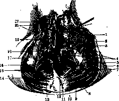
Fig. 3. Nerves of the female perineum: 1—ischiocavernosus muscle; 2—bulbospongiosus muscle; 3 and 17—perineal branch of posterior femoral cutaneous nerve; 4—dorsal nerve of clitoris; 5—perineal nerve; 6—posterior femoral cutaneous nerve; 7—sacrotuberous ligament; 8 and 15—inferior hemorrhoidal nerves; 9—gluteus maximus muscle; 10 and 14—middle inferior clunial nerves; 11—coccygeal nerve; 12—external anal sphincter; 13—ischiorectal fossa; 16—perineal nerve; 18—superficial transverse perineal muscle; 19—vaginal vestibule; 20—posterior labial nerves; 21—clitoris. (After Spalteholz.) deep transverse perineal muscle in women is connected with the sphincter urethrae muscle, but the latter is much less developed in women and its fibers are not arranged as strictly circularly around the urethra: many of them enclose both the urethra and the vagina together, others go separately to these organs, part of them disappear in the connective tissue of the pelvis. They pass into the transverse fibers of the deep transverse perineal muscle, leaving a free space for the passage of vessels and nerves between the upper edge of the muscle and the pubic bone (dorsal artery and vein of the clitoris and two small nerves of the same name). The levator ani muscle and the deep transverse perineal muscle converge in the pubic angle; in addition, the posterior transverse fibers of the deep transverse perineal muscle intricately interlace in the areas between the pubis and the anus with the pubic part of the levator ani muscle and the anterior fibers of the external anal sphincter, which significantly strengthens the female perineum. The deep transverse perineal muscle is covered above and below by very dense fasciae (superior and inferior fasciae of the urogenital diaphragm). A feature of the musculature of the female perineum is
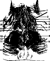
Figure 4. Arteries of the female perineum: 1—posterior labial arteries; 2—perineal artery; 3—deep artery of the clitoris; 4—dorsal artery of the clitoris; 5—urethral artery; 6—artery of the clitoris; 7—artery of the vestibular bulb; 8—internal pudendal artery; 9—inferior hemorrhoidal artery; 10—gluteus maximus muscle; 11—pudendal nerve; 12—levator ani muscle; 13—external anal sphincter; 14—superficial transverse perineal muscle; 15—urogenital triangle; 16—bulbospongiosus muscle; 17—ischiocavernosus muscle. (After Spalteholz.)
the significantly developed bulbospongiosus muscle, or constrictor cunni, or sphincter vaginae; the first, old name of the muscle in women is inappropriate because they lack a "bulb"; it makes some sense only from the point of view of its genesis and analogy with this muscle in men. The constrictor cunni muscle with its numerous fibers is embedded in the wall of the vagina. These fibers connect with each other along the midline in front and behind the pudendal cleft. Far from all the fibers of this muscle reach the crura of the clitoris, as follows from the name of the muscle; some of them in the form of tendinous cords attach either to the corpora cavernosa of the clitoris or in the vicinity. The fibers of the external anal sphincter also embed here into the constrictor cunni muscle; intertwining mutually, they form a figure-of-eight shape, with the vagina passing through the anterior opening of such a figure-of-eight, and the anus through the posterior one; lying also near the vestibular bulbs, the constrictor cunni muscle upon its contraction compresses simultaneously the Bartholin's glands. The compressing force of the constrictor cunni muscle is small, but it increases significantly due to the joint action with it of other accessory muscles, especially the levator ani muscle; mutually
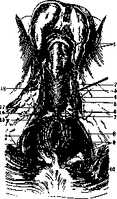
Figure 5. Arteries of the male perineum: 1—posterior scrotal arteries; 2—perineal artery; 3—deep artery of the penis; 4—dorsal artery of the penis; 5—urethral artery; 6—artery of the penis; 7—artery of the urethral bulb; 8—internal pudendal artery; 9—inferior hemorrhoidal artery; 10—sacrotuberous ligament; 11—gluteus maximus muscle; 12—pudendal nerve; 13—levator ani muscle; 14—external anal sphincter; 15—superficial transverse perineal muscle; 16—urogenital triangle; 17—ischiocavernosus muscle; 18—bulbospongiosus muscle. (After Spalteholz.)
earlier contraction of the levator ani muscle in women can also contribute to the narrowing of the vagina, since it is located on both sides of it before its transition to the rectum. The Bartholin's glands, which are partially embedded between the fibers of the constrictor cunni muscle, can be rapidly emptied during intercourse under the influence of the contraction of this muscle's fibers. The ischio-cavernosus muscle in women constitutes a barely noticeable, small, mostly tendinous plate; it begins at the ischial tuberosity, lies directly on the crus of the clitoris on its side and therefore, by its weak contraction, can contribute to the engorgement of the cavernous bodies of the clitoris (hence its old name - m. erector clitoridis). The superficial transverse perineal muscle in women is poorly developed or absent altogether. The vessels and nerves of the female perineum correspond to those in males, although their names are partly different (Figs. 3 and 4). The blood supply to the perineum is carried out via the internal pudendal artery, which emerges from the pelvic cavity through the infrapiriform foramen, and then through the greater sciatic foramen enters the region of the perineum (through Alcock's canal). Along the way, the artery gives off branches to the anus (1-3 inferior hemorrhoidal arteries). At the urogenital diaphragm, the main trunk of the artery divides into two secondary and at the same time terminal branches - the perineal artery (to the muscles of the penis and to the scrotum - posterior scrotal arteries) and the artery of the penis (to its cavernous bodies) (Fig. 5). The veins precisely (in pairs) follow the arteries of the same name; the pudendal artery is accompanied, as usual, by two veins, but upon entering the lesser pelvis, the veins unite into a large trunk (internal pudendal vein). The lymphatic vessels of the perineum drain into the superficial medial inguinal lymph nodes. The nerves of the perineum muscles originate from the pudendal nerve, which with its branches innervates all the muscles of the perineum. The nerves are located together with the vessels of the perineum in a single neurovascular bundle. Those branches that emerge along the course of Alcock's canal and enter the mucous membrane of the anus also innervate the skin of the perineum (inferior hemorrhoidal branches); then further anteriorly, the perineal and posterior scrotal branches depart. In addition, sensory nerves from the posterior surface of the thigh approach the perineum (perineal branch of the posterior cutaneous nerve of the thigh); they emerge from beneath the lower edge of the gluteus maximus muscle. See also Rectum, Prostate gland, Urethra, Urinary bladder, Vagina. Pathology of the perineum - see Hernia, Uterus, Colporrhaphy, Labor.
Related articles
Mentioned in
Cite this page
“Perineum.” Soviet Medical Encyclopedia. English translation of Bolshaya Meditsinskaya Entsiklopediya, 1st ed. (Moscow, 1928–1936), ed. N. A. Semashko. https://sovietmedicalencyclopedia.pages.dev/article/perineum/