Pelvis
Historical document, translated for reference. It reflects medical knowledge of the 1920s–30s and is not medical advice.
Summary
The pelvis is a skeletal structure forming the pelvic girdle that supports the trunk and transfers weight to the lower limbs. It consists of paired hip bones, sacrum, coccyx, and fifth lumbar vertebra, forming a bony ring that contains pelvic organs and serves as the birth canal.
Encyclopedia article (1928–1936)
PELVIS. Contents: I.
267 II.
306 I. Anatomy of the pelvis. The pelvis (pelvis), part of the skeleton forming the so-called pelvic girdle or girdle of the lower (in animals, posterior) limbs and represented by a bony ring of two (right and left) hip bones, sacrum, coccyx, and V lumbar vertebra. The pelvis represents an important static-dynamic part of the skeleton, bearing a supporting function for the trunk and in turn resting on the lower limbs. The vertical position of the human body causes the transfer of pressure from the mass of abdominal organs from the anterior abdominal wall to the pelvis, in relation to which the latter carries a supporting function, and for some serves as a container. The pelvic ring serves as an attachment site for muscles: anterior-lateral walls of the abdomen, back, pelvic girdle, thigh, and perineum. Together with soft tissues, the pelvis forms a canal through which the fetus passes during childbirth. The bones that make up the pelvis limit the pelvic cavity (cavum pelvis), which is divided by the so-called linea innominata (linea innominata, s. terminalis) into two parts: 1) the upper-large pelvis [pelvis major, s. superior, s. spuria, as well as labrum pelvis (Hyrtl)] and 2) the lower-small pelvis (pelvis minor, s. inferior). In the linea innominata, according to its formation from three bones, three segments are distinguished: 1) pars iliica-represented by linea arcuata, s. semicircularis (ilium), 2) pars pubica-pecten pubis (pubic bone) and 3) pars sacralis-promontorium of the sacrum. The small pelvis has an inlet, bounded by the aforementioned line (apertura pelvis superior), a canal, the proper cavity (cavum pelvis), and an outlet (apertura pelvis minor, s. inferior). The outlet is bounded by the lower edge of the pubic symphysis, inferior rami of the pubic and ischial bones, ischial tuberosities, lig. sacrotuberosa (see below), the edge of the sacrum and coccyx. The innominate, pelvic bone, os coxae (syn. os innominatum, s. anonymum, s. coxale, s. coxendicis, s. pelvis laterale) (fig. 1), is a paired bone, connected behind with the lateral masses of the sacrum, and in front with its pair, divided into three large parts (bones): 1) the ilium (os ilium, s. ilii), 2) the ischium (os ischii, s. coxendicis, s. ischium) and 3) the pubic bone (os pubis, s. pectinis). This division is based on the principle of bone formation during its development from three main separate ossification centers (see below) (fig. 1). With their most massive parts, called bodies (corpus os. ilii, corpus os. ischii and corpus os. pubis), these three bones connect with each other into a common mass, forming on the outer surface a cup-shaped depression-the acetabulum, and the boundaries of the bones forming it are distinct only in childhood. Upward and outward from the body of the ilium extends a wide, somewhat concave plate, sharply thinned in the middle and thickened along the upper edge, called the wing, ala os. ilii (pars abdominalis of the ilium). The upper S-shaped curved outward to inward edge of the ilium is marked as the crest (crista iliaca), on which three lines are distinguished to varying degrees for attachment of muscles of the anterior-lateral abdominal wall (mm. obliquus abd. ext. int. et transv. abd.): 1) external (labium ext. s. laterale), 2) internal (labium int., s. mediale) and 3) intermediate (between the first two) (linea intermedia). Crista iliaca forward passes into a projection-the anterior superior spine (spina iliaca ant. sup.), below which, separated from it by a notch (incisura iliaca ant.), is located the anterior inferior spine (spina iliaca ant. inf.). Behind, the iliac crest also ends with a projection-the posterior superior spine (sp. iliaca post. sup.); below the latter, separated from it by incisura iliaca post., is the posterior inferior spine (spina iliaca post. inf.). The listed
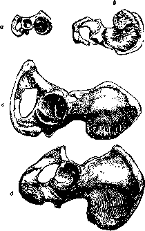
Figure 1. Formation and ossification centers of the pelvic bone: a-in newborn; b-at 2 years; c-12 years; d-20-22 years.
These projections are sites of attachment for muscles and ligaments. The inner surface of the ilium is concave, forming the lateral wall of the pelvis and is called the iliac fossa (fossa iliaca) because part of the loops of the ileum (intestinum ileum) is located there. The outer surface of the ilium, where the gluteal muscles originate, has faintly expressed ridge-like lines: slightly above sp. iliaca ant. inf. extends posteriorly the short linea glutaea inf., near sp. iliaca post. sup. goes also the short linea glut. post., and finally the third, longest linea glut. ant. obliquely crosses in an arched direction from below upward and from behind forward the middle of the outer surface of the ilium. The part of the ilium adjacent to the sacrum and called pars sacralis has an uneven surface, with its contours resembling the auricle (facies auricularis), for connection with the lateral masses of the sacrum (see Sacroiliac joint). Sometimes near the anterior and posterior edges of the auricular surface, grooves (sulcus paraglenoidalis ant. et post.) are noticeable for the attachment of the iliolumbar ligaments. Anterior to the auricular surface along the inner surface of the ilium extends a ridge toward the superior ramus of the pubic bone - linea arcuata (see above). The part of the ilium located below this ridge and fusing with the ischium is called pars pelvina. On the ilium, anterior and lateral to facies auricul., more or less constant foramina nutricia can be found (see Bone, blood vessels). In addition to the mentioned body (corpus os. ischii), the ischium has thick, short plates arranged at an angle to each other: the posterior superior ramus, connected to the body of the ilium (ramus sup. os. ischii, s. ramus descendens), and the anterior ramus, connected to the pubic bone (ramus inf., s. ascendens). At the angle between the mentioned rami of the ischium, a massive tubercle (tuber ischii) develops, a site of attachment for muscles and ligaments, and a support for the body when sitting. Somewhat above the ischial tuberosity on the posterior edge of the superior ramus, a sharp tubercle - the ischial spine (spina ischiadica) - is marked, which from tuber ischii is separated by the so-called lesser sciatic notch (incisura ischii minor), and from spina iliaca post. inf. - by a second, deeper notch (incisura ischii major). The lesser sciatic notch together with 2 ligaments - one stretched from the ischial tuberosity to the sacrum (lig. sacrotuberosum), and the second going from the ischial spine also to the sacrum (lig. sacrospinosum) - forms the lesser sciatic foramen (foramen ischiad. minus); the greater sciatic notch, the edge of the sacrum and lig. sacrospinosum limit the second - greater sciatic foramen (foramen ischiad. majus). Both foramina serve for the exit from the cavity of the small pelvis of blood vessels, nerves and muscles. The outer surface of the body and rami of the ischium is studded with numerous vessel openings, the inner surface is smooth. In the pubic bone, the above-mentioned body (corpus os. pubis) and two rami are distinguished: 1) the superior, horizontal ramus (ramus superior, s. horizontalis), departing from corpus ossis pubis and directed forward and inward, and 2) the inferior ramus (ramus inf., s. descendens), fusing with the ascending ramus of the ischium. At the junction of both rami (tuberositas os. pubis) there is a surface (facies symphyseos) by which the pubic bone of one side connects with that of the opposite side (symphysis). Along the upper edge of ram. sup. os. pubis extends a sharp ridge - pecten os. pubis, s. linea iliopectinea, which is as it were a continuation of the lineae arcuatae of the ilium. At the transition of the latter into pecten os. pubis there is a rounded elevation - eminentia iliopectinea. The pubic crest near the symphysis of the pubic bones forms a distinctly protruding (easily palpable in the living) tubercle (tuberculum pubicum), an important landmark in examining the external opening of the inguinal canal. The rami of the ischium (from the dorsal and caudal sides) and the rami of the pubic bone (from the ventral and cranial sides) limit the large opening of the innominate bone - the obturator foramen (foramen obturatum, s. obturatorium), which is closed in its greater part by a fibrous membrane (membrana obturatoria), as well as by the obturator muscles (mm. obturator, ext. et int.) located inside and outside the membrane. In the upper-lateral edge of foram. obtur. a small tubercle (tuberculum obturat. post.) is noticeable; a similar tubercle (tubercul. obturat. ant.) is marked on the lower edge of the superior ramus of the pubic bone. Between the mentioned tubercles and the somewhat concave at this place superior ramus of the pubic bone, a groove (sulcus obturatorius) is observed, the posterior end of which is bounded by a small ridge (crista obturatoria). Sulcus obturatorius, membrana obturatoria and the obturator muscles form a short canal (canalis obturatorius), through which from the cavity of the small pelvis into the thigh region pass the blood vessels and nerve of the same name (see Obturatoria arteria, vena). In the acetabulum (acetabulum) its bottom (fossa acetabuli), the remaining surface (facies lunata) and on the lower edge a notch (incisura acetabuli) are noted. The innominate bone is subject to certain variations, which include e.g.: 1) the presence of a transverse bony bar in foramen obturatorium, 2) the absence of bony fusion (a cleft) between the inferior ramus of the pubic bone and the inferior ramus of the ischium, 3) the absence of incisurae acetabuli, 4) the formation of an opening in fossa iliaca and a deep groove on the outer surface of the ilium for vasa glutaea sup. Constant are the variations of the innominate bone associated with age, sex, body type, individual. - The innominate bone is laid down in the primary skeleton as the proximal part of the lower limb; in the initial stage it consists entirely of cartilage. Earlier than other parts, the cartilaginous primordium of the ilium appears, fusing with the lateral parts of the cartilaginous sacral vertebrae, later - the primordia of the ischium and pubic bones, growing in the ventral direction. The process of ossification begins with the appearance of three main ossification centers, first in os ilii on the 2-3rd month of intrauterine life [on the 56th day, according to Mall, on the 60-65th day, according to Adair]; later - on the 4-5th month of intrauterine life, according to Braus, on the 105th day (Mall), on the 94-98th day (Adair) - in the ischium; finally on the 5-6th month of intrauterine life (Braus), on the 129th day (Adair) a nucleus appears in the pubic bone. In all three parts of the innominate bone, the process of ossification proceeds mainly endochondrally. By the time of birth, the bony parts of the three mentioned bones converge to the area of the forming acetabulum, leaving for a long time after birth a Y-shaped cartilaginous layer of varying width (depending on age) (Fig. 1). In addition to the three main ossification centers in individual areas of os coxa, additional centers appear, most often nine in number (Figs. 2-4). These additional centers arise relatively late (after
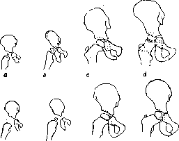
Fig. 2. Ossification of the pelvic bone and proximal end of the femur in girls (top row) and boys (bottom row): a-4 m. 10 d., 60 cm; b-0 mos. 7 d., 66.0 cm; c-3 years; d-4 y. 4 mos, 109 cm; e-4 mos. 24 d., 60 cm; f-6 mos. 7 d., 62.5 cm; g-3 y. 4 mos; h-4 years.
Fig. 3. Ossification of the pelvic bone and proximal part of the femur in girls (top row) and in boys (bottom row): a-9 y. 10 mos, 136.2 cm; b-11 y. 6 mos, 144.2 cm; c-13 y. 3 mos, 143.6 cm; d-10 y., 140 cm; e-12 y. 3 mos, 146 cm; f-13 y. 3 mos, 147.2 cm.
Fig. 4. Ossification of the pelvic bone and proximal end of the femur in girls (top row) and in boys (bottom row): a-15 y. 6 mos, 155.5 cm; b-17 y. 9 mos., 163 cm; c-19 y. 8 mos, 159.7 cm; d-15 y., 168.5 cm.; e-17 y. 8 mos, 155.7 cm; f-19 y. 3 mos, 162.5 cm.
the appearance of the three main centers), more often in the number of nine (Figs. 2-4). These additional centers arise relatively late (after
Fig. 2. Ossification of the pelvic bone and proximal end of the femur in girls (top row) and boys (bottom row): a-4 m. 10 d., 60 cm; b-0 mos. 7 d., 66.0 cm; c-3 years; d-4 y. 4 mos, 109 cm; e-4 mos. 24 d., 60 cm; f-6 mos. 7 d., 62.5 cm; g-3 y. 4 mos; h-4 years.
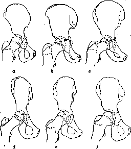
Fig. 3. Ossification of the pelvic bone and proximal part of the femur in girls (top row) and in boys (bottom row): a-9 y. 10 mos, 136.2 cm; b-11 y. 6 mos, 144.2 cm; c-13 y. 3 mos, 143.6 cm; d-10 y., 140 cm; e-12 y. 3 mos, 146 cm; f-13 y. 3 mos, 147.2 cm.
Fig. 4. Ossification of the pelvic bone and proximal end of the femur in girls (top row) and in boys (bottom row): a-15 y. 6 mos, 155.5 cm; b-17 y. 9 mos., 163 cm; c-19 y. 8 mos, 159.7 cm; d-15 y., 168.5 cm.; e-17 y. 8 mos, 155.7 cm; f-19 y. 3 mos, 162.5 cm.
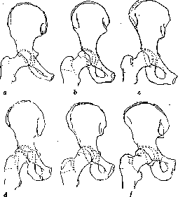
the appearance of the three main centers), in individual areas of os coxa appear additional centers, most often nine in number (Figs. 2-4). These additional centers arise relatively late (after
before 10 years, and some even by the end of the 2nd decade). Of the mentioned accessory nuclei, three belong to the Y-shaped cartilage of the acetabulum. One of them, the largest (anterior), bordering the superior branch of the pubic bone and the ilium, when it transforms into a clearly expressed bony area, is denoted by a special term - os acetabuli; the other two are called epiphyses acetabuli. In girls they appear at the age of 10, in boys at 12-13 years (Pratje). The fusion of the three main parts of os coxae occurs relatively early. First - at 7-8 years (Waldeyer) - the branches of the pubic and ischial bones fuse (by synostosis) (see Articulation). The Y-shaped cartilage in the acetabulum disappears in girls between 12-16 years, in boys between 13-18 (Hasselwander). The other 6 accessory ossification nuclei are observed in various places of the innominate bone, mainly on the projections. The largest of them and appearing earlier than the others is the so-called epiphysis marginalis (fig. 1), forming in the place of the future cristae iliaca and spina il. ant. sup. (according to Pratje at 13-14 years in girls and 14-15 years in boys) (fig. 2-3). But, having developed earlier than others, epiphysis marginalis fuses with the ilium later than others (by 21-25 years according to Waldeyer). Separate ossification nuclei (apophyseal centers) are also found in spina il. ant. inf., sp. os. ischii, tuber isch., angulus pubis and tuberculum pubicum. In men of 24 years and in women of 21 years, Waldeyer observed an ossifying area in the region of tuber isch. not yet fused with the latter; bony nuclei in angulus pub. and tuberculum pub. form around 18-20 years and retain their independence in the first until 20-21 years and in the second until 20 years. The nuclei in spina ischii remain unconnected with os ischii until 17-18 years, in the region of sp. il. ant. inf. - until 16-17 years. The number of accessory ossification nuclei of os coxae is variable. By 22-25 years all the main and accessory ossification areas merge into a common bony mass - os coxae is formed (fig. 1). Along with the course of ossification processes, the growth and shaping of the bones forming the P. proceed, which determine the presence of certain age-related features of the P. as a whole. The ilium of the fetus and newborn is set more steeply, ala os. ilii are flatter, the iliac fossae are less deep. The acetabula of newborns are smaller, flatter and relatively larger (Le Damany, 1904; Harrenstein). The pubic bones are wider than in the adult. In the symphysis (of the pubic bones) a fissure appears from 2 years (according to Fick). Foramina obturata are small. The innominate bone, and indeed the entire P. in childhood is less structured than in the adult: growth proceeds more in height than in width. The P. of a child has relatively small dimensions and a small cavity in the form of a funnel-shaped depression, not cylindrical like in adults. The growth of the pelvic ring in width occurs mainly due to the growth in width of the sacrum; in the longitudinal direction the pelvis grows due to the growth of the pelvic bones. The growth of bony mass also occurs in the region of acetabuli, whereby the depth of the acetabulum increases. The peculiarities in form and size of the innominate bones together with those of the sacrum determine the sexual differences of the P. These differences can be detected already in the newborn (mainly in the individual parts of the P.). For example, the width of the cavity and outlet of the lesser P. is greater in girls; the P. of girls is less deep; in them the lateral masses of the sacrum are more developed, but the differences in the region of the symphysis are already clearly evident at birth. The superior branches of the pubic bones of boys are more strongly developed; the pubic bones of both sides converge at an acute angle (in girls - at a more obtuse one). In childhood, specifically in the 1st decade, parallel with the growth of the entire body (skeleton), the P. grows, but sexual differences in this period are not sharply manifested and not intensified. From 8-10 years (according to Megger) the sexual differences of the P. begin to appear again, which by the period of sexual maturity lead to the characteristics that in the subsequent life characterize the male or female P. Individual points of the bones of the P. protrude more distinctly under the soft tissues in the living, especially when the subcutaneous fat layer is poorly developed. These protrusions are used as anthropometric points, as well as in obstetrics (when determining the external dimensions of the P., e.g. spinae iliacae ant. sup.). Some bony protrusions of the P. in topographic and clinical anatomy are taken as points between which conditional lines are drawn, dividing the large area of the body into a number of separate areas where certain anatomical formations, or certain organs lie (for example a conditional line connecting the anterior superior iliac spines of both sides separates in the abdominal region regio mesogastrica from regio hypogastrica; a line connecting the ischial tuberosities divides the perineal region into regio rectoanal and regio urogenitalis). The innominate bones with their articular surfaces (facies auriculares) connect with the articular surfaces on the lateral masses of the sacrum, forming the sacroiliac joint (see). The narrow articular fissure, the strong articular capsule with a large number of auxiliary ligaments stretched between the last lumbar vertebrae, the sacrum and the iliac bones (about the ligaments see also Sacroiliac joint and Sacrum) allow one to speak of almost complete absence of mobility in this joint. Facies symphyseos of the pubic bones of both sides connect with each other with the help of a fibrous cartilage called the pubic symphysis (symphysis os. pubis). Due to the presence inside the cartilage of a fissure-like cavity (1 mm in diameter) the pubic symphysis is considered a half-joint (hemiarthrosis), in which only some stretching is possible. In the region of facies symphyseos the fibrous cartilage turns into hyaline cartilage (in childhood only hyaline cartilage is present). The posterior surface of the cartilage forms a small protrusion directed into the cavity of the lesser P. [eminentia retropubica (Waldoyer)]. The height of the cartilage in men is about 5.5 cm, in women - 4.5 cm; its thickness in men is less significant than in women. The cartilage fissure in women is wider. The pubic symphysis is strengthened in front by a dense fibrous plate (perichondrium) and auxiliary strong ligaments: above - the lig. pubicum sup. stretched between the pubic tubercles, below - the lig. arcuatum. The latter ligament rounds the pubic angle, especially in women. Besides the ligaments of the sacroiliac joint and the pubic symphysis, the most important ligaments of the pelvis are the above-mentioned lig. sacrotuberosum and lig. sacrospinosum. The lower end of the sacrum is connected to the coccyx by cartilage (synchondrosis, s. symphysis sacrococcygea). This connection appears immobile and is reinforced by auxiliary sacrococcygeal ligaments located in front and behind (see Sacrum, Coccyx). Ossification of the ligaments can lead to an even greater restriction of mobility of the coccyx. The immobile connections of the bones forming the pelvis allow one to consider it as a single whole in terms of mobility in relation to the spine and to the free lower limbs. In the cavity of the P. are located and take their origin on the inner surface of the innominate bone, sacrum and coccyx certain muscles, functionally related to the movements of the free lower limb (in the hip joint), as well as the muscles of the pelvic floor, perineum and muscle bundles that help to strengthen the pelvic organs. From the lumbar region into the region of the greater P. goes, starting from the lateral surface of the bodies of the intervertebral cartilages and transverse processes of all lumbar vertebrae, the major psoas muscle . This muscle in the cavity of the greater P. is located along the linea arcuata of the ilium, approaches the Poupart's ligament, where, having previously merged into a common muscle mass with m. iliacus int. (see below), it exits through lacuna musculorum (together with the n. femoralis) (see Femoral triangle) onto the thigh and ends by attaching to trochanter min. femoris. Over m. psoas major and along it goes an accessory muscle (often absent), m. psoas minor, s. parvus, which originates on the lateral surface of the body of the L12 and L1 vertebra and the intervertebral cartilage, its terminal tendon fibers are woven into the fascia m. psoatis and together with the latter attaches to eminentia iliopectinea and to lig. pubicum sup.. The fossa iliaca is filled with the muscle originating here - m. iliacus (internus), which, as indicated, connects with m. psoas major and attaches together with it to the femur. Both muscles are united under the common name - m. ilio-psoas; it is covered on the surface by fascia, which is part of the general fascia of the walls of the abdominal cavity (fascia endoabdominalis). Adhering relatively tightly to the anterolateral surfaces of m. psoas maj. (et min.) (on this section the fascia is called fascia psoatica), fascia iliaca in the region of fossa iliaca seems to overhang m. iliacus, leaving beneath it a slit-like space filled with loose adipose tissue. Among this tissue pass some branches of the lumbral plexus (e.g. n. cutaneus fem. lat.).
On the inner surface of the ilium bone around the obturator foramen and from the obturator membrane begins a fan-shaped muscle called the internal obturator muscle (m. obturator int.). Through the lesser sciatic foramen, the internal obturator muscle leaves the pelvic cavity, entering the gluteal region (under the m. glutaeus max.), where it ends in the trochanteric fossa of the femur. In the gluteal region, the tendon of the m. obturator int. is accompanied by and joined to it by additional muscle bundles, the so-called twin muscles (m. gemelli sup. et inf.). The upper one originates from the ischial spine, the lower one from the upper slope of the ischial tuberosity. The outer surface of the obturator membrane, the edges of the obturator foramen, and the outer surface of the ilium are the site of origin of the external obturator muscle (m. obturator ext.). The latter (the smallest of the adductor muscle group) attaches with its end in the trochanteric fossa and to the capsule of the hip joint. From the pelvic surface of the lateral parts of S1-4 and between the anterior sacral foramina, partly from the sacroiliac joint capsule, the ilium bone, and the sacrotuberous ligament originates the piriform muscle (m. piriformis), which through the greater sciatic foramen leaves the pelvic cavity and exits into the gluteal region (under the m. glutaeus max.) in order to attach to the apex of the greater trochanter of the femur. The m. piriformis in the greater sciatic foramen leaves above and below itself through slits: the upper one (suprapiriform foramen) serves for the exit from the pelvic cavity into the gluteal region of the superior gluteal artery with the vein of the same name and the superior gluteal nerve; the lower one (infrapiriform foramen) for the passage of the inferior gluteal artery and vein, the internal pudendal artery and vein, as well as branches of the sacral plexus (n. ischiadicus, n. cutan. fem. post., n. pudendus). The a., v. et n. pudend., after curving around the sacrospinous ligament, enter the lesser sciatic foramen, through which they pass into the perineal region. The exit of the lesser pelvis is closed by soft tissues (muscles and fasciae), which form the pelvic floor, the diaphragm (diaphragma pelvis, see also Perineum). Diaphragma pelvis is perforated by the terminal portion of the rectum, the urethra, in women by the vagina, and is an important support for the pelvic organs. It is formed by two muscles: m. coccygeus and m. levator ani (with their covering fasciae), and in the anterior part, corresponding to the pubic angle, by the so-called urogenital diaphragm (diaphragma urogenitale). The pelvic diaphragm separates the cavity of the lesser pelvis and its contents from the perineal region. The muscles, fasciae, and areolar tissue of the latter complement the soft pelvic floor. M. coccygeus originates from the ischial spine, goes together with the sacrospinous ligament, and ends on the lateral parts of the last sacral and coccygeal vertebrae. M. levator ani originates mainly on the dense tendinous arch of the fascia of the internal obturator muscle (arcus tendineus), at the anterior end of the tendinous arch, on the descending branch of the pubic bone as far as the symphysis (pars pubica m. lev. ani), and at the posterior end of the arch: on the ilium below the terminal line (pars iliaca m. lev. ani). The pars pubica of both sides converge, forming a tendinous tract (3 cm long, 1.5 cm wide), extending from the anal opening to the apex of the coccyx (lig. ano-coccygeum). Pars iliaca ends with a tendinous plate on the anterior surface of the sacrum, coccyx, and anterior sacrococcygeal ligament. The bundles of m. lev. ani also interweave with the m. sphincter ani ext. In the thickness of diaphragma urogenitale lies the deep transverse muscle of the perineum (m. transv. perinei prof.). Besides the muscles of the pelvic wall, the pelvic floor is formed by fascia (fascia pelvis, s. endopelvina). This fascia is a direct continuation of the endoabdominal fascia (namely: the fascia transversalis from the anterior abdominal wall passes to the anterior wall of the pelvis; the fascia iliaca of both sides, bending over the linea innominata, passes to the lateral walls of the lesser pelvis). The part of fasciae pelvis covering the m. obturator int. is called fascia obturatoria. Its thickened part, the mentioned arcus tendineus, divides the fascia obturatoria into the upper part, which passes into the fascia covering the pelvic surface of m. lev. ani, called fascia diaphragmatis pelvis sup., and the lower part (below the arch), which faces the cavity of the perineal region, filled with areolar tissue (fossa ischio-rectalis, s. cavum, s. spatium ischio-rect.). From the inner surface of the pubic bones and symphysis, fascia pelvis passes into a fascial plate covering the diaphragma urogenitale from above. This fascial plate can be called fascia trigoni urogenitalis superior. The described part of fasciae pelvis is considered its parietal leaf in contrast to the dense connective tissue plates and tracts (dense pelvic areolar tissue) associated with it, forming partitions between the pelvic organs, connective tissue sheaths for them, the so-called visceral leaf of fasciae pelvis (see Rectum, Urinary bladder, Prostate gland, Uterus). The transitional tracts are given the name of ligaments (for example lig. pubo-prostatica lat.). The muscle bundles of the longitudinal layer of the rectum (its lower end) partially interweave with fascia pelvis, forming m. tensor fasciae pelv. The cavity of the greater and lesser pelvis (with the organs lying in it) is covered by peritoneum (see Peritoneum, Urinary bladder, Uterus, Rectum), in this area divided into parietal and visceral. The peritoneum from the pelvic walls passes onto the organs. Between the urinary bladder and rectum, the peritoneum forms a deep pocket, an excavation (excavatio recto-vesicalis), limited on the sides in men by folds of peritoneum (plicae recto-vesicales), caused by the presence in the subperitoneal layer of mm. recto-vesicales. The latter are bundles of smooth muscle fibers, which connect the outer longitudinal muscle layer of the urinary bladder with the muscle layer of the rectum. In women, they correspond to mm. recto-uterini, s. retractor uteri and also cause the presence of two similarly named folds of the peritoneum of the lesser pelvis. Outward from plicae recto-vesicales on the sides of the rectum are small depressions (fossae recto-pelvinae), on the sides of the urinary bladder are fossae (fossae paravesicales). The topography of the peritoneum of the female pelvis is complicated by the position between the urinary bladder and rectum of the uterus with its appendages and ligaments. In women, the peritoneum forms two depressions: 1) between the urinary bladder and uterus (excavatio vesico-uterina) and 2) posterior-between the uterus and rectum, limited on the sides by the mentioned plicae recto-uterinae-excavatio recto-uterina. Between the peritoneal covering of the pelvic cavity and diaphragma pelvis there is a layer of areolar tissue to varying degrees developed. The location of pelvic areolar tissue, its relation to pelvic organs, blood vessels, and connection with the areolar tissue of adjacent topographic areas are of considerable practical interest to the physician, particularly from the point of view of the spread of inflammatory processes (purulent). The nature of pelvic areolar tissue in terms of its greater or lesser density varies in different places (near organs, blood vessels, etc.). Its thickened parts form, as noted above, the visceral leaf-fasciae pelvis. Along the course of the ureters, blood vessels, round ligament of the uterus, etc., the areolar tissue forms thickenings in the form of tracts or plates, which divide the entire pelvic areolar tissue into separate chambers filled with looser areolar tissue (see Parametrium, Urinary bladder, Uterus, Rectum). The areolar tissue of the lesser pelvis through the greater and lesser sciatic foramina enters into connection with the areolar tissue of the gluteal region, through the obturator canal-with the areolar tissue of the thigh region. In the pelvic areolar tissue pass the ureter, nerves, arteries, venous plexuses are located, groups of lymph nodes with afferent and efferent lymph vessels. VESSELS AND NERVES OF THE PELVIS. From the lumbar region into the pelvis goes a large artery (with its accompanying vein)-a. iliaca comm. (dex. et sin.), which at the level of the sacroiliac joint divides into a. iliaca externa and a. iliaca int., s. hypogastrica (under the umbilical). The first of them together with the vein of the same name lies along the arcuate line of the ilium, medial to m. psoas major, and, approaching the Poupart's ligament, passes under it through the lacuna vasorum to the thigh; the second, a. hypogastrica, along the lateral wall of the lesser pelvis descends downward, soon dividing into its branches. The branches of the hypogastric artery are divided into two groups: 1) parietal branches and 2) visceral. The parietal branches include the following arteries: aa. ilio-lumbalis, glutaea sup., sacralis lat., glutaea inf., obturatoria (see Blood vessels, arteries); to the visceral ones (for internal organs): aa. umbilicalis, vesicalis sup., vesicalis inf., deferentialis, uterina, haemorrhoidalis media, pudenda int. with its branches (in men): a. haemorrhoid. inf., perinaei, penis, bulbi urethrae, urethralis, dorsalis penis, profunda penis; in women, the same branches, only respectively aa. scrotales post. are aa. labiales post., respectively a. bulbi urethrae-a. bulbi vestibuli (vaginae), respectively a. penis-a. clitoridis (a. dorsalis et prof. clitor.) (see Blood vessels, arteries).
Along the anterior surface of the sacrum into the pelvis descends the a. sacralis media. - The parietal veins accompany the corresponding arteries, the veins of internal organs form the richest venous plexuses (for example plex. utero-vaginalis, vesico-prostaticus, pudendalis, praevesicalis, s. Santorini), their terminal branches, as well as the veins of the parietal group, flow mainly into the system of v. hypogastricae. - The lymph nodes with lymphatic vessels are located mainly along the course of blood vessels (see Lymphatic system). - The nerves of the pelvis belong to the lumbar, sacral, and coccygeal plexuses (see Lumbo-sacral plexus), then to the pelvic division of the autonomic nervous system with its sympathetic and parasympathetic parts (see Autonomic nervous system). The term pelvis is also used to denote the common reservoir formed by the union of the major renal calyces, passing into the ureter and called pelvis renalis (borrowed from Greek pyelos) (see Kidneys, Ureter).
Pelvis. Pathology of the pelvis. Congenital anomalies of the pelvis. The pelvis is a bony ring of varying density and varying elasticity. The wide wings of the ilium are joined by strong synchondroses to the massive dense sacral bone. The connection, due to the extensive uneven surface and dense strong ligaments, is completely immobile. The anterior synchondrosis (symphysis and pubic arch) is a more elastic connection, however, in mature age (in men) it is little susceptible to separation, and great force is needed to rupture the pubic arch in the symphysis. Congenital anomalies of the pelvis are located mainly in the posterior parts of the pelvic ring (see Sacrum, Coccyx). Anomalies of the anterior part of the pelvic ring are extremely rare, in the form of the so-called "split" pelvis (Spaltbecken). An enlarged symphysis fissure (Symphysenspalte), sometimes a complete non-closure of the pelvic ring, is usually associated with ectopia of the bladder and underdevelopment of the anterior abdominal wall. These cases die early, and therefore are described mainly in childhood. In adults, a split pelvis is described as a rarity; underdevelopment of the bones of the anterior part of the pelvis creates a fissure 10 cm in size. Injuries to the pelvic area can be divided into: 1) injuries to the soft tissues surrounding the pelvic ring, 2) injuries to the pelvic ring itself, 3) injuries to the organs contained within the pelvic ring. All these injuries can be open and closed. Open injuries proceed like ordinary wounds, and the course depends on the size and nature of the wound and the infection that has entered. In this sense, injuries located in the perineal area and around the anus are especially dangerous, as well as injuries that damage the bladder and organs of the abdominal cavity. With deep wounds in the gluteal area, especially gunshot wounds, injury to the aa. glutaeae (sup. et inf.) is possible, which causes the most severe bleeding (mortality 20-52%). Ligation of the aa. glutaeae in the early stage presents great difficulties due to the fact that the main trunks of these vessels, emerging from the pelvic cavity, are very short (especially with the scattered type of structure). The best access for exposing the a. glutaeae is opened by the method of Radzievsky-Hagen-Thorn. A semilunar incision is made along the beginning of the m. glutaeus maximus along the posterior two-thirds of the cristae ilii and along the edge of the sacrum. The place of origin of the m. glutaei is cut almost at the bone; a large skin-muscle flap is turned outward; sometimes it is necessary to additionally separate the m. piriformis. If ligation of the artery is not possible, a clamp is applied to it for two days. With injuries to the pelvic area, injury to the a. and v. hypogastrica is possible, most often with fatal bleeding. Aneurysm formation is possible. Injury to the art. obturatoriae is sometimes accompanied by a fracture of the pelvis. Large nerve trunks, due to their elasticity, often escape injury, however, with injuries to the gluteal area, damage to the sciatic nerve with complete or partial rupture is possible, with injuries to the inguinal area - damage to the femoral nerve in the area of the lacunae musculorum, which often accompany injury to the vascular bundle passing through the lacuna vasorum. Open injuries to the pelvic bones are rare due to the powerful muscular layer covering the pelvic ring. Most often these are gunshot wounds that damage soft tissues, pelvic bones, pelvic organs, large vessels, and nerves. Depending on the injured organs, the course of these injuries is extremely severe. Associated in the vast majority of cases with infection, they lead to the development of prolonged and persistent osteomyelitis and often end in general sepsis. Closed injuries. Bruises of soft parts are often accompanied by extensive hematomas and fractures of the pelvic bones. Dislocations of the pelvis are rare. Hematomas that form deep in the gluteal muscles resolve slowly, become easily infected, and lead to phlegmon. Extensive hematomas appearing in the perineum, inguinal and gluteal area 1-2 weeks after a bruise suggest a fracture of the pelvic bones. In difficult cases, the diagnosis is decided by X-ray. Treatment in non-infected cases - massage, blue light, heat; in infected cases - wide incisions. Fractures of the pelvis - the result of severe trauma; according to previous statistics and old textbooks they are rare; thus, according to the statistics of Bruns, Malgspa (1806-08), fractures of the pelvis constitute 0.3% of other fractures, according to the statistics of Steinthal - 1.6%. According to modern statistics, fractures of the pelvis constitute about 5% of all fractures. Hirsch considers 5.5%; according to materials from the Sklifosovsky Institute from 5% (Kurgan, 1921-25) to 5.8% (Ratner, 1932-33). The increase in the frequency of pelvic fractures is explained by improved diagnosis for corresponding injuries: thanks to X-ray, it became possible to diagnose not only severe fractures of the pelvis, but also relatively mild, isolated fractures. This is also confirmed by the fact that the mortality rate for pelvic fractures according to modern authors is significantly lower than previously (Holii-76% mortality, Kaufman-40% mortality, Sklifosovsky Institute-20.8%). The frequency of pelvic fractures also increases with the increase in railway and unregulated street traffic - a large number of fractures are caused by trams, automobiles, and railways. The importance of these factors is noted by Western European, American, and our authors. Occurring mainly as railway and street trauma, fractures of the pelvis are rare in industry; exceptions are the coal mining and mining industries, as well as construction work. In miners, fractures of the pelvis constitute 10% of all bone injuries (Donbass; Afanasyev). Factors causing fractures of the pelvis: pressure in the sagittal or frontal direction - compression between car buffers, being crushed by a car, cart against gates, fence, wall, etc. Falling when jumping from the footboard of a moving car, being struck by the footboard of a moving train or tram, falling on the side, on the trochanter under the influence of a push or when carrying a load, falling from a height onto the ischial tuberosities, etc. - these are the factors causing fractures of the pelvic bones. Hence it is clear that fractures of the pelvis occur in persons engaged in heavy physical labor - construction workers, miners;roofers, loaders, railway workers, conductors, couplers, drivers, etc. Mechanism of fracture of the pelvis. The weakest points in the pelvic ring are the horizontal branch of the pubic bone in the area of the eminentia pectinea, the ascending branch of the ischium at the place of its transition into the pubic or the descending branch of the pubic bone. These places, being the thinnest, most often break when the pelvis is compressed in the lateral and anteroposterior directions. A typical fracture of the pelvic ring was described in 1847 by Malgaigne and was experimentally produced on a corpse by Messerer and Kuzmin. When force is applied in the transverse-lateral direction, i.e., when the pelvis is compressed from the greater trochanters, the pelvic ring takes the form of an ellipse (egg-shaped shape) and can rupture in its anterior part in the area of the symphysis. More often, however, instead of a rupture of the symphysis, a fracture of the horizontal pubic and ascending ischial bone occurs; with severe trauma, the pelvic ring ruptures simultaneously in the posterior part due to the fact that the converging iliac bones, rotating around a vertical axis, rupture the posterior ligamentous apparatus - a gap of the sacroiliac synchondrosis occurs in the posterior part. Due to the strength of the ligaments and the sacroiliac joint itself, instead of a rupture of the synchondrosis, a fracture of the wings of the ilium more often occurs. When trauma acts in the anteroposterior direction, the sagittal size of the pelvic ring decreases, the wings of the iliac bones as if unfold (the pelvis flattens); if the ligaments covering the anterior surface of the sacroiliac joint do not withstand, the sacroiliac joint ruptures and gapes (anteriorly) on the anterior surface from the side facing the pelvic cavity. If the ligaments are strong enough, a fracture-disruption of the wings of the sacral bone occurs (Rissbiegungsfractur). These are the main factors causing typical fractures of the pelvic ring of the Malgaigne type (fig. 5). Malgaigne described a typical fracture of the anterior and posterior parts of the pelvis on both sides, i.e., fracture of both branches, ascending and descending, of the pubic bones of both sides, fracture of the anterior part of the pelvis in 4 places and in the area of the posterior part, fracture of both iliac bones, in short, a fracture of the pelvic ring in 6 places. All 6 fractures do not always occur. Depending on the force and direction of the blow, the aforementioned fractures occur in one or another combination. Fractures of the anterior part of the pelvis most often occur in its thinnest places - fractures of the horizontal branches of the pubic (or ascending ischial) bones. In these cases, the anterior part of the pelvis breaks out in the form of the letter X and, as a fragment, seems to be driven inward into the pelvic ring (fig. 6). Fractures of the posterior parts of the pelvis do not always occur and are not always bilateral. A complete bilateral Malgaigne fracture assumes a wide surface of application of a large moving force (weight).
The fracture lines described by Malgaigne (and experimentally obtained by Messerer and Kuzmin) are typical (Fig. 5 and 7). In severe cases with complete fractures, due to muscle contraction, the entire
Figure 5. Vertical fracture of the pelvis of the Malgaigne type.
lateral side of the P. is displaced upward by 1-3 cm by the action of the psoas, quadratus lumborum, and oblique muscles; by the action of the adductors, the thigh is pulled toward the midline, on the other hand, due to gravity and the action of the gluteal muscles, the entire limb is rotated and falls outward (lies on its outer surface) (Fig. 7). When the P. is compressed in the frontal direction, when falling on the greater trochanter, especially in cases where the impact acts along the axis of the femoral neck through its head into the acetabulum, central or marginal fractures of the latter occur, which in a number of cases leads to a simultaneous dislocation of the hip (Fig. 8). In addition to combined fractures of the pelvis, which result from the action of great forces on the pelvic ring as a whole, isolated fractures of the bones of the P. are possible: fragments and avulsions
Figure 6. Fracture of the anterior part of the pelvis in the form of the letter X.
of protruding areas of the pelvic bones - crests of the ilium, their anterior superior spines, ischial tuberosity, transverse fractures of the ilium described by Duverney, etc. Such fractures can also occur as "avulsive" fractures under the influence
a
ь
Figure 8. a-fracture of the acetabulum; b-fracture of the acetabulum with displacement. of sudden strong muscle contraction with detachment of their attachment point (Fig. 9). The following classification of P. fractures seems more correct: 1) isolated fractures of individual bones of the P. without violation of the pelvic ring, resulting from direct impact, from a relatively small compressive force acting for a short time, or from detachment of muscle attachment points (due to muscle contraction) (iliac crest - Fig. 9, fracture of one
Figure 9. Transverse fracture of the ilium.
pubic bone, more rarely one ischial, transverse fracture of the ilium, etc.); 2) fractures of the anterior part of the pelvic ring with displacement, unilateral and bilateral (Fig. 6) (in the form of a butterfly or the letter x); fractures from compression of the P. in the anterior-posterior direction (direct depression of the anterior part of the pelvic ring) (Fig. 6); 3) fractures of the acetabulum (marginal and central), often associated with dislocation of the hip (Fig. 8); 4) vertical fractures of the P. of the Malgaigne type - they can result from compression of the P. both in the frontal and sagittal directions (Fig. 6 and 8); 5) ruptures of the symphysis (Fig. 10) and synchondrosis (Fig. 11) and dislocations of the P.; 6) in addition, fractures of the P. accompanied by damage to internal organs and other bones of the skeleton should be singled out into a separate group. Based on the material of the Sklifosovsky Institute of Traumatology and Emergency Medical Aid, only in 29% (out of 120 pelvic fractures in 35 years) were P. fractures not accompanied by damage to other parts of the skeleton or other organs. The more severe the injury, the faster the moving force (or the more rapid the fall of the body itself), the wider the surface of application of this force, the greater the number of fractures, the greater their displacement, and the worse the prognosis.
Figure 10. Rupture of the symphysis.
Figure 11. Rupture of the right sacro-iliac synchondrosis, fracture
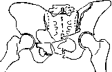
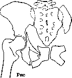
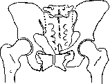
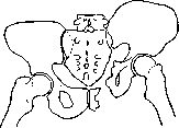
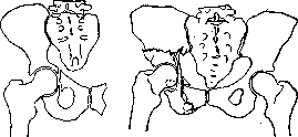
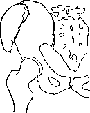
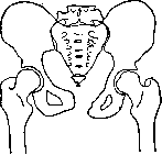
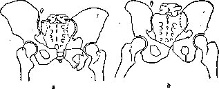
of the left pubic and ischial bones and avulsion of the transverse process of the V lumbar vertebra: a-before treatment; b-after treatment. Diagnosis of fracture of the P. in severe injuries to the P., accompanied by damage to pelvic organs, does not present great difficulties, although in cases when the patient is brought in unconscious, especially if he has many other injuries, fractures of the P. may remain unnoticed. The diagnosis of fracture of the P. is especially difficult in cases of isolated fractures and without displacement. Familiarity with the mechanism of trauma is often of decisive importance at the very first orientation. Such moments as "crushing" of the P., falling from a height, being hit by a tram, automobile, train, etc., should immediately suggest the possibility of a fracture of the P. Next, a characteristic feature is the typical position of the victim. In case of rupture of the symphysis in the patient, both legs are bent at the knees and adducted; the patient does not allow them to be separated, experiencing severe pain in the process. In fractures of both pubic and ischial bones, as well as in vertical fractures of Malgaigne, a characteristic feature is the "frog position" (according to Volovich), i.e. bent knees in a state of abduction. In fracture of the horizontal branch of the pubic bone, the patient cannot lift the corresponding leg in an extended position, cannot lift the heel off the bed (if his leg is raised, the patient raises it himself and holds it without outside help). This symptom is explained by the fact that the m. ilio-psoas, crossing over the horizontal branch of the pubic bone, is traumatized by the fragments of this bone during contraction and causes painful sensations. This sign is almost infallible: an error can only occur in cases of hemorrhage into the muscle itself, the iliopsoas, due to which the patient cannot contract the injured muscle and does not raise the extended leg. Furthermore, when proceeding to palpation, the P. is first compressed in the sagittal and frontal directions, which, in case of disruption of the pelvic ring, gives abnormal mobility and rare tenderness at certain points (sometimes crepitus). Knowing the mechanisms of trauma and the typical lines of fracture of the bones of the P., one can discover the fractures themselves by palpating typical sites. For this purpose, it is necessary to palpate the area of the symphysis, the horizontal branches of the pubic bones, the ascending branches of the ischial bones, the crests and the iliac bones, behind the sacroiliac joint (sometimes it is possible to directly palpate the displaced fragments of the iliac bone or sacrum). Palpation should be supplemented by examination per rectum and in women per vaginam; in this way, fractures of the sacrum, coccyx, pubic arch, etc., can be detected. Very valuable additional data are provided by comparative measurements of both legs and both halves of the body, proposed by Dr. Bejoul. Shortening of the lower limb on the affected side may be due to fracture of the femoral neck or fracture of the acetabulum. This same measurement, in case of avulsion of the anterior superior spine with upward displacement of the fragment by contraction of the external and internal oblique muscles, gives apparent lengthening of the affected limb; to verify these data, it is necessary to compare measurements from the greater trochanters to the ankles and from the spina ilii ant. sup. to the greater trochanter of the same side. An increase in the specified distance on the affected side indicates avulsion of the anterior superior spine, pulled upward by the muscles. Measurement from the sternoclavicular joint to the anterior iliac spines of both sides reveals shortening on the affected side in case of avulsion of the iliac crest. Shortening of the distance from the anterior iliac spine to the symphysis indicates fracture of the pubic bones. Lateral measurements are not made in the first days, as turning on the side is extremely painful for severely ill patients. These measurements are important not only for diagnosis but also for assessing the final results. The diagnosis is finally confirmed by X-ray, and it is desirable to take an X-ray of the entire pelvis. Associated damage to internal organs. The prognosis in fractures of the P. depends on damage to internal organs. Characteristic are injuries to pelvic organs, and in fractures of the anterior part of the symphysis ring and pubic bones, damage to the bladder and urethra is observed. Damage to the rectum is extremely rare. In injuries to the sacral region, root phenomena are possible, mainly from the sciatic nerve. Damage to the bladder, intestines, and sexual organs (female) must be diagnosed immediately, as delay in diagnosis, and therefore in appropriate surgical intervention, can cost the patient's life. First of all, one must think of damage to the bladder or urethra. An important diagnostic sign is the presence of blood in the urine (see Bladder and Urethra). Course. As for the course of fractures of the P., it is determined first by the nature of the fracture of the pelvic bones, displacement of fragments, and contusion of adjacent soft tissues, formation of hematomas due to rupture of deep vessels; second, by damage to pelvic organs; and third, by associated injuries to other parts of the body, which are the result of the trauma itself. The serious condition of a patient with a fracture of the P. upon admission to the hospital may be due to shock, which is observed in Malgaigne fractures and in fractures of the posterior parts of the P. Cases of death of patients are observed where at autopsy nothing but extensive damage to the P. is found. Bleeding is also of great importance: the richly vascular area of the sacrum in fracture of the P. gives large collections of blood in the retroperitoneal tissue, descending into the pelvis and after several days appearing as a hematoma (in the shape of a butterfly) on the perineum. In these severe cases, patients can be saved by immediate blood transfusion. In cases of complications from the bladder and rectum, the entry of infection into the blood-soaked tissue can easily be a source for the development of sepsis or osteomyelitis. In most cases, death in the first hours is associated with the severity and multiplicity of injuries (simultaneous fracture of several ribs, contusion of the lung, fracture of the spine, kidney injury, finally fracture of the skull, etc.). Uncomplicated fractures of the pelvis are not so dangerous to the patient's life, although (except for isolated fractures of the P.) they usually proceed as severe diseases, accompanied by increased pulse, elevated temperature, and severe general condition. All this is explained by extensive hemorrhages in rupture of large vessels and crushing of soft parts, which always accompany fractures of the pelvic ring. Extensive internal hemorrhages accompanying fractures of the P. are what classify fractures of the P. as severe diseases. However, in the absence of serious complications, the absorption of the spilled blood gradually ends, after 1-2 weeks the temperature drops, the pulse normalizes, the patient's condition improves and returns to normal; after 2-3 weeks only tenderness in the area of the fracture and difficulty in movements remain. Treatment. In the treatment of uncomplicated fractures of the P., which do not pose a direct threat to the patient's life, one should from the very first days care about the complete restoration of the patient's working capacity. The pelvic ring is the support of the trunk when sitting, standing, and walking; therefore, to restore working capacity in fracture of the pelvic ring, it is necessary to achieve the most secure connection of the fragments. Based on this, the patient should be placed in a position in which the muscles attached to the bone fragments would be in a relaxed state and the fragments could be freely held in place. For this, in fractures of the anterior part of the pelvic ring, the patient is placed in a supine position (watch for bedsores). Legs-in slightly bent and abducted position, rollers under the knees (fig. 12). In cases with displacement-slight traction with adhesive plaster on the corresponding thigh (2-3 kg) (fig. 13). In vertical fractures of the Malgaigne type, accompanied by displacement of half of the P. backward and upward, it is necessary to apply stronger skeletal traction on the thigh (up to 10 kg) with elevation of the foot end of the bed (countertraction by body weight). In ruptures of the symphysis, accompanied by divergence of the anterior pelvic ring, to the above measures is added the application of a circular corset or, even better, suspension of the P. in a hammock with crossed traction in opposite directions. This measure eliminates the divergence of the symphysis (fig. 14). It is necessary to achieve correct closure of the pelvic ring and, with the help of traction, fight against the tilting of the P. Failure to observe these measures results in improper healing of the pelvic ring, which affects the gait ("duck gait"), causes rapid fatigue and subsequently disrupts the entire posture of the victim. Particular importance is attached to the disruption of the pelvic ring in women, where this can lead to disruption of the act of childbirth, about which the patient should be warned.
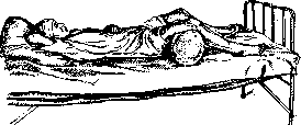
Figure 12.
pelvic ring, it is necessary to achieve the most secure connection of the fragments. Based on this, the patient should be placed in a position in which the muscles attached to the bone fragments would be in a relaxed state and the fragments could be freely held in place. For this, in fractures of the anterior part of the pelvic ring, the patient is placed in a supine position (watch for bedsores). Legs-in slightly bent and abducted position, rollers under the knees (fig. 12). In cases with displacement-slight traction with adhesive plaster on the corresponding thigh (2-3 kg) (fig. 13). In vertical fractures of the Malgaigne type, accompanied by displacement of half of the P. backward and upward, it is necessary to apply stronger skeletal traction on the thigh (up to 10 kg) with elevation of the foot end of the bed (countertraction by body weight). In ruptures of the symphysis, accompanied by divergence of the anterior pelvic ring, to the above measures is added the application of a circular corset or, even better, suspension of the P. in a hammock with crossed traction in opposite directions. This measure eliminates the divergence of the symphysis (fig. 14). It is necessary to achieve correct closure of the pelvic ring and, with the help of traction, fight against the tilting of the P. Failure to observe these measures results in improper healing of the pelvic ring, which affects the gait ("duck gait"), causes rapid fatigue and subsequently disrupts the entire posture of the victim. Particular importance is attached to the disruption of the pelvic ring in women, where this can lead to disruption of the act of childbirth, about which the patient should be warned.
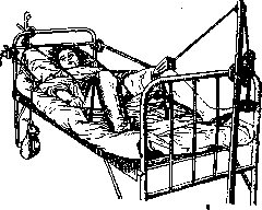
Figure 13.
thigh (2-3 kg) (fig. 13). In vertical fractures of the Malgaigne type, accompanied by displacement of half of the P. backward and upward, it is necessary to apply stronger skeletal traction on the thigh (up to 10 kg) with elevation of the foot end of the bed (countertraction by body weight). In ruptures of the symphysis, accompanied by divergence of the anterior pelvic ring, to the above measures is added the application of a circular corset or, even better, suspension of the P. in a hammock with crossed traction in opposite directions. This measure eliminates the divergence of the symphysis (fig. 14). It is necessary to achieve correct closure of the pelvic ring and, with the help of traction, fight against the tilting of the P. Failure to observe these measures results in improper healing of the pelvic ring, which affects the gait ("duck gait"), causes rapid fatigue and subsequently disrupts the entire posture of the victim. Particular importance is attached to the disruption of the pelvic ring in women, where this can lead to disruption of the act of childbirth, about which the patient should be warned.
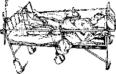
Figure 14.
In fractures of the pelvis, taking into account its supporting and static significance, as well as the fact that the pelvic ring is the site of attachment of large muscle groups that move the lower limbs and keep the torso in a vertical position, it is necessary to strengthen these muscle groups. It is necessary throughout the patient's stay in the hospital (even better if this can be continued on an outpatient basis and after discharge) to conduct strictly dosed therapeutic physical exercise. The main task in the application of therapeutic physical exercise is to 1) raise the general tone of the entire organism, 2) with special, gradually increasing exercises, strengthen those muscle groups that will bear a heavy load during standing and walking and maintaining the deformed pelvic ring in a stable position. In cases not complicated by damage to internal organs, these exercises should be started from the first days after the injury, as soon as normal temperature is established, gradually increasing their intensity, amplitude, and duration. By the end of the first month in mild cases and in two months in severe cases, the patient's musculature is strengthened by gymnastics to the extent that it is no longer difficult for him to get out of bed and move without crutches and without a cane. Experience in conducting therapeutic physical exercise according to the method of E. F. Dreving, carried out in the traumatology department of the Sklifosovsky Institute, convinces us that therapeutic physical exercise in fractures of the pelvis excellently trains and prepares the worker to return to work, making it unnecessary in a whole series of cases to transfer to temporary disability. Special importance for the complete restoration of function has the early and persistent application of gymnastics in intra-articular fractures of the acetabulum. Being convinced advocates of early active movements in all kinds of fractures (especially intra-articular), considering that active movements contribute to the most rapid restoration of function, rapid resorption of hematomas, improvement of lymphatic and blood circulation, and consequently, more rapid formation of bone callus, we nevertheless, in fractures, are opponents of "early rising," i.e., early uneven loading of the bone callus (especially dangerous in fractures of the pelvis). Premature loading of the still soft bone callus contributes to displacement of fragments, curvature of the pelvis, and as a result, the rocking "duck gait" about which much is written in the works of old authors and which can be completely avoided with proper treatment of pelvic fractures. Proceeding from these premises, in the treatment of fractures of the pelvis, the following periods should be adhered to: in isolated fractures of the pubic bones, in which the integrity of the pelvic ring as such is not violated, patients are kept in bed for 3-4 weeks. As for fractures with disruption of the pelvic ring, so-called X-shaped fractures or vertical Malgaigne fractures, even incomplete ones, as well as partial fractures of the acetabulum, etc., the minimum period of bed rest is 2 months. Of course, these periods are extended depending on the age of the patient, the presence of complications from other organs and parts of the skeleton, and the displacement of fragments. It is especially necessary to keep in bed ruptures of the anterior and posterior symphyses for a long time, since achieving union in a rupture of the symphysis is much more difficult than in a fracture. In order to correctly approach the solution of the question of the timeframes for restoration of working capacity, of temporary and permanent disability in fractures of the pelvis, one should first consider fractures of the pelvis not as an isolated injury, but as a severe general, often multiple injury, simultaneously disrupting the function of a whole series of organs and parts of the body; secondly, one should differentiate cases of fracture of the pelvis depending on their anatomical localization, based on the data presented above and remembering that fractures of the thick and thin pubic and ischial bones form bone callus much faster than fractures of flat bones—the ilium and especially the posterior parts of the pelvic ring, the sacrum. Fractures of the acetabulum require special attention: here one should strive from the very beginning for full mobility, since limitation of mobility, and even more so ankylosis of the hip joint, can lead to permanent loss of working capacity. In this, one should take into account the age and sex of the patient. Fractures of the pelvis in women should be treated with special care, since premature loading can lead to deformation of the pelvic ring, which has specific significance for women. Finally, when determining the timeframes for leaves (in fractures of the pelvis), one should take into account the nature of the labor processes and the severity of the upcoming work in terms of load on the pelvic ring. When evaluating the long-term results of fractures of the pelvis and when determining loss of working capacity, attention should be paid 1) to changes in the skeleton that give shortening of the limb (checked by measurements), sagging of one half of the pelvis (Trendelenburg symptom), secondary curvature of the spine; 2) to improper unions or absence of unions at the site of fracture or at the sites of rupture of symphyses, which causes rocking when walking—the "duck gait," uncertainty and rapid fatigue when walking and standing; 3) to functional disorders—limitation of movement in the hip joint (stiffness and even ankylosis) in fractures of the acetabulum; difficulty in spreading the legs, inability to squat. All these points not only extend the timeframes of loss of working capacity but sometimes require transfer to temporary disability or a change of profession. On the other hand, one should remember that one or another deformation of the pelvis, visible on an X-ray, in itself does not give the right to judge the loss of working capacity of the injured person, since ultimately the decision on returning to work depends on the completeness of functional restoration. One can often observe people with large deformities of the pelvis after improperly united fractures, coping with any work thanks to well-developed musculature that compensates for all defects of the deformed skeleton. Hence it is clear what enormous importance functional treatment of fractures of the pelvis has with the application of methods of therapeutic physical exercise. If with respect to the first five groups the timeframes and completeness of restoration of working capacity depend to a greater extent on the early and systematic application of the functional method of treatment, then with respect to fractures of the last group (see the classification above), the matter is considerably more complicated. Here the complications associated with damage to other organs decide the prognosis; damage to pelvic organs—urinary fistulas, disorders of urination, pyelitis, suppurations, etc., which worsen the prognosis, accompanying damage to peripheral nerves (n. ischiadicus and n. peroneus), which are often overlooked at first and make themselves known with persistent pains and sometimes with pareses. Proceeding from functional indicators, covering in one's assessment the entire traumatized patient as a whole, knowing the nature of his work, one can in each case correctly determine the timeframes for restoration of working capacity, the possibility of returning to work and the place he should occupy in production. Inflammatory processes of the pelvic region can originate from the bones of the pelvis—osteitis, periostitis, and osteomyelitis, from the joints—coxitis, inflammation of the sacroiliac joint or inflammatory processes of the symphysis (see Coxitis, Sacroiliac joint, Pubic symphysis). Further, inflammatory processes can have as their point of origin the pelvic organs—urinary bladder, urethra, female genital organs (see Parametritis), rectum (see Paraproctitis) and finally the extra-peritoneal parts of the cecum (see Cecum, Typhlitis). Inflammatory processes can develop in the pelvic cellular tissue, originating from retroperitoneal glands; the source of infection can be inflammatory processes on the lower limb. Finally, inflammatory processes penetrate into the region of the pelvis, descending along the muscles and interfacial spaces—cold abscesses. In addition, inflammatory processes can develop in the soft parts surrounding the pelvic ring—in the gluteal region, in the perineum, in the inguinal region, penetrating into the depth as a result of damage to the external coverings—infected wounds or in inflammatory and necrotic processes of the skin—FURUNCles, eczema and finally bedsores. Inflammatory processes of the pelvic skeleton, osteomyelitis, most often arise as complications in open (gunshot) fractures of the pelvis, in injuries to the urinary bladder in connection with urinary extravasations. More rarely, osteomyelitis of the pelvis is observed as a complication of infectious diseases (typhoid, angina, scarlet fever, measles, etc.); in a number of cases it is difficult to determine the portal of entry of the infection. The causative agent of osteomyelitis is usually staphylococcus. Localization—most often the ilium, which is affected more often than any other flat bones of the skeleton. The sacrum is affected relatively rarely (see Sacrum). A diffuse and limited form of osteomyelitis of the pelvis are distinguished.
The diffuse form begins in the anterior part of the ilium: the entire bone is penetrated by small abscesses, which gradually merge and detach the periosteum from the outer or inner surface and eventually extend to neighboring joints (hip or sacroiliac). The limited, focal form is localized in the area of the acetabulum, the iliac crest, or in the area of the sacral wings and very rarely in the anterior parts of the pelvic ring. The focal form runs comparatively mildly. The course of diffuse osteomyelitis of the pelvis is extremely severe, often ending in death. The onset is sudden: a shaking chill, high temperature, loss of consciousness, pains in the depth, radiating to the sacrum or hip joint. After some time, a tumor appears, later multiple fistulas open. The intense general picture often obscures the local phenomena. Acute osteomyelitis of the pelvis should be differentiated from typhoid and acute articular rheumatism. The diagnosis is confirmed by the appearance of an inflammatory tumor and fistulas. It is especially difficult to make a diagnosis when the process is localized on the inner surface of the pelvic bones. An X-ray of the pelvis can be of great help, however, it must be remembered that changes on the X-ray are detected only in a developed and advanced process. Chronic osteomyelitis of the pelvis either arises from acute or begins gradually. In chronic osteomyelitis of the pelvis, the affected bones thicken, osteophites form on the ilium, severe changes occur in the neighboring joints—subluxations, ankyloses, as a result of which deformation and change in the configuration of the entire pelvis occur. Abscesses of the pelvis, arising in connection with osteomyelitic processes in the pelvic bones or in connection with tuberculosis of the pelvic bones, exit from the pelvic cavity to the surface along certain paths, which are predetermined by the course of the fasciae and muscle groups; they rupture outward, forming fistulas localized in typical places. These paths are as follows: 1) the path along the course of the iliac muscle—iliac abscesses. The abscess originating from the bones of the sacrum, the inner surface of the ilium or from the area of the joint socket, descends along the iliac muscle to the inguinal canal, in rare cases rupturing the skin at the height of the eminentia pectinea. More often the abscess passes under the inguinal ligament, in the space between the outer edge of the iliac muscle and the inner edge of the rectus femoris, exiting here outward; in other cases the abscess descends further, rupturing between the m. rectus femoris and the m. tensor fasc. latae or near the sartorius muscle. The second path of pelvic abscesses—through the foramen ischiadicum, under the gluteal muscles, with exit directly in the gluteal region or on the posterior surface of the thigh. In addition to abscesses originating from the pelvic bones, abscesses (most often of tuberculous origin) pass through the pelvic cavity, descending from the spine along the m. psoas, which in these cases assumes the same leading role as the m. iliacus in abscesses coming from the pelvic ring. Fistulas in this case are located on the anterior surface of the thigh, medial to the m. psoas. In the pelvic region, subserous abscesses are also located, not associated with the bones of the pelvis, originating from organs that are partially covered by peritoneum: the cecum, female genital organs, urinary bladder. These abscesses are located in the area of the inner two-thirds of the inguinal ligament, displacing the peritoneum upward; fistulas with them are above and below the inguinal ligament, sometimes near the femoral ring (see Cecum, Urinary Bladder, Parametritis, etc.). For a correct differential diagnosis, a thorough examination of the skeleton (spine and pelvis) as well as appropriate examinations of internal organs is necessary. Abscesses originating from the outer surface of the pelvic bones are easier to determine. Here one should differentiate with abscesses of the gluteal muscles, suppurated hematomas, phlegmons of the gluteal region. The localization of pains, soreness on percussion and pressure on the pelvic bones, the course of the process itself, and above all the X-ray of the pelvis help to make the diagnosis. One should not forget the examination per rectum and per vaginam. Phlegmons of the gluteal region and intramuscular phlegmons develop from infected hematomas or as a complication of intramus medicinal injections conducted with insufficient asepsis. Treatment. Abscesses and phlegmons of the gluteal region are opened with wide incisions and treated according to the general rules of purulent surgery. Acute osteomyelitis of the pelvis is also treated surgically. If the general condition is not impaired, one can wait for the abscess to be delimited, and then it is sufficient to open it. With high temperature and severe pains, one should not wait for fluctuation, but should make an incision at the site of the swelling. The matter may be limited by this, a large amount of pus is discharged with the separation of a small superficial sequestrum. With extensive changes of the ilium and sacrum, the matter is much more complicated. Schede, Bergmann, and others attempted in such cases to remove a large area of bone, to perform a total resection of the ilium. Larghi (1845) proposed an extensive incision from the spina ilii ant. to the spina ilii post., going directly under the iliac crest. The gluteal muscles are displaced along with the periosteum. The ilium is chiseled out with a wide chisel and is removed subperiosteally, the huge defect is subsequently completely restored from the periosteum. In chronic osteomyelitis with fistulas, the fistulas are incised and small sequestra are removed. Removal of large sequestra from the inner surface of the pelvis presents particular difficulties. If one cannot approach from the outer surface and trephine the bone, then from the inner surface the peritoneum should be detached, the large vessels should be pushed aside with a blunt instrument, and, having detached the periosteum, approach the sequestrum. Tuberculous osteitis of the pelvic bones, except for the sacrum and coccyx, is found on the ilium, localizing in its dense part, directly behind the acetabulum, subsequently extending to the hip joint. Often the tuberculous process localizes in the area of the symphysis on the pubic and ischial bones, i.e. just where osteomyelitis does not occur due to the absence of spongy substance. Treatment is first of all general, locally—incision of fistulas, curettage of foci with a sharp spoon, introduction of iodoform emulsion. In recent years, most surgeons, even in cases of tuberculosis of the pelvic bones, lean toward conservative methods of treatment (especially in children). Great importance is attached to treatment with a quartz lamp, etc. Syphilis of the bony pelvis is rare. Early forms of gummatous periostitis and osteoperiostitis are easy to diagnose when there are also other signs of fresh syphilis; great difficulties are presented by the diagnosis of tertiary syphilitic osteitis, which localizes on the ilium as well as on the sacrum and leads either to thickening of the affected bone or to a carious process with the formation of abscesses. Treatment is specific, softened gummas are curetted with a sharp spoon. Among other diseases of the pelvic bones, one should mention deforming osteitis (see), fibrous osteitis (see), osteomalacia (see). Tumors of the pelvis can originate 1) from pelvic organs, 2) from the soft parts surrounding the pelvic ring, 3) from the pelvic ring itself and 4) from the pelvic cellular tissue. Tumors originating from the skin coverings of the pelvis and from the soft tissues surrounding the pelvic ring—atheromas, dermoids, cysts of mucous bursae, lipomas of various sizes on a stalk and without a stalk—present nothing characteristic for this area. Tumors developing from the bones of the pelvis—exostoses, enchondromas, fibromas, sarcomas. Exostoses are located on the inner surface of the pelvic bones, usually on the ilium, near the sacroiliac joint. When these benign formations reach large sizes, they can mechanically cause suffering and disruption of functions, compressing the pelvic organs. Compression of the rectum can cause complete obstruction and require the creation of an anus praeternaturalis to save the patient's life. Exostoses of the pelvis are especially dangerous during childbirth, which is why they should be removed in advance in women. Exostoses developing on the outer surface of the ilium are less dangerous, however, even with significant growth they can hinder movement in the hip joint or can press on the sciatic nerve, and therefore should be removed in a timely manner. Enchondromas, initially benign tumors of dense consistency, when reaching large sizes, soften and give metastases; with the underlying parts of the enchondroma are associated a thin or thick stalk. Enchondromas develop more often on the inner surface of the ilium, rarely in the area of the sacroiliac fossa. External enchondromas originate from the pubic or ischial bone. When reaching a large size, enchondromas stretch the skin and ulcerate. In connection with twisting of the stalk, ichorous decay of the tumor occurs. Fibromas of the pelvis are rare, usually arising from the connective tissue layers of the periosteum. The favorite localization—the spina ilii ant. sup., from where they descend into the iliac fossa. The consistency of such fibromas is dense, 293
Tuberculous osteitis of the pelvic bones, except for the sacrum and coccyx, is found on the ilium, localizing in its dense part, directly behind the acetabulum, subsequently extending to the hip joint. Often the tuberculous process localizes in the area of the symphysis on the pubic and ischial bones, i.e. just where osteomyelitis does not occur due to the absence of spongy substance. Treatment is first of all general, locally—incision of fistulas, curettage of foci with a sharp spoon, introduction of iodoform emulsion. In recent years, most surgeons, even in cases of tuberculosis of the pelvic bones, lean toward conservative methods of treatment (especially in children). Great importance is attached to treatment with a quartz lamp, etc. Syphilis of the bony pelvis is rare. Early forms of gummatous periostitis and osteoperiostitis are easy to diagnose when there are also other signs of fresh syphilis; great difficulties are presented by the diagnosis of tertiary syphilitic osteitis, which localizes on the ilium as well as on the sacrum and leads either to thickening of the affected bone or to a carious process with the formation of abscesses. Treatment is specific, softened gummas are curetted with a sharp spoon. Among other diseases of the pelvic bones, one should mention deforming osteitis (see), fibrous osteitis (see), osteomalacia (see). Tumors of the pelvis can originate 1) from pelvic organs, 2) from the soft parts surrounding the pelvic ring, 3) from the pelvic ring itself and 4) from the pelvic cellular tissue. Tumors originating from the skin coverings of the pelvis and from the soft tissues surrounding the pelvic ring—atheromas, dermoids, cysts of mucous bursae, lipomas of various sizes on a stalk and without a stalk—present nothing characteristic for this area. Tumors developing from the bones of the pelvis—exostoses, enchondromas, fibromas, sarcomas. Exostoses are located on the inner surface of the pelvic bones, usually on the ilium, near the sacroiliac joint. When these benign formations reach large sizes, they can mechanically cause suffering and disruption of functions, compressing the pelvic organs. Compression of the rectum can cause complete obstruction and require the creation of an anus praeternaturalis to save the patient's life. Exostoses of the pelvis are especially dangerous during childbirth, which is why they should be removed in advance in women. Exostoses developing on the outer surface of the ilium are less dangerous, however, even with significant growth they can hinder movement in the hip joint or can press on the sciatic nerve, and therefore should be removed in a timely manner. Enchondromas, initially benign tumors of dense consistency, when reaching large sizes, soften and give metastases; with the underlying parts of the enchondroma are associated a thin or thick stalk. Enchondromas develop more often on the inner surface of the ilium, rarely in the area of the sacroiliac fossa. External enchondromas originate from the pubic or ischial bone. When reaching a large size, enchondromas stretch the skin and ulcerate. In connection with twisting of the stalk, ichorous decay of the tumor occurs. Fibromas of the pelvis are rare, usually arising from the connective tissue layers of the periosteum. The favorite localization—the spina ilii ant. sup., from where they descend into the iliac fossa. The consistency of such fibromas is dense, 293
The tumor is sometimes soft and fleshy (giving false fluctuation); over time, the tumor may ossify and become calcified (should not be confused with sub-peritoneal fibromas of pelvic organs). Removal of fibromas of the pelvis is associated with great difficulties and requires resection of the pelvic bones (Kuttner). Sarcoma grows endostally or periostally or from the bone marrow. In contrast to fibromas, sarcoma grows from the inner layers of the periosteum—the outer layer remains as a fibrous covering. Sarcoma originating from the bone marrow is a richly vascular, pulsating tumor. If the sarcoma grows on the outer surface of the pelvis, diagnosis presents no difficulties; if on the inner surface, it can be confused with an aneurysm of large vessels. Localization is the middle of the iliac fossa outward from the vessels and nerves, less commonly the sacro-iliac fossa, sacrum, or pubic bone. Usually, the sarcoma is closely connected with the bone and has no sharp boundaries, although sometimes it has a capsule. Sarcoma of the pelvis can have various consistencies: hard, uneven, elastic, fluctuating. Sometimes a parchment-like crack is felt on palpation. Usually the skin is unchanged, but with rapid growth, decay and ulceration may appear. Unbearable pain from compression of nerve trunks by the sarcoma is an early sign, which however is often underestimated and considered as neuralgia, sciatica. The tumor is palpated significantly later, metastases develop quickly and death occurs. Surgical removal is possible only with early diagnosis and requires extensive resections of the pelvis. The operation is amputation interilio-abdominalis (see Ampuiatio interilio-abdominalis). Echinococci, developing as single-chamber or multi-chamber cysts, are located either in the bones of the pelvis or in the pelvic cellular tissue—in women in the area of the parametrium, in men between the bladder and rectum. Echinococci of the pelvic cellular tissue reach a large size and, if not recognized in time, rupture into nearby organs (bladder). When recognized in time—removal according to general rules. In the bones of the pelvis, echinococcus develops rarely, localizing in the ilium or in the area of the acetabulum (rupture into the hip joint is possible). Usually the tumor is the size of a fist, however destruction can spread to half of the pelvis. The operation must be performed in a timely manner. Late operations give recurrences or profuse suppurations, from which patients die. For other operations see Amputatio interilio-abdominalis, Pelviotomy. V. Gorineietskaya. III. Female pelvis. Anatomically, the female pelvis compared with the male (see above) has a number of features (secondary sexual characteristics). These features in general consist in the fact that the female pelvis is much more capacious than the male (fig. 15-18); it is wider in the transverse direction, lower in the vertical; the pubic angle in the female pelvis is straight or obtuse, i.e. much larger than in males (acute); the general shape of the small pelvis in a woman is cylindrical, whereas in a man it is cone-shaped narrowing downward (funnel-shaped pelvis). The size and shape of the female pelvis have enormous importance in obstetrics, however this importance for a long time escaped the attention of obstetricians and anatomists, and only at the beginning of the 18th century appeared the work of Deventer, in which for the first time attention was drawn to the importance of a narrow pelvis in obstetrics. Since then and to the present time, the female pelvis has been the subject of such detailed study that a special discipline pelviology—the science of the pelvis (pelvis in Greek—pelikos)—has been created.
Figure 15. Male pelvis
Figure 16. Female pelvis
from the front and above.
from the front and above. The study of the pelvis proceeds both dynamically—in the sense of studying the laws of its development and formation in normal and pathological conditions, and statically—in the sense of studying the influence which this or that established form of the pelvis exerts on the reproductive functions of the female body—pregnancy and childbirth. It must however be said that the special shape and size of the female pelvis determine certain peculiarities of the female organism outside its reproductive function. For example, the width of the female pelvis in the transverse direction to a large extent determines certain peculiarities of the external
Figure 17. Male pelvis
Figure 18. Female pelvis
from below and behind.

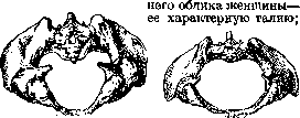
below and behind. further, the same width of the pelvis determines the difference in the attachment of the lower limbs (thighs): in a woman, the thighs approach the pelvis at an angle from within, whereas in a man the thighs have an almost vertical and parallel arrangement. This feature of the attachment of the lower limbs to the pelvis is reflected, among other things, in such an important function as walking - the female gait sharply differs from the male. In what follows, mainly those aspects of the female pelvis that present obstetric interest are considered. It is quite natural that obstetricians are primarily interested in the dimensions of the female pelvis and their correspondence to the dimensions of the fetus, which must pass through the pelvis during childbirth (see Childbirth). Since these dimensions can be determined accurately only on a pelvis skeleton, and on a living woman they cannot be subjected to exact direct measurement, for their determination use is made of measuring the so-called external dimensions of the pelvis (see Obstetric examination). There are four such dimensions: 1) the external conjugate size (conjugata externa) - the distance from the spinous process of the last lumbar vertebra to the upper edge of the pubic symphysis; this distance equals 20 cm; 2) the distance between both anterior superior iliac spines - distantia spinarum (ossium pubis) equals 26 cm; 3) the distance between the crests of both iliac bones - distantia cristarum equals 29 cm; 4) the distance between the greater trochanters of both femora - distantia (inter-) trochanterica equals 32 cm. These four dimensions give the external index of the female pelvis. For a normal pelvis it will therefore be as follows: 20: 26: 29 : 32. If there were an exact and constant correlation between the external dimensions of the pelvis and the dimensions of the inlet, the task of obstetric measurement would be very simple. Unfortunately this is not so. however in practice it is customary to proceed from the premise that such a correlation exists and the pelvis is characterized precisely by this external index. In addition to the inlet of the pelvis, in obstetrics great importance is attached to the cavity of the small pelvis and the outlet of the pelvis. The outlet of the pelvis is characterized by two dimensions: 1) the conjugate diameter of the outlet - the distance from the lower edge of the pubic symphysis to the tip of the coccyx equals 9 cm, but when the coccyx is bent backward this dimension increases to 11 cm; 2) the transverse diameter of the outlet is the distance between both ischial tuberosities - also equals 11 cm. Knowing the dimensions of the inlet and outlet of the pelvis, one can form a fairly accurate idea also of the cavity of the pelvis - both as to its dimensions and as to its shape. Upon more detailed study in the cavity of the pelvis a wide and narrow part are noted and these parts of the pelvis are characterized by the corresponding dimensions, however these dimensions on a living woman cannot be measured, and for practical purposes it is sufficient to know that in the cavity of the pelvis the largest dimension is not the transverse but the oblique. - Besides the dimensions of the pelvis and its shape determined by these dimensions, the so-called inclination of the pelvis (inclina-tio pelvis) also has generally recognized significance, namely the angle formed by the true conjugate with the horizontal plane when the woman is in the vertical position. This angle in normal conditions equals 60°. Finally in obstetrics the so-called conducting line, or axis of the pelvis (axis pelvis), has significance - a curved line connecting the midpoints of the conjugate diameters of the inlet, cavity, and outlet. The curvature of this line almost corresponds to the curvature of the sacrum. The above-given dimensions of the female pelvis characterize the so-called normal pelvis. Variational fluctuations of these dimensions within 2 cm allow considering as normal also those pelves that are somewhat larger or somewhat smaller than the given figures; but if any of these dimensions of the pelvis is reduced by 2 cm or more, then from the obstetric point of view the pelvis is already considered narrow. The greatest significance in obstetrics is attached to the conjugate diameter of the inlet. If this dimension is less than 11 but more than 9 cm, one can speak of a narrowed pelvis, if it equals 9 cm and less, then the pelvis is already narrow. Since conjugata vera cannot be subjected to direct measurement, it is conventionally accepted to consider as narrow those pelves in which conjugata externa equals 18 cm and less. Such a conventional definition of a narrow pelvis is fully justified by the needs of everyday obstetric work. To clarify the narrowing of the pelvis one can measure the distance from the lower edge of the pubic symphysis to the tip, the so-called diagonal conjugate (conjugata diagonalis). Narrow pelves according to the degree of their narrowing and the shape they have at the same time are of the most diverse character; for a long time attempts have been made to work out one classification or another of narrow pelves, however to this day there is no classification that would satisfy all the requirements of scientific methodology. The simplest classification and at the same time quite sufficient for practical purposes is the division of all pelves according to the degree of narrowing: to the 1st degree of narrowing belong pelves with conjugata vera between 11-9 cm, to the II-9-7 cm, to the III- 7-5 cm and to the IV degree of narrowing all pelves in which conjugata vera is below 5 cm (Bumm). The last category of pelves is called absolutely narrow pelves, since with these pelves delivery through the natural birth canals (per vias naturales) is practically impossible and the only possible operation is cesarean section for so-called absolute indications. With the third degree of narrowing delivery through the natural canals is possible, but as a rule only with the help of destructive operations on the fetus, and finally with the second degree of narrowing birth is possible also through the natural canals, however compared with a normal pelvis birth with a narrow pelvis of the second degree is protracted, operative assistance is relatively frequently required and in connection with this with such narrow pelves there is a relatively greater stillbirth rate, a relatively greater morbidity and mortality of mothers. Since narrowing of the second degree occurs significantly more frequently than narrowing of the third and especially fourth degree, and such pelves are encountered in general in 10% of women, it is precisely these narrow pelves that constitute one of the main problems of practical obstetrics. It is quite clear that the given classification is far from satisfactory. A second simplified classification is the classification according to the shape of the pelvis, connected with its narrowing (fig. 19). The most frequent form of narrowing is narrowing in the conjugate diameters. Such pelves are called flat (pelvis plana). Besides narrowing in the conjugate diameters and along with it narrowing in other dimensions of the pelvis also occur. If the pelvis is narrowed equally in all dimensions, then the pelvis retains its shape but turns out to be smaller than normal. Such pelves are called generally narrowed (or uniformly narrowed) (pelvis simpliciter, s. aequabi-liter justo minor; pelvis nimis parva). If the pelvis is narrowed mainly in the transverse dimensions, which occurs rarely, then this is a transversely narrowed pelvis. If the narrowing of the pelvis is expressed mainly in the outlet, then the pelvis is called funnel-shaped (pelvis infundibuliformis angusta). All the enumerated forms of the pelvis are characterized by the fact that the narrowing of the pelvis affects both halves of the pelvis equally, without disturbing its symmetry, why all these pelves can be grouped into one large group of symmetrical pelves. Besides such pelves there occur pelves where the narrowing affects one half of the pelvis more than the other, i.e. where one oblique dimension is smaller than the other. Such pelves naturally lose their symmetrical shape and are referred to the group of asymmetrical pelves. In the group of asymmetrical pelves the main place is occupied by the so-called obliquely narrowed pelves. To this group also belong pelves, completely disfigured by osteomalacia or by markedly expressed rickets. Both given classifications - according to the degree of narrowing and according to shape - do not satisfy the requirements of strict science, but attempts to build a fully satisfactory classification have to this day not been crowned with success. The most developed at present is the classification of Friedrich Schauta, although it was proposed almost half a century ago. Classification of narrow pelves according to Schauta. I. Anomalies of the pelvis due to defects (intrauterine) of development: 1) generally uniformly narrowed, non-rachitic pelvis: a) generally narrowed (of female type), b) male pelvis, c) dwarf pelvis;
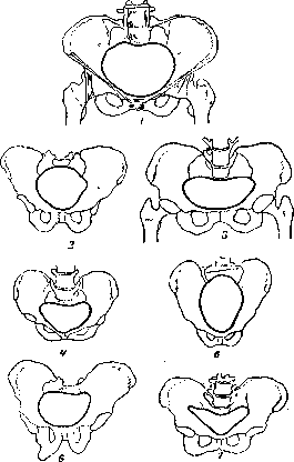
Figure 19. Various forms of narrow pelvis compared to normal: 1-normal pelvis; 2-general uniformly narrowed pelvis; 3-flat pelvis; 4-uniformly narrowed flat pelvis; 5-transversely narrowed pelvis; 6-obliquely narrowed pelvis; 7-collapsed (osteomalacic) pelvis. 1) simple flat, non-rachitic; 2) general narrowed flat, non-rachitic; 3) narrow, funnel-shaped, embryonic or Liegebecken; 4) pelvis with underdevelopment of one wing of the sacrum (Negel); 5) pelvis with underdevelopment of both wings of the sacrum (Robert); 6) pelvis wide in all dimensions; 7) cleft pelvis. II. Anomalies due to diseases of the pelvic bones: 1) rachitic P., 2) osteomalacic P., 3) neoplasms of pelvic bones, 4) fractures of pelvic bones, 5) atrophy, caries, necrosis of pelvic bones. III. Anomalies due to abnormalities in the joints of the pelvis: 1) synostoses: a) pubic symphysis, b) one or both sacroiliac joints, c) sacrum with coccyx; 2) too weak connection of pelvic bones: a) in pelvic joints, b) dislocation of the tip. IV. Pelvic anomalies due to diseases of the spine: 1) spondylolisthesis, 2) kyphosis, 3) scoliosis, 4) kyphoscoliosis, 5) assimilation. V. Pelvic anomalies due to diseases of the lower limbs: 1) coxitis, 2) dislocation of the hip (unilateral), 3) dislocation of the hips (bilateral), 4) bilateral or unilateral clubfoot (Klumpfuss), 5) absence or curvature of one or both lower limbs. The first section of Schaut's classification includes anomalies that could be called constitutional anomalies, however, it is not excluded that some of the anomalies listed here may develop on the basis of one or another fetal disease. The remaining sections of the classification do not meet objections, but here too there are controversial points (see below). 1) The general uniformly narrowed pelvis is characterized by a reduction of all dimensions of the pelvis by 2-3 cm (rarely more). Such pelvises have a perfectly correct shape and differ from normal ones only in their dimensions. They are observed in women of small stature, of miniature build. Often these pelvises, despite their reduced size, turn out to be relatively spacious, which is explained by the thinness of their bones. Sometimes, but rarely, such P. can be explained not by constitutional causes, but as a consequence of mild rachitis, suffered in childhood. The male pelvis is characterized by the roughness of the bones, the height (vertical position of the iliac bones), the sharp pubic angle and finally, as a rule, by a funnel-shaped form. In other words, the male pelvis is narrowed more in the outlet than in the inlet and in the cavity,
Figure 20. Obliquely narrowed
Figure 21. Obliquely narrowed Negelian pelvis (por
Negelian pelvis (por
tion of the left half
of the pelvis).
of the pelvis).
Male P. are observed in women of inter- and heterosexual type, often sturdy, stocky, sometimes even athletic build. - Dwarf P. (pelvis pumila) is observed in dwarfs. Usually such P. are completely narrowed. Schaut had every reason to place the dwarf P. in section I of his classification, but with modern views on the pathology of growth, it must be recognized that some dwarf P. are formed not congenitally, but on the basis of disease, e.g., of the pituitary gland, both intrauterinely and in early childhood. Dwarf pelvises are very rare in the population, and even rarer in obstetric practice. Simple flat, non-rachitic P. is the most common and most frequently occurring form of narrow pelvis. It is characterized by the fact that all direct dimensions in it are uniformly reduced, the sacrum as a whole is as if pushed inward into the pelvis and thereby brought closer to the pubic symphysis. The other dimensions of these P. are normal. These P. can be attributed to the first section only with great reservations, since experience shows that in a large number of carriers of these P. one can note as an etiological factor excessive undue labor load in childhood or early adolescence. In other words, in most cases these pelvises are not of congenital, constitutional origin, but are caused by the effects of harmful factors in extrauterine life. It is obvious that a pelvis of such origin cannot be attributed to section I of the classification. - General narrowed flat, non-rachitic P. in form resembles a simple flat one with the difference that in it not only direct but all other dimensions are reduced, however the latter to a lesser extent than the direct ones. These P. are observed in women of small stature, and in their history many of them can note undue load in adolescence. - Negelian obliquely narrowed P. is characterized by underdevelopment of one of the wings of the sacrum (fig. 20 and 21). This form of P. is very rare. Its origin is explained both by a congenital anomaly of the sacrum of constitutional character and by intrauterine disease of one of the sacroiliac joints, which leads to ankylosis of this joint and subsequently to atrophy of the corresponding sacral wing. From this it is clear that not all Negelian P. can be attributed to section I of Schaut's classification. In contrast to all previous and subsequent P. of this section, Negelian P. are asymmetrical. - Robert's transversely narrowed P. is an extremely rare form. Like the Negelian P., the origin of Robert's P. is explained by underdevelopment of the sacrum, but here both wings are underdeveloped. - Broad P. - pelvis wide in all dimensions - is observed in women of tall stature, but of typically female build. - Cleft, or open in front P. (pelvis fissa, s. aperta) is characterized by the absence of pubic symphysis. This form of P. is closely connected with such a deformity as congenital eversion of the bladder (ectopia vesicae). From this it is understandable that these P. are extremely rare, since girls born with such a defect rarely reach sexual maturity. To section II of his classification Schaut attributes P., changed in their structure due to diseases of the bones of the pelvis itself. Here rachitic pelvises (fig. 22) are rightly placed in first place. Rachitis can affect the development of the skeleton and in particular the pelvis in the most diverse way, depending both on the course of the rachitis itself and on various kinds of additional factors. In view of this, in pelvises of the most diverse form, one can note the influence of rachitis suffered in childhood. However, in obstetrics it is customary to call rachitic P. of one definite form. These are flat P., but, unlike simple flat P., rachitic flat P. has a number of characteristic features, which are determined both on the skeletonized P. and on a living woman. These features are as follows: in rachitic flat P. mainly only the direct dimension of the inlet into the pelvis is reduced, and the dir
Fig
Figure 2::. Pelvis achoi
neshshl Hiociini ra\n
plastic (chondro-
tatic pelvis.
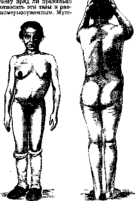
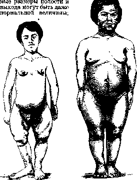
distrophic). This depends on the fact that in the rachitic flat pelvis, the sacrum is as if tilted forward along its horizontal axis, and only the tip is brought close to the pubic symphysis, while the lower half of the sacrum is deviated backward, and thus the direct diameters of the cavity and outlet are not shortened at all or are shortened much less than the direct diameter of the inlet. Besides this main and very important feature of the rachitic flat pelvis, it also has other characteristics: thus, the iliac fossae are flattened and the iliac crests are as if pressed outward, as a result of which the pelvis really has a flat shape. Due to such flattening, the distance between the iliac crests equals the distance between the anterior superior spines, whereas in a normal pelvis these distances differ by 2-3 cm in favor of the intercrestal line. This feature of the rachitic flat pelvis is already striking when measuring the pelvis externally on a living woman and is one of those signs by which the rachitic origin of a given pelvis is determined. While in simple flat pelves the narrowing of direct diameters is generally small and rarely exceeds 3-31 cm, in rachitic flat pelves there is often very great narrowing, but this narrowing is limited mainly to the inlet. The connection between the narrowing and deformation of the pelvis with rickets suffered in childhood is so clear that it was precisely in the study of rachitic pelves that various assumptions were made about the influence of one or another influences on the formation of the pelvis, both under normal and pathological conditions. The chondrodystrophic (achondroplastic) pelves should also be included in this section, the origin of which in the 19th century was explained by intrauterine development (Fig. 23). In second place in section II are placed the osteomalacic pelves. This form of pelvis also does not raise doubts regarding its origin, since osteomalacia is too characteristic a disease of the bone system and moreover it is closely connected precisely with pregnancy. It is therefore quite possible for the obstetrician to observe how under the influence of the disease of the pelvic bones its shape changes. Naturally, these changes will be different depending on the nature and course of the disease. However, obstetricians understand osteomalacic pelves mainly as those pelves where, due to great softening of the pelvic bones, they are as if pressed into the pelvic cavity and the pelvis as a result becomes collapsed. The shape of the inlet in such pelves approaches triangular, the pubic symphesis protrudes forward like a beak, and the pubic angle is sharpened to an extreme degree. New growths of the pelvic bones are observed comparatively very rarely, especially in young, childbearing age. Usually such new growths are malignant and naturally quickly lead to a fatal outcome. Thus these pelves would not have great practical obstetric significance if not for those pelves on the inner side of which there are bony, often very small, outgrowths in the form of small spurs or tubercles. Since these growths are most often located on the sacrum in the area of its joints or on the pubic symphysis, it is natural that even a small outgrowth can sharply narrow precisely the direct diameter of the inlet and thereby strongly disrupt the course of labor. Such pelves bear the name pelvis spinosa, s. acanthopelvis. The 4th and 5th rubrics of section II of Schaut's classification do not require explanation. One can only say that the changes in the pelvis in this case can be expressed very differently, and the degree and form of these pelves depend entirely on the extent and size of the bone lesion, as well as on the age when the patient suffered an injury or disease process. Section III of Schaut's classification covers such anomalies which are almost never encountered in life and therefore have no practical significance. It seems that the entire section is rather a product of theoretical assumptions and is not based on observations in life; however, it was sometimes possible to see an abnormal connection of the sacrum with the coccyx both in the sense of synostosis and in the sense of 'dislocation'. In the first case, an obstacle to labor may be encountered if the coccyx is bent inward and in this form is fused with the sacrum. A dislocation of the coccyx has no significance. Section IV of Schaut's classification encompasses anomalies of the pelvis that arose on the basis of diseases of the spine. Here, first place in frequency and practical significance should be given to kyphosis, but we will adhere to the order established by Schaut. The spondylolytic pelvis is characterized by the fact that the last lumbar vertebra as if slips from the sacrum into the inlet and even into the pelvic cavity, which of course shortens the inlet to the pelvis (Fig. 24); sometimes such a slip can be so great that the inlet to the pelvis appears as if closed (pelvis obtecta).
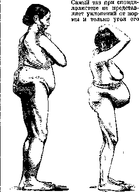
Figure 24. Spondylolisthetic
Fig. 25. Kyphotic
pelvis.
pelvis. The inclination can change. In pronounced cases of spondylolisthesis, it can be easily suspected and recognized already upon examination of the lumbar region, where a saddle-shaped depression is observed. The diagnosis is fully clarified by radiography. Kyphotic pelves are encountered comparatively often - these are the pelves of hunchbacks (Fig. 25). It should be said that not every kyphosis causes changes in the pelvis: if the kyphosis is localized in the upper part of the spine, then in the lower part of the spine a compensatory lordosis is formed, as a result of which the pressure of the trunk on the sacrum and pelvic ring does not differ from normal. On the contrary, if the kyphosis is in the lower, lumbar part of the spine, then the pelvis acquires characteristic features, and it is precisely such pelves that are called kyphotic. In a typical kyphotic pelvis, the upper part of the sacrum is deviated backward, as a result of which the direct diameter of the inlet1 is as if increased; the lateral pelvic bones are brought closer together, so that the transverse diameters of the pelvis are shortened. In form, the kyphotic pelvis is therefore transversely narrowed and funnel-shaped, since its outlet is narrowed in both the direct and transverse diameters. In addition, the inclination angle is decreased - the kyphotic pelvis lies as if horizontally. Recognition of a kyphotic pelvis presents no difficulties. Scoliotic pelves develop on the basis of scoliosis of the spine (Fig. 26). Here one can repeat what was said about kyphosis. If the scoliosis is located in the upper part of the spine, then it is usually compensated
_ish_
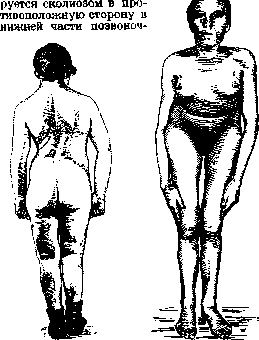
Figure 26. Obliquely narrowed
Figure 27. Kyphoscoliotic
scoliotic pelvis.
Czech pelvis. In the case of scoliosis, the pressure of the trunk on the pelvis is equalized and the pelvis does not undergo changes. If the scoliosis is located in the lumbar region, the compensatory curvature already occurs within the pelvic cavity and the pelvis changes its symmetrical form—it becomes obliquely narrowed. Scoliosis of the spine most often develops on the basis of rachitic changes in the vertebrae, but it can, as is believed, also develop from improper loading in childhood and adolescence. For example, the so-called schoolchildren's scoliosis, which develops in schoolchildren during prolonged improper sitting, especially during writing lessons, was included here. It should however be kept in mind that in individual cases scoliosis can also be caused by congenital anomalies in the structure of the vertebrae, which by the way is usually easily detected on X-ray images. Thus it is obvious that according to the etiological principle, all scoliotic pelves cannot be classified in the same department.—Kyphoscoliotic pelves, as the name itself shows, are pelves that combine the characteristics typical of both kyphosis and scoliosis (fig. 27-29). Assimilation pelves according to modern views should be classified in the first section of congenital anomalies. Assimilation is called the likening of the vertebrae of one section of the spine to the vertebrae of a neighboring section. Thus, if the last lumbar or first coccygeal vertebrae are likened to the adjacent sacral vertebrae and fuse with the sacrum, we will have assimilation: in the first case—upper, in the second—lower. Assimilation can also occur from the side of the sacral vertebrae—from above the first sacral vertebra can be likened to the V lumbar, from below—to the coccygeal. Assimilation can be complete and incomplete. It is clear that the assimilation process can reflect on the form and dimensions of the pelvis in the most varied ways. In recent years with the development of X-ray diagnostics of bone diseases and anomalies, it has been clarified that assimilation processes in the lumbosacral region are by no means rare. However not all assimilation pelves present obstetrical interest. Such interest is presented mainly by those assimilation pelves where the sacrum has assimilated an extra vertebra and therefore consists of six vertebrae. Such a sacrum is naturally longer than normal, and the pelvis with such a sacrum will obviously be higher, its cavity will be more elongated, which can naturally reflect on the course of labor in an unfavorable direction. The last, V section of Schaut's classification covers those narrow pelves, the narrowing and deformation of which are caused by diseases of the lower extremities. Of these pelves, the so-called coxalgic, i.e., pelves narrowed due to tuberculous coxitis (fig. 30), presents the greatest practical interest. Regarding this disease, it should be said that it causes characteristic changes only in those cases when the process affects the hip joint in childhood or
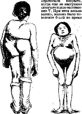
Figure 28. Kyphoscoliotic
Figure 29. Kyphoscoliotic-
pelvis.
pelvis. diseases: if a girl suffering from tuberculous coxitis underwent appropriate orthopedic treatment, then it is possible that the coxitis will have little effect on the form and dimensions of the pelvis. More often however even with correct treatment, the coxitis affects the diseased half of the pelvis and it develops worse than the healthy one, as a result—narrowing of the pelvis on the diseased side. If the child is left to himself and the process proceeded 'on the move', then usually when walking the child protects the diseased side and the entire weight of the trunk falls on the healthy half of the pelvis. The unformed bones of the child's pelvis yield under pressure, and the healthy half of the pelvis is narrowed. In both cases the pelvis will be obliquely narrowed. Thus the coxalgic pelvis is an asymmetrical, obliquely narrowed pelvis. If coxitis affects a woman who has already formed, with an ossified pelvic ring, then the form and dimensions of the pelvis do not change, but the position of the pelvis will be changed, it will be obliquely positioned. The changes characteristic of the coxalgic
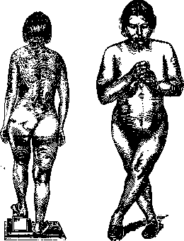
Figure 3 0. Coxalgic
Figure 31. Pelvis in double-
pelvis.
sided congenital dislocation of the hip. Pelves are also observed in all cases when one of the extremities was shortened in childhood for one reason or another and of course if the sick child in this case was left to himself. Thus obliquely narrowed pelves of the coxalgic type are pelves of the lame.—Congenital bilateral dislocation of the hip or curvature of both lower extremities usually leads to a flattening of the form of the pelvis, while the degree of narrowing is usually not very large (fig. 31). Therefore such kind of deformed women as a rule give birth quite successfully. Of other classifications, the following classification by Martin deserves attention, based not only on etiological factors but also providing for practical considerations. I. Incorrect pelves formed on the basis of violations of embryonic development: a) malformations, b) assimilation, c) shortened rachitic, d) dwarf rachitic, e) generally narrowed, f) funnel-shaped, g) flat, h) male. II. Incorrect pelves developed on the basis of diseases of the pelvic bones and their synchondroses: a) due to tripsin (rachitic and osteomalacic pelvis), b) due to osteitis (Negeli's, Robert's, synchondrotic pelvis), c) with exostoses, d) with fractures, e) with ruptures. III. Incorrect pelves caused by changes in the spine: a) spondylitic, b) kyphotic, c) kyphoscoliotic. IV. Incorrect pelves caused by changes in the lower extremities: a) after dislocations, b) after coxitis, c) due to unequal length of the lower extremities. V. Incorrect pelves caused by anomalies of the central nervous system.
Ludwig u. Savor . 3,8» Stolypinsky . . 8,0
Franz......24.0 Shakhshatinskaya 37.5 On average, it should be considered that pelvic narrowings occur in 15-20% (Pobedinsky), of which only about 5% are truly narrow pelvises requiring any surgical intervention or presenting obstacles during childbirth. Undoubtedly, the data provided can only have approximate value, and they certainly cannot give an exact idea of the frequency of narrow pelvises in general. Diagnosis of a narrow pelvis. The initial, presumptive diagnosis of a narrow pelvis is established during pregnancy, while the final diagnosis, except in cases of obvious narrowings or deformity of the pelvis, is determined only during childbirth, because along with the numerical data obtained, it is necessary to a large extent to consider a number of factors that can only be taken into account when the act of childbirth begins. Thus, besides certain pelvic sizes characterizing one or another anatomical structure of it or the degree of narrowing, the entire musculature of the body, the structure and contractile capacity of the uterine musculature, and finally the fetus itself play an important role. Proper labor activity, which has enormous significance in each case of childbirth with a normal pelvis, acquires exceptional importance in its narrowing, since abnormalities in expulsive forces, disruption of the tempo and rhythm of labor activity (primary, secondary labor weakness) can be, and not infrequently are, a source of various complications precisely with a narrow pelvis. The role of the fetus is no less important: its size, dimensions, weight, and especially the head, the structure of the cranial bones and its ability to configure are of significance. All this shows that the combination of these data, along with numerical dimensions, is of great importance in the clinic of a narrow pelvis, and that the final diagnosis can and must be established only during childbirth. Numerical dimensions with all available methods of measuring the pelvis cannot give an exact representation, and in practice cases are not infrequent where, with the presence of a large fetus, large head size, density of cranial bones, and insufficient ability to configure, even a normal pelvis may appear narrowed or further narrow. Consequently, in practical life the concept of 'narrowed, narrow' pelvis should be expanded and should be considered as a mismatch between the fetal head and the given pelvis. And deeply right is the well-known German gynecologist Martin (Martin), who says that 'in practical terms it is indifferent what particular form of pelvis exists', 'only the fact is important that the fetal head is too large for the mother's pelvis and childbirth cannot end spontaneously.' In view of all the above, the obstetrician should not overestimate the figures obtained when measuring the pelvis and should not base one or another method of therapy on them. Although pelvimetry is of course necessary in every pregnant woman, Martin's opinion is completely correct that measurement of the pelvis should not be a reference point for the obstetrician in diagnosis, that 'the pelvimeter should recede into the background, that it is sufficient only for the sense of touch inherent in every obstetrician to establish whether there is a narrow pelvis, how great the mismatch is, and whether the natural forces of the organism are sufficient for the head to pass through the narrow place of the pelvis.' The initial, approximate diagnosis of a narrow pelvis, as indicated, is established during pregnancy. This diagnosis should be based not only on measurements of the pelvis but also on examination of the structure of the entire body, height (height has a certain significance, but one must keep in mind that even with tall stature deviations, and quite significant ones, from the normal structure of the pelvis can occur), face, head shape, chest, the relationship between the trunk and extremities, comparison of the width of the pelvis and shoulder girdle, etc. It is necessary to form a clear idea of the nature of the pubic joint-its height and thickness. It is also necessary to clarify the nature of the gait, which can give an idea of joint mobility, lameness, degree of shortening of the leg if present, etc. Examination of the trunk should be done in the lying and standing (from the front and back) positions. For the diagnosis of a narrow pelvis, it is not sufficient to limit oneself only to external examination of the body, measurements of the pelvis, but it is necessary to take into account anamnestic data. The anamnesis has exceptional value, it should precede the examination, obstetric investigation, and should also be a guiding factor for the obstetrician in the subsequent examination of the pregnant woman and measurement of the pelvis. When collecting the anamnesis, one must especially consider the 'first step' and generally moments related to the first months of life. Various deviations of early and late childhood, childhood infections, and finally the time of puberty and sexual development, the year of the first menstruation, the time of its final establishment, its type and character of course may be of significance. Late appearance of menstruation, anomalies of the ovarian-uterine cycle can be an indicator not only of shortcomings in development and infantilism of the sexual sphere, but also of general insufficiency of the entire organism, its infantilism, in particular insufficiency and deviations in the development of the bone system and bony ring. Thus, for example, some (Voron) point out that the late appearance of menstruation (at 16, 18, even at 20 years) is a frequent phenomenon with a narrow pelvis, just as is its improper character (large intervals-several months to a year). When questioning, one must also take into account the living conditions, conditions and character of labor, socio-hygienic factors, housing, nutrition, way of life, since these moments, especially early and heavy physical labor in childhood, can have a definite and essential significance in the etiology of a narrow pelvis. If in the future many of these moments will lose their acuteness in connection with the widely developed network of preventive institutions and with the protection of infancy, prohibition of labor for adolescents, etc., then at the present time these factors must still be taken into account, since all these increasingly expanding preventive measures have not yet been able to completely eliminate the narrow pelvis-one of the typical social diseases, which is often a consequence of difficult social conditions. Further, in the anamnesis, attention should be paid to pains. Pains are by no means a rare accompanying symptom of a narrow pelvis, they arise not only in the last months in connection with overstretching of the abdominal walls, pendulous abdomen, but also significantly earlier. These pains can be observed (especially during movement) in the spine, in the bones of the pelvis, in the lower extremities; the constancy of pains, their more intense character, soreness (increasing during walking) is one of the initial and characteristic symptoms of the onset of osteomalacia. Pains can also often be caused by painful uterine contractions arising on the basis of abnormal position of the uterus, its abnormal mobility, pendulous abdomen, irregularities in accommodation on the part of the fetus. In multiparas, great importance has the clarification of their obstetric past-course of previous pregnancies and childbirth. Abnormalities of labor activity, prolonged, protracted labor, complications encountered-early or premature rupture of membranes, prolapse of small parts, cord, weight of the fetus, its abnormal positions, stillbirths, one or another surgical interventions-should attract special attention of the obstetrician, since the possibility of their repetition can never be excluded, and when taking them into account, one can often prevent them in advance by preventive measures. Taking into account to a large extent the data of the obstetric anamnesis, the obstetrician should not, however, upon receiving a favorable impression from the course of previous pregnancies and childbirth, remain calm, since even under these conditions, with the presence of a narrow or even narrowed pelvis, complications in subsequent childbirth are always possible (incorrect insertions, positions, larger size of the fetus, etc.). After preliminary questioning and thorough examination of the pregnant woman, one proceeds directly to obstetric investigation and measurement of the pelvis (see Obstetric investigation). Special attention must be paid to Michaelis' rhombus, the structure of which changes considerably depending on one or another anomaly of the pelvis (thus, for example, in a flat pelvis instead of the normal parallelogram form it takes the shape of a triangle, in obliquely narrowed pelvises the rhombus occupies an oblique position and finally in individual cases is even completely unnoticeable) (fig. 32-33), in consequence of which it helps to orient oneself in one or another form.
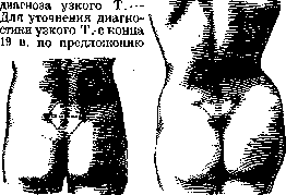
Fig. 32. Normal
Fig. 33. Michaelis' rhombus in
Michaelis' rhombus in a woman with a flat rachi-
well-developed woman
tic pelvis has a triangular shape.
a triangular shape. Varnier (1896) also resorts to measuring the pelvis using X-rays—radiography and radio-pelvimetry, which has been sufficiently perfected in recent times thanks to a whole series of ingenious inventions, specially equipped instruments and apparatus (Levy and Thumin, Bouchacourt, Fabre, Guibert, Levy-Solal, Glagoleva-Arkadyeva, Arkhangelsky, etc.). But even with regard to X-rays in terms of the possibility of predicting childbirth, the same can be said as with regard to the usual methods of obstetric examination and measurement of the pelvis: if X-rays undoubtedly have great significance for the scientific study of various complications in the structure of the pelvis, a more detailed study of individual pathological forms, then they can hardly be used to guide the course of impending childbirth in each individual case or serve as a basis for choosing one therapy or another. No matter how ideally an X-ray might reflect a particular anomaly of the pelvis, how vividly it might reproduce the size of the fetal head and its relationship to the pelvis, it still in no way can speak to the prognosis and character of impending childbirth. In clinical practice, for a general idea of the nature of a given pelvis, for determining its form and degree of narrowing, all narrow pelves can be divided into separate groups (see above). Narrow pelvis and fertility. The influence of a narrow pelvis on pregnancy. The course of pregnancy, hygiene and dietetics of the pregnant woman with a narrow pelvis. The question of the possible influence of a narrow pelvis on a woman's fertility must be decided not in the plane of the presence of only a narrow pelvis as such, but the presence of a number of pathological symptoms indicating general insufficiency of the organism, its underdevelopment, general and local infantilism. It is quite understandable that in cases of general infantilism or with hypo- and aplasia of the sexual sphere, anomalies in the structure of the skeletal system as a whole and of the bony ring in particular are caused by the same causes as this or that infantilism. For this reason, the decreased ability to conceive (or even infertility) observed with a narrow pelvis, and the late onset of the first pregnancy, must be attributed not to the narrow pelvis, but to those general causes that caused the general retardation in the development of the organism, including the anomalies of the pelvis. Due to the relationships that develop during pregnancy with a narrow pelvis, it is quite natural to expect that its course in such cases must differ from the course with a normal pelvis and present certain peculiarities both with respect to the mother and the fetus. These peculiarities, except in rare cases of severe degrees of narrowing, begin to manifest themselves only in the last months of pregnancy, whereas in the first months the course of pregnancy does not affect the condition of the pregnant woman and presents no deviations. Approximately from the seventh month of pregnancy, in connection with the significant increase in the size of the uterus, characteristic symptoms begin to develop, which accompany as a rule the course of pregnancy with a narrow pelvis. One of the most characteristic symptoms is the change in the abdomen—the so-called pendulous abdomen (venter propendens) (fig. 36 and 37). In primigravidas, due to the elastic walls of the abdomen, a so-called pointed abdomen forms and the upper wall of the abdomen protrudes predominantly, whereas in multigravidas the sharply anteflexed uterus in the presence of soft, flaccid, yielding abdominal walls forms a pendulous abdomen, which is almost completely absent in primigravidas. The rhombus of Michaelis is poorly expressed or almost not expressed at all in women with a narrow pelvis. The rhombus of Michaelis has a sloping shape. Kyphoscoliosis in the lower part of the spine. A pendulous abdomen is formed, on the one hand, because the presenting part, due to its incompatibility with the dimensions of the pelvis, cannot engage in the inlet, remains mobile and is positioned high above the pubic symphysis, and on the other hand—because often with a narrow pelvis we have general infantile structure and a smaller volume of the abdominal cavity. The great mobility of the uterus, the incompatibility of the presenting part with the dimensions of the pelvis, its high position, by causing general overstretching of the abdominal wall and the formation of a pointed or pendulous abdomen, reflecting on the pregnant woman, cause various deviations on the part of the fetus. Thus, along with pains, more acute painful sensations compared to a normal pelvis, mechanical complications are observed on the basis of compression of the abdominal organs by the pregnant uterus—greater tendency to constipation, stagnant phenomena, circulatory disorders, Fig. 36. Pendulous abdomen (venter propendens) in a woman with a narrow pelvis and flaccid, yielding abdominal wall.
Fig. 34. In a woman with a narrow pelvis the rhombus of Michaelis is poorly expressed.
Fig. 35. Rhombus of Michaelis.
with a narrow pelvis the rhombus is almost not expressed at all.
The rhombus of Michaelis has a sloping shape.
Kyphoscoliosis in the lower part of the spine.
A pendulous abdomen is formed, on the one hand, because the presenting part, due to its incompatibility with the dimensions of the pelvis, cannot engage in the inlet, remains mobile and is positioned high above the pubic symphysis, and on the other hand—because often with a narrow pelvis we have general infantile structure and a smaller volume of the abdominal cavity.
The great mobility of the uterus, the incompatibility of the presenting part with the dimensions of the pelvis, its high position, by causing general overstretching of the abdominal wall and the formation of a pointed or pendulous abdomen, reflecting on the pregnant woman, cause various deviations on the part of the fetus.
Thus, along with pains, more acute painful sensations compared to a normal pelvis, mechanical complications are observed on the basis of compression of the abdominal organs by the pregnant uterus—greater tendency to constipation, stagnant phenomena, circulatory disorders,
Fig. 36. Pendulous abdomen (venter propendens) in a woman with a narrow pelvis and flaccid, yielding abdominal wall.
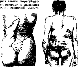
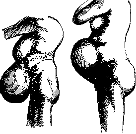
Figure 37. Acute abdomen in a woman with a narrow pelvis with an abdominal wall of normal elasticity. Breathing difficulties, shortness of breath. From the side of the fetus, incorrect positions (oblique, transverse) are encountered more frequently, and various types of incorrect presentations are also more often observed (for example, Bumm pointed out that incorrect positions of the fetus in a narrow pelvis occur four times more often than with a normal one, and face and brow presentations increase 3-6 times). The influence of a narrow pelvis on premature termination of pregnancy, its greater or lesser duration, and the development and weight of the fetus at this time is denied by the overwhelming majority of obstetricians. As for the question, for example, of the possible more frequent termination of pregnancy, the data of recent times to a large extent refute the previous point of view, and La Torre indicates that it is observed even less frequently with a narrow pelvis (22% with a narrow pelvis, 25% with a normal one). Similarly, it is now refuted the prevailing opinion for a long time that supposedly children with a narrow pelvis are born with less weight. All those changes and deviations that accompany the course of pregnancy with a narrow pelvis compel the obstetrician to conduct in-depth prevention; this prevention should aim not only to alleviate the observed pathological changes, but also to prevent more serious complications that could adversely affect the nature and course of the act of labor itself.
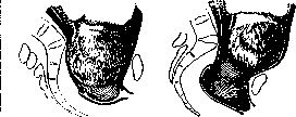
The principles of hygiene and dietetics for a pregnant woman with a narrow pelvis in their main features do not differ from those preventive measures that modern clinicians conduct in each case of pregnancy under normal conditions, but they must be carried out with special pedantry in pregnant women with a narrowed or narrow pelvis. Some individual questions deserve special mention, specifically relating to the hygiene and dietetics of pregnancy with a narrow pelvis—these are questions of nutrition, walks, and baths. The previously prevailing and fairly widespread view that pregnant women with a narrow pelvis need some special lifestyle, increased exercise, long walks, limited or even special nutrition, frequent baths, is now shared by far from all obstetrical schools. And this is quite understandable, since our modern concepts of pregnancy and the deviations observed in it are now much broader; in particular, various anomalies of the pelvis are considered not only as processes of a local nature, but as general processes, connected with the structure of the entire organism, its constitutional features, the nature of the entire bone system, etc. For these reasons, one must take a negative attitude toward all those advice given to pregnant women with a narrow pelvis (not finally abandoned even at the present time) that concentrate only on the narrow pelvis as such, do not take into account the entire organism, the processes occurring in it, the condition of individual organs, and finally do not take into account those conditions and that environment surrounding the pregnant woman. For this reason, for example, we must completely change our point of view on the nutrition of a pregnant woman with a narrow pelvis. The previously widespread view that pregnant women with a narrow pelvis need to limit their nutrition, that they need some special diet, now has few followers; many believe that it is hardly possible to regulate the weight, volume of the fetus, promote the construction of the head, its better configuration, and that generally all this is hardly rational from the point of view of the completeness of the growing fetus. Therefore, the diet proposed at one time by Prokhovnik (mainly limitation of fluids, carbohydrates and generally fat-forming substances) is irrational. Its inadequacy is also revealed in the light of our modern knowledge, is proven by clinical observations, and should be completely abandoned both in minor and in degrees of narrowing; hardly for this reason can one agree with Traugott, who proposes to leave it only for minor narrowing of the pelvis, since its therapeutic effect is unreliable, or with Seitz, who believes that sometimes by appropriate nutrition of the pregnant woman one can delay the development of the fetus (although he says that the development of the fetus does not depend entirely on the nutrition of the mother). The Moscow obstetrical school has always been critical of Prokhovnik's diet, and N. I. Pobedinsky as early as 1913 at the V Congress of Gynecologists and Obstetricians in his report on the treatment of a narrow pelvis pointed out that the reserved attitude toward this diet can easily be explained by the fact that, on the one hand, it is difficult to carry it out strictly and control it in each individual case, the scientific explanation of the diet appears insufficiently substantiated, and considering that owners of a narrow pelvis are often of weak health, this reduced nutrition applied continuously (for 4-5 months) can hardly pass without harm. Proceeding from all these considerations, one must come to the conclusion that no special diets, limitation of nutrition are needed for pregnant women with a narrow pelvis: without giving any real results, they can also adversely affect both the mother's organism and the development of the fetus; the prescription of nutrition should be individual in each case and based on the same principles that are usually followed in clinical practice.
Just as negatively must one relate to those still existing advice to pregnant women 'to walk more'. Long, tiring walks bring nothing but harm. If it is generally considered that rational walks, exercise are necessary for every pregnant woman, it is hardly correct to believe that similarly prolonged walking for 3-4 hours in a row (and even more), allegedly aimed at promoting a reduction in the size of the fetus, better course and facilitation of the act of labor, can be beneficial. The prescription of walks, determining their duration should proceed from the peculiarities of each individual case, the nature of the living conditions of the pregnant woman and the nature of her productive labor. Long, aimless walks undoubtedly weaken the organism, can contribute to an intensification of those pathological symptoms and painful sensations that accompany the course of pregnancy with a narrow pelvis, can affect the body musculature, the uterine musculature (in general more excitable due to its incorrect position, greater mobility, greater stretching of the abdominal walls), can lead to a number of other disorders, to an intensification of phenomena from the side of the heart, its overload, already existing in a narrow pelvis. Just as cautiously should pregnant women with a narrow pelvis approach various types of physical exercises—very often the most innocent of them should be directly contraindicated. Long walks should not have a place in the practice of a narrow pelvis; alleviation of the painful symptoms observed in it, ensuring less painful labor pains, a more correct rhythm of labor activity, better accommodation of the fetal head to this pelvis are achieved only by regular movements, sometimes even by limiting them with the simultaneous prescription of mandatory rest during the day.
For the same reasons, one must completely abandon the use of frequent baths. The prescription of baths of high temperature 2-3 times a week and even more often was a routine phenomenon in pregnant women with a narrow pelvis (unfortunately, some still carry it out to the very last time); the starting point of this was a completely incorrect assumption that supposedly under the influence of frequent hot baths the organism and uterine musculature are prepared for childbirth, the pelvis 'expands', its bones become 'more pliable', labor proceeds more easily. This opinion is of course incorrect, baths with a narrow pelvis should be prescribed just as with a normal one, not more than once a week. More frequent baths, especially hot ones, undoubtedly adversely affect the entire organism, the functions of individual organs (especially the heart), and do not pass without a trace for the body musculature in general and the uterine musculature in particular. The influence of frequent baths on the bones of the pelvis in the sense of its possible 'expansion' is based on nothing; if this expansion does take place, then of course not under the influence of baths or any other mechanical or thermal factors, but exclusively as a consequence of the process of pregnancy and the growth and further development of the entire organism occurring in it.
The course and management of labor with a narrow pelvis (Figures 38 and 39). Prognosis. The final diagnosis of a narrow pelvis (except for significant or sharp narrowing), as has already been said, is established only during labor and can be made only with the most careful and regular observation of the parturient. The diagnosis in its main features should be based on whether the fetal head can engage in the pelvic inlet at the beginning of labor, and in primiparas even at the end of pregnancy and serves as it were a valve protecting the fetal sac from premature rupture.
Figure 38.
Figure 39.
Figure 38. With a normal pelvis, the fetal head engages in the pelvic inlet at the beginning of labor, and in primiparas even at the end of pregnancy and serves as it were a valve protecting the fetal sac from premature rupture.
Figure 39. With a narrow pelvis, the head does not engage in the pelvic inlet and does not serve as a valve protecting the
fetal sac from premature rupture.
the fetal sac from premature rupture. The influence of the newly arisen conditions in connection with labor activity can change those unfavorable relationships between this head and this pelvis, which existed before the onset of labor. These conditions consist of two basic moments—the character, rhythm, and tempo of contractions and the character of insertion of the fetal head into the pelvis and its ability to configure. These moments mutually complement each other and are often decisive in the clinic of a narrow pelvis. Pathological manifestations from these factors can significantly affect the course of the labor process and the change in the line of behavior, since when the fetal head is unyielding, when it has no ability to adapt to this pelvis, or finally with incorrect insertion, despite good proper labor activity, the abnormal relationships between the head and pelvis are not eliminated; just as well with various anomalies of expulsive forces, despite a clearly expressed ability of the head to configure, the result can be the same. These conditions are necessary for a successful completion of labor. Of course, these favorable conditions also have their limit and can only apply to cases where the discrepancy between the head and pelvis is not particularly great. These factors must be fully taken into account, since in many cases with certain degrees of narrowing, the outcome of labor for the mother and fetus depends on them, and taking them into account in a practical sense gives much more than guidance only by the bare representation of digital dimensions and orientation to a certain degree of narrowing of the pelvis. This approach is shared by many authors, and among others Bumm already said that difficulties during labor are not always in exact proportional relationship to the degree of narrowing and that sometimes, when all other factors are favorable, even with significant degrees of narrowing, labor 'surprisingly quickly ends,' whereas when there is any complication there can be significant disorders even with relatively insignificant narrowing. ' Consequently, only regular observation of the course of the labor process can allow one to say whether those abnormal relationships between the head and pelvis that existed before the onset of the first contractions can change or are changing. Only such observation should guide the obstetrician and indicate to him the paths of therapy. Underestimation of these factors, careless, inattentive attitude toward the parturient, neglect of any observed pathological symptoms (prolonged high standing of the head, its deviation to one side or the other from the midline, premature rupture of membranes, anomalies of labor activity, etc.), not taking in time those measures necessary for each individual case can prove fatal not only for the fetus but lead to complications for the mother and be accompanied by severe irreversible consequences.
When conducting labor, it is always necessary to remember that their course with a narrow pelvis has its characteristic features, differs significantly from the course of labor with a normal pelvis, and that for individual forms of narrowed or narrow pelvis there exist special laws of the mechanism of labor, special types of progressive and rotational movement of the head. Only when taking into account these peculiarities, only with exact knowledge of the type of insertion of the head and in general of its further progression through the pelvis with certain forms of narrow pelvis, can one form a concrete idea of the nature of the course of labor in each individual case, speak of deviations and make a prognosis about their possible favorable outcome. Thus, the mechanism of labor with a uniformly narrowed pelvis and a flat pelvis is completely different from each other. And in both cases, the head standing above the entrance to the pelvis strives to insert into it and pass through the narrowed place. The insertion of the head, its adaptation, configuration with a uniformly narrowed pelvis will differ significantly from the insertion of the head with a flat pelvis, since with a uniformly narrowed pelvis the entire pelvis is narrowed, the entire bony ring, whereas with a flat pelvis only the direct dimension of the entrance to the pelvis is narrowed. The fetal head with a uniformly narrowed pelvis at the beginning of labor inserts either in the transverse dimension or (according to some authors this happens more often) in one of the oblique dimensions. With a uniformly narrowed pelvis the head can pass only with extreme flexion (provided there is sufficient possibility of its configuration—the so-called sugar head, significant club-like elongation of the fetal skull). Flexion of the head occurs also with a normal pelvis (see Labor), but with a uniformly narrowed pelvis its flexion is expressed more sharply, the chin is almost pressed against the chest, and as a result the head passes through the narrowed place with the smallest dimensions, and the small fontanel descends so low that it reaches the middle of the pelvis (fig. 40 and 41). This low, sharply expressed position of the small fontanel in the longitudinal plane of the pelvis is the basic characteristic moment for the insertion of the head with a uniformly narrowed pelvis and is the only favorable one (in a prognostic respect). Only with such a position of the small fontanel can the head (with club-like elongation) insert with its smallest circumference (suboccipito-bregmatic), in contrast to its insertion with the suboccipito-frontal circumference in cases of usual labor with head presentations. It is completely natural that for this form of pelvis only such a mechanism is favorable and that
Figure 40. Favorable insertion of the head with a uniformly narrowed pelvis—sharp flexion of the head.
Figure 41. Unfavorable insertion of the head with a uniformly narrowed pelvis—absence of flexion.
of it (e.g. face, brow, vertex presentations), especially in cases of more severe narrowing, significantly worsen the prognosis. In general, in view of the fact that the presenting part has to overcome obstacles throughout the entire pelvis, labor is often prolonged, and in some cases (with large degrees of narrowing of the pelvis or large discrepancy between the head and pelvis) a more prolonged course of labor is almost the rule. The course of labor with a uniformly narrowed pelvis with breech presentations in cases of insignificant narrowing can be quite favorable, but with an increase in narrowing the extraction of the subsequent head can present significant difficulties, especially in cases where the chin cannot be pulled down or where it is distant from the chest of the fetus, and also because the unprepared head (without configuration) passes more difficultly through the narrowed place.
The mechanism of labor with a flat pelvis appears completely different, since here there is narrowing only of the entrance to the pelvis and in addition this narrowing is limited only by the direct dimension (conjugate). With a flat pelvis in the initial stage of insertion of the head, its relatively strong extension and entry (by the direct dimension) into the transverse dimension of the pelvis occur. Of course, the configuration of the fetal head, which after birth has an incorrect shape (often characteristic depressions from pressure by the promontory, called spoon-shaped, grooved) are observed on it (fig. 42). Due to the occurring extension of the head (when entering the pelvis) the large fontanel descends, the small fontanel is on the same level
Figure 42. Spoon-shaped depression on the parietal bone from pressure by the promontory with a flat pelvis. S18 removed higher, and the head enters the direct dimension of the pelvis with its smallest dimension—the transverse (distantia biparietalis, in contrast to insertion of distantia biparietalis with a normal pelvis). Subsequently the anterior parietal bone begins to descend (lateral Naegele's inclination), the posterior one is pressed against the promontory, while the sagittal suture approaches it (fig. 43 and 44). Following the descent of the anterior parietal bone, the posterior parietal bone descends, the occurrence
Figure 43.
Figure 44.
Figure 43. Favorable configuration of the head with a flat pelvis—Naegele's anterior parietal insertion. Figure 44. Further descent of the anterior parietal bone with Naegele's insertion (the sagittal suture approaches the promontory). of the parietal bones passing one over the other (configuration of the head), the ever greater approach of the sagittal suture to the midline, establishment of the head in a synclitic position, subsequent flexion of the head and descent of the small fontanel. The characteristic feature of the mechanism of labor with a narrow pelvis is thus the insertion of the head into the transverse dimension of the pelvis, the descent and low position of the large fontanel (and the anterior part of the head) and asynclitic insertion. Unfavorable deviations are insertion with the posterior parietal bone—Ricci's lateral inclination (although some authors, for example N. Z. Ivanov, do not attach special importance to this inclination and believe that there is nothing bad in it) and brow presentations (fig. 45 and 46).
Figure 45.
Figure 46.

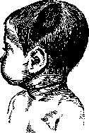
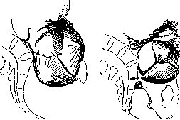
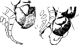
Figure 45. Unfavorable configuration of the head in a flat pelvis—Litsman's posterior parietal insertion. Figure 46. Further descent of the posterior parietal bone in Litsman's insertion (the sagittal suture approaches the pubis). The mechanism of labor in a generally narrowed flat pelvis presents many variants, depending on the nature of the pelvic narrowing, but the characteristic feature of the mechanism is the insertion of the head (with the sagittal suture) in the transverse

Figure 47. The head of a newborn in a flat pelvis of the mother. The left parietal bone is flattened due to pressure against the
size of the pelvic inlet (fig. 47). It is clear that for the head, along with its ability to configure and adapt to a given pelvis, to follow certain laws of the mechanism, there must also be a certain character of the force (uterine muscular motor) that promotes its advancement and overcoming of the obstacle in the narrowed place. The prognosis of labor actually depends on the sum of these components. The prognosis of labor thus depends both on the degree of narrowing (on the form of the pelvis—it appears more favorable in a flat pelvis) and the mismatch of the head with the pelvis, as well as on the timely establishment of diagnosis, timely detection of various deviations, and the associated choice of one or another therapy. The prognosis is significantly clouded in prolonged, protracted labors, in premature rupture of the fetal membranes and discharge of waters, in prolapse of the umbilical cord or small parts of the fetus. For the diagnosis of a narrow pelvis, for further observation of the course of labor, it is often quite sufficient (of course, with the experience of the obstetrician) to perform an external examination of the pregnant woman and external research methods (some authors, e.g., Martin, advise using exclusively them). A good aid are also special obstetric manual techniques (see) of Müller and Hofmeyer, but at-
va
y
B
birth tumor. They should be changed not roughly, but carefully, since damage to the fetus is possible. Of course, greater effectiveness can be obtained from these methods when the narrowing of the pelvis is not sharply pronounced. If external methods fail to establish a diagnosis and create a clear understanding of the relationship between the head and the pelvis, and the course of labor, then it is necessary to perform an internal examination, which is not only not superfluous but a necessary auxiliary aid, providing reference points for the further management of labor, for the selection and performance of one or another operative intervention. In individual unclear cases, it is necessary to resort to the help of anesthesia and to examination with half a hand, because, as Wumm quite rightly notes, before deciding on one or another aid, it is necessary to obtain a clear understanding of the nature of the narrowing and the nature of the head's engagement. Thus, the main points in the management of labor with a narrow pelvis are the establishment of the diagnosis of a narrow pelvis, the determination of the relationship between the fetal head and the pelvis, and unremitting observation of labor and the nature of labor activity. The latter has exceptional significance, and yet, as is known, it is precisely with a narrow pelvis that various anomalies of expulsive forces, primary and secondary labor weakness, weak, short, spasmodic contractions are more frequently encountered. These various anomalies can significantly affect the course of labor, creating a prolonged nature, disrupting the engagement and advancement of the head. Timely diagnosis of one or another disorder of the rhythm of labor activity, timely therapy can not only prevent their disorderly nature, reduce heightened sensitivity, alleviate pain, regulate them, but also shorten the duration of labor and prevent various subsequent complications characteristic of a narrow pelvis. Among these complications, one must first remember about the frequently observed premature rupture of the fetal membranes and premature escape of waters, occurring due to the presence of special favorable conditions for this with a narrow pelvis: 1) significant stretching of the fetal membranes and 2) the high position of the fetal head, whereby the amniotic fluid in the uterine cavity directly contacts the forewaters of the fetal membranes. Premature escape of waters, especially at the height of a contraction and if labor activity is abnormal, can lead to subsequent complications-prolapse of the umbilical cord, small parts, which occurs with a narrow pelvis five times more often than with a normal one. These complications, of course, significantly worsen the course of labor, the prognosis for the fetus, and give a higher percentage of maternal morbidity. A serious consequence of premature rupture of the fetal membranes, especially if the escape of waters occurs at the very beginning of the first stage of labor, are not only disorders from the side of cervical dilation (more prolonged opening, edema of the cervix) but also the possibility of entrapment of the anterior lip between the head and the pelvis. With a narrow pelvis, injuries to the soft birth passages, tears of the perineum, lacerations and ruptures of the uterus, injuries to the symphysis pubis, traumatic neuritis are more frequently observed. All this creates more favorable conditions for infection, which can occur even before the end of labor. Prevention and therapy of a narrow pelvis. Taking into account the fact that a fairly significant part of various forms of a narrow pelvis arises already in the antenatal period, that there are various congenital anomalies of it, that often the cause of a narrow pelvis are shortcomings of early and late childhood, and finally of the school period, the entire center of attention must be shifted to early prevention of a narrow pelvis, to the struggle with it as a social disease. Therefore, one must think about the prevention of a narrow pelvis not when a woman is already pregnant and not when labor begins, but when there is not yet pregnancy, long before its onset, when the bony skeleton begins to form, when the female organism begins to form. One of the main and most essential preventive factors is not only the prevention of the period of a girl's puberty, not only rational upbringing and nutrition in earlier years, the struggle with childhood infections, which play a significant role in underdevelopment, general and local infantilism of the organism, but also prevention of the intrauterine period, consisting in a more rational regime of pregnancy, improvement of social-hygienic conditions and generally a series of measures carried out by the widely extended network of preventive institutions for the Protection of Motherhood and Infancy. Prevention in later years aims to create such conditions for a pregnant woman with a narrow pelvis as to facilitate the course of labor for her. This prevention can be carried out both already in the presence of pregnancy and even before its onset (see Pelviotomy). Prevention of a narrow pelvis with an existing pregnancy can consist in a series of preventive measures taken during pregnancy itself (see above) or already with the onset of labor in certain of its periods. Among the measures taken in advance are also artificial premature labors (technique see Labor, artificial and premature), aiming to facilitate labor with a narrow pelvis by conducting them before term with a smaller volume and size of the fetus. Artificial premature labors from their very beginning to the most recent time have met with far from uniform attitude towards them. Along with their principled supporters, there are no few opponents, and far from a few modern obstetrical schools completely do not resort to them as a method of therapy for a narrow pelvis. The negative side of artificial premature labors is that it is not always possible to take into account and correctly choose the term necessary for interrupting pregnancy, and the fetus may turn out to be too small or too large. This operation also does not take into account the above-mentioned factors of labor activity and in a number of cases may turn out to be futile and unnecessary. On the other hand, cases of birth of a too small fetus, insufficiently viable, and the associated quite high percentage of mortality both soon after birth and in more remote time are not rare (thus, Martin points out that only 50% of premature infants survive to a year, while the others often suffer from various disorders of the central nervous system). One cannot also say that artificial premature labors are completely safe for the mother, and to the present day with them a certain percentage of maternal morbidity and mortality is noted even in the best obstetric institutions (thus, one of their convinced supporters, Donald, cited 0.1% mortality for mothers and 12% for children). Despite this, artificial premature labors under certain circumstances can occur in practical life, but for their conduct it is necessary to carefully weigh the indications and take into account the contraindications. They should not first of all be applied to primiparas (with which, however, not all supporters of this method agree) and with more sharply expressed degrees of narrowing with the true conjugate less than 7½ cm. An indication for the operation can be cases with moderate degrees of narrowing in multiparas, in whom the history notes one or another operative interventions in previous labors and stillborn children; artificial premature labors should not be performed before the 34-35th week of pregnancy, because before this term the fetus is not yet viable. The best term is the 36th week. Direct therapy of a narrow pelvis should begin before the onset of labor activity. A significant preventive factor according to the observations of individual clinics (Zeits, Selitsky) for a favorable course and outcome of labor with a narrow pelvis is the timely placement of such pregnant women in a pregnant ward 7-12 days before the expected date of delivery. Clinical practice to a significant extent confirms the beneficial significance of preliminary stay of the pregnant woman in an obstetric institution. On the one hand, the pregnant woman herself is studied in more detail, day by day the relationship between the pelvis and the head is checked, changes occurring in these relationships are recorded, on the other hand, the pregnant woman is gradually prepared for labor, while at the same time being free from involuntary household duties and harmful domestic conditions for her. This premature hospitalization is of no less importance for the prevention of various factors complicating the course of labor with a narrow pelvis (e.g., premature rupture of the fetal membranes); the fact that contractions begin in a medical institution, observation of them is carried out from the very beginning, and that the pregnant woman is in more aseptic conditions in general, allows the obstetrician to more widely choose one or another operative aid, and in case of necessity with greater confidence to resort to more serious interventions (e.g., to cesarean section). The course of action with the onset of labor activity appears to be extremely diverse.
This diversity depends not only on the one or another degree of narrowing or form of the pelvis, but is formed from the interrelationships of the fetus itself, its head to the given pelvis, and from the ability of the organism to produce a certain force to overcome the obstacle and the further forward movement of the presenting part. The basic principles of therapy in childbirth with a narrow pelvis are patience, endurance, observation, and the ability to take into account the developing situation: not to do what is not necessary, and not to provide this or that assistance either too early or too late. If the total sum of the data obtained allows for the possibility of spontaneous labor, it is necessary to wait; but with absolute narrowing of the pelvis or with relative narrowing, but in the presence of a number of unfavorable factors - (incorrect positions, incorrect insertions, obvious mismatch of the head with the pelvis) - waiting is out of the question, and the only assistance is cesarean section (concerning the so-called preventive turn, see Obstetric turn and Pelviotomy). Therefore, the question of expectant therapy can only arise in cases where spontaneous labor can be expected (the percentage of such cases reaches up to 90%, and in some clinics even more). There can and should be no stereotyped approach; each case must be individualized. All our measures must be based on what is generally known about the course of labor with a narrow pelvis, and on the peculiarities of each individual case. The main point is the therapy of various anomalies of labor activity, more often of its insufficiency. In such cases, one can never resort to strengthening contractions with strongly acting agents (e.g., pituitrin), and especially not pursue the goal of promoting the insertion of the highly situated presenting part into the pelvis or its passage through the narrowed area. This is dangerous for both mother and fetus. One should also not apply and practice widely used vaginal douches with rather high temperatures, as they not only fail to achieve their purpose but are even harmful, weakening the organism, affecting the heart, and besides can be a source of infection. Hot vaginal douches to strengthen uterine contractions are also not rational - they are pointless and hardly harmless. Quinine, administered in small doses, periodically, with certain intervals (0.1-0.2 every 1-2-3 hours), often gives great benefit. The advisability of using quinine in small doses is shared not by individual authors (e.g., Martin). Weak labor activity, primary and secondary weakness of expulsive forces can often be caused, especially in cases of prolonged labor, by fatigue and exhaustion of the forces of the parturient. In such cases, one should strive not to strengthen contractions or bearing down, but to give a respite, rest to the overtired uterine musculature. And indeed, clinical experience shows that artificially induced respite, the administration of narcotics (morphine subcutaneously or per os, sometimes repeatedly, an enema of T-ra Valerianae aet. 20-25 drops + 8-10 drops of T-ra Opii simplex in a 7g glass of water) often gives a good therapeutic effect, contribute after a certain time to the resumption of labor activity, restoration of its disturbed rhythm, and thus to the acceleration and more rapid termination of labor. Great importance during labor, especially in the dilatation period, is also the hygiene of the parturient, in particular the timely emptying of the bladder and rectum, since non-compliance with this, especially in long-lasting labors, can significantly affect the course of the labor act and even serve as a cause of the occurrence of this or that deviation. The appropriate position of the parturient can also have definite significance: it can facilitate the course of labor, promote the insertion of the head into the pelvis and contribute to its further forward movement. When choosing one or the other lateral position (position on the right or left side), one must be precisely oriented to the position of the fetus, to clearly visualize the position, and always remember that simultaneously with placing the parturient on one or the other side, the uterus also changes its position. The therapy of secondary labor weakness, therapy of weakness of bearing down in the expulsion period are based on the same principles as the therapy of the mentioned anomalies in the dilatation period. In cases of tight insertion of the head into the pelvis, pituitrin can be tried with sufficient opening of the os. Traction and Verbova's forceps (see) undoubtedly bring benefit. To facilitate the mechanism of labor, more rapid termination can also the appropriate position of the parturient, the so-called Walcher's hanging position (see). But with these measures the desired result is not always achieved, and in a number of cases one has to resort to operative assistance - the application of forceps or perforation of the fetal head (of course, if there are no corresponding conditions for performing a cesarean section or if there are any contraindications). The use of forceps in the modern clinic is permissible only in those cases when the fetal head is already firmly fixed at the entrance to the pelvis. Forceps with a mobile, high-lying head should not be used, since under such conditions they almost never achieve their purpose, but only significantly traumatize the soft maternal birth passages, damage the symphysis pubis, give a high percentage of maternal morbidity, and are very often accompanied by severe trauma to the fetus and give a high percentage of infant mortality. Forceps should not serve as an instrument aimed at promoting the head when there is an obvious mismatch with the mother's pelvis. In a number of cases, craniotomy of a living fetus is still performed, but this operation, with the increasingly preventive direction in obstetrics, should be completely excluded from the routine of operations used in cases of a narrow "pelvis."
Related articles
Mentioned in
Cite this page
“Pelvis.” Soviet Medical Encyclopedia. English translation of Bolshaya Meditsinskaya Entsiklopediya, 1st ed. (Moscow, 1928–1936), ed. N. A. Semashko. https://sovietmedicalencyclopedia.pages.dev/article/pelvis/