Pubic Symphysis
Historical document, translated for reference. It reflects medical knowledge of the 1920s–30s and is not medical advice.
Summary
The pubic symphysis is a fibrocartilaginous joint connecting the pubic bones of the pelvis. This article describes its anatomical structure, differences between sexes, changes during pregnancy, and potential complications.
Encyclopedia article (1928–1936)
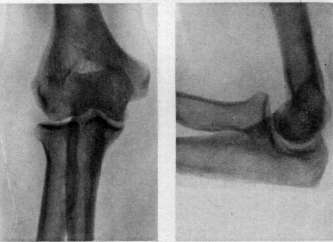
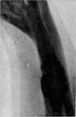
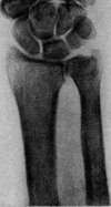
Pubic Symphysis, symphysis os-sium pubis, serves to connect the two pubic bones of the pelvis. The connection is formed by means of the fibrocartilaginous interpubic plate (lamina fibro-cartilaginca interpubica) and fibrous ligaments: superiorly - lig. pubicum superius, inferiorly - lig. arcuatum pubis, anteriorly (Luschka) - lig. fibrosum ant. and posteriorly - lig. fibrosum post. Anteriorly, the pubic symphysis appears almost twice as wide as posteriorly; at the arcus pubis it is wider than at the superior edge (Waldeyer). In a properly formed pelvis, the articular ends of the pubic bones should be distinguished (according to Luschka) into two parts: the posterior part, which has an elliptical, slightly convex surface and occupies the posterior half of the articular surface, and the anterior part, which is longer and at the same time narrower, forming a direct continuation of the posterior one. The posterior oval surfaces of the joint lie parallel to each other, while the anterior surfaces, conversely, diverge at various angles and form a triangular space with the base open anteriorly. The distance between the posterior bony parts of the joint (according to Luschka) averages 8 mm, while between the anterior parts it is 2 cm. The bony surfaces appear rough. Between them is located the fibrocartilaginous plate - lamina fibrocartilaginea interpubica, which is wider anteriorly and inferiorly than posteriorly and superiorly. On its posterior surface, it forms a cartilaginous prominence (eminentia retropubica), expressed to a greater or lesser degree. In women, this prominence can be well palpated from the vaginal side. In the middle of the interpubic cartilaginous plate, in most cases (according to Spalteholz), there is a small articular cleft-like cavity, located closer to the posterior edge and in the upper half of the plate. This cavity is usually simple, but sometimes it is divided by a transverse bridge into two parts - superior and inferior (Luschka). In men, this cavity is smaller and occurs less frequently (according to Aeby, in 20%), while in women it is larger and more frequently observed (in 93%, according to the same author). In large cavities, sometimes a viscous synovial fluid is present, while in small ones it is absent. Such cavities are observed predominantly in pregnant women, but not always. Thus, Aeby reports one case of pregnancy where he could not find any cavity in the pubic symphysis. Regarding the origin and presence of these cavities in the pubic symphysis, anatomists have different opinions. The pubic symphysis in men has greater height than in women; the interpubic cartilaginous plate is thicker and shorter in women, while in men it is narrower and longer. On average (according to Waldeyer), the height of the pubic symphysis in men is 5.4 cm, while in women it is 4.5 cm. Topography of the pubic symphysis. Anterior to the pubic symphysis is the mons pubis with its fatty padding, immediately below which in women are the lig. suspensoria clitoridis; lower, under the symphysis, pass next to each other the n. clitoridis et vasa dorsalia clitoridis; posterior to the symphysis lie, separated from it by areolar tissue and a fascial plate, the venous plexuses - plexus venosus pudendus et plexus venosus vesicalis inferior and the urinary bladder. During pregnancy, the pubic symphysis undergoes significant changes; there is increased vascularization of it, edema and loosening of the entire ligamentous apparatus. According to research by Brosike, Luschke and others, slits form in the symphyseal cartilage, which may serve as a predisposing factor for various types of traumatic injuries in it during childbirth or for the formation of abscesses during pregnancy. Luschke describes these slits as typical birth injuries in women and compares them with tears on the portio vagin. colli uteri after childbirth. This same author also points to the increase and growth of new elements of the ligamentous apparatus, as well as the neoplasm (growth) of bony elements in the pubic bones at the border with cartilage tissue. As a result, during pregnancy there is an increase in the overall size of the pubic symphysis, greater stretchability, and an increase in the cartilaginous prominence on the posterior surface. Figure 1. Divergence of the pubic symphysis (ninth month of pregnancy). Pain occurred with the strictest bed rest, with the slightest movements of the lower limbs and was accompanied by general nervous disorders and insomnia. (Observation by Selatsky.) Figure 2. Divergence of the pubic symphysis during pregnancy. The same patient three weeks after a cesarean section with subsequent sterilization (without any therapy). Long-term results (after four years): no complaints, normal gait, occasional slight pain and some "awkwardness" in the pelvis. (Observation by Selitsky.) Figure 3. A healed intra-articular fracture of the humerus in the upper third. Figure 4. Normal elbow joint in the extended position. The center of the olecranon and the center of both epicondyles are on a straight line. Figure 5. Normal elbow joint in the flexed position. Figure 6 and 7. False joint in the upper third of the tibia. Figure 8. Normal wrist joint. (For illustrations see articles: False joint, Elbow joint, Pubic symphysis, Wrist joint.)

fi шш
iff/
See articles: False joint, Elbow joint, Pubic symphysis, Symphysis pubis. The posterior surface of the pubic bone (eminentia retropubica). Leschke found that during pregnancy the bones of the pubic symphysis can grow by up to 2 cm. Due to the loosening of the symphysis during pregnancy, the cavity located in the interpubic cartilaginous plate can increase to considerable sizes, and sometimes the bones of the symphysis can also diverge by 1-2 cm. Therefore, in some cases, a greater or lesser mobility of the bones in the pubic symphysis can be observed in a pregnant woman. In such cases, a finger placed on the symphysis of a pregnant woman can easily detect 'vibration, trembling' in the symphysis and displacement in the vertical direction; this is especially noticeable if the woman alternates her weight from one leg to the other. Subjectively, women in such cases feel a dull pain in the pubis, which intensifies especially with changes in position and when walking. The gait becomes unsteady, swaying. Such relaxation of the pubic symphysis can be predisposed to by factors other than pregnancy, such as general weak constitution and certain diseases, for example, jaundice, hemorrhagic diathesis (Kephen and others). Some authors believe that this phenomenon in the pubic symphysis and generally in the bony pelvis can also be considered from the point of view of metabolic disorders and interpreted as the initial stage of osteomalacia. This is not contradicted by the observation that these deviations are more common in multiparous women and in pregnancies that follow one another quickly. Based on these data, Selitsky considered it possible, when considering indications for surgical sterilization of a woman in obstetric pathology, to include cases of marked divergence of the pubic symphysis among the indications (see separate table, fig. 1 and 2). As a result of changes in the pubic symphysis under the influence of pregnancy and other etiological factors (syphilis, tuberculosis, jaundice, hemorrhagic diathesis, arthritism), the force of adhesion in it weakens so much that childbirth, especially accompanied by the application of forceps or rotation and extraction of the fetus, leads to a rupture of the pubic symphysis. Sometimes such ruptures are also observed during normal childbirth. Ruptures of the pubic symphysis are rare: once in 10, 30, and even in 60,000 births. Stumpf (1907) cites 124 cases of rupture of the pubic symphysis, of which in 95 cases surgical intervention was performed. Jordania (1929), when describing two cases of rupture of the pubic symphysis, states that according to his research in foreign and Russian literature, about 150 cases of rupture of the pubic symphysis have been described to date, of which in 15% they were observed during normal childbirth, and in 85% after surgical assistance. Clinical manifestations in ruptures of the pubic symphysis can be different. In some cases, at the moment of rupture of the pubic symphysis, the patient feels a sharp pain in the pubis and a feeling of rupture, and a special crunch can be heard by those present, after which the presenting part of the fetus, which had been stationary above the pelvic inlet, rapidly descends into the pelvis. In other cases, pain in the pubis appears only several hours, or even days after childbirth, when patients begin to complain of severe pain when moving their legs; the lower limbs are slightly bent at the knee, and the thighs are turned outward. In some cases, rupture of the pubic symphysis is accompanied by ruptures of the sacroiliac ligaments (one or both). Ahlfeld considered that with every rupture of the pubic symphysis, there is also a rupture of one of the sacroiliac joints. However, research in this direction undertaken by Meyer and Dimont showed that for rupture of the symphysis and one of the sacroiliac joints, it is necessary for the pubic bones to diverge by more than 7 cm. With small degrees of divergence of the symphysis (3-5 cm), no violations of the integrity of the sacroiliac joints are observed. Ruptures of the pubic symphysis can be complicated by damage to the clitoris, urethra, bladder (cases of Krivsky, Kozorozov and others) and the formation of subcutaneous hemorrhages (a case of death based on significant hemorrhage has even been described). In cases where ruptures of the pubic symphysis are not accompanied by suppuration and septic infection, they usually end in recovery. According to statistics from older authors (Stumpf), suppuration was observed in 35% of cases, and in 74% of these they ended in death, but most of these cases belong to the pre-antiseptic era. Treatment of closed ruptures of the pubic symphysis consists of rest, application of ice, tight bandaging of the pelvis; some authors used plaster and starch bandages and the application of bone sutures. For open ruptures, drainage and a demeure catheter must be used; in cases of suppuration, timely incision of the abscess is indicated. In some cases, after healing of the rupture of the pubic symphysis, the pelvis became wider, and subsequent births occurred normally. Inflammatory processes and suppuration of the pubic symphysis during pregnancy have also been described. Thus, Kaboth reports on one case of suppuration in the pubic symphysis, where infection penetrated from the bladder (the patient suffered from cystitis) apparently through lymphatic or blood vessels. In the pus of this abscess, Gram-negative rods and diplococci were found. In other cases, the cause of suppuration was the bacillus coli. More often, tuberculous infection is encountered. Wirz described a case of tuberculous lesion of the pubic symphysis and cited statistics from Peremans, covering 85 cases of tuberculosis of the pubic symphysis for the period 1769-1924. The disease affects both sexes equally: it occurs at ages 5-15 years and 20-30 years. In 11 cases of rupture of the pubic symphysis during childbirth, a tuberculous process was discovered (10 of them ended in death). The symptom that initially attracts attention and causes the patient to consult a physician is an abscess. Other complaints, such as pain, disturbance of gait at the beginning of the disease, are very rare and not characteristic. The abscess is usually located above the pubis, on both sides of the midline and can be confused with a hernia; it can pass into a cold abscess and appear in the area of the labia majora and minora, the anus, and the inner surface of the thigh. Outside the state of pregnancy, the prognosis of the disease is generally good. Most authors recommend surgical treatment - removal of the affected tissue, especially when sequestra are formed. To this is added treatment with X-rays, quartz. Other authors obtained good results from conservative treatment, applying rest, X-rays, irradiation, dietary and climatic treatment at the onset of the disease. Tumors observed in the pubic symphysis - exostoses, bone-cartilage protrusions on the posterior surface of the pubic symphysis, which can sometimes reach considerable sizes and serve as a major obstacle to the passage of the fetal head. In such cases, it is necessary to resort to their surgical removal. Frangenberg reports on one case of formation of such a bone-cartilage exostosis at the site of a previous symphysis sawn off several years ago. Among malignant tumors of the pubic symphysis, osteosarcomas have been encountered, which can sometimes be difficult to differentiate from tuberculosis of the pubic symphysis.
Related articles
Mentioned in
Cite this page
“Pubic Symphysis.” Soviet Medical Encyclopedia. English translation of Bolshaya Meditsinskaya Entsiklopediya, 1st ed. (Moscow, 1928–1936), ed. N. A. Semashko. https://sovietmedicalencyclopedia.pages.dev/article/pubic-symphysis/