Uterus
Historical document, translated for reference. It reflects medical knowledge of the 1920s–30s and is not medical advice.
Summary
The uterus is the central female reproductive organ responsible for menstruation and the development of the fetus. This 1930s encyclopedia entry details its anatomical position, supporting ligaments, dimensions, and physiological changes.
Encyclopedia article (1928–1936)
UTERUS, the organ that serves as the source of menstrual blood (see Menstruation) and the site of development of the fertilized ovum (see Pregnancy, Childbirth), occupies a central position in the female genital apparatus and the pelvic cavity; it lies in the geometric center of the pelvis, at an equal distance from the symphysis and the sacrum, from the right and left lateral walls of the pelvis, and at such a height that its upper end, the uterine fundus, does not protrude beyond the plane of the pelvic inlet (Fig. 1). Usually, in the standing position of the woman, this end is directed upward and anteriorly, while the lower end, the vaginal portion of the uterine cervix, is directed downward and posteriorly, i. e., the uterus is inclined anteriorly, being in a position of anteversio. In this case, the longitudinal axes of both main parts of the uterus, its body and cervix, either form an almost straight line or the axis of the uterine body intersects with the axis of the uterine cervix at an obtuse angle open anteriorly—anteflexio uteri. Thanks to such a position,

Figure 1. Sagittal section through the pelvis of an adult woman with the soft parts contained within it.
the uterus is usually easily palpable during a bimanual examination, wherein the outer hand of the examiner palpates the posterior wall of the uterus through the abdominal wall, and the fingers of the inner hand palpate the anterior wall of the cervix and uterine body through the anterior vaginal fornix. The aforementioned position of the uterus is brought about by a whole series of factors: on the one hand, genetic factors play a role here; on the other hand, the elasticity of the walls of both the uterus itself and the associated vagina, into the lumen of which the uterus is inserted by its narrowed part, then the action of the uterine ligaments, intra-abdominal pressure, the influence of neighboring organs, especially the urinary bladder, support from the pelvic floor, etc. As for the role of the uterine ligaments, the round ligaments (ligamentum teres, s. rotundum uteri), which are muscular cords 13–14 cm long and as thick as a pencil, depart from the uterine angles and run first in an arched manner to the lateral walls of the pelvis, and then ascend obliquely upward and anteriorly and pass through the inguinal canals, terminating in the thickness of the labia majora; they slightly pull the uterine fundus in an anterior direction; the broad uterine ligaments (ligamenta lata uteri)—peritoneal folds departing from the edges of the uterus and attaching to the lateral walls of the pelvis—pull the uterus to the right and left with equal force, as a result of which it occupies a median position; finally, the uterosacral ligaments (ligamenta sacro-uterina), or retractors of the uterus (see Douglas pouch), which are muscular bands, depart from the posterior wall of the uterus in the region of the isthmus, run in an arched bend posteriorly, and attach to the anterior surface of the sacrum; they pull the lower part of the uterine body and the upper part of the cervix posteriorly, which, in combination with the pulling action of the round ligaments on the uterine fundus in an anterior direction, determines the anteversion and anteflexion of the uterus. The stated view on the significance of the uterine ligaments in the origin of the position of the uterus is far from shared by all authors. Among the organs neighboring the uterus, the urinary bladder exerts a particularly large influence on its position: upon filling of the bladder (Fig. 2), the uterus rises slightly, and the degree of its anterior inclination decreases, whereas upon emptying of the bladder (Fig. 3), it descends, and its anteversion increases. The filling and emptying of the rectum also exert an influence on the position of the uterus, although a lesser one than the filling and emptying of the bladder. In addition, the uterus changes its position under the influence of fluctuations of

Figure 2. Position of the uterus with a filled urinary bladder and emptied rectum.
intra-abdominal pressure and certain other moments (Fig. 3). In appearance, the uterus is usually compared to a pear, the thicker part of which is flattened in the anteroposterior direction. The dimensions of the uterus in an adult woman exhibit considerable variations: the length of the entire uterus ranges from 6 cm (Hoffmann) to 9.4 cm (Krause), the width in the region of the fundus from 3.4 to 6.1 cm (Krause), and the weight from 25 to 120 g (Hoffmann). These variations depend chiefly on whether the woman has given birth or not. During pregnancy, not only significant hypertrophy but also multiplication of muscle cells is observed in the uterus; in the postpartum period, the size of the latter
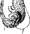

Figure 3.
Figure 3. Position of the uterus with an empty bladder and emptied rectum. Fig. 4. Parts of the uterine cervix. (After Slaviansky.) returns to normal, while the number of cells does not decrease; therefore, the uterus in women who have given birth is larger in volume and weight. Corresponding to the external contours of the uterus, two main sections are distinguished in it: the expanded upper one—the body of the uterus (corpus uteri) and the narrowed lower one—the uterine cervix (cervix uteri). Each of these sections in turn is divided into several parts, namely, in the body are distinguished: 1) the fundus of the uterus (fundus uteri)—the very upper part, located above the site of origin of the Fallopian tubes; 2) the upper segment; 3) the lower segment; and in the cervix: 1) the supravaginal part (portio supravaginalis), lying above the site of attachment of the vaginal vaults, and 2) the vaginal part (portio vaginalis), protruding into the lumen of the vagina. Since the posterior vaginal vault lies higher than the anterior one, some authors (e.g., Slaviansky) divide the uterine cervix not into two, but into three parts (Fig. 4), namely: I) the proper supravaginal part, located above the horizontal plane drawn through the site of attachment of the posterior vault; 2) the middle part, lying between this plane and the plane drawn through the site of attachment of the anterior vault, and 3) the proper vaginal part, lying below this latter plane. The vaginal part of the cervix has a different shape in nulliparous women and in parous women: in the former, its shape is more conical, in the latter—cylindrical. The question of the lower segment of the uterine body until recently belonged to the most controversial in obstetrics. Many authors, with K. Schroeder at the head, held the view that the lower segment is both physiologically and anatomically an isolated part of the uterus, specifically a part of its body, which can be distinguished even in the non-pregnant state of a woman. Others (Bandl, Küstner) derived the lower segment from the upper part of the cervix. Still others (Schatz, Fehling, and others) completely denied the existence of the lower segment, distinguishing in the uterus only two sections: the body and the cervix. Fourthly, e.g., Zweifel, recognized that the lower uterine segment is formed only during childbirth. Bayer, denying the existence in the non-pregnant uterus of the lower segment as a separate segment, expressed the view that it can arise during pregnancy either from the body of the uterus or from the cervix. Finally, in recent times, the teaching of Aschoff has become widespread in science, according to which the internal uterine os, long considered the border between the cavity of the cervix (cervical canal) and the cavity of the body of the uterus, represents not an opening, but a narrow canal up to 1 cm long. This canal macroscopically lies in the narrowed part of the uterus, i.e., in the cervix, although during pregnancy, expanding, it enters into the composition of the cavity containing the ovum, i.e., the cavity of the body of the uterus; upon microscopic examination of its walls, it turns out that the mucous membrane lining them has a structure similar to the structure of the mucosa of the body canal of the uterus. The intermediate part of the uterus containing this canal, to which Aschoff gave the name of the isthmus (isthmus), he considers to be Schroeder's lower segment. This view of Aschoff was generally confirmed by the studies of a number of authors, among whom the recent detailed studies in Germany by Stieve and in the USSR by Tarlo particularly deserve mention. The uterus is a hollow organ, and the cavity enclosed within it, corresponding to the two main sections of this organ, is also divided into 2 sections: in the body of the uterus there is the proper cavity of the uterus (cavum uteri), and in the cervix—the cervical canal (canalis cervicis uteri). The uterine cavity on a frontal section has the shape of a spherical triangle, the sides of which are convex outwards (see separate table, Fig. 19), and both its outlines and its size in nulliparous women are somewhat different than in parous women (see separate table, Fig. 20). In the three corners of the triangle are openings communicating the uterine cavity with neighboring cavities, namely: in the upper two—the uterine openings of the Fallopian tubes, leading to the channels of the oviducts, in the lower—the so-called "anatomical internal os of the uterus" (orificium uteri internum), communicating the uterine cavity with the isthmus canal. This canal is the narrowest place of the uterine cavity. In its lower part, it communicates with an opening bearing the name of the "histological internal os", with the cervical canal, which in nulliparous women has a spindle-shaped form (see separate table, Fig. 19), i.e., towards the middle it expands, and then narrows again and finally opens at the apex of the vaginal part with a round or transversely oval (in nulliparous women) opening (see separate table, Fig. 11)—the external uterine os (orificium uteri externum). At the very first birth, the external os in a woman usually tears, more often on the sides, and therefore in parous women it appears in the form of a transverse slit (see separate table, Fig. 12), less often—in the form of a star-shaped opening; as a result of these tears, the shape of the cervical canal itself also changes in parous women: instead of spindle-shaped, it becomes conical, with the apex
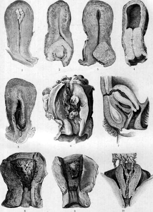
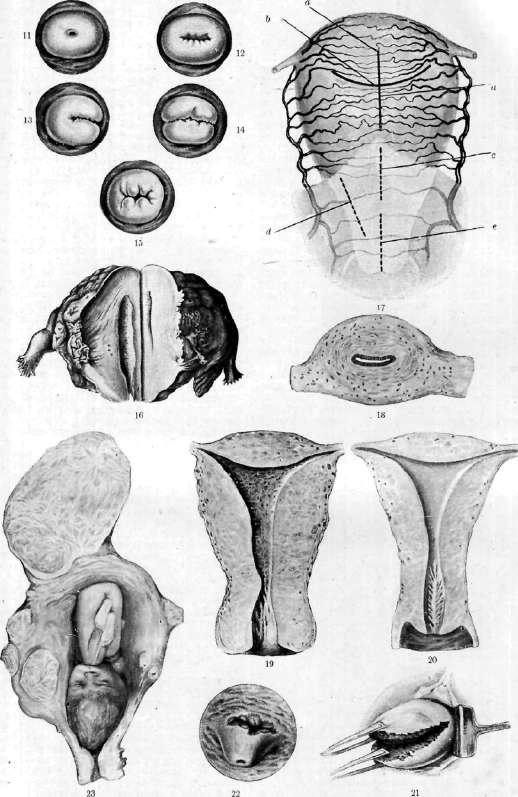
Figure 1. Cancer of the uterine body. Fig. 2. Cancer of the portio vaginalis. Fig. 3. Cancer of the posterior lip. Figure 4. Cancer of the uterine cervix. Figure 5. Polyp of the uterine body. Figure 6. Sarcoma growing through the left corner. Fig. 7. Abdominal-uterine fistula after classical Caesarean section. Silk ligatures are visible. Figure 8. Tuberculosis of the uterus. Figure 9. Placental polyp. Figure 10. Double perforation with extraction of the omentum. Fig. 11. Os in a nulliparous woman. Figure 12. Os in a parous woman. Fig. 13. Unilateral tear. Fig. 14. Bilateral tear. Fig. 15. Star-shaped tear. Figure 16. Actinomycosis of the uterus. Figure 17. Diagram of blood vessels and incisions of the uterus: a - longitudinal incision; b - fundal incision of Fritsch; v - retrovesical incision; d - incision in extraperitoneal Caesarean section according to Döderlein; e - incision in vaginal Caesarean section according to Dührssen. Fig. 18. Transverse section through the body of the uterus of an adult woman. Fig. 19. Uterine cavity of a nulliparous woman. Figure 20. Uterine cavity of a parous woman. Fig. 21. Exposure of a fresh tear. Figure 22. Fistula cervico-vaginalis laqueatica. Fig. 23. Pregnancy with a uterine fibroma.
el «2 cone is directed upwards, and the base downwards. The length of the uterine cavity, calculated from the inner surface of the fundus to the external os, varies in an adult woman within the same limits as the total size of the uterus. On average, in nulliparous women, this length is about 7 cm, in parous women 8 cm, of which 5-10 mm fall on the canal of the isthmus, and of the remaining ones approximately 2/3 fall on the uterine cavity and 1/3 on the cervical canal. Despite the fact that the uterine cavity in an adult woman has fairly significant dimensions in length as well as in width, its capacity under normal conditions is very small: according to Sappey, Krause, and Testut, it is equal to 2-3 cm3 in nulliparous women and 3-5 cm3 in parous women. This is because the anterior wall of the uterine cavity closely adjoins the posterior, and therefore on cross sections this cavity appears as a narrow transverse slit (see separate table, fig. 18). Throughout its entire extent, the uterine cavity is lined with a mucosa, which forms the innermost layer of the uterine wall—the endometrium, having a different thickness in various parts: from 1/2 to 7 mm (in the body of the uterus this thickness varies depending on the various phases of the ovarian-uterine cycle). The next layer, to which the mucosa closely adheres without any submucosal layer, is a powerful muscular layer—the myometrium, from 11/2 to 2 cm thick, constituting the main part of the entire mass of the uterus. Finally, on the outside, the body of the uterus is clad with the peritoneum, which can be regarded as the third layer of the uterine wall—the perimetrium. The endometrium in various parts of the uterus already looks macroscopically dissimilar: in the cavity of the body of the uterus in adult women it appears smooth, whereas in the cervical canal clearly expressed folds are noticeable on its surface, arranged like palm leaves; hence their old anatomical name "plicae palmatae". The mucosa in different parts of the uterus also differs in its histological structure. In particular, as regards its covering epithelium, in all parts of the uterine cavity this is a single-layered cylindrical epithelium, in places provided with cilia (the oscillations of which occur in the direction from top to bottom), and in other places consisting of secreting cells; in the body of the uterus its cells are lower (their height is 25-30 µ), wider, provided with ovoid nuclei lying in the middle of the cells; the covering epithelium in the canal of the isthmus has a similar morphological structure, whereas in the cervical canal the cells of the covering epithelium are taller (their height ranges from 35 to 60 µ), narrower, their nuclei have a rod-like shape and are located at the base of the cells. Furthermore, the epithelium of the mucosa of the cervical canal stains with mucicarmine (reaction for mucus), whereas the epithelium of the mucosa of the canal of the isthmus and the cavity of the body of the uterus does not stain. The stroma of the mucosa in all parts of the uterus consists of a network of thin connective tissue fibrils, in the meshes of which cells of various shapes are located. At the same time, in the stroma of the mucosa of the body of the uterus, one can histologically distinguish two layers: a more superficial one, periodically changing in various phases of the menstrual cycle, the so-called functional layer, the stroma of which is looser and contains partly spindle-shaped, partly stellate cells, and a deeper one, not undergoing special changes, the basal layer, the stroma of which appears denser and contains only spindle-shaped cells. Some authors (Hitschmann, Adler) distinguish not two, but three layers in the mucosa of the body of the uterus: a superficial compact, a middle spongy, and the deepest basal. The stroma of the mucosa of the cervical canal is less rich in cells and more fibrous, and the fibers in it are somewhat thicker and coarser. The stroma of the mucosa lining the canal of the isthmus, in its histological structure, represents something intermediate between the stroma of the mucosa of the body of the uterus and that of the cervical canal. The mucosa of the body of the uterus is very rich in glands (glandulae uterinae), and they have the form of long, narrow tubules, completely unbranched or dichotomously branching at the end, penetrating the entire thickness of the mucosa right up to the myometrium; however, both the appearance of these glands and their dimensions vary sharply in various stages of the menstrual cycle. The mucosa of the isthmus contains the same glands as the mucosa of the body of the uterus, but their number is much smaller here and they are arranged differently than in the latter: whereas in the mucosa of the uterine body the glandular tubes have a direction from below and inside upwards and outwards, in the mucosa of the isthmus they run obliquely from above and inside downwards and outwards. As for finally the glands of the cervical mucosa, they are distinguished by a wider lumen, and most importantly—they branch repeatedly like antlers and in places form expansions that give them the appearance of acinar glands (Fig. 5). The epithelium in the glands
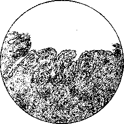
Figure 5. Mucosa of the cervix uteri.
of the various parts of the uterus presents the same differences as the covering epithelium. The uterine glands secrete a secretion which, in contrast to the vaginal secretion, has an alkaline reaction. Also by the nature of this secretion, the glands of the cavity of the body of the uterus differ from the glands of the cervical canal: the secretion released by the former is more liquid, watery, while the latter secrete a viscous, glassy mucus which, accumulating in the cervical canal, forms something like a plug here (the mucous plug of Kristeller). This plug is ascribed great importance. While the cavity of the vagina even in completely healthy women contains a mass of various microbes, the uterine cavity under normal conditions is completely sterile; it is apparently this mucous plug that is the obstacle to the penetration of microbes from the vagina into the uterus. In the region of the external os, the mucosa of the cervical canal, covered with a single-layered cylindrical epithelium and rich in glands, passes into the mucosa of the vaginal part, which in its structure resembles the mucosa of the vagina: like the latter, it is lined with a stratified squamous epithelium, the cells of which are rich in glycogen, and is devoid of glands. However, in the vicinity of the external os, glands are often encountered in it, but these are glands belonging strictly to the cervical mucosa. The myometrium, the middle, most powerful layer of the uterine wall, as its very name shows, consists chiefly of muscular tissue, namely of smooth muscles with an admixture of fibrous connective tissue and elastic fibers. The content of connective tissue in it increases in various parts of the uterus in the direction from top to bottom: in the cervix, its content is greater than in the walls of the body of the uterus; the connective tissue structure of the myometrium is especially pronounced in the vaginal part; likewise, the cervix of the uterus, especially its vaginal part, is richer in elastic fibers than the body of the uterus. As for the muscle bundles themselves, if one examines sections from the uterus of an adult woman, it is difficult to notice any order in the arrangement of the bundles. To form an idea of the architecture of the myometrium, one must begin studying it from the early stages of intrauterine life, when the uterine musculature is simpler and the order of arrangement of its individual layers is disturbed neither by vessels nor by the admixture of muscle bundles of the uterine ligaments; then it is easier to trace step by step the gradual complication of its architecture up to the age of puberty of the woman. The musculature of the uterus, like the musculature of some other hollow organs, basically consists of three layers (Werth, Gruzdev): an outer longitudinal (subserous), a middle circular, and an inner longitudinal (submucosal). In the body of the uterus, the most developed is the circular layer, consisting of rings which in the upper part of the uterine body surround the uterine horns perpendicularly to their axis and obliquely to the midline of the uterus, and the further downwards, the more horizontal a position they occupy. What has been said, however, refers only to the upper segment of the uterine body, while in the lower segment and in the cervix, longitudinally running muscle bundles are relatively more pronounced, which, going downwards, partly lose themselves in the thickness of the vaginal part, and partly, curving around the fornices, pass into the walls of the vagina, closely connecting it with the uterus. The indicated relationships are also inherent in the myometrium of adult women, although here the regularity of the arrangement of individual layers, especially in the peripheral parts of the myometrium of the body, is disturbed, firstly, thanks to the passage of large vessels, and secondly, thanks to the ingrowth of muscular layers that are a continuation of the musculature of the uterine ligaments. The length of individual muscle cells of the myometrium, according to the latest research by Tarlo, ranges from 60 to 80 µ in the body of the uterus and from 30 to 40 µ in the uterine cervix. In places in the myometrium of the lateral parts of the uterus, especially in the myometrium of the supravaginal part of the cervix, remnants of Wolffian ducts (Gartner's ducts) are encountered. These ducts, which in the embryo are the excretory ducts of the primitive kidneys (Wolffian bodies), in adult women become part of the epoophoron, or appendage of the ovary, run in an arched fashion in the broad uterine ligaments towards the borders of the uterus, penetrate the myometrium in the region of the isthmus, and descend downwards, then passing into the lateral walls of the vagina. Under the microscope
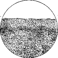
Figure 6. Vaginal portion covered with stratified squamous epithelium.
remnants of Gartner's ducts appear as epithelial inclusions of various shape and size, sometimes solid, sometimes provided with a lumen lined by a single layer of high columnar ciliated epithelium, passing in places into cubic and even squamous epithelium, and furthermore devoid of ciliated cilia (Fig. 6). The perimetrium, or uterine peritoneum, forming the outermost layer of the uterine wall, represents the direct continuation of the peritoneum of the bladder. Passing from the latter onto the uterus in the region of the 'histological internal os', the peritoneum covers the anterior wall of the uterine body, and in the region of the isthmus it is loosely attached to the underlying myometrium and can easily be separated from it, whereas at the border of the isthmus with the upper segment of the uterine body it is firmly attached to the myometrium and in this form reaches the fundus of the uterus, having lined which, it passes to the posterior wall of the uterus. Here it descends significantly lower than in front: it reaches not only the border between the body of the uterus and the cervix, but also lines the supravaginal part of the cervix and even the upper third of the posterior wall of the vagina. From the sides of the uterus, the uterine peritoneum forms duplications extending from the borders of the uterus to the lateral walls of the pelvis—the aforementioned broad uterine ligaments. Thus, the body of the uterus, with the exception of narrow clefts on the sides, where the layer of cellular tissue separating both leaves of the broad ligaments lies, is everywhere clothed with the perimetrium. As for the cervix of the uterus, the peritoneum is present only on the posterior wall of the supravaginal part, while in front and on the sides the cervix is surrounded by paracervical cellular tissue, and the vaginal part of the cervix, as already mentioned above, is covered on the outside by a mucosa resembling the mucosa of the vagina. The main blood vessel nourishing the uterus is the uterine artery (a. uterina). Having departed from the internal iliac artery, it passes in the base of the broad uterine ligament, where it crosses the ureter, being located anterior to it, approaches the uterus approximately at the border of the isthmus with the upper segment of the uterine body and here divides into two branches: one, a. cervico-vaginalis (cervico-vaginal), goes downward, supplying blood to the cervix and vagina; the other, winding like a corkscrew, goes along the border of the uterus upward, to the uterine horn, where it anastomoses with the ending of the internal spermatic artery; rising upward, this second, main branch of the uterine artery gives off a series of horizontal secondary branches in the anterior and posterior walls of the uterus, from which branches depart in a radial direction; the horizontal branches of the right and left uterine arteries anastomose with each other along the midline of the uterus in its posterior and anterior walls. In addition to the uterine artery, the uterus, namely its very upper part, the region of the fundus, is supplied with blood from the internal spermatic artery, which, having departed from the abdominal aorta or renal artery (the right internal spermatic artery more often departs from the aorta, the left from the renal artery), goes downward along the psoas muscle, crosses the ureter along the way, and, having descended into the lesser pelvis, enters the very lateral section of the broad ligament—the suspensory ligament of the ovary; having reached the funnel of the tube between the two leaves of this ligament, the internal spermatic artery gives off a branch to nourish the latter, then goes immediately below the place of attachment of the ovary, giving off a whole series of corkscrew-like winding branches into the hilum of the ovary, and heads to the horn of the uterus, where it anastomoses with the ending of the uterine artery. In addition to these two main vessels, the upper part of the body of the uterus also receives blood from the artery of the round ligaments. The region of the isthmus is the poorest in vessels among the various sections of the uterus, according to Moshkov. The arteries nourishing the uterus are accompanied by corresponding veins, usually each artery by two veins, and from a practical point of view it is not without significance that of the two veins accompanying the uterine artery, one usually lies together with the artery anterior to the ureter, the other posterior. The lymphatic vessels draining lymph from the uterus are arranged in the broad uterine ligaments in two bundles: one bundle, which consists of vessels carrying lymph from the cervix and the lower part of the body of the uterus, is located at the base of the broad ligament together with the uterine artery, and the vessels making it up flow into the iliac lymph nodes lying at the lateral walls of the pelvis, on the hypogastric and iliac vessels; the other bundle, formed by lymphatic vessels draining lymph from the upper parts of the body of the uterus and the lymphatic vessels of the Fallopian tubes and ovaries, lies in the upper section of the broad ligament, along the course of the internal spermatic artery, and empties into the lumbar lymph nodes lying on the aorta and inferior vena cava near the place of their bifurcation. In the very thickness of the uterine walls, the flow of lymph occurs from the mucosa towards the subserous layer, i.e., from the inside out (Fridolin), wherein the subserous layer is distinguished, according to Merzheevsky, by an extreme richness of lymph. As for the innervation of the uterus (see Autonomic nervous system and the related figures of Table I, Fig. 3, Table II, Fig. 6 and Table III, Fig. 12), the uterus, like other organs belonging to the internal section of the female genital apparatus, receives both sympathetic nerves and spinal nerves. The former come from the inferior hypogastric plexus and the sympathetic trunk, the latter from the II–IV sacral nerves. The indicated nerves form a dense plexus in the cellular tissue to the side and behind the uterine cervix and the upper part of the vagina, with numerous nerve ganglia embedded in it. Frankenhaeuser, who first investigated it, took this plexus for one extensive nerve ganglion and gave it the name of the cervical ganglion (ganglion cervicale). Subsequent works, mainly by Russian researchers, made it clear, however, that the so-called Frankenhaeuser's cervical ganglion is not at all a ganglion in the proper sense of the word, but a plexus of sympathetic and spinal nerves containing many nerve ganglia. It is from this plexus that the nerves of the uterus depart—both sensory and motor, both myelinated and unmyelinated. The distribution of these nerves in the thickness of the uterine walls was the subject of numerous works, among which a prominent place belongs once again to the works of Russian authors. Sinitsyn, who investigated the distribution of nerves and nerve endings in the wall of the uterus and vagina in some small mammals, found that both myelinated and unmyelinated trunks enter the wall of the uterus, with the latter in a relatively small number. These trunks go first along the connective tissue layers between the muscle bundles of the outer layer of the myometrium, mostly accompanied by vessels, and having reached the boundary between the outer longitudinal and inner circular layers of muscles, they diverge in a longitudinal direction, giving off side branches, which in turn give branchlets going in various directions. If one then follows the fate of exclusively unmyelinated fibers, it is easy to be convinced that ultimately, thanks to gradual division, a dense nerve plexus arises from them, from which the finest terminal threads depart, penetrating between individual muscle cells and breaking up into side varicose branchlets enmeshing each cell. Having traced the unmyelinated fibers in the mucosa of the uterus, Sinitsyn found that around the uterine glands there are networks of thin nerve fibers, from which the finest threads depart, piercing the limiting membrane of the glands, directly adjacent to the cells of the glandular epithelium, and ending with pericellular terminal apparatuses in the form of lumps, buttons, or clusters. Approaching the surface epithelium of the mucosa of the uterus, unmyelinated fibers go some distance beneath it and give off varicose threads ending in thickenings or swellings between the epithelial cells. Myelinated fibers form, according to Sinitsyn, sensory endings in the muscles of the uterus; in the mucosa of the cervix, Sinitsyn managed to discover a special form of their sensory endings—in the form of creeping bushes. The various sections of the uterus are supplied with nerves, according to Sinitsyn, rather evenly. Naidich, who investigated the nerves of the human uterus, could establish the presence of two plexuses in it: extramural and intramural; the former lies in the parametrial cellular tissue, being a direct continuation of the hypogastric plexus, the latter is located directly on the myometrium, penetrating into the superficial layers of the muscles. The special interest of researchers has long been aroused by the question of the presence of nerve ganglia and cells in the thickness of the walls of the uterus. Like the heart, the uterus is an organ capable of automatic contractile activity. True, the movements of the uterus also depend on the central nervous system: experimental studies make it indisputable that there is a center in the lumbar part of the spinal cord controlling the movements of the uterus, while in the medulla oblongata there is an inhibitory center, and perhaps a second, accelerating one; it is further indisputable that reflex contractions can easily be induced in the uterine muscle by various irritations of the central ends of transected spinal nerves, the skin in various areas of the body, the nasal mucosa, the clitoris, the nipples, etc. However, on the other hand, precise experiments, at the head of which can be placed the flawless classical experiments of Kurdinovsky on the isolated uterus, just as irrefutably testify that even being outside of any connection with the central nervous system, the uterus retains the ability to contract and even the capacity for labor activity.
There are, furthermore, not a few clinical observations indicating that even in cases where the conductivity of the spinal cord in a pregnant woman is impaired, for example in tabes dorsalis, labor nevertheless proceeds normally. Although some authors state that to explain all such facts there is no need at all for automatic centers of a ganglionic structure, and that the uterus, like any other muscular organ, can contract independently of such centers under the influence of the direct action of mechanical, thermal, hormonal, and other stimuli on its muscle, the majority are inclined to relate these facts to the circumstance that the uterus possesses automatic nerve centers of a ganglionic structure. Sechenov already pointed out that uterine ganglia should be sought in the thickness of the myometrium itself. Numerous studies have been carried out in this direction, which, however, have not yielded consistent results: while earlier researchers working with less sophisticated methods (among Russians, Gladkovsky, Patenko, Razumovsky, and Gavronsky) encountered not only nerve cells but also ganglia in the myometrium, recent observers (in the USSR, Sinitsyn and Naidich) found neither nerve ganglia nor even nerve cells in the uterine musculature. Oofoter, using Ehrlich's methylene blue staining on sections of a freshly extirpated pregnant uterus, was able to find an unquestionable nerve cell in one of the sections. Of the physiological processes taking place in the uterus, regarding processes connected with the ovarian-uterine cycle, see Menstruation, and regarding the processes that make up the act of reproduction, see Pregnancy, Labor, and Puerperium. The question of the internal secretion of the uterus remains controversial to this day. It has long been known that women with a congenital absence of the uterus, who nevertheless have ovaries present, upon reaching the age of puberty begin to suffer from monthly recurring pains, the so-called molimina menstrualia; these pains are sometimes so severe that they compel gynecologists to resort even to castration. Molimina menstrualia are sometimes also observed in women who have undergone removal of the uterus with preservation of the ovaries; moreover, in such women, disorders analogous to those that arise after castration sometimes occur, only to a lesser degree. These facts served as the basis for raising the question of whether the uterus, like the ovaries, possesses the capacity for internal secretion. To clarify this question, Fedorov performed a series of experiments involving the injection of a glycerin extract from the uterine mucosa into the blood of rabbits, and obtained a noticeable physiological effect from such injections, which led him to attribute the capacity for internal secretion to the uterine mucosa. Other authors attributed this capacity to the myometrium. Ancel and Bouin found connective tissue cells in the myometrium of the pregnant rabbit uterus, which they described as glande endocrine myométriale, while Fornero attributes endocrine properties to epithelioid cells found by him in the myometrium of the human uterus. The internal secretory capacity of the uterus has not yet received general recognition.
V. Gruzdev. II. Embryology. In 1825, the anatomist Rathke described in the human embryo a previously unknown formation having the appearance of two parallel tubules located side by side with the Wolffian ducts. The famous Johannes Müller understood and explained that these tubules are the primordia of the future Fallopian tubes; after this, these tubules were given the name of Müllerian ducts (Müllerian tracts), which has survived to the present time. Further study of these formations in embryos of an earlier age and in the process of their subsequent development showed that these tubules are the primordia not only of the Fallopian tubes, but also of the uterus itself, as well as the vagina. At the present time, the development of the uterus is represented in the following form: the appearance of the primordia of the Müllerian ducts is noted for the first time in embryos of about 10 mm in length, even before the differentiation of the gonad. These primordia initially have the appearance of an oblong depression on the outer (lateral) side of the Wolffian body (mesonephros) and are located at the level of the III-IV thoracic segment; but even before the appearance of this depression on the corresponding section of the Wolffian body, the epithelium covering it separates and stands out by its height ("Müllerian epithelium"). Soon after its appearance, the aforementioned depression closes into a tubule, which narrows somewhat toward the caudal end of the embryo (caudally), penetrates into the very substance of the Wolffian body and ends blindly here, whereas the head end (cranially) remains open, representing in the future the abdominal opening (funnel) of the Fallopian tube. At this age, the entire length of the Müllerian duct is approximately 300 µ. Subsequently, the Müllerian duct grows in the caudal direction with its blind end in the form of a solid strand, which, however, very quickly acquires a lumen and thus in turn turns into a duct. In embryos 20 mm in length, the direction of the strand-duct changes from vertical to horizontal; it crosses the Wolffian duct from the front and now grows toward the median plane of the body towards a similar duct of the opposite side. At this time, the length of the Müllerian duct already reaches 1,500 µ. At the place of contact of both Müllerian ducts, their direction changes again from horizontal to vertical, and growth goes again toward the caudal end of the embryo, both ducts lying closely adjacent to each other and placed already inwards (medially) from the Wolffian ducts. All four ducts now lie in a secluded section of mesodermal tissue, which bears the name of the "sex cord." In an embryo of about 50 mm in length, the Müllerian ducts approach the wall of the urogenital sinus (sinus urogenitalis) and bulge into it in the form of the so-called "Müllerian tubercle." Finally, in embryos of about 70 mm in length, the Müllerian ducts open into the lumen of the urogenital sinus (sinus urogenitalis). Now their length exceeds 3,000 µ. Simultaneously with the further growth of the Müllerian ducts, soon after their contact, their gradual fusion also begins; by this time, the gonads also differentiate toward the female sex, i.e., the ovary is formed. In the case where the embryo develops toward the male sex, the fate of the Müllerian ducts will be completely different: they are doomed to gradual regression, and in adult male individuals only rudiments remain of them (the appendix of the testis on the epididymis and the utriculus masculinus in the prostate gland). The fusion of the Müllerian ducts begins at the end of the 2nd, beginning of the 3rd month of embryonic life, precisely at the border of the middle and lower third of their extent, namely, in that place where the cervix of the uterus will develop in the future. From here, the fusion of the Müllerian ducts proceeds in both head and caudal directions, forming the uterus in the former, and the vagina in the latter. In the head direction, the fusion does not reach the end, because the upper (head) ends of the Müllerian ducts, being at a great distance from each other, do not fuse—the Fallopian tubes are formed from them. However, the ducts fuse not only in their vertical section, but partially also fuse the closest sections of the horizontally located segments. This part goes to the formation of the upper part of the body and fundus of the uterus. As the Müllerian ducts fuse during the 4th and 5th months of intrauterine development, their final separation into the tubes, uterus, and vagina takes place, and the vagina at this stage of development does not yet have a lumen, but represents a solid strand consisting of large epithelial-like cells. The uterus in turn is divided into the body and the cervix, and the body is sharply demarcated from the cervix, which in its dimensions exceeds the dimensions of the body by 3 to 4 times and projects into the upper section of the vagina, gradually forming here the "vaginal part" (portio vaginalis cervicis uteri). The thin wall of the newly formed uterus gradually thickens due to the formation of muscle fibers, first longitudinal, and then circular; the mucosa retains a simplified structure for a long time and only at the end of intrauterine development do glands appear in it, and much earlier in the cervix than in the body. The embryonic development of the uterus does not end by the moment of birth, because the uterus continues to grow and complicate in its structure after birth, reaching its final development only by the time of the onset of puberty. In the development of the uterus after birth, the following stages should be noted: 1) soon after the birth of a girl, her uterus decreases rather sharply in its dimensions—from 4 cm in a newborn to 2.5 cm in an infant; 2) the girl's uterus retains such dimensions almost unchanged throughout the neutral childhood age, i.e., up to 7–8 years; 3) from this time, accelerated growth of the uterus begins and its transformation in size and shape into the uterus of an adult woman (see above—anatomy). The decrease in the uterus soon after birth is explained apparently by the fact that after birth the organism of the newborn girl, and consequently her uterus, fall out from under the influence of maternal hormones and placental hormones. The invariability (stabilization) of the uterus in neutral childhood is in all probability connected with the fact that at this age only the growth of eggs occurs in the ovaries, but their maturation has not yet begun, and the endocrine activity associated with this does not yet manifest its effect on the uterus. Finally, with the beginning of the maturation of eggs, accelerated growth of the uterus also begins, and its body grows especially strongly, so that the ratio between the dimensions of the body and the cervix is soon established in the form that takes place in adult women, i.e., the length of the body relates to the length of the cervix as 4 or 5 to 3. The main fact of the embryonic development of the uterus must be considered the fact that it is entirely formed from the middle germ layer (mesoderm), and even in the adult state it retains many properties of "mesenchymal" tissue and in particular the huge ability of mesenchyme for metaplasia and hypertrophy, which is important both in changes observed during the ovarian-uterine cycle, and especially during pregnancy, when the uterus undergoes completely exceptional hypertrophy for 10 months. III. Malformations of the uterus. The history of the development of the uterus determines and predetermines those anomalies of its structure in adults that are called "malformations" of the uterus. These malformations are expressed very sharply if the delay in the normal development of the uterus occurred at a very early age of intrauterine life—in its very first weeks; on the contrary, the malformation will be barely noticeable if the development of the uterus stopped shortly before the onset of puberty. In other words, malformations can be expressed, on the one hand, by the complete absence of the uterus in an adult woman (anhystoria in the old gynecological terminology), on the other hand, in an adult woman one can meet the uterus of an adolescent or a girl, differing little from the normal uterus of an adult woman (uterus pubescens, uterus virgineus). One could imagine even deeper violations in the development of the Müllerian ducts, but practically they are not observed, because embryos with such deep anomalies of the reproductive system, and consequently of the Wolffian body, as a rule, turn out to be unviable already in intrauterine life and die long before birth, perhaps in the very first months or even weeks of embryonic development. If the violation in the development of the uterus captures it in that period of embryonic life (2nd-3rd months) when the very fusion of both Müllerian ducts takes place, then malformations of its development are obtained that bear the character of "duplication," complete or partial. Finally, the arrest of development already in extrauterine life will give an insignificant malformation, again depending on the age when this arrest occurred.—The cause of the formation of various malformations of the uterus is unknown. Formerly, some explained them by phenomena of atavism, since some forms of malformations of the uterus are encountered as a norm in certain species of animals; others—by the action of some pathological, most likely inflammatory, process in embryonic life. At the present time, new ones can be added to these explanations: some speak of the constitutional-hereditary origin of malformations, others of "mutations" at the moment of their formation.
Probably in each individual case, both one of these presumed causes and various combinations of them may play a role. Elucidating this issue in each given case appears very difficult, not to say impossible. Despite attempts to give an exhaustive classification of developmental defects of the uterus (and of the female reproductive system in general), we do not yet have such a classification at the present time. For practical purposes, developmental defects of the uterus can be divided into five groups, adhering to the chronological moment as the principle for classification. These groups are as follows: 1) complete aplasias, 2) duplication, 3) partial (unilateral) aplasias, 4) atresias and displacements, 5) hypoplasias. 1. Complete aplasias. Their formation should be attributed to that very early period of intrauterine development when the Müllerian ducts are just beginning to form. In aplasia, the uterus in an adult woman is either not palpable at all or is palpable per rectum as a small, solid, cylindrical connective-tissue cord (uterus rudimentarius solidus). The lateral (distal) segments of the Fallopian tubes are usually present. The ovaries may be well developed, and the woman may appear completely healthy and correctly formed in her external constitution and appearance. 2. Duplication (Fig. 7) is observed either along its entire extent, such that two completely separate uteri with two separate cervices are produced, i.e., essentially two hemiuteri (uterus didelphys, uterus bipartitus, uterus duplex separatus), or this duplication is expressed merely by a depression in the fundus of the uterus in the form of a more or less deep arc, with there being only a single uterine cavity and a single cervix (uterus arcuatus - "saddle-shaped" uterus; uterus incudiformis - anvil-shaped uterus). Between these two extreme degrees of uterine duplication, a whole series of intermediate ones can be observed. Thus, a uterus with a pronounced saddle shape will already be called bicornuate (uterus bicornis), and such a uterus may have either a single common or a separate cavity. The septum itself, which divides the uterine cavity, can either be along its entire extent, i.e., from the fundus to the external os, or be partial (uterus septus and uterus subseptus). The division of the uterus into two cavities, both partial and complete, can also be encountered in a uterus that in its external shape and size differs almost not at all from normal. All the enumerated developmental defects of the uterus are observed
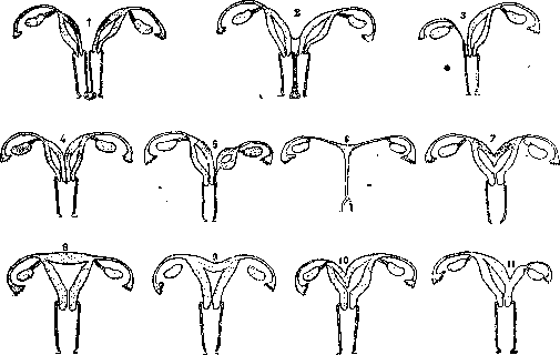
Figure 7. 1 - uterus didelphys; 2 - uterus et vagina duplex; 3 - complete atresia of the right duct (uterus pseudounicornis); 4 - uterus bicornis bicollis; 5 - uterus bicornis with a rudimentary left horn; 6 - uterus bicornis rudimentarius solidus; 7 - uterus bicornis unicollis; 8 - uterus incudiformis; 9 - uterus introrsum arcuatus simplex; 10 - uterus bicornis with unequal horns; 11 - uterus bicornis with a rudimentary horn.
as with a normal vagina, so too with one or another defect of its development, in particular with its duplication; hence all sorts of new combinations. 3. Partial unilateral aplasias of the uterus manifest themselves by the fact that one of the Müllerian ducts does not develop at all or is underdeveloped over a certain extent. In the first case, there will be as if a hemi-uterus, which is conventionally called a unicornuate uterus (semiuterus, uterus unicornis); in the second, various forms of a bicornuate uterus with an underdeveloped rudimentary horn (uterus bicornis rudimentarius). Naturally, such a rudimentary horn may be underdeveloped to a greater or lesser extent, which in turn will give various forms of developmental defects of the uterus, which may also manifest themselves clinically in various ways. 4. Atresias of the uterus occur because no lumen has formed over a certain extent of the Müllerian ducts. Here, secondary "occlusion" on the basis of an inflammatory process that took place both during intrauterine development and in childhood or adolescence is sometimes also assumed. To this same group can be attributed those developmental defects that are expressed in the congenital displacement of the uterus posteriorly, laterally, or downward (prolapse), as well as the displacement of the uterus from the pelvic floor into one hernia or another that is present. 5. Hypoplasias of the uterus. The fifth group of developmental defects of the uterus consists of those of its defects in which we have an arrest in the development of the uterus already at one stage or another of extrauterine (or late intrauterine) life. They are, as a rule, closely associated with the complete or partial underdevelopment of the entire organism of the woman—with so-called infantilism (see). Here it is customary to distinguish the following degrees of underdevelopment: a) embryonic uterus (uterus fetalis), b) infantile uterus (uterus infantilis), c) adolescent uterus (uterus pubescens), d) virgin uterus (uterus virgineus). The least pronounced expression of the underdevelopment of the uterus will be its small size with a normal shape (hypoplasia uteri), although to this is often added either a conical cervix (collum conicum), or an excessive bend (flexion) of the uterus anteriorly (hyperanteflexio uteri congenita), or both simultaneously. Developmental defects of the uterus, as a rule, manifest themselves by specific symptoms only with the onset of puberty or even pregnancy; but some of them may manifest themselves in no way throughout the entire life of a woman and are sometimes discovered quite accidentally during a gynecological examination, during operations on the living, or at autopsies of the deceased. On the other hand, mildly expressed developmental defects of the uterus are easily overlooked and go unrecognized even by experienced specialists and upon a sufficiently careful examination of sick women. Every obstetrician-gynecologist of a certain standing has had to encounter women who throughout their lives have visited many specialists without the developmental defect of the uterus present being recognized or even suspected. The very manifestation of a developmental defect is determined by both its degree and its form. Thus, hypoplastic uteri manifest themselves by a late onset of menstruation, insufficiency, and painfulness of menstruation; complete absence of menstruation is observed with complete aplasia and atresias, and in the latter case, the menstrual discharge accumulates above the site of atresia (haematodermia, gynaetresia). With duplications of the uterus, one half, which is more developed, may menstruate quite correctly, while the second either does not menstruate at all or does not excrete the menstrual discharge outward due to atresia in the lower segment [for example, a bicornuate uterus with a rudimentary horn, in which menstrual discharge may accumulate with the subsequent formation of haematosalpinx (see)]. In the presence of menstruation, a developmental defect of the uterus may be discovered with the onset of sexual life, if it gives cause for sterility. A significant number of mildly expressed developmental defects belong here. The onset of pregnancy in an underdeveloped uterus can often lead to habitual miscarriages or habitual premature births; during labor, weakness of labor activity, a tendency to placenta accreta, hypotension and atony of the uterus, etc., may manifest themselves. On the other hand, it must be borne in mind that with mildly expressed underdevelopment of the uterus, pregnancy itself is the most powerful stimulus contributing to the further development of both the uterus itself and the entire organism of the woman as a whole. Since a certain underdevelopment of the uterus (infantilism) is present in a large number of young women, the artificial termination of a first pregnancy in such infantile women without serious medical indications cannot be justified by anything, since it often permanently fixes the existing underdevelopment of the uterus. Diagnosis. Some forms of developmental defects of the uterus are recognized without difficulty. Thus, a bicornuate or saddle-shaped uterus can be recognized already by the sensation received by the external examining hand; a conical cervix, by routine internal examination; hypoplasias of the uterus, and excessive anterior flexion, by bimanual examination. In other cases, recognition, as stated above, may not be easy even when a developmental defect is suspected. Here, in addition to the clinical analysis of the existing symptoms, more detailed methods of investigation must be applied: speculum examination, sounding (sometimes with two sounds), hysterosalpingography, and the like. Recognizing a developmental defect of the uterus is sometimes especially difficult when pregnancy has occurred, and this particularly concerns uterine duplications. A developmental defect of the uterus recognized in one way or another should always draw the physician's attention to the state of the urinary system, since it is closely connected in its embryonic development with the development of the genital system—hence the frequent coincidences of defects and developmental anomalies in both systems simultaneously. As for the treatment of developmental defects of the uterus, we are still rather powerless in this matter, although in individual cases even thermal treatment alone (especially mud therapy) can lead to the further development of the uterus. It is only necessary that such treatment be initiated in young years. In other cases, the underdevelopment of the uterus is closely connected with metabolic disorders. In these cases, one has to observe the further development of the uterus and pregnancy after a sharp change in the regimen of sterile women with an underdeveloped uterus. In other cases, indications for surgical assistance also arise. This includes, for example, the excision or dissection of fused sections of the cervix of the uterus in its atresia, or the excision of the underdeveloped half of the uterus in the presence of a rudimentary horn. Surgical intervention may be required upon the onset of pregnancy in the presence of a developmental defect of the uterus (e.g., pregnancy in a rudimentary horn). Surgical (obstetric) intervention is also frequently required during labor in women with a developmental defect of the uterus. Here assistance can be most diverse depending on the indications in each individual case. However, one should not think that assistance in labor is necessary always and in all cases of underdevelopment of the uterus: with a double and unicornuate uterus, quite successful and multiple pregnancies and labors are frequently observed. Recently, operations have been proposed to correct even such developmental defects of the uterus as its duplication; thus, Strassmann and Kakushkin proposed suturing both halves of the uterus after excising the septum. Prevention of developmental defects of the uterus should begin before birth (antenatally) and, with the current state of our knowledge, can consist exclusively in improving the living conditions of pregnant women. Underdevelopment of the uterus in extrauterine life, insofar as it may depend on diseases, inadequate nutrition, and improper upbringing of children and adolescents, can be prevented to a certain extent by observing the rules of hygiene and protecting children from unbearable labor. To the developmental defects described above, it remains to add certain other forms not included in the given scheme. Underdevelopment of the organism in intrauterine life may, by the time of birth and sometimes subsequently, manifest itself by the presence of a whole series of hernia orifices and true hernias. It must be borne in mind that in inguinal and femoral hernias of such congenital origin, one or another internal genital organ is frequently found (in more than half of the cases). Most commonly encountered is the ovary, then the tube, the uterine horn, and the uterus itself (hernia uteri). Congenital prolapse can also be classified among hernias. As a very great rarity, such a prolapse is described in newborn girls; but prolapses in nulliparous women, especially young women and girls, should probably be attributed here. The close proximity of the Müllerian and Wolffian ducts in the early stages of embryonic development can lead to the fact that part of the Wolffian duct is as it were captured into the wall of the growing and transforming uterus. Such remnants of the Wolffian ducts, included in the wall of the uterus (or vagina), are called Gartner's ducts. Cysts of the uterus (or vagina) can develop from them; such formations are rightly classified among developmental defects of the uterus. Among the developmental defects of the uterus, one must also include so-called congenital erosion of the vaginal portion of the uterus (erosio s. pseudoerosio congenita). There are still some other developmental defects of the uterus, but in view of their very great rarity, they are of little practical interest. Thus, for example, as an exceptional rarity, individual cases of excessive size of the uterus in newborn girls and the unusual thin-walled nature of the uterus have been described.
(uterus membranaceus) and so on. In conclusion, it should be mentioned that the most diverse developmental defects of the uterus can be found in cases of hermaphroditism (see) along with the abnormal development of the entire reproductive system in these individuals. Malformations of the same kind in the reproductive system and particularly in the uterus can also be found when studying monstrosities of the most diverse character. Unfortunately, this issue has not yet been covered at all in modern teratology. M. Kolosov. IV. Abnormal positions of the uterus. The uterus, being a mobile organ, can easily leave its normal position for a time. When the change in position is persistent, it is spoken of as a pathological abnormal position of the uterus; in special cases, it is accompanied in addition by a change in the shape of the uterus. Accordingly, the following anomalies of uterine position are distinguished: - I. Abnormal position of the uterus without special changes in its shape. A. Positions of the uterus: 1) anteposition (antepositio uteri), 2) retroposition (retropositio uteri), 3) and 4) dextro-, sinistroposition (dextro-, sinistropositio uteri), 5) elevation of the uterus (elevatio uteri), 6) descent and prolapse of the uterus (descensus, prolapsus uteri). B. Inclinations (versions) of the uterus: 1) anteversion (anteversio uteri), 2) retroversion (retroversio uteri), 3) and 4) dextro-, sinistroversion of the uterus (dextro-, sinistroversio uteri). C. Rotation of the uterus (rotatio uteri). - II. Abnormal position of the uterus accompanied by a change in its shape. A. Flexions (changes in the bend between the body and the cervix of the uterus): 1) anteflexion (anteflexio uteri), 2) retroflexion (retroflexio uteri), 3) and 4) dextro-, sinistroflexion of the uterus (dextro-, sinistroflexio uteri); B. Torsion of the uterus (torsio uteri). C. Inversion of the uterus (inversio uteri). D. Hernia of the uterus (hysterocel'e uteri). - Often individual forms of abnormal positions occur in various combinations. In some cases, abnormal positions of the uterus, being a secondary phenomenon of another disease, are devoid of clinical significance. Anteposition of the uterus is the displacement of the entire uterus anteriorly; in some cases, along with this, there is also an upward displacement of the uterus. As a physiological and transient phenomenon, anteposition is observed with excessive filling of the rectum. In pathological cases, anteposition is caused by various kinds of tumors located in the posterior pouch of Douglas (ovarian tumors, subserous uterine myomas) and accumulations of encysted fluids behind the uterus (hemorrhages in ectopic pregnancy, purulent effusions in the posterior pouch of Douglas). Symptoms characteristic of the anterior displacement of the uterus itself consist in frequent urges to urinate; usually symptoms associated with the underlying condition that caused the displacement of the uterus prevail. Retroposition of the uterus - displacement of the entire uterus posteriorly - as a temporary phenomenon is observed when the bladder is overfilled. In pathological cases, retroposition of the uterus is caused by an inflammatory process of the peritoneum behind the uterus, of the uterine appendages. Particularly often, inflammatory processes in the uterosacral ligaments, respectively in the tissue behind the uterus, leading to subsequent wrinkling and the formation of scars pulling it posteriorly, lead to the displacement of the uterus posteriorly. Frequently, such shortening of the tissues behind the uterus is congenital as an expression of underdevelopment. Less commonly, retroposition of the uterus is observed depending on tumors developing from the anterior wall of the uterus (myomas) or located anterior to the uterus (ovarian cysts). Symptoms are associated with the underlying condition. Lateral displacement of the uterus, lateroposition - displacement of the body of the uterus together with the cervix to the right or left (dextropositio - displacement to the right, sinistropositio - displacement to the left) - is often combined with posterior displacement of the uterus (retroposition of the uterus). The most frequent cause of this position of the uterus is inflammatory processes in the uterine appendages. In inflammatory processes in the pelvic tissue, the uterus is initially displaced in the direction opposite to the inflammatory process; with the onset of wrinkling, it is pulled in the direction corresponding to the inflammatory process. Lateral displacement of the uterus can be caused by intraligamentously located myomas, ovarian cysts, etc.; in these cases, some elevation of the uterus is also often noted. Symptoms depend on the underlying condition. Elevation of the uterus - displacement of the entire uterus upward - as a temporary phenomenon occurs with the simultaneous filling of the bladder and rectum; as a persistent phenomenon, it is observed with the accumulation of blood in the vagina (haematocolpos), with tumors of the cervix, with intraligamentously located tumors. Descent and prolapse of the uterus - see Prolapse of the uterus, vagina. Anteversion of the uterus is a pathological position in cases where the uterus is deprived of its mobility depending on inflammatory processes in the region of the peritoneum, uterine tissue, and its appendages. Symptoms depend on the underlying condition; due to the immobility of the uterus, increased blood loss during menstruation is possible due to poor contractility of the uterus. - With retroversion, the body of the uterus is deflected posteriorly, and the cervix anteriorly; the axes of the body and cervix form an almost straight line. Often (but not always) the uterus is lowered. Such a position is rare and is usually combined with flexion of the uterus. As a temporary phenomenon, retroversion of the uterus is observed in some cases when the bladder is overfilled, then in the postpartum period with insufficient involution of the uterus and ligamentous apparatus. Retroversion of the pregnant uterus - see below (retroversion during pregnancy, incarceration of the uterus). - With lateroversion (dextroversio - inclination to the right, sinistroversio - inclination to the left), the body of the uterus is deflected to one side of the pelvis, and the cervix to the opposite side, wherein the axis of the body of the uterus forms an almost straight line with the axis of the cervix. The most frequent cause is unilateral inflammation of the uterine appendages, the tissue of the upper section of the broad ligament, and the tissue around the cervix on the side opposite to the inclination of the fundus of the uterus. Symptoms and treatment - depending on the underlying condition. - Rotation of the uterus - rotation of the uterus together with the cervix around its longitudinal axis. The rotation of the uterus with its anterior surface to the right is called dextrorotation, to the left - sinistrorotation. Weakly expressed dextrorotation is considered a normal position. The origin of uterine rotation is associated with inflammatory processes in the parametrial tissue in the region of one or the other uterosacral ligament; with the wrinkling and shortening of the right uterosacral ligament, the anterior surface of the uterus turns to the left. Unilateral inflammatory processes of the peritoneum covering the uterine appendages also lead to rotation of the uterus. Rotation of the uterus very often occurs as a result of the twisting of the pedicle of a tumor originating from the genital sphere (subserous myomas, ovarian tumors). Symptoms are associated with the underlying condition. Acute-angled, pathological anteflexion (hyperanteflexion) is present when the body of the uterus in the region of the internal os bends sharply anteriorly, wherein the axis of the body of the uterus forms an angle of less than 90° with the axis of the cervix. In some cases, the body of the uterus is so strongly bent forward that the anterior wall of the uterus lies as it were on the cervix; in addition, retroposition of the uterus is also often present. The vaginal part of the uterus usually has a conical shape, the entire cervix is elongated, taking the shape of a trunk bent anteriorly. The body of the uterus is small, underdeveloped, its tissue is poorly pliable. Patients with pathological anteflexion almost always have certain signs of infantilism or other constitutional anomalies. The most frequent cause of pathological anteflexion is insufficient ovarian function, and in these cases, anteflexion is mostly a congenital condition. But the forward bend of the uterus can also form as a result of a chronic inflammatory process in the region of the uterosacral ligaments and secondary ovarian insufficiency. Often with hyperanteflexion, dysmenorrhea (see), frequent or rare abundant or scanty menstruation are observed in connection with the underdevelopment of the uterus, inadequacy of its muscle, and ovarian insufficiency. Infertility is often present; with pregnancy having occurred, abortion often occurs due to poor attachment of the fertilized egg. In connection with constitutional defects, easy fatigue, weakness, phenomena of hysteroneurasthenia, lowered libido are noted. - Treatment consists in the use of general strengthening measures, preparations of the ovary and the anterior lobe of the hypophysis. Locally - diathermy, hot vaginal douches. Good results are given by regular sexual intercourse, especially - the occurred pregnancy, which stimulates the development of the uterus. Of surgical aids, sounding of the uterus, dilation of the cervical canal, curettage of the uterine cavity can be recommended. Plastic operations (incision or discission of the cervix, wedge-shaped excision) should be resorted to only in extreme cases, since such operations usually do not give favorable results and are often the cause of subsequent complications (catarrh of the cervix, etc.). If the inflammatory process plays the main role in the etiology of the bend, one should eliminate catarrh of the cervix, treat erosion, posterior parametritis. Anteversion during pregnancy with a pendulous abdomen is observed with a narrow pelvis, weakened abdominal walls, divergence of the rectus muscles. Retroflexion of the uterus, usually in combination with a backward bend of the uterus in the region of the internal os (retroversion-flexion of the uterus), is found in 16-20% of all gynecological patients.
The degree of anteversion and retroflexion of the uterus can vary; a more detailed subdivision depending on these signs has not become widespread; the term retrodeviation of the uterus is used, uniting all forms of deviation and bending of the uterus backwards. In general, it is customary to divide bends of the uterus backwards into mobile (retroflexio uteri mobilis) and immobile, fixed (retroflexio uteri fixata). Mobile retroflexion does not represent a strictly outlined anomaly; with a mobile uterus, inflammatory changes in the uterine appendages and the pelvic peritoneum may also be observed; other objective data also vary according to the etiology. The etiology of uterine retroflexion is diverse. Congenital cases are extremely rare. On the contrary, a major role is played by the relaxation of tissues, a decrease in their tone, both acquired (damage to the pelvic floor, subinvolutio uteri post partum, weakening of the organism in connection with various kinds of diseases, etc.) and congenital (asthenic constitution). Under such conditions, the cervix easily departs anteriorly, intestinal loops penetrate into the excavatio vesico-uterina, and intra-abdominal pressure under such conditions must act on the anterior surface of the uterus. In such cases, one can observe weakening of the pelvic floor, lowering of the vagina, urinary bladder, flabby abdominal walls, and a pendulous abdomen; the uterus is deprived of its usual tone, flabby, edematous, and often lowered. The uterine appendages are lowered, and the ovaries are located low. Subsequently, prolapse of the uterus may develop. Very often, the origin of uterine retroflexion is associated with inflammatory processes in the region of the uterine appendages and in the posterior pouch of Douglas; existing adhesions and fusions disrupt the mobility of the uterus to varying degrees; less commonly, the origin of uterine retroflexion is associated with the formation of scar tissues in the anterior parametrium. The origin of uterine retroflexion exclusively under the influence of mechanical strokes (traumatic retrodeviations)—after a fall on the buttocks, on the back, etc.—is subject to great doubt. In some cases, retroflexion of the uterus may be caused by a tumor located in the excavatio vesico-uterina or originating from the anterior wall of the uterus. Not long ago, uterine retroflexions were attributed exceptionally great importance in the origin of various symptoms. At present, the clinical significance of this anomaly of uterine position is evaluated significantly lower. The diverse symptoms noted in retroflexion of the uterus should be considered in accordance with the etiological moments playing a role in the origin of uterine retroflexion. Heaviness in the lower abdomen, in the groin, pain in the sacrum and lower back, pain during menstruation, during sexual intercourse, etc.—all this can be attributed partly to the manifestation of an asthenic constitution (general ptosis, neurasthenia, psychasthenia), partly to shortcomings in the development of the pelvic organs and the entire organism, as well as inflammatory processes in the pelvic region. Undoubtedly, in the origin of pain in many cases, a role is played by those conditions under which the uterus, deprived of support, gets the opportunity to easily drop downwards. As a result, tension of the ligaments and folds of the peritoneum occurs. Hence, those favorable results in the sense of the cessation of pain which are obtained after the insertion of a pessary, regardless of the anteverted or retroflexed position of the uterus, are understandable. When evaluating such complaints as fatigue, headaches, palpitations, radiating pain in various organs, phenomena from the gastrointestinal tract (nausea, vomiting, belching, heaviness in the stomach area, etc.), it is always necessary to take into account the constitutional characteristics of the patient and the state of her nervous system. - Disorders of menstrual function in retroflexion occur no more frequently than in the anteverted position. The assumption that heavy and longer than usual discharges are due to stagnant phenomena as a result of twisting and compression of the venous vessels of the broad ligaments has not yet received factual confirmation. Stagnant phenomena in uterine retroflexion can be attributed to other causes existing independently of uterine retroflexion (prolapse of the uterus, relaxation of the pelvic floor, enteroptosis, sluggish bowel activity, coitus interruptus, improper ovarian activity, innervation disorder, sedentary lifestyle, etc.). There are no sufficient grounds to explain the origin of leukorrhea by the presence of uterine retroflexion. The gaping of the external opening of the cervical canal noted in some cases is a consequence of the lowered tone of the uterine musculature or depends on an inflammatory state that developed independently of the position of the uterus; here one must also take into account what was said about the origin of stagnant phenomena in uterine retroflexion. Symptoms from the urinary bladder—frequent, painful urination; from the rectum—painful defecation; all this finds its explanation in the displacement of the bladder, in inflammatory processes in the tissues behind the uterus; constipation in women with retroflexion of the uterus is often associated with asthenia, weakening of the pelvic floor, enteroptosis, a sedentary lifestyle. Infertility in retroflexion of the uterus occurs no more frequently than in its anteverted position. The opinion of former authors that retroflexion of the uterus leads to abortions has also not been confirmed. According to Schröder (E. Schröder), in 25%, and according to Vedeler (Vedeler), in 40%, retrodeviation of the uterus proceeds without any symptoms. Recognition of uterine retroflexion usually presents no difficulties. It is very important to determine the mobility of the uterus, as well as to establish the presence of adhesions and inflammatory changes in its appendages. For this purpose, the uterus is repositioned according to Schultze; in doubtful cases, reposition is done according to Küstner: the vaginal part of the uterus is grasped with bullet forceps and pulled downwards; with a hand introduced into the vagina, the body of the uterus is pushed forward from the posterior vault; when this is achieved, the vaginal part is translated as much as possible backwards using bullet forceps; through the abdominal wall, the body of the uterus is grasped and translated anteriorly. The use of a uterine probe for reposition is a dangerous procedure (perforations!). A well-taken anamnesis, accurate clarification, and proper evaluation of complaints usually make such a measure unnecessary, often dangerous in subacutely proceeding inflammation in the pelvic region. In mobile retroflexion, it is necessary to clarify details important for elucidating the etiology of uterine displacement (asthenia, infantilism, etc.) and the complaints existing with it. One should not jump to conclusions, attributing all the complaints of the patient to uterine retroflexion; one should be especially careful with psychasthenic patients. Therapy. Accidentally discovered retroflexion of the uterus, not accompanied by any complaints, is not subject to any treatment. In cases where there are signs of infantilism, one should first of all do everything possible to improve the general condition and development of the genital apparatus, stimulating ovarian activity with appropriate measures (diathermy, curettage of the uterine cavity, etc.)
82, etc.). When retroflexion of the uterus is complicated by an inflammatory process, the latter must be eliminated, after which correction of the position of the uterus often becomes unnecessary. If the cause of retroflexion and its accompanying symptoms is tissue relaxation, then by using a pessary one can ascertain how effective surgical assistance can be. If relief is not achieved after the introduction of a pessary, attention should be directed to the underlying diseases. General strengthening treatment and a well-fitted binder are appropriate here. Where the cause of the painful phenomena undoubtedly lies in retroflexion or prolapse of the uterus, surgical treatment should consist of appropriate plastic operations on the pelvic floor followed by ventrofixation (see Ventrofixation, Alexander-Adams operation, Gilliam-Doléris operation) and attention to improving the general condition of the organism. Formerly, treatment with pessaries enjoyed great success. The most commonly used pessaries are those of Hodge and Thomas (Hodge, Thomas), made of ebonite or celluloid (see Uterine rings). The percentage of cures by pessaries is negligible; the greatest success is achieved in cases where a retroflexed uterus is subjected to pessary treatment 6-7 weeks after childbirth. At the present time, a pessary is used mainly for diagnostic purposes, in the event of a patient's unwillingness to undergo surgery, or when there are contraindications to the latter. In the presence of inflammatory infiltrates, treatment with gynecological massage combined with appropriate thermal procedures is appropriate. Frequently, massage yields good results in pelvic congestion; painful phenomena in such cases disappear regardless of the position of the uterus. When tissue tone is sluggish, massage exerts only a temporary effect. When prescribing massage, it is necessary to take into account its drawbacks, especially the irritation it causes in nervous patients. The knee-chest position has a limited, symptomatic significance, favoring the elimination of congestion in the pelvic region, if such exists, and also eliminating the tension of ligaments and peritoneal folds in uterine prolapse; in retroflexion of the uterus during pregnancy, the knee-chest position favors spontaneous correction of the uterus. Retroflexion and retroversion of the pregnant uterus usually correct themselves spontaneously, and the uterus assumes the correct position. In rare cases, the uterus has to be manually corrected. Even rarer is the incarceration of a retroflexed or retroverted uterus (incarceratio uteri). In retroflexion of the pregnant uterus, the latter fills the entire lesser pelvis, closely adjoining the surface of the sacrum; the urinary bladder is pushed far upward, the cervix of the uterus is pressed against the symphysis, and the urethra is compressed. In this connection, urination is first of all hindered; subsequently, urine flows out involuntarily from the maximally stretched urinary bladder (ischuria paradoxa); compression of the rectum prevents defecation and the free passage of gas. Cystitis develops, then gangrene of the urinary bladder, and septic peritonitis; in rarer cases, a fatal outcome is caused by a septic process originating from the uterus or from the rectum that has undergone gangrene. In those cases where only the posterior wall of the uterus or a part of it remains fixed as a result of adhesions, while the anterior wall stretches and rises into the abdominal cavity, it is termed retroflexio uteri gravidi partialis. Under such conditions, incarceration of the uterus rarely occurs, and pregnancy may reach a normal termination. Complications are possible: delayed dilation of the cervix, ruptures of the anterior wall of the uterus; necessary interventions in such cases are Caesarean section and posterior colpohysterotomy. Diagnosis of uterine incarceration usually presents no difficulties; disorders of urination in the first half of pregnancy always compel one to think of incarceration of the pregnant uterus. In treatment, it is first of all necessary to take care of emptying the urinary bladder using a long male catheter, and then to perform correction of the uterus in the knee-chest position, sometimes under anesthesia. With two fingers introduced into the vagina, one tries to push the uterus out through the lateral fornices; if correction does not succeed completely, one should apply correction with a colpeurynter filled with mercury or fluid. The most reliable and careful method of correcting an incarcerated uterus is operative laparotomy. Lateroflexion, a lateral bending of the uterus in which the cervix retains its position while the body is bent to the side, is rare. Usually, such a position and change in the shape of the uterus are mistaken for an anterior or posterior bend of the uterus along with an existing rotation of the uterus around its longitudinal axis. The causes of lateroflexion are identical with the causes of lateroversion. Torsion of the uterus is a twisting of the uterus along its axis, with its body twisting around its axis by 180° and more, while the cervix participates in this rotation only to a certain degree. Dextrotorsion and sinistrotorsion are distinguished depending on where the anterior surface of the uterus is turned. Such a position and change in the shape of the uterus are caused mainly by the twisting of the pedicle of a tumor associated with the uterus (subserous myoma, ovarian cyst), especially with a short, wide pedicle. Symptoms depend on the degree of twisting and the changes that have occurred as a result; with severe twisting, peritoneal irritation, bladder disorders, gangrene, and necrosis develop. Diagnosis is difficult, and uterine torsion is usually determined during laparotomy. Hernia of the uterus. The uterus may be displaced into the hernial sac of an inguinal hernia (hysterocele inguinalis) or femoral hernia (hysterocele cruralis), through hernial gates formed at the site of pathological clefts and defects in the abdominal wall (hysterocele ventralis). A predisposing factor is significant mobility of the uterus and developmental defects (duplication, lateral inclination). During pregnancy, incarceration of the uterus is possible. Diagnosis is determined by gynecological examination, in which the uterus is absent from its usual place, and the transition of the cervix into the body located in the hernial sac is established. Treatment. During pregnancy—timely correction and herniotomy. Antefixation of the uterus—the position of the uterus corrected by surgery. Complications are possible towards the end of pregnancy and during labor. (See Ventrofixation, Vaginofixation, Wertheim operation.)
D. Gudim-Levkovich. V. Injuries of the Uterus. Tears of the cervix of the uterus (laceratio colli uteri) are very common and represent injuries associated with the physiological act of labor; not a single primary labor occurs without minor injuries to the cervix of the uterus; therefore, the presence of notches of the cervix is a distinguishing feature of any woman who has given birth. Deep tears of the cervix, extending to the vaginal fornices and splitting the uterine os, must be definitely classified as pathological injuries. Their causes are a large presenting part, malposition of the fetus, rigidity of the cervical tissue (infantilism, old primipara), inflammatory and ulcerative processes on the cervix, placenta previa, and the like. Injuries to the cervix often occur in rapid labor, and even more often in forced delivery with insufficiently dilated uterine os. Much less frequently, tears of the cervix are the result of direct trauma by instruments during one or another obstetric (Bossi dilator, metreurynteur, digital or manual dilation of the cervix, etc.) or gynecological operation (dilation by bougies in a rigid cervix, enucleation of a large submucous fibromyoma (Gruzdev), when slipping bullet forceps or Museux forceps, etc.). Tears of the uterine os during labor usually spread sideways, in one direction or another, turning the oval opening of the external uterine os into a transversely located asymmetrical fissure. Tears can also be bilateral; then the vaginal part appears as if transversely sectioned, forming the anterior and posterior lips. Multiple small, radially arranged notches give the external os the appearance of an irregular starry figure [see separate table (p. 59-60), Fig. 13, 14, and 15]. Tears can extend extremely deep, exposing the parametrial cellular tissue and extending to the vaginal fornix. Even in these cases, they may not produce any symptoms and therefore remain unrecognized in most cases after labor. Only if a significant arterial branch (from the uterine artery) gets into the tear area, the bleeding can be of a threatening nature and require urgent surgical assistance. Tears of the cervix (unsutured) always heal by secondary intention, since the tear surfaces diverge and gape. Deeply penetrating tears during their healing and scarring lead to significant disfigurement of the cervical canal and eversion of its mucosa outward (ectropion). Most authors believe that eversion depends mainly on the contraction of torn muscle fibers, while inflammatory processes and scarring processes are of only secondary importance. The everted mucosa of the cervical canal is easily exposed to harmful external influences, is very predisposed to inflammatory processes and the formation of erosions, and is prone to atypical epithelial proliferation, which in turn prepares the ground for the development of cancer. The wide gaping of the cervical canal facilitates the entry of microbes into the uterine cavity. Only with sharp disfigurement of the cervical canal with eversion of its mucosa do various symptoms appear, associated with the inflammatory process developing here. Fissures of the cervix (fissura colli), longitudinal notches of the cervix, capturing the inner layers of its wall. They can be isolated or combined with external tears of the cervix, can be limited only to the region of the cervix or, passing beyond the area of the internal os, also extend higher onto the body of the uterus or penetrate into the parametrial cellular tissue. The causes of their occurrence are the same as for tears of the cervix. They can also give threatening hemorrhages after labor. Necroses of the cervix of the uterus (necrosis colli) are most often the result of prolonged compression of its tissues during difficult labor. They are observed with disproportion between the fetal head and the pelvis, with prolonged labor, with prolonged dilation of the uterine os, a rigid cervix, and the like. The anterior lip is more often subjected to incarceration and necrosis, but cases of circular detachment of the entire vaginal part of the cervix have also been described. As a result of subsequent scarring, disfigurement and gaping of the cervical canal are obtained. Cervico-vaginal fistulas (fistula cervico-vaginalis laqueatica). Peculiar tears of the cervical canal are observed in cases where the external uterine os remains intact and a fistula is formed between the cervical canal and the vaginal fornix, bearing the name fistula cervico-vaginalis laqueatica. An analogy can be drawn between these tears of the cervix and central rupture of the perineum. Fistulas of this kind occur both after abortions and after term labor. In the vast majority of cases, they are localized in the posterior vaginal fornix, stepping back 2-3 fingerbreadths from the external uterine os. More often the tear has a transverse direction, which is explained by the more powerful transverse circular layer of muscles in comparison with the outer and inner longitudinal layers. These fistulas are more common in primiparae; there are cases where subsequent labor occurred through the fistula formed during the first labor (Ushkov, 1910). In some cases, the formation of a fistula can be associated with trauma inflicted during inept manipulation of the genital organs in attempts to terminate pregnancy. According to Ottow, the mechanism of these tears is analogous to the mechanism of cervical abortions. In labor, a predisposing factor is the disproportion between the presenting part and the pelvis. According to Freund, in the area of the cervix crushed by the presenting part, prolonged anemia sets in, then venous stasis and edematous swelling of the external uterine os, preventing its dilation. Nevermann believes that this is necrosis on the basis of thrombosis in the compressed place. The significance of early rupture of membranes and surgical intervention is noted. Among the factors predisposing to the formation of a fistula, constitutional features, infantilism of the sexual sphere (Wiczynski), hyperanteflexio, excessive density of the elongated cervix with an unyielding external os (in persons with late onset of menstruation, prolonged barren marriage, as well as in old primiparae), eccentric location of the external os in the period of dilation are noted. Pathological changes in the tissue of the uterus itself play an essential role. These include ulcerative processes (lues, tbc, cancer, etc.), inflammatory changes (metritis), loss of tissue elasticity, excessive proliferation of cervical glands leading to depletion of tissues in muscle and connective tissue elements, and finally benign tumors in the cervical wall. Diagnosis usually does not encounter difficulties, although in some cases the appearance of the fistula with jagged and everted edges of the inflamed mucosa of the cervical canal gave reason to suspect an ulcerative process or decaying cancer (Gurtovoy). In diagnostic terms, the presence of an intact external os, usually located in front of the fistula, is of great importance. Cervico-vaginal fistulas are quite rare. Thus, G. Matveyev (1910) could collect in world literature only 18 cases (in Russia only 2 cases), of which in 6 the etiological factor was instrumental perforation. Touching upon the question of the danger of the presence of a fistula for subsequent labor, Matveyev, based on the described cases of repeated miscarriages and labor through a fistula (Neugebauer et al.), inclined to the view that the possibility of a normal course of labor is not excluded, but, on the other hand, complications and surgical intervention are also possible (one case of dissection of the bridge between the external orifice and the fistula during the application of forceps is described). Subsequent statistics belong to Tsovyanov, who, including the case described by him, numbers 28 cervico-vaginal fistulas (1914). The clinical picture of the disease resembles that in tears of the cervix. The treatment of injuries of the cervix can be divided into the treatment of fresh tears and old scarred ones. In all cases of fresh tears, bleeding usually serves as the indication for intervention. Characteristic of all types of injuries of the cervix is that bleeding from them does not yield to any conservative measures, since agents that cause muscle spasm and stop uterine bleeding are not able to give sufficient retraction of the muscle-poor tissue of the cervix. The use of cold and heat (irrigation, ice, etc.) and tamponade also remain ineffective. From this it is clear that it is necessary, without losing time, immediately upon recognizing the injuries, to resort to suturing. This operation can present significant technical difficulties. It is necessary to be able to well expose the cervix, and therefore it is necessary to use large specula and elevators (Ott set). The best technique is the following: upon applying Museux forceps to the anterior and posterior lips, pull the cervix out as much as possible in front of the external genitalia; then the surgical field becomes well accessible to the eyes (see separate table, Fig. 21 and 22). A fissure is easier to sew up by turning it into a complete tear with an incision. A cervico-vaginal fistula can be sutured after trimming its edges (Silin).
Neck necroses do not require surgical treatment and are left to heal by secondary intention. In cases of old tears, the only rational treatment is operative. It would be erroneous both to consider every cervical tear as fundamentally subject to surgery and to neglect this operation in appropriate cases. Among operative methods, the old operations of Schröder and Emmett have not lost their significance. The Schröder operation aims to remove the diseased mucosa of the cervical canal. In deep lateral tears, the Emmett operation is used. The operation can be performed for both unilateral and bilateral tears. It is frequently necessary to perform a combined Schröder and Emmett operation. Recently, Sturmdorf proposed a new modification for severe lesions of the cervical mucosa. He forms a kind of vaginal cuff, then removes all the mucosa of the cervical canal, and pulls the cuff from the vagina over the resulting mucosal defect. In necrosis of one of the lips of the vaginal portion, the undamaged lip usually protrudes strongly forward, and its mucosa appears markedly altered. In such a case, the simplest method is to amputate it. It must be noted that during operations on the cervix, it is sometimes necessary to wound fairly large arterial branches, and therefore special attention must be paid to meticulous hemostasis. Narrowing and obliteration of the internal uterine os (stenosis et atresia orificii interni uteri). Cicatricial stenoses and atresias of the internal os after curettage operations must also be classified among uterine injuries. They are formed due to multiple fissures during the dilation of the os with bougies, from excessive scraping of its mucosa, and from pressure sores formed after prolonged retention of tampons. As a result of os atresia, haematometra and bilateral haematosalpinx may develop. With deep scraping of the mucosa of the uterine body, cicatricial changes may form within it, leading to menstrual disorders, a tendency to spontaneous abortion, anomalies of placental attachment, and permanent sterility. Uterine incisions (see separate table, Fig. 17) (sectio, hysterotomia) are performed either to evacuate the uterus during pregnancy or to remove tumors from its cavity or walls. Uterine incisions for the removal of the ovum (various types of Cesarean section, see) are of the greatest practical importance. After the incision of the fundus and body of the uterus, a large scar remains, which can lead to severe complications in the future. To the same complications, although less frequently, can the scar after perforation also lead. These complications are extensive adhesions of the abdominal cavity organs and adhesions of the uterus to the parietal peritoneum. The most careful peritonealization and asepsis cannot always guarantee against the formation of these adhesions. They have repeatedly been the cause of ileus. The wider and coarser the scar in the uterine wall, the lower its strength and the greater the danger it presents in the sense of deep ingrowth of chorionic villi, which can grow through its entire thickness. Improvement in the technique of Cesarean section has significantly reduced the frequency of scar rupture in subsequent pregnancies and deliveries; nevertheless, this danger remains formidable at the present time. Cases of endometrial implantation into the thickness of the uterine scar have been described. Scars from tears in the lower segment and cervix of the uterus present a lesser danger of rupture in subsequent deliveries and can be better covered by peritoneum. Incisions of the lower segment and cervix are used in extraperitoneal Cesarean section according to Latzko, Döderlein, and others. It must be mentioned that during anterior vaginal hysterotomy, healing by secondary intention can lead to complete dehiscence of the entire wound with eversion of its edges. Fistulas between the uterine cavity and the abdominal wall (Bauchdeckenfisteln) are observed after operations on the uterus involving the opening of its cavity. Delivery through an artificially created uterocutaneous fistula by suturing the uterine walls into the abdominal wound was proposed by Sellheim for delivery in knowingly infected parturients in whom delivery through the natural birth canal cannot be performed. Upon completion of the infectious process, sutures are applied to the uterine fistula, and the uterus, after separation of adhesions from the abdominal wall, is immersed in the abdominal cavity. Even more radical for infected cases is the Portes operation, in which not only is the uterine wound left open, but the entire uterus is placed completely extraperitoneally. Small abdominouterine fistulas, bearing the character of a narrow, tortuous tract, are observed during the healing of a uterine incision by secondary intention. The inflammatory process along the suture line is the cause of adhesion of the uterus to the abdominal wall, and a further suppurative process in the abdominal wound leads to the formation of a fistula. The formed fistula may not heal for a very long time until a foreign body—which in these cases is most often silk ligatures—comes out (see separate table, Fig. 7). Sometimes one has to observe the monthly discharge of menstrual blood through the fistula. Such fistulas are most often observed after classical Cesarean section, as well as after uterine fixation according to Leopold and after other operations on the uterus. The treatment of these fistulas can only be surgical. Foreign bodies (corpora aliena) in the uterine cavity can be divided into the following groups. 1. True foreign bodies. These include various objects left in the uterine cavity and introduced there most often for the purpose of producing an artificial abortion. These also include the so-called threads and other objects introduced for the purpose of pregnancy prevention into the uterine cavity, where they can remain for a long time. 2. Remnants of the ovum, which can become calcified (see Lithopaedion). 3. True pathological formations, so-called uterine calculi. They can be of various origins. Thus, they can result from the deposition of salts around a foreign body (Horwitz), especially in urinary fistulas; then they can be of phlebolithic origin (calcified thrombus-phlebolith). According to other authors, stones can form as a result of calcification of submucous fibromas. Foreign bodies in the uterus can cause severe disorders, provoking pain attacks and hemorrhages; in other cases, they are the cause of severe injuries to the uterine walls and infection of the genital apparatus. Diagnosis may be difficult. Treatment consists in the removal of the foreign body, and in case of uterine injury, in the removal of the latter as well. Chemical and thermal injuries of the uterus are observed with the careless use of high temperatures and chemical agents during certain therapeutic measures. Strong solutions of chemicals used as caustics for the mucosa of the cervical canal and uterine cavity can leave deep burns and necroses behind with subsequent cicatricial changes in the form of stenoses and atresias. These include zinc chloride, fuming nitric acid, formalin, silver nitrate, and tincture of iodine. Cases of necrosis of the vagina and vaginal portion after leaving tampons moistened with ferric chloride are known (paravaginitis dissecans; Bodenstein, Busse). Deep necroses with the formation of fistulas into neighboring organs, stenoses, and atresias of the uterus after the application of radium have been described (Vignes, Lucien). Among injuries caused by thermal irritants, it is necessary to mention extensive necroses with subsequent obliteration or cicatricial changes of the uterus after the application of vaporization (Snegirev) or scalding with hot air. G. Tsomakion. VI. Inflammatory processes. Inflammatory processes can involve various anatomical parts of the uterus. It has long been customary to subdivide these processes into endometritis, respectively endocervicitis (inflammation of the uterine mucosa and its cervix), metritis (inflammation of the myometrium proper), perimetritis (inflammation of the peritoneum surrounding the uterus), and parametritis (inflammation of the parametrial cellular tissue). At the present time, in this chapter of female genital pathology, there is much that is unclear and contradictory, not only in individual details but also regarding the essential questions included in it. To this day, there is no complete agreement even on which uterine diseases should be classified as inflammatory. We know that there are pathological states of the uterus that clinically proceed under the guise of inflammation and are treated as such by many, whereas anatomically they have nothing in common with an inflammatory process [chronic parenchymatous endometritis (see) (hypertrophic and hyperplastic according to former terminology), chronic metritis, metritic changes in so-called work hypertrophy (Arbeitshypertrophie of German authors), and many others]. The cause of true inflammatory processes of the uterus is most often infection. Among infectious agents in this case, the main role is played by gonococci, septic microbes, Escherichia coli, certain anaerobes, and others. Acute inflammations of the uterus, as well as of the surrounding peritoneum and cellular tissue, are observed in certain acute infectious diseases (cholera, typhus), especially in childhood infections (diphtheria, scarlet fever). Frequently, such inflammatory processes are based on a mixed infection.
Specific infectious granulomas are also found in the uterus [tuberculosis, syphilis (very rarely), actinomycosis]. Tuberculosis endocervicitis and tuberculosis of the vaginal portion of the uterus are encountered significantly less often than tuberculosis of the uterine body (see separate table, fig. 8). Even in tuberculosis arising for the first time after childbirth, during which injuries in the cervical region are especially frequent, tubercular lesions usually bypass the latter. As a rule, tuberculosis of the vaginal portion has to be observed with a sharply pronounced tubercular lesion of the upper sections of the genital apparatus. Primary tuberculosis of the portio vaginalis must be considered an extreme rarity. Only three or four cases have been described in which one can admit with a certain degree of probability the primary manifestation of infection in the region of the uterine cervix (Schneider). Tubercular lesions of the uterine cervix most often manifest in the form of tuberculous ulcers (ulcerative form), usually located on the posterior lip or in the region of the external os. The ulcer has a flat bottom with a grayish coating, and sharp, often undermined edges; at the bottom, as well as around the ulcer, yellow miliary tubercular nodules can frequently be seen; the edges of the ulcer are painful. Significantly rarer than the ulcerative is the so-called papillary form of tuberculosis of the vaginal portion of the uterus. With it, there are growths resembling carcinomatous ones, the true nature of which can be recognized only by trial excision. In far-advanced tuberculosis of other parts of the genital sphere, on the cervix, resp. vaginal portion of the uterus, yet a third form is encountered, the so-called miliary tuberculosis, characterized by the presence of small, closely spaced tubercles, not always, however, noticeable to the naked eye. Syphilis of the uterus is preferentially localized on the vaginal portion and very rarely affects the body of the uterus. In both cases, one encounters both a primary syphilitic lesion (primary sclerosis, hard chancre) and secondary and tertiary manifestations of syphilis. On the vaginal portion of the uterus, primary syphilitic sclerosis is more frequently described. According to the latest data (Fürth, Oppenheim, Neymann, Matzenauer), a hard chancre with localization in the region of the vaginal portion of the uterus on average occurs in 10-15% of all cases of primary localization of syphilis on the female genitalia. Syphilitic chancres of the portio vaginalis result from spermatogenic infection (inoculation of contagious discharge sub coitu). Compared to the vagina, where hard chancres are observed significantly less frequently, the vaginal portion of the uterus obviously presents a more favorable soil for the introduction of syphilitic infection (erosions, wound surfaces after the evacuation of Nabothian glands, maceration of the epithelium as a result of abundant cervical leucorrhea, tears and traumatic injuries of the cervix sub partu, etc.). In the majority of cases, primary syphilitic sclerosis on the vaginal portion of the uterus has the appearance of a round erosion, dark red in color, up to 2-3 centimeters in diameter. In some cases, the hard chancre has a papular and even rarer ulcerative character. In contrast to simple erosion, the primary syphilitic affect is more frequently encountered on the anterior lip, obviously due to the fact that the latter, with normal anteversion of the uterus, protruding further forward, sooner and more frequently undergoes infection than the posterior. One feature characteristic of primary sclerosis of the vaginal portion of the uterus must be noted: frequently at the site of the former initial sclerosis, a typical erosion subsequently develops, stubbornly recurring, which should be regarded as a recurrence of sclerosis in situ (Matzenauer). The practical significance of such erosions in terms of spreading syphilis is self-evident. Secondary manifestations of syphilis (syphilids) are encountered on the vaginal portion of the uterus rarely and as a rule at a young age. Here, erythematous forms are observed (exceedingly rarely, some deny them altogether), erosions (in contrast to the usual accompanying inflammatory phenomena from the side of the vagina and cervical canal), papular syphilids, ulcerative (very rare), and others. All of them share common features: a characteristic appearance, a definite localization (only on the cervix), a regular unchanged type of menstruation, the presence of syphilitic lesions in other places of the organism, and so on. The prognosis is good, recurrences are observed rarely. Tertiary manifestations of syphilis on the portio vaginalis are also not frequently encountered. They are described here either in the form of solitary gummosises or in the form of a gummatous diffuse infiltrate. After the opening of the gumma, a gummatous ulcer is formed. From the tertiary manifestations, the so-called syphilitic leukoplakia should also be noted. The cervix in this case looks whitish. Leukoplakia is considered a disease predisposing to the development of cancer. Syphilitic gummas on the portio vaginalis are difficult to distinguish even microscopically from tubercular lesions localized in the region of the uterine cervix. Syphilis of the uterus itself (the body), according to literary data, is an extremely rare phenomenon. Initial syphilitic sclerosis, according to some authors (Ozenne, Matzenauer), has never been observed in the uterine cavity, whereas others, predominantly Italians, admit such a possibility, having described in individual cases a primary affect in the uterine cavity. Even less is known about secondary manifestations of syphilis of the uterine body; Ozenne indicates that the body of the uterus itself is not affected by syphilis; it suffers only indirectly, especially during secondary manifestations of syphilis. Tertiary forms of syphilis are more frequently encountered in the body of the uterus, which can be reduced to two main forms: ulcerative-gummatous and sclerotic. The first is extremely rare, with patho-anatomical descriptions either in the form of a gummatous diffuse infiltrate or in the form of true isolated gummas with a preferential localization in the lower segment of the uterus. Syphilitic sclerosis is of great clinical interest. Here, too, two forms can be distinguished (Ozenne): true sclerosis (syphilomas) and syphilitic angiosclerosis in the form of obliterating endarteritis and phlebitis. The cardinal clinical symptom in syphilitic sclerosis of the uterus is uterine bleeding of the menorrhagia type, yielding neither to styptics nor to local therapy (curettage). Only specific therapy helps. Actinomycosis of the uterus (see separate table, fig. 16). To date, about 50 cases of actinomycosis in the region of the lesser pelvis have been published, with only isolated cases falling to the share of the uterus. In the majority of cases, the disease has a secondary metastatic character. The cases described by Giordano and Kolle admit the possibility of primary infection of the uterus with actinomycosis (infection of the cervix with sharply pronounced prolapse). Secondarily, the fungus is carried into the uterus per continuitatem from the intestines, appendix, rectum, or it gets there by being carried by the bloodstream (the fungus spreads through blood and lymphatic vessels). Macroscopically, the disease appears as a diffuse infiltrate, characterized by extreme density and numerous abscesses. The latter open outward in the form of fistulous purulent tracts. Recognition is based on microscopic examination of pus (see Actinomycosis). Treatment: medical (injections of potassium iodide), surgical (removal of the uterus), and X-ray irradiation. Non-microbial parasitic inflammations of the uterus. Echinococcus is encountered very rare, especially the so-called endogenous forms thereof ("cystes uterins primitifs" according to Doléris), i.e., those when the echinococcus develops from the very beginning in the uterus. Exogenous echinococcal cysts of the uterus ("cystes primitifs juxta-utérins" according to Doléris), developing not in the uterus, but only territorially connected with it, are encountered somewhat more frequently. At one time, the existence of endogenous echinococcus of the uterus was denied altogether; at present, such a view should be considered erroneous, since cases of endogenous echinococcus of the uterus have been established in the literature with certainty. Echinococcus can primarily get into the uterus via the circulatory system (intestine - portal vein - liver - inferior vena cava - lungs - greater circulation). Some (Küster) admit the possibility of the direct penetration of the parasite through the rectum into the mesentery, then into the pelvic cellular tissue and into the uterus. If such a pathway of echinococcus carriage exists, it nevertheless occurs as a rare exception. Most often, echinococcus of secondary origin is observed in the uterus - after the rupture of a hepatic echinococcus, splenic echinococcus. Theoretically, one cannot deny the possibility of the direct rupture of an echinococcal cyst into a blood vessel, the dissemination of the parasite throughout the organism and ultimately its carriage into the uterus. Of the two varieties of echinococcus - single-chambered, E. cysticus, s. hydatidosus, and multi-chambered, or alveolar, E. multilocularis, s. alveolaris (see Echinococcus) - only cases of single-chambered echinococcus have been described in the uterus. Getting into the uterus by one way or another, the echinococcus is localized here subperitoneally or within the myometrium (intramurally) or closer to the mucosa (submucosally). Clinically, an echinococcal tumor resembles in its shape, consistency, location, and so on, a usual type of uterine tumor (myoma, cyst), with which it is usually confused during diagnosis. The size of the tumor varies, from a goose egg to the head of an adult human.
An echinococcal cyst usually has a tight elastic consistency, is slightly movable, and is painless. The latter is considered (not entirely correctly) to be a characteristic feature of an echinococcal cyst. The so-called hydatid thrill, another characteristic sign of echinococcus, has not yet been documented even once in echinococcal cysts of the uterus. The echinococcal tumor of the uterus grows extremely slowly, over the course of years. With such growth, the cardinal functions of the female genital apparatus—menstrual, secretory, and reproductive—usually suffer very little. For diagnosis, apart from clinical signs, which are indefinite and vague, a number of biological reactions have been proposed and used (eosinophilia, complement fixation reaction, precipitation reaction, meistagmin reaction, antitryptic reaction, anaphylactic reaction, etc.). Their practical significance (in view of their complexity) is not great. In practice, a more accessible and surer measure is exploratory laparotomy. A trial puncture of the cyst followed by examination of the obtained contents (hooklets of the parasite, membrane, traces of succinic acid) should be considered a dangerous procedure. Treatment of uterine echinococcus is exclusively surgical. Bilharziasis of the uterus is infection with the parasite Schistosoma (Distoma) haematobium (Bilharz, 1851). Entering the uterus, Bilharzia haematobia causes an inflammatory process there, pathologically and anatomically characterized by abundant proliferation of papillary (polypoid) masses, resembling cancer in appearance. There are cases in the literature where bilharziasis of the uterine cervix, mistaken for carcinoma, served as the reason for removing the uterus. The parasite has not yet been found in the body of the uterus. Clinically, bilharziasis manifests as hemorrhages. In modern treatment, injections of emetine into the vein (0.02-0.1) are recommended. Other parasites of lesser clinical significance are those not so frequently encountered in the uterus (pinworms, roundworms, tapeworm segments). They can give rise to catarrhal irritation of the mucosa. Yeast fungi, which have been found in the uterus, can cause similar irritation. Erosions of the uterine cervix. In the exact sense of the word, erosion (abrasion) denotes the loss of the epithelial covering. Macroscopically, such an abrasion appears as a border located in the area of the external os, velvety to the touch, bright red in color, slightly protruding above the surface. Sometimes such an eroded area bleeds upon touch. Erosions in the overwhelming majority develop on the basis of corrosive leukorrhea, one of the main symptoms of catarrhal inflammation of the cervical mucosa (endocervicitis). The constant presence of leukorrhea macerates the epithelium; it eventually sloughs off, exposing the underlying tissue. The formal genesis (histogenesis) of erosions remains insufficiently clarified to this day. A distinction is made between so-called true and false erosion; in true erosion, in a certain limited area of the external os of the uterus, the destruction of all stratified squamous epithelium occurs, and a small defect (ulcus) forms with inflammatory infiltration of the stroma. The inflammatory process underlying erosion does not always clearly come to the fore in the histological picture. Usually, it is more distinctly expressed around the so-called erosion glands and beneath the denuded surface of the erosion. Microscopic examination of such areas reveals round-cell infiltrates of varying depth, sometimes the development of true granulation tissue with the development of a large number of small vessels (Fig. 8). The practical physician does not encounter true erosion very often. The so-called false erosion (pseudoerosion) is observed more frequently. In this form, we have the replacement of stratified squamous epithelium by columnar epithelium within the bounds of the erosion, often again accompanied by inflammatory infiltration of the stroma; in addition, papillary proliferations of the stroma and the development of so-called erosion glands are noted. The latter represent (Fig. 9) glandular cavities of various shapes and sizes or branching passages that can penetrate deep into the underlying tissue, in some cases even reaching the muscular layer of the uterine wall. The glands are arranged without a definite order. All of them are lined with a single layer of columnar epithelium. The question of the origin of the columnar epithelium that replaced the stratified squamous epithelium in the area of the erosion has not been finally resolved to this day. The view of Ruge and Robert Meyer that in this case we are dealing with a true metaplasia of squamous epithelium into columnar is currently disputed by the majority of gynecologists
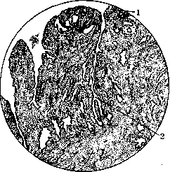
Figure 8. Erosion: 1—edge of erosion (stratified epithelium); 2—well-developed erosion glands.
and pathologists. Some believe that the columnar epithelium in pseudoerosions originates from the epithelium of glands that end up in the thickness of the cervix even during embryonic life. It should still be considered more likely the view of R. Meyer (as well as Gebhard, Frankl, and others), who established that in false erosions, the columnar epithelium proliferates from the cervical canal, gradually taking the place of the dying squamous epithelium. In essence, such proliferation should be seen as the beginning of the healing of true erosion, which is why R. Meyer proposes, instead of the not entirely apt term "pseudoerosion," to call it the first stage of erosion healing. This stage is followed by a second, in which regeneration and proliferation of the squamous epithelium occur, which again begins to oust the columnar epithelium, taking its place. Thus, in this stage of erosion healing, we will have under the microscope a typical stratified squamous epithelium that still covers the as-before-infiltrated stroma with erosion glands embedded in it. Subsequently, this infiltration also disappears, the erosion glands gradually disappear due to the ingrowth of squamous epithelium into their lumen, and the erosion heals completely (third stage of healing). Pseudoerosion must be distinguished from so-called congenital erosion (other designations: fetal, embryonic, pseudoerosio congenita). This form is found only in newborn girls; already in childhood, it is almost never observed. Here we are dealing with a disruption of the normal boundary between the columnar and stratified squamous epithelium, which normally passes, as is well known, right at the external os. In congenital erosion, this boundary passes too low, protruding beyond
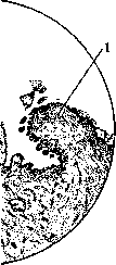
Figure 9. Rough surface of erosion with the development of papillae (1); 2—erosion glands.
the external os onto the vaginal portion of the cervix. This condition is explained by the fact that during embryonic life, the columnar epithelium covers a much larger area of the cervix, and after birth, this boundary gradually retreats upward due to the proliferation of squamous epithelium. The formation of congenital erosion is thus nothing more than a delay in the normal physiological process of replacing the columnar epithelium with squamous epithelium. True and false erosions can be complicated by the formation of cysts (ovula Nabothi). The latter arise when the mouths of the erosion glands become blocked by proliferating epithelium, leading to the retention of secretion and the formation of cavities filled with mucus (Fig. 10). Erosions of the cervix are a very common disease; they are found in up to 30% and more of all gynecological patients. They are observed almost exclusively in women who have given birth; in nulliparous women, they are extremely rare. The symptoms of erosions are leukorrhea, contact bleeding, and sometimes drawing pains in the lower abdomen and sacrum. Differential diagnosis must keep in mind syphilis (hard chancre), tuberculosis, and initial forms of cancer. Treatment consists in destroying the altered epithelium (cauterization with silver nitrate, zinc chloride, thermocautery, galvanocautery, surgical excision, etc.). Carcinoma of the uterus (cancer). Uterine cancer occurs with extreme frequency; among all localizations of cancer in women, cancer of the uterus takes first place. According to statistical data (Prague, Vienna, Munich), uterine cancer constitutes from 25 to 33% of all cancers in women. Such a high frequency of this disease makes it one of the most important problems in gynecology. Uterine cancer is observed at any age, but most frequently between the ages of 40 and 50 (climacteric period); however, cases of cancer in young women and even in girls are not rare. There are two distinct forms of uterine cancer: cancer of the cervix and cancer of the body. Cancer of the cervix occurs incomparably more frequently than cancer of the body. Statistically, cancer of the cervix accounts for up to 95% of all uterine cancers, while cancer of the body accounts for only about 5%.
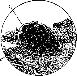
Figure 10. Erosion: 1—Nabothian cyst; 2—proliferated erosion glands.
margins of the external os in the direction of the vagina. R. Meyer calls such an erosion a congenital histological eversion. To avoid confusion with the term "pseudo-erosion", it is better to call erosion in the healing stage, if the term false erosion is to be retained, "adult pseudo-erosion" (pseudoerosio adultorum), retaining the term fetal, embryonic pseudo-erosion (pseudoerosio congenita) for that form which develops during the first differentiation of the epithelial tissue. Clinically, false erosion of adults can manifest in various forms. It is customary to distinguish: 1) simple erosion (erosio simplex), in which the eroded surface is covered with columnar epithelium with a small inflammatory infiltrate in the stroma and a few small-sized glands; 2) glandular erosion (erosio glandularis), the most frequently encountered form: it exhibits epithelization of the raw surface and abundant development of glands (Fig. 10); 3) glandulo-cystic erosion (erosio glandularis cystica) with abundant development in place of the erosion glands of small cysts developing by the type of retention tumors (mechanical retention of mucus); 4) papillary erosion (erosio papillaris), the massive formation of papillary outgrowths from the stroma. The columnar epithelium does not plunge into the depth of the stroma, but covers the grown papillae. Erosions are of great practical importance, firstly as indicators of a past or existing inflammatory process in the cervical canal and secondly in recognizing early cancer of the cervix of the uterus. In the latter case, particularly great difficulties in terms of differential diagnosis are presented by erosions in the third stage of healing (sprouting of squamous epithelium into the erosion glands). Here there is the possibility of error in several directions: failing to recognize incipient cancer proceeding under the guise of erosion, failing to recognize cancer developing from erosion, and mistaking a vulgar erosion for true cancer. Even microscopic examination, as practice shows, does not always guarantee against errors, although it remains the best method of differential diagnosis. For details see below—cancer of the uterus. In treating erosions, all efforts should be directed toward eliminating the underlying disease (cervical catarrh). In clinical practice, cauterization of erosions is usually employed (silver nitrate, tincture of iodine, formalin, red-hot iron, thermocautery, etc.). Gubarev recommends cauterizing erosions with potassium permanganate powder (Kalii hypermanganici). Cotton on a splint is taken, smeared with petroleum jelly, and sprinkled with crushed dry potassium permanganate powder. The procedure is repeated after a few days; generally, two or three such cauterizations are sufficient for the final healing of the erosion. Erosions should not be confused with the so-called eversion of the mucous membrane of the cervical canal (ectropion). Such an everted mucous membrane in its external appearance completely resembles erosion. Eversion is more often observed on the basis of tears of the cervix during childbirth (laceration-ectropion) with subsequent cicatricial shrinkage. In addition, ectropion can occur in chronic inflammatory processes, as can erosions. In eversion, the mucous membrane either remains in place, especially at first, or, losing its epithelium, becomes covered with granulations. Sometimes abrasions of the vaginal part of the uterus (erosions) and eversion of the mucous membrane can occur simultaneously. In its time, Heitzmann proposed a method by which erosion can be distinguished from eversion. He advises applying a piece of cotton moistened with a 10% solution of copper sulfate to the affected areas (for 1-2 minutes). In eversion, the mucous membrane will turn pale under the influence of cauterization with such a strong solution, while in erosion the surface of the mucous membrane will be covered with a coating in the form of a bluish-whitish film. With repeated cauterizations (intervals of 2-3 days), benign erosion should heal in 1-2 weeks. In the presence of blood when wiping the cauterized abrasion with dry cotton, especially if blood appears systematically and during repeated cauterizations, such an erosion should be considered suspicious in the sense of incipient cancer. Eversion is subject to surgical treatment. Nabothian eggs, or glands (ovula Nabothi), are observed in that form of false erosion which is designated as glandulo-cystic. If in such an erosion gland the excretory duct or its branches are closed for some reason, a small cyst arises, the size of a millet seed or a pea, which bears the name of the Nabothian gland. If such a retention cyst is opened, its contents will flow out in the form of a small drop of vitreous mucus. In some cases, Nabothian glands contain colloid masses. Nabothian glands are more often located on the apex of the vaginal part in the form of translucent small bubbles, which, becoming thinner, either burst or sometimes suppurate. To destroy these glands, their evacuation using incisions (lancet puncture) is used.
M. Malinowsky. VIII. Perforation of the uterus. Perforation of the uterus (laceration), a through-and-through injury to its wall, occurs during various types of intrauterine interventions, e.g., during sounding, dilation, and curettage of the uterine cavity. Most often, perforation occurs during the performance of an artificial abortion or the evacuation of the uterus from the remnants of the ovum in an incomplete miscarriage. The frequency of perforations occurring during curettage is difficult to establish, because some cases of perforation are not recognized, while others are hushed up. According to published data, the percentage of perforations fluctuates within small limits and averages 0.5% (1:200). Statistics by Madjuginsky (material from Moscow gynecological clinics for several years, ending October 1, 1929) show a significant decrease in the percentage of perforations: 1 perforation per 1,330 operations—0.07%. In 1931, Moscow clinics reduced this percentage to 0.04% (1 perforation per 2,788 operations). Perforation can be produced by any instrument introduced into the uterine cavity: a sound, dilator, Bozeman forceps, abortion forceps. Rare cases of perforation of the uterus by a finger during digital evacuation of a pregnant uterus are known. In clandestine abortions, cases of perforation using sharp objects such as a knitting needle, nail, etc., have been observed. Regarding the question of which surgical instrument most often perforates the uterus, opinions differ. According to the majority of authors, the highest percentage of perforations falls on the curette. Since perforation is the result of a certain violence applied by the operator when introducing the instrument into the uterus, it is also natural to think that the perforation is produced by the dilator, and the curette or sound enters an already ready-made hole. The most severe injuries are observed in perforations produced by grasping instruments, such as abortion forceps and especially Bozeman forceps. The main reason for perforation is that curettage has to be performed not under visual control, as any surgical intervention requires, but blindly. A predisposing factor for perforation is changes in the uterine wall (softening, thinning, loss of elasticity), which can arise on the basis of uterine developmental defects (Peham), inflammatory (especially septic) and degenerative processes of the myometrium, malignant neoplasms, old scars, spongy structure of the uterine wall in its limited area due to the development here of a huge number of thin-walled dilated veins (Bockman), or destructive growth of elements of the fetal membranes (hydatidiform mole, chorioepithelioma). Not only local but also general diseases, such as tuberculosis, heart defect with circulatory disorder, etc., can lead to degenerative changes in the uterine wall. Thus, the cause of uterine perforation is not always the use of brute force and the operator's insufficient experience. Cases of perforation occur in very experienced doctors. It can be said that there is no gynecologist who has grown old without having perforated the uterus at least once. In most cases, the cause of perforation is still technical errors in the operation or insufficient orientation of the operator regarding the position of the uterus, the length and direction of its canal. Perforation can be easily produced in unrecognized retroflexion or pathological anteflexion. The site of perforation can be either the cervix or the body of the uterus. When the cervix is perforated, its lateral wall is most often damaged, with the instrument penetrating into the parametrial cellular tissue. Cases of perforation of the anterior or posterior wall of the cervix are rarer, and the urinary bladder may be injured or the instrument may penetrate into the posterior pouch of Douglas. When the body of the uterus is perforated, the instrument usually penetrates into the abdominal cavity. Lateral perforation of the body of the uterus also occurs, when the instrument enters the cellular tissue between the leaves of the broad ligament. In retroflexion, the perforation opening is more often located in the anterior wall; in anteflexion, in the posterior wall or the fundus of the uterus. The size of the opening depends on the caliber of the instrument, as well as on the mechanism of the perforation. With a sound perforation, the opening will be punctiform; if the perforation is produced by abortion forceps or Bozeman forceps, the perforation opening is a jagged wound, often of a large size and with crushed, uneven edges. Uterine perforation may pass unnoticed. In most cases, it is a severe complication of the curettage operation. Everything depends on the nature of the perforation, i.e., on the size and location of the perforation opening and, mainly, on whether the perforation is accompanied by infection and secondary injuries, especially of the abdominal organs, or not. In case of injuries to a larger blood vessel, which is observed predominantly when the lateral wall of the uterus is damaged, the threatening symptom is bleeding. With large openings in the uterus, bleeding is also always profuse. In infected cases, uterine perforation threatens peritonitis. Bleeding and infection pose an immediate threat to the patient's life in case of injuries to the abdominal organs. The literature is rich in various casuistics of uterine perforations, of which each case may have its own features. Thus, cases of avulsion of the cecum and ovary (Braude), avulsion of the ureter from both the kidney and the urinary bladder (Wertheim), avulsion of a piece of the promontory with forceps (Frank), avulsion of the mesentery of the small intestine, which the doctor removed from the uterus and cut off over a length of almost 2 m (Sadovsky), and the like, have been described. Diagnosis of perforation. The most serious attention must be paid to the diagnosis of perforation, because the prognosis of perforation largely depends on whether it is recognized in time or not. Perforation during curettage is recognized mainly by the sudden entry of the instrument into the uterus to a depth that does not correspond to the length of its cavity determined before the start of curettage by means of bimanual examination and sounding. The operator does not have the feeling at the same time that the instrument introduced into the uterus encounters the resistance that the uterine wall should provide to it. The instrument as it were falls through and goes "into the boundless." A sudden shock can sometimes indicate that a perforation has occurred. It should be noted, however, that there may be no shock even after severe and complicated uterine perforations. The extraction of the omentum (see separate table, Fig. 10), an intestinal loop, or another abdominal organ (appendix, ovary, etc.) from the uterus serves as undoubted proof of uterine perforation. The extraction of adipose tissue from the uterus by the instrument also speaks unmistakably for a perforation that has occurred, complicated by damage to the omentum, mesentery, or appendices epiploicae. However, it is not always easy to correctly recognize the nature and anatomical structure of the extracted piece of adipose tissue among the mass of scrapings. Establishing an accurate diagnosis, especially in terms of the size of the injury, is of very essential significance: such a diagnosis predetermines further measures, because if conservative treatment is still possible in an exceptional case with a simple and uncomplicated uterine perforation, damage to an abdominal organ categorically dictates immediate surgical intervention. In most cases, determining the size of the damage in uterine perforation is very difficult. Complicated injuries, which are accompanied by profuse bleeding or shock, and often also by the prolapse of abdominal organs through the perforation opening, are diagnosed more easily and more often than uncomplicated cases of perforation. Bimanual examination can, in the case of profuse internal bleeding, reveal the appearance of free blood in the abdominal cavity or a rapidly growing hematoma in the posterior pouch of Douglas or between the leaves of the broad ligament (in case of damage to a branch of the uterine artery). Of the objective diagnostic methods, control sounding of the uterus is most often used. Regarding the value of this method, it should be noted that the sound, even with repeated introduction into the uterine cavity and careful probing of its walls, may not detect the perforation opening, especially if the latter is small. On the other hand, with a persistent desire to find the perforation opening, the sound can produce new perforation openings or cause a perforation in a uterus that was only erroneously considered perforated. Control sounding is contraindicated where the uterine cavity is infected or where there is even just a suspicion of infection. Posterior colpotomy can be used for diagnostic purposes. This operation in the indicated cases has significant advantages in many respects over exploratory laparotomy, because it is technically simple and relatively safe. In the latter respect, it compares particularly favorably with exploratory laparotomy when dealing with an infected case. Posterior colpotomy has the advantage of making it possible to also carry out the necessary surgical and therapeutic measures if the perforation is established. Great difficulties for the diagnosis of perforation arise in cases where the patient is admitted already with phenomena of peritoneal inflammation.
In cases where patients are admitted in a severe condition due to diffuse purulent peritonitis or general sepsis, determining the cause of the disease no longer has decisive significance: therapy in these cases remains identical and equally unpromising of success. It is more important to recognize perforation at the onset of peritonitis. Clinical data here are indefinite and scanty. Sometimes for differential diagnosis, one can find supporting points in the anamnesis. Thus, if it is reliably known that before the curettage the patient had no inflammatory process at all, and peritoneal phenomena began immediately after curettage, then in such a case the suspicion of perforation receives some reinforcement. Along with cases of perforation in which peritoneal inflammation begins to develop rapidly and immediately after curettage, there are also cases where the inflammatory process develops slowly and does not immediately lead to a severe condition. Questions of differential diagnosis lose their urgency where the picture of peritonitis has already developed; no matter what caused the peritonitis, surgical intervention, whether colpotomy or laparotomy, is a method that, in the opinion of the majority of authors, can most contribute to saving the patient (see Peritonitis). Prophylaxis and treatment of perforations. To prevent perforations, measures are taken to combat clandestine abortions and the application of correct technique in curettage operations (see Abortion and Curettage). Some (Relat, Katz, and others) advise abandoning curettage altogether in abortion and using only digital emptying of the uterus. Once perforation is detected, all manipulations inside the uterus must be immediately ceased. Conservative treatment (bed rest, opium, ice on the abdomen) is permissible in cases where the perforation opening is small, there is no internal hemorrhage (external can be stopped by vaginal tamponade), the case is uninfected, there are no side injuries, and the omentum or a loop of intestine has not prolapsed into the uterus. The first condition for non-surgical treatment is complete awareness of all circumstances under which the perforation of the uterus occurred. Knowing the case well, fully accounting for all stages of the operation at which the detected perforation could have happened, and correctly evaluating the dimensions of the damage based on all data and establishing a correct prognosis can only be done by the person who performed the curettage himself. Therefore, a physician can limit himself to the conservative method of treatment and take responsibility for not using surgical intervention only if he personally performed the curettage during which the perforation occurred, or at least was present at it (Latzko, Braude). Every case of conservative treatment must be accompanied by medical observation so as not to miss the moment when bleeding or incipient peritonitis will require surgical intervention. In perforations accompanied by the extraction or damage of abdominal cavity organs, laparotomy is indicated. This also includes those cases of perforation that were produced by forceps-like instruments, especially a dressing forceps, since most of these cases are accompanied by damage to abdominal organs, which cannot always be immediately detected by the operator. In those cases of perforation where a damaged intestinal loop has been extracted from the perforation opening, no attempts should be made to reduce it before laparotomy so as not to contaminate the abdominal cavity and so that one does not have to rummage through the intestines to search for the damaged area (Ott). The intestinal loop subject to resection need not be extracted from the perforation opening either before or after resection: the resected segment of the intestine, lying in the vagina, is extracted from there as well (Bumm). In cases of perforation requiring surgical intervention but not accompanied by damage to abdominal cavity organs, vaginal methods of surgery are also used. The question of what to do with the perforated uterus is resolved depending on the individual characteristics of the case: with extensive and multiple injuries of the organ, especially in infected cases, the uterus is removed. Extirpation of a perforated uterus is indicated in cases of destructive mole and malignant neoplasms. In uninfected cases, especially in young women, efforts are made to preserve the uterus by suturing the perforation opening. If the opening has uneven, bruised edges, they are smoothed with scissors or a scalpel before suturing. The uterine wound is sutured using several interrupted sutures penetrating through the entire thickness of the uterine wall, with the exception of the mucosa. Over this row of sutures, a continuous superficial suture is applied, capturing the serous cover of the uterus. On a pregnant uterus, one can additionally apply another continuous suture resembling the Lembert sero-serous suture, and where the peritoneum is tightly attached to the muscular coat, the underlying layer of the latter is also caught by the suture. When peritonealizing the sutured perforation opening, it is necessary to individualize each separate case, using adjacent peritoneal surfaces for this purpose: the peritoneum of the uterovesical fold, the broad and round ligaments, etc. If there is no certainty in complete hemostasis or the asepsis of the case, drainage can be applied through the abdominal wound or through the posterior vaginal fornix. Some (Bumm, Sigwart, and others) applied suturing of the uterus with extraperitoneal placement even in cases knowingly infected or suspicious of infection. If the uterus is extirpated during the operation by the abdominal or vaginal route, the question of removing the ovum from the uterine cavity disappears by itself. If the perforation opening is sutured and the uterus is left, the question arises of what to do with the parts of the ovum remaining in its cavity. To avoid complications in the postoperative course, these remnants must be removed. Performing curettage after the perforated uterus is sutured is dangerous because the sutures may be damaged in the process. Performing curettage before suturing is also dangerous because the dimensions of the damage may increase and, moreover, new injuries may appear, especially in cases where the uterine wall is predisposed to perforation by its structure. Therefore, emptying the perforated uterus by means of curettage through the cervical canal should be done only under the control of the eye or a hand grasping the body of the uterus through an incision in the vaginal fornix or through the abdominal wound. However, this method presents great inconveniences, especially during laparotomy, when the patient has to be moved from one position to another. It is therefore preferable to perform curettage of the perforated uterus through the perforation opening, which can be enlarged if necessary for this purpose by means of a small incision. I. Braude.
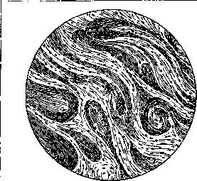
Figure 11. Myoma rudiment. (After R. Meyer.) VIII. Neoplasms. Neoplasms of the uterus according to their origin are divided into connective tissue neoplasms (myomas, fibromas, fibromyomas, lipomas, angiomas, sarcomas, myosarcomas, and others) and epithelial neoplasms (adenomas, cancers). Of the first group, myomas and fibromyomas are of particular importance. Myomas and fibromyomas of the uterus (for details, see the respective entries). These tumors were known in antiquity and are mentioned by Hippocrates (as a "uterine stone" discharged from the vagina of a sixty-year-old woman) and Galen (as sclerosis of the uterus). As for the frequency of this tumor, according to modern statistics, it is observed in approximately 20% of women over 35 years of age; according to various authors, fibromyomas make up approximately 5% of all gynecological diseases. Racial predisposition to these tumors does not lend itself to statistical accounting, although the view exists that Jewish women and Negresses are particularly predisposed to the formation of fibromyomas. The question of the relationship of fibromyomas to childbearing provoked particularly lively discussion; specifically, Bayle in 1813 gave it a definite solution, expressing the view that fibromyomas are the consequence of sterility, and this point of view remained dominant for a very long time.
Only in [o]ur 1894 Hofmeier came forward with new data, finding even that fibromyomas enhance the ability to conceive; Albrecht also comes to the conclusion that sterility as one of the etiological moments of the occurrence of fibromyomas must be rejected. However, even now some hold a different view on this matter; for example, Troell places the development of fibromyomas in direct dependence on the number of previous births; specifically, according to his data, 26% of patients with fibromyomas had no births at all, 8% had one birth, 6% had three births, and so on; according to Pinard, sterility is also a predisposing circumstance to the development of fibromyomas, and its prevention should consist in ensuring that correct sexual life does not begin...
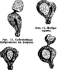
Figure 13. Fibro...
Figure 15. Fibro-...
myoma.
fibroids. Later than 25 years and at least every 5 years pregnancy followed with full-term labor and breastfeeding. Regarding the influence of age on the occurrence of fibromiyoma, it is considered established that the most favorable age is starting from 35 years and until the onset of the climacteric period, although isolated cases of finding fibromiyomas even in young children have been described; with the cessation of menstruation, the growth of fibromiyomas ceases, and along with other organs of the genital apparatus, they undergo atrophy and may even disappear entirely; in any case, at this age, new fibromiyomas do not arise. Fibromiyoma of the uterus can develop from any part of the uterine wall (Figs. 11, 12, 13, 14, and 15)—both its body and the cervix, but in the latter it is found much less frequently (in 4% of cases) and is always solitary, whereas in the body of the uterus it is observed in 96% of cases and is often multiple; since the fibromiyoma develops from the muscle fibers of the uterine wall, at the beginning of its formation it is always located in its thickness, sometimes closer to its peritoneal covering, sometimes, conversely, closer to the mucous membrane of the cavity; upon further growth, the fibromiyoma stretches the muscle fibers and simultaneously begins to shift in the direction of least resistance. It is pointed out that such a shift occurs under the influence of the contraction of the musculature of the uterus surrounding the tumor. In the case where the myoma was originally located closer to the peritoneal covering of the uterus, it protrudes under the peritoneum on the outer surface of the uterus and bears the name of subserous fibromiyoma if it protrudes above the surface of the uterus by no less than half of its volume; in the case where the tumor was originally located closer to the mucous cavity of the uterus, it gradually protrudes above its surface and acquires the name of submucous fibromiyoma; finally, if the tumor is originally located at approximately an equal distance both from the peritoneal covering and from the lumen of the cavity, upon contraction of the muscular wall of the uterus it encounters equal resistance from all sides and remains interstitial, or intramural. Subserous fibromiyomas can gradually protrude more and more above the peritoneal surface of the uterus and finally remain connected to it only by a more or less thin stalk. The same can happen with submucous fibromiyomas—they also protrude into the uterine cavity, remaining connected to it only by a stalk; in these cases, such a submucous fibromiyoma receives the name of a polypoid tumor; thanks to the contractions of the uterus, such tumors gradually begin to be expelled from the uterine cavity, or "born"; the cervix of the uterus is smoothed out, the internal and external os open, and the tumor with a larger or smaller segment protrudes into the vagina; sometimes the entire tumor is born into the vagina and may even protrude from the external genitalia. According to available statistical data, these tumors are observed in the following proportions: interstitial tumors are encountered more often than others, giving approximately 70% of fibromiyomas; submucous tumors stand in second place, giving 20%, and subserous tumors stand in third, giving 10%. In addition to these tumors typical in their location, such ones are observed which protrude above the surface of the uterus, but are located between the peritoneal layers of one or the other broad ligament; such tumors are called intraligamentary and upon further growth occupy the lateral pelvic cellular tissue, displacing the uterus upward and in the opposite direction; besides this, fibromiyomas can also develop from various parts of the uterine cervix. In those cases when they arise from the upper parts of the cervix, they fill the lesser pelvis, and the body of the uterus is displaced upward; if they arise from the tissues of the vaginal part of the uterus, they have the appearance of a polyp on a stalk.—Depending on the location of the tumor in the uterus, there is a change in the shape of the uterus, as well as the size of its cavity and the state of the mucosa; thus, for example, in subserous fibromiyoma, the uterus appears nodular and on its outer surface it is possible to determine protruding nodes; with interstitial development of the tumor, the entire uterus will be enlarged, and the appendages and the site of origin of the round ligaments will turn out to be displaced either anteriorly or posteriorly depending on the location of the node; with submucous fibromiyoma, the uterus will also be enlarged, but this enlargement is expressed uniformly in all directions, and the uterus acquires a spherical shape resembling the shape during pregnancy. The size of its cavity will be far from identical with various forms of fibromiyomas; thus, for example, with subserous development of the tumor, the uterine cavity either remains unchanged or changes little; with an interstitial tumor, the uterine cavity can turn out to be significantly enlarged both in length and in width; but it changes especially sharply with submucous fibromiyoma, and in these cases the degree of its enlargement will directly depend on the size of the node located in the cavity. Regarding those changes that occur in the uterine mucosa during fibromiyoma, Frankl's studies refuted the previously existing views on its hyperplasia and hypertrophy in this kind of tumor; based on his works, it must be considered established that all changes in the uterine mucosa occur solely under the influence of two causes: mechanical stretching of the mucosa by the growing tumor and the congestion phenomena caused by it. The former takes place chiefly during submucous growth of the tumor and manifests itself in thinning and atrophy of the uterine mucosa (from pressure by the tumor) and in the fact that the glands decrease in number and shorten; congestion phenomena manifest themselves in edema of the mucosa caused by the pressure of the tumor, and in the development of a network of tiny vessels covering the tumor and becoming engorged with blood in the premenstrual period. These phenomena are also expressed most sharply in submucous fibromiyomas; in interstitial fibromiyomas they can be expressed to varying degrees depending on the size and location of the tumor; in subserous tumors these changes in the uterine mucosa are usually completely absent. The size of individual fibromiyoma nodes can fluctuate within significant limits: from microscopic, barely noticeable tumors to those that reach enormous sizes; thus, for example, Kuznetsky described a fibromiyoma removed by him weighing about 43 kg, and Hunter discovered at autopsy a fibromiyoma weighing 56 kg. Fibromiyomas belong to tumors that usually occur multiple, sometimes up to several tens in one uterus, and simultaneously they can develop subserously, interstitially, and submucously; however, single tumors of any character also frequently develop.—By their localization, multiple tumors are most often observed in the region of the body of the uterus and are subserous or interstitial; submucous nodes in the majority of cases are solitary; the same solitary nodes are usually observed in the cervix of the uterus, as well as in those cases when they grow into the cellular tissue of the broad ligament. The shape of the tiniest fibromiyomas is always spherical due to the uniform pressure of the surrounding tissues; with interstitial development of single fibromiyomas, this shape can be preserved even with a significant size of the tumor. With the simultaneous existence of several nodes, if they are in contact with each other and exert mutual pressure, their shape can be most varied: in submucous tumors, their shape is most often oval, elongated corresponding to the long axis of the uterine cavity; in subserous fibromiyomas, which do not encounter counter-pressure from the surrounding tissues during their growth, the shape of individual nodes usually appears as an oval or resembles a kidney, and the stalk of such a tumor can be very diverse both in its width and in length.—The color and consistency of fibromiyomas largely depend on the tumor's richness in muscle or connective tissue fibers, as well as on its blood supply. A pure myoma with an abundant content of muscle tissue is usually somewhat denser in consistency than the muscular wall of the uterus and has a whitish-pink or grayish color; a pure fibroma has a very dense consistency due to the abundant development of connective tissue and on cross-section appears glossy and whitish; however, quite often the consistency of the tumor becomes very soft and elastic, which is caused by its cystic degeneration or congestion phenomena; the color of the tumor under these conditions can also change significantly and acquire the most diverse shades from reddish to brown or black. Color and consistency can also change depending on the tumor's richness in blood and lymph vessels, in connection with necrobiotic changes, lime deposits, the development of bone trabeculae, etc. (see below). By their structure, fibromiyomas must be divided into two main forms: the first, significantly more frequent, is a tumor enclosed in a well-defined capsule, the latter consisting of stretched layers of the muscular wall of the uterus and more or less closely connected with the tumor itself; the second form has no capsule and represents a diffuse formation growing into the uterine wall without clear boundaries or encompassing the entire uterus.
Usually the capsule is well-expressed only in interstitial fibromyomas, while subserous and submucosal tumors in their further growth may lose it and remain connected to the uterus only by means of a stalk. Pathological changes in the tissues of the fibromyoma can be very diverse; they depend on various causes, among which the most important are, firstly, the disturbance of blood and lymph circulation, secondly, the disturbance in metabolism, thirdly, the degeneration of the fibromyoma cells, and fourthly, the ingrowth of epithelial elements into the tumor, thanks to which the latter becomes a so-called mixed tumor. The most severe circulatory changes in fibromyomas occur as a result of their twisting; in this case, the veins undergo greater compression due to the pliability of the walls, and stagnant phenomena develop in the tumor, which sometimes can be very markedly expressed and even lead to the rupture of individual vessels. If such a ruptured vessel is located on the surface of the tumor, significant, sometimes fatal bleeding into the abdominal cavity can occur; in the event that the hemorrhage occurs in the thickness of the tumor, an infarction is formed; the necrotic area of the tumor acquires a peculiar coloration, and this condition bears the special name of "red degeneration". Sometimes the circulation is not disturbed so sharply, and then changes are observed that develop gradually; their essence comes down to the fact that due to the impaired blood supply to a certain area of the tumor, necrosis develops here, the further fate of which can be very different. With the formation of necrosis in submucosal tumors, where it develops due to contractions of the uterus and where the tumor, upon its birth into the vagina, comes into close contact with the vaginal flora, such a necrotized area usually becomes infected, and the matter reaches suppuration and disintegration of the tumor, and individual parts of it can separate, which is accompanied by the appearance of purulent, dirty discharges with an odor. With necrosis of subserous nodes, fibrinous deposits form on their surface, causing adhesions with neighboring organs, and infection can also develop from here. Interstitial fibromyomas are exposed relatively frequently to necrosis during pregnancy or in the postpartum period, and if infection does not occur in these cases, the necrotized tissues gradually die off and disintegrate, and small cavities form in their place, which can merge into more or less voluminous cysts; their contents are usually pultaceous and have a bloody or brown color. The change in blood circulation is usually also accompanied by a violation of lymph circulation, which is manifested primarily in the edematousness and softening of the tumor; the delayed outflow of lymph causes the expansion of lymph clefts and the formation of lymphangiectases, which in some cases, especially in single tumors, spread to the entire tumor (so-called lymphangiectatic fibromyomas); in other cases, a significant expansion in a certain area of the lymph vessel can form, and such an expanded cavity takes the form of a cyst. It differs from a cyst formed as a result of necrosis in that its contents are usually transparent and have a gelatinous character and a yellowish color. The disturbance of metabolism in the fibromyoma is observed very frequently, and regressive processes to a greater or lesser extent are encountered in almost every case, for example, simple atrophy of myomatous cells occurs under the influence of the cessation of ovarian activity with the onset of the climacteric period or as a result of treatment with X-rays; it is also observed not infrequently in the postpartum period in connection with the involution of the uterus; simple atrophy is expressed in a decrease in the volume of the tumor, which sometimes reaches such dimensions that a tumor that had reached the size of a three-month pregnant uterus completely ceases to be palpable; this depends on the gradual deterioration of the blood supply and the thus impaired nutrition of the tumor. In addition to the just-mentioned simple atrophy, pictures of tumor sclerosis are often observed in the fibromyoma. They consist in the fact that an enhanced development of fibrous tissue and its hyaline degeneration occur. Such an enhanced formation of fibrous tissue occurs, according to R. Meyer, not as a result of its proliferation, but due to the degeneration of the muscle fibers of the tumor and is observed especially frequently in subserous fibromyomas, and the consistency of the tumor becomes extremely dense. In most cases, such fibrous transformation is combined with hyaline degeneration, which in some cases can be limited to connective tissue layers in the tumor; but usually it also affects muscle bundles. This degeneration is encountered especially frequently in subserous fibromyomas, but it is also not infrequently observed in interstitial and submucosal tumors, and here it can begin from the central areas, gradually disappearing towards the periphery; macroscopically, it is characteristic in that on the cut section of the tumor, a stratification is obtained consisting of cartilage-like shiny tissue. Such fibrous-hyaline change in the tumor can lead to a violation of blood and lymph circulation and cause the onset of necrosis, but especially often it leads to the deposition of lime. Calcification of the fibromyoma usually begins from its central areas, and in cases where it is weakly expressed, the grains of lime are located in small piles or rows and sometimes resemble bone formations; true ossifications of fibromyomas also sometimes take place and have been described by Lubarsch and R. Meyer. In addition to such diffuse calcification, cases are observed where a calcareous shell is formed either on the surface of the entire tumor or on some area of it around the necrotic parts of the myoma; submucosal or subserous fibromyomas are subject to calcification more often than others, and the former lead to the formation of so-called womb stones; petrified subserous fibromyomas often entail the formation of pressure necroses with perforation into the intestine or bladder. In addition to the mentioned changes in fibromyomas, fatty degeneration is sometimes observed (relatively rarely), which is the result of prolonged nutritional disturbance; on the cut section, such a tumor has a yellowish appearance throughout or in individual areas; upon examination under a microscope, individual fat droplets can be discovered. Under the influence of the same conditions, mucous, or myxomatous, degeneration of the fibromyoma is sometimes observed, and its muscle and connective tissue cells turn out to be strongly swollen and transformed into a gelatinous mass. The third change in the histological structure of the fibromyoma consists in the malignant, i.e., sarcomatous, degeneration of its cells. There are disagreements on this score now; thus, R. Meyer and Borst completely reject the possibility of sarcomatous degeneration of a fibromyoma. According to literature data, 70% of uterine sarcomas develop from already existing fibromyomas, but in general, uterine sarcoma is a rather rare disease and accounts for an average of only about 1.4% of all uterine fibromyomas. The fourth and final change in the tissues of the fibromyoma is the ingrowth of epithelial elements into the tumor, thanks to which it acquires the character of a fibroadenoma. Combination with endometrioid growths—see Adenomyoma, which is also a rather rare complication (according to available statistics, observed from 2% to 8% of adenomyoma cases); exceptionally rarely, a fibromyoma also degenerates into cancer. The combination of fibromyoma and cancer is not frequently observed (about 3%); a combination with cancer of the uterine body takes place more often. Clinic of fibromyomas. Fibromyomas cause a number of very diverse symptoms of both a local and general nature, and some of the symptoms depend on the presence of the tumor in the uterus and are expressed in irregular periods, pain, etc., while others consist in changes in the heart, thyroid gland, etc. As for the disorders of the first group, the most severe ones are caused by submucosal fibromyomas or those that develop from the lower segment of the uterus; interstitial fibromyomas cause less significant changes, while subserous ones often produce no symptoms at all. With fibromyomas in general, one most often has to deal with one or another change in menstrual function; thus, for example, there is a view that women suffering from a fibromyoma continue to menstruate significantly longer and that menopause in them often occurs after fifty years of age; for example, according to Trell's statistics, in healthy women, menopause occurred at an age older than 50 in 34%, while in those suffering from a fibromyoma, in 43%; at an age older than 55, it occurred in the former in 1% and in the latter in 20%. One of the most frequent complaints forcing a patient to seek medical help are changes in the menses, consisting in the fact that they become more abundant and prolonged, without losing their cyclical character; such menstrual bleeding bears the name of polymenorrhea.
These bleedings develop gradually; at first they become only slightly heavier and more prolonged, but with the growth of the tumor they become increasingly heavy, and finally the situation reaches a point where patients do not have time to recover during the intermenstrual period, and severe anemia develops: the hemoglobin level may drop to 20% or even 10% of normal, and the red blood cell count decreases to 1,000,000. With the prolongation of menstruation, the impression is created that they change their type and from four-week become three-week, but this only seems so due to the prolongation of blood loss; in reality, it turns out that in approximately 80% of cases the menstrual type remains the same. The explanation for such menorrhagia in fibromyomas is that, firstly, the surface of the uterus covered by the mucous membrane, which bleeds during menstruation, is significantly enlarged; secondly, before menstruation, there is an intensification of the hyperemia of the vascular apparatus of the uterus, which increases depending on the volume of the tumor; thirdly, some tumors located intramurally or intraligamentously impede the venous outflow of blood, and finally, fourthly, under the influence of tumors located in the uterine wall, an obstacle is created to the adequate and uniform contraction of the uterine muscles. In rarer cases, accounting for about 15%, the bleedings lose their cyclical character and acquire the form of metrorrhagia, and they are caused by some complications in the tumor, for example, a violation of the integrity of the mucous membrane covering it due to necrosis, torsion of the tumor, or the formation of bedsores; this is observed most often in submucous fibromyomas or in interstitial ones deeply protruding into the uterine cavity. Sometimes with fibromyomas, an increased secretion of liquid serous leukorrhea is observed, which occurs with large tumors and depends on stagnant phenomena; in some cases, one also has to deal with pain sensations in the area of the tumor, which depends either on an unfavorable location or excessively rapid growth of the node; sometimes the pain is caused by incarceration, twisting of the pedicle, or the resulting inversion of the uterus. The sensation of increased pressure in the pelvis is usually localized in the area of the bladder, ureters, and rectum, and the passage of urine can sometimes be strongly hindered or even completely interrupted. The ureters possess the ability to resist the pressure of the tumor well, and sometimes on large intraligamentous tumors one can see a groove corresponding to the location of the ureter; yet in some cases, the fibromatous node can completely compress the ureter, and then the formation of hydronephrosis is observed. The rectum is also subjected to pressure, but it tolerates it much easier. Finally, one must also mention those pain sensations observed in the abdominal cavity during various inflammatory changes arising in the tumor in connection with the torsion of its pedicle, with the rupture of its peritoneal cover, and with other processes that can in severe cases end in peritonitis. Of the general symptoms observed in fibromyoma, great attention has been paid to those changes observed on the part of the heart. The first to draw attention to this was Hofmeier (1884), but systematic processing of this issue begins in 1898 with Strassmann, who found heart disease in 40% of patients suffering from fibromyoma. In 1924, Winter again subjected it to a thorough study; according to his data, fatty degeneration and dilation of the heart, which must be considered a consequence of anemia, and brown atrophy, which occurs with very large tumors, are often observed in fibromyomas. As for functional heart disorders and its hypertrophy, their direct connection with myomas has not yet been established; an increase in blood pressure also cannot be attributed to fibromyoma—it remains even after the removal of the latter. Of particular attention are the so-called basedowoid heart disorders, such as palpitations, pulse arrhythmia associated with a tendency to sweating, and easy excitability; W. Freund was one of the first to draw attention to the frequently encountered swelling of the thyroid gland in fibromyoma; at present, the majority adhere to the view that the cause of such cardiac disorders is impaired secretion of the thyroid gland. There are indications of a tendency of female owners of uterine fibromyomas to atherosclerosis, obesity, diabetes; in relation to tuberculosis, on the contrary, comparative resistance is noted. The diagnosis of uterine fibromas is established by palpation of one or more nodes of spherical shape and dense consistency, more or less closely connected with the uterus; the recognition of uncomplicated fibromyoma belongs to the simplest tasks of gynecological diagnostics, and if any doubts arise here during the initial examination, in most cases the patient can be taken under observation for repeated examination. The recognition is simplest in subserous fibromyomas protruding above the outer surface of the uterus and showing an undeniable connection with the latter; in most cases, these nodes are multiple, and the pedicles connecting them with the uterus can have various thicknesses and lengths; diagnosis appears more complex when the node is single and has a long pedicle; in these cases, the fibromyoma can often be edematous, and then it is quite difficult to distinguish it from an ovarian tumor having the same consistency with it; a correct diagnosis can be made here only after the appendages are determined and both ovaries are clearly felt; difficulty in recognizing a subserous node also arises in the case when it is incarcerated in the posterior pouch of Douglas; under these conditions, as the tumor grows, the small pelvis is filled with a dense tumor, and the cervix of the uterus turns out to be highly elevated and pressed against the pubic bone. Submucous fibromyomas also do not present great difficulties in diagnostics if they reach such a size that they smooth out the cervix of the uterus and open the external os, and the finger manages to determine the lower segment of the tumor; during the birth of a submucous node into the vagina, it is also not difficult to recognize it by the features of the mucous membrane covering it; however, one must remember that such a birthed node can often simultaneously cause inversion of the uterus, and it is extremely important to recognize this in a timely manner, prior to the surgical removal of the node; a correct diagnosis can be made here thanks to the state of the tumor pedicle; namely, in a submucous myoma, it gradually becomes thinner towards the top, whereas in uterine inversion, the pedicle is thick and widening upwards. In some cases, a submucous node can be confused with a fibromyoma developing from one or the other lip of the cervix of the uterus, but here recognition is helped by determining the shape of the external os, which in a cervical node appears in the form of a sickle. Recognition becomes significantly more difficult with a submucous tumor that is still located in the uterine cavity and shows no tendency to birth; sometimes some opening of the os is observed here with the appearance of menstruation, and in such cases, it turns out to be possible to feel the lower segment of the tumor; but in most cases, this is not observed, and the diagnosis has to be made only presumptively; for more precise recognition, one has to use uterine sounding or trial curettage; however, this does not always give reliable results, since a small submucous fibromyoma on a pedicle can slip away from the probe or curette. In those cases where the node protrudes into the uterine cavity, sitting on a short, thick pedicle, the curette, in the words of Stoeckel, "stumbles", and this serves as a diagnostic sign; recently Mikulicz-Radecki proposed to use a hysteroscope for this purpose, with the help of which he obtained an image in the mirror of small polyps and submucous fibromyomas; larger formations cannot be seen as a whole, since the hysteroscope can give only a small field of illumination, but in these cases, one can form an accurate idea of the tumor by the shadow formed at the boundary of the transition of the tumor into the uterus. While in subserous and submucous tumors, recognition often presents no difficulties, since one can feel the tumor itself, in interstitial fibromyomas this is significantly more difficult, since the node is embedded in the uterine wall itself and, increasing in volume, causes an enlargement of the entire uterus; the differential diagnosis in these cases has to be made most often between fibromyoma and pregnancy, uterine fibromyoma, or an ovarian tumor.
As for pregnancy, diagnosis is especially difficult in its first months, since menstruation may sometimes appear at this time; moreover, in the first months such obvious signs of pregnancy as fetal heartbeat and movements are absent; here, in difficult cases, reliable results can be provided by the Zondek-Aschheim reaction, which makes it possible to recognize pregnancy with undoubted accuracy (this reaction consists in injecting the urine of the woman being tested into young, not yet sexually mature mice and obtaining blood spots in their ovaries after a certain time; formerly, prior to the introduction of this reaction, a correct diagnosis could be established only through prolonged observation of the patient and repeated examination). Along with cases where it is necessary to determine whether a pregnancy or a fibromyoma is present, one also encounters those where the question is of the simultaneous existence of pregnancy and a fibromyoma (see separate plate, Fig. 23); here diagnosis proves possible only by observing the growth of the tumor, which during pregnancy corresponds to a certain regularity; in addition, the softness of a certain area of the uterus, typical of pregnancy, turns out to be characteristic. In diagnosing an interstitial tumor and an ovarian cyst, difficulties are observed mainly with tumors of considerable size; in doubtful cases here, one must first of all try to identify the separately situated uterus, and if this succeeds, to establish its connection with the tumor. This is done in two ways: the external hand firmly fixes (Figs. 16, 17, and 18) the tumor against the pelvic inlet,


Figure 16. Diagnosis of fibromyoma by lifting it upward. Figure 17. Diagnosis of fibromyoma by displacing it into the pelvis.
while the internal hand checks whether the vaginal part and cervix are displaced relative to the tumor and whether the uterus is located anteriorly, posteriorly, or laterally to the lower segment of the tumor; or the external hand tries to penetrate deeply between the symphysis and the tumor and push the latter as high upward as possible, while the internal hand controls whether the vaginal part of the uterus and the cervix are simultaneously pushed upward. Palpation of the appendages with large tumors is far from always successful, nor is it
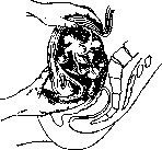
Figure 18. Diagnosis of fibromyoma by palpation.
may have significance, since they can be displaced and do not fall under the examining finger; determining the point of origin of the round ligaments is far from always possible, but if it is successful, it greatly facilitates recognition. In some cases, examination through the rectum and palpation of the uterosacral ligaments and determining their relationship to the tumor are of importance. For the recognition of intraligamentary tumors developing beneath the peritoneum, the circumstance that the fibromyoma in these cases becomes completely immobile is of importance, and the uterus is deviated in the opposite direction, and sometimes it turns out to be significantly elongated; with a large tumor size and thin abdominal walls, it is sometimes possible to determine the corresponding appendages and palpate the round ligament. A much more difficult and responsible task is the recognition of any pathological changes occurring in the fibromyoma, e.g., necrosis and infection of the tumor, torsion of its stalk and rupture of the capsule or its sarcomatous degeneration; here one has to reckon with phenomena of peritoneal irritation, with an increase in temperature, with excessively rapid growth of the tumor and phenomena of developing anemia, and sarcomatous degeneration of the fibromyoma usually becomes accessible to recognition only in far-advanced cases. Prognosis in fibromyoma of the uterus. Here, on the one hand, one must take into account the circumstance that after the onset of menopause, fibromyomas usually stop growing and even decrease in volume, so that a sort of self-cure occurs; on the other hand, one must keep in mind that each such tumor can cause severe phenomena both as a result of hemorrhages, resulting in life-threatening phenomena of anemia, and due to pressure on neighboring organs with a significant increase in the tumor; in addition, various complications can often be observed, such as tumor necrosis, its infection, etc. Therefore, although a fibromyoma is essentially a benign tumor, it is impossible to establish a general prognosis for all cases in general, and each case must be strictly individualized and appropriate treatment must be outlined for each individual case. Treatment of fibromas. In cases where the fibromyoma is not large (does not exceed the size of the uterus at three months of pregnancy), when the patient does not experience any unpleasant symptoms and when the patient's age is such that it is not long to wait until the onset of menopause, one can generally refrain from any treatment, but it is necessary that such a patient be under the observation of a physician, which should consist in the patient coming to the physician for an examination every three or four months so that he can control the growth and condition of the tumor. As a prophylactic agent, supposedly exerting an inhibitory effect on tumor growth, mammin, or breast gland extract, is prescribed by some. Since bleeding during menstruation is the most frequent complaint of patients with fibromyomas, one has to reckon with it. In cases where there are no suspicions of sarcomatous degeneration of the tumor or the presence of cancer, when at the same time the fibromyoma is small and the patient is close to the onset of menopause by age, one of the ergot preparations can be prescribed to regulate menstruation; in Germany, for example, hynergen is very successful. One can also use the ordinary liquid extract of ergot with the addition of some other hemostatic agent; in former times, when there were no other methods of treatment for fibromyomas and their surgical removal gave a very poor prognosis, such treatment with ergot was very widespread; now it is rarely used; it should be prescribed a few days before and during menstruation. Of local remedies, hot douches are used, but they are of very little effect. As for curettage of the uterine cavity, which in former times was also very widely used, it is now almost completely abandoned, since in fibromyoma it presents comparatively great dangers, depending firstly on the possibility of traumatizing the tumor if it develops submucosally, and secondly on introducing infection; now curettage is done in fibromyomas not for a therapeutic purpose, but only for diagnostic purposes to exclude cancer and determine whether there is a submucosal myoma in the given case or not. In all cases where a fibromyoma requires treatment, and the measures just cited do not lead to the goal, the question of radical treatment must be raised, which may consist either in the use of radiation therapy or in surgery. Comparing these two methods of treatment, it should be kept in mind that when using radiant energy, we do not directly eliminate the diseased organ, but in a roundabout way, by destroying the secretory activity of the ovaries, we cause the cessation of certain symptoms of the disease, specifically hemorrhages, and at the same time force the tumor to decrease in size. Making a comparative evaluation of both methods, two questions must be posed: 1) which of them is the safest and 2) which cures the disease more reliably. As for the first question, it must be decided in favor of radiant energy, if we consider only the fact that mortality in fibromyoma surgery is now calculated at 2%, while with the skillful use of X-rays or radium, no deaths are observed at all. However, a correction must be made here that this mortality during surgery is calculated in relation to myomas with various complications, for example, inflammatory phenomena, etc., which cannot be treated with radiant energy and at the same time give the highest mortality percentage. Regarding the second question, it must be kept in mind that both of the treatment methods mentioned above cannot replace each other at will in every case, and there are cases where the use of only one of these methods is possible. For example, in fibromyoma in a young woman, approximately around the age of 30, the majority of gynecologists will prefer to use the operation of tumor removal while leaving the ovaries. At the present time, the majority considers it established that the use of radiant energy for the treatment of fibromyomas should generally not take place in women younger than 40 years; this is how Bumm, Franz, and a number of gynecologists view it, but not everyone is in solidarity on this issue either. For example, Schickele considers 30 years to be the minimum age at which X-ray therapy is indicated, and some, during the surgical treatment of fibromyomas, simultaneously remove the ovaries (for example, Faure, Opitz), finding that all the above-mentioned phenomena depend not on the removal of the ovaries, but on the cessation of menstruation. Statistics show the following in this regard: with the use of radiant energy, severe symptoms are observed in 50%, with the removal of the ovaries together with the fibromyoma - in 48%, and with the removal of the fibromyoma with the preservation of the ovaries - in only 17%. Thus, when choosing one or the other method, the patient's age must be taken into account. The second very important circumstance when choosing a method of treatment is the localization of the tumor, namely, whether it is located subserosally, interstitially, or submucosally; and in this regard, a definite view has also been established now, and the majority considers the use of radiant energy contraindicated in subserous and submucosal myomas having a stalk, since in this case the nutrition of the tumor is disturbed and it undergoes various changes - up to necrosis with subsequent infection. One must think that subserous and submucosal myomas sitting on a broad stalk are also not subject to treatment with radiant energy, since upon its application the uterus begins to decrease in size, and the tumor is expelled in the direction of least resistance, and its connection with the uterus becomes less close. Thus, only interstitial fibromyomas are subject to treatment with radiant energy. The third fundamentally important circumstance is the size of the tumor at which treatment with X-rays or radium can give reliable results. Opinions diverge somewhat more on this score, but still the overwhelming majority believes that large fibromyomas, the upper border of which is located above the navel, are not subject to treatment with radiant energy; others consider the limit of such treatment to be the size of the tumor whose upper border is in the middle between the navel and the pubis; finally, others believe that tumors whose size is no larger than the uterus at three months of pregnancy are subject to X-ray treatment. In addition, great controversy was caused by the question of the treatment of fibromyomas with pronounced anemia. In former times, many preferred to operate out of fear that with a prolonged wait for the results of X-ray treatment, the patient could be lost, but now, with the improvement of X-ray techniques and the possibility of obtaining amenorrhea after a single irradiation, the majority has changed its attitude, and now such cases are not operated on. Finally, it should be said that where there is a pregnancy that it is desirable to preserve, or some other complications exist, for example, inflammation of the appendages, surgical treatment is indicated. Surgical treatment of fibromas.
Indications for the surgical removal of uterine fibromyomas are found in deep antiquity, in the Corpus Hippocraticum, though here it could only be a question of removing fibrous polyps on a pedicle located in the vagina. For the first time, a subserous fibromyoma sitting on a pedicle was removed by abdominal celiotomy in 1830 by Chelius, although the operation was performed under the diagnosis of an ovarian tumor; with a correct diagnosis, a subserous myoma was removed for the first time in 1855 by Kimball in America, and only ten years after this was the radical operation for uterine fibromyomas introduced via the abdominal wall by Kœberle in Strasbourg, wherein he performed supravaginal amputation with ligation of the uterine stump using wire; total extirpation of the uterus via the abdominal wall was performed by Bardenheuer and developed into a standard operation by A. Martin. The removal of a fibromyoma via vaginal celiotomy was performed for the first time by Amussat and subsequently developed further by Doyen, Ott, and others, with Ott developing this method to perfection. In former times, mortality in operations for uterine fibromyomas was extremely high; thus, for example, from 1825 to 1864 it ranged from 80% to 67%; from 1865 to 1874 it equaled 52%; from 1875 to 1885 it decreased to 33% and, gradually falling, reached 3% or 2% at the present time, with statistics citing 100 cases without a single percent of mortality; however, it must be remembered that in celiotomy operations the possibility of a fatal accident always remains, and therefore indications for the operation must be strictly established. In this operation, a particular danger is represented by cases of thrombosis and embolism, which, according to the statistics of Franz and Döderlein, are calculated at 0.5–0.45%. At the present time, the surgical treatment of fibromyomas should be divided into conservative, when only the myoma is removed and the uterus is preserved, and radical, when a supravaginal amputation of the uterus is performed or it is removed together with the vaginal portion. Both the former and the latter operation can be performed by means of abdominal celiotomy or via the vaginal route; however, now, thanks to fully developed technique and proper asepsis, both the vaginal and abdominal methods give an identical mortality rate, and therefore a vast number of operators predominantly use the second method, since the operation is performed faster thereby and the entire operative field is well accessible for inspection; at the present time, the vaginal method is used only when the size of the tumor does not exceed the size of the uterus at a 3-month pregnancy and when the vagina is wide and the uterus is easily brought down. The conservative method of abdominal removal of a myoma is used, firstly, when there is a subserous tumor on a pedicle; here the operation consists in that the pedicle is cut at the base and the incision is sutured with catgut; secondly, it is used in a subserous tumor sitting on a wide base, or even in an interstitial tumor, wherein here one has to incise the capsule of the tumor with an incision made along the long axis of the uterus. After this, the tumor is enucleated bluntly or with scissors, and first a series of buried sutures is applied, and then the incision of the capsule is closed with a continuous or interrupted suture. Such conservative treatment makes sense only in young women where it is desirable to preserve the possibility of childbearing, since it can never be guaranteed that a small myoma nodule has not remained in the uterus which will give a recurrence; furthermore, according to some statistics, it turns out that the number of complications with this operation is greater than with radical operations. The vaginal conservative method is usually applied in submucous myomas, especially in those cases when they are on a pedicle and have a tendency to be born into the vagina. The operation here consists in the removal of the fibrous polyp, which is performed either by twisting it off or cutting the pedicle; in some cases, it is necessary to preliminarily incise the external os, and the incision is usually made through the anterior lip of the vaginal portion of the uterus; sometimes the enucleation of interstitial and subserous nodes is also performed from the side of the vagina, with the anterior vaginal fornix being preliminarily opened in a transverse direction, the urinary bladder separated, and the uterus brought out into the vagina; however, this method is more risky than the abdominal one, because hemostasis is not achieved as perfectly and infection from the vagina is possible. As for radical operations, at the present time the majority prefers supravaginal amputation of the uterus (see Hysterectomies), considering it a simpler and faster operation, wherein the topography of the urinary bladder and pelvic cellular tissue is less disturbed and the possibility of infection from the vagina is completely excluded; furthermore, in this operation it is possible to preserve menstruation if a high amputation is performed and at least a small part of the body of the uterus is left. However, the question of the advantage of supravaginal amputation and complete extirpation of the uterus cannot yet be considered finally resolved, and some (e.g., Weibel) suggest completely abandoning supravaginal amputation, since inflammatory changes in the uterine stumps supposedly occur more frequently after it and, furthermore, cancer of the vaginal portion may develop in the future. According to literature data, cancer after supravaginal amputation has been described only 50 times, and there are many gynecologists who, despite very large surgical material, have never once encountered cancer of the cervix uteri after supravaginal amputation. In the opinion of the majority, infiltrates in the uterine stump are so insignificant and observed so rarely that it is still preferable to perform supravaginal amputation in those cases where the cervix of the uterus presents no pathological changes. Supravaginal amputation can be performed both by the abdominal route and by the vaginal method, in which latter case it bears the name of Ricca's operation. But both this operation and the complete removal of the uterus through the vagina are now performed only in the case when the uterus in size does not exceed a 3-month pregnancy and when it is easily brought down and the vagina is wide. (On the technique of these operations, see Hysterectomia.) Treatment of uterine fibromyomas with radiant energy. The first attempt to use X-rays for the treatment of uterine fibromyomas was made by Foveau de Courmelles in 1904, but they began to be systematically used for this purpose by Albers-Schönberg. However, the attitude toward them at first was not particularly favorable, since he observed two cases of death from hemorrhage, which in his opinion was the result of irritation, and furthermore, cases of skin damage due to burns were encountered; recurrences of hemorrhage observed sometimes, which often occurred after the initially obtained success, also caused distrust of this method. A big step forward was made in 1911 by Krönig's Freiburg clinic with the introduction of the aluminum filter, after which it became possible without the risk of getting skin damage to apply large doses of rays, and they were introduced through numerous fields to obtain a crossfire of rays. At the present time, when using filters of zinc, aluminum, and wood, they have switched to the cross application of rays from a small number of fields, no more than 6–8 (each field has a surface of 20×20 cm). Having determined in how much time 1 HED is obtained with the operation of a given tube, the dosage can be precisely established, guided exclusively by the duration of the session, and it has been established that the ovary requires 35% HED for complete castration; but from the studies of Seitz and Wintz it follows that when X-raying in the first days of the intermenstrual period, the menstruation in the majority of cases no longer appears, while when X-raying in its last days, they still come two or three times. It must also be taken into account that when treating fibromyomas with X-rays, the latter also exert some effect on the tissues of the tumor itself, since the tumor decreases in size more strongly than upon the onset of natural menopause. As for the use of radium for the treatment of fibromyomas, theoretically it is entirely possible and preferred by some, but practically it presents certain inconveniences. When applied from the side of the vagina, the radium must be positioned in such a way that it is located at a distance of not less than 1 cm from the surrounding tissues and especially from the rectum; for this purpose, the rubber balloon or wax pessary proposed by Döderlein is used; but in their dimensions these devices are so large that they cause pain upon insertion. When using the intracervical method, the use of radium also presents certain inconveniences, since the cervical canal must be artificially dilated, for which anesthesia is often required, and furthermore, small tissue injuries occur during this, giving cause for infection. To achieve amenorrhea, from 1,000 to 3,000 mg/hours of radium are required. Angiomas, especially lipomas and rhabdomyomas of the uterus, are a great rarity.
L. Ovipchits. Cancer of the uterus (carcinoma uteri, cancer uteri). Cancer most frequently affects the female genital tract in women. M. Hofmeier, on the basis of his observations, asserts that 1/3 of all women who contract cancer suffer specifically from uterine cancer. It is possible that racial differences may also have an influence on the frequency of cancer incidence; thus, Theilhaber and Kleinwachter point out that Jewish women contract cancer significantly less often than Slavic and German women. Although women of almost all ages (even children) contract uterine cancer, age undoubtedly is of great importance. Thus, cancer of the cervix (see separate table, Figs. 2, 3, and 4), according to the compiled statistics of Kermauner, is encountered most frequently in the fifth decade of a woman's life—36.7% of all cases of this form of cancer. According to the same statistics, the fourth decade accounts for 24.7% and the sixth decade for 23.8% of all cases of cervical cancer. Before the age of 30, uterine cancer is encountered very rarely. It also rarely affects women after 60 years of age. Cancer of the body of the uterus (see separate table, Fig. 1) is encountered more often at a later age: its maximum frequency falls already in the 6th decade of a woman's life. It must be thought that the number of childbirths, or more correctly the trauma accompanying childbirth, influences to a certain degree the origin of cancer of the uterine cervix. Of all women who contract cancer of the uterine cervix, only 5-8% fall among nulliparous women. True, this figure does not have special significance, since at the age when a woman contracts cancer, the number of unmarried and nulliparous women is comparatively very small. More conclusive in this sense is the figure of Gusserow, who points out that women suffering from cancer on average have more childbirths (4-5) than the average figure of childbirths for a woman in Germany (3-4). The influence of birth trauma in the origin of cervical cancer is supported by observations according to which 1/5 of all patients who contracted this form of cancer have a history of deliveries with deep tissue damage. The study of statistical figures for cancer of the body of the uterus gives a different picture for this form of cancer compared to cervical cancer. Cancer of the body is encountered significantly less often than cervical cancer—it accounts for only 8-11% of all uterine cancers. Nulliparous women relatively more often contract cancer of the body than multiparous women. The general incidence of uterine cancer fluctuates within the limits of 3-4% of all gynecological diseases. Old indications that cancer progressively increases in quantity from year to year can hardly be considered proven. As for the place of uterine cancers in general cancer mortality, for Moscow (1923-27) it is expressed as 6.98%; among other cancers of the female genital sphere, uterine cancers constitute about 57%. Significant successes in the field of combating uterine cancer over recent decades have been expressed in a rather clear and moreover progressive decrease in the figures of general mortality from gynecological cancers. Thus, instead of 20% in 1892-96, we already have the following figures for subsequent five-year periods: 19; 16; 15; 12.2 (Arkhangelsky, Davydovsky).
S. Skrobansky. Pathological Anatomy. The variety of microscopic pictures characterizing uterine cancer is conditioned by the character of the epithelium covering its various sections. Thus, the outer section of the vaginal part of the cervix, covered by stratified squamous epithelium (Fig. 19), is the site of origin of squamous cell carcinoma; in the cervical canal, as well as in the region of the endometrium, cancer develops from cylindrical glandular epithelium and yields glandular cancer—adenocarcinoma. Of course, having begun in the outer section of the vaginal part, the process can spread upward along the mucosa and muscles or downward onto the fornices and vagina, and also along lymphatic pathways into the pelvic cellular tissue and adjacent lymph glands. Glandular cancer that has begun in the cervical canal can spread upward along the endometrium or, having arisen
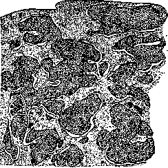
Fig. 19. Squamous cell carcinoma.
anywhere on the wall, uterine cavity, or at the fundus, can seize the entire cavity with greater or lesser immersion into the muscular wall. Squamous cell carcinoma of the vaginal part. Microscopically it is characterized by atypical growth of the epithelium, testifying to a violation of the physiological boundary. Among the usual connective tissue making up the thickness of the mucosa of this section of the cervix, more or less infiltrated by lympho- and leukocytes, lie nests and trabeculae of various shapes and sizes, consisting of solid accumulations of epithelial cells. Under high magnification, the designated nests are formed by cells extremely diverse in their shape and size: small, medium, and large, spherical, oval, octahedral, polygonal, and other cells lie side by side, disorderly mixing among themselves; differentiation into layers, proper to normal stratified squamous epithelium, is absent. Thus the described nests and trabeculae, consisting of complexly arranged cells of the epithelial tissue type, are distinguished by extreme polymorphism. In the cell nuclei, which are also distinguished by a diversity of size and shape, a large accumulation of chromatin is noticed, which also constitutes a feature of tumor nests—hyperchromatosis. At the same time, numerous division figures are noted in the nuclei; both regular and irregular mitoses are observed in them. It is necessary to touch upon those changes that are observed in squamous cell carcinoma in the process of growth and that exert an influence on its microscopic picture. If the epithelial nests and trabeculae characterizing the microscopic picture of cancer are rather thick, then their central sections, lying farther from the source of nutrition (connective tissue), undoubtedly suffer and undergo degenerative changes. The latter are expressed more often in simple disintegration of centrally located cells, and then cavities filled with detritus appear in the nests and cords. In rare cases, among the epithelial nests, pictures of pearls are encountered when the central cells of the nest become larger, lose the ability to stain and, stretching out, are located one next to the other like leaves of an onion bulb. As for the division of cancers according to the degree of maturity into mature, medium maturity, and immature, whereby the degree of differentiation of epithelial cells is taken into account, it does not have that practical significance which they try to find in it for dosage in radium treatment, as well as for prognosis. And indeed in almost every case of cancer one can to a certain degree find both mature and immature cells. Dwelling on the differential diagnosis of squamous cell carcinoma of the cervix, one should take into account what enormous significance the earliest possible recognition of cancer has for treatment and prognosis. Therefore, when evaluating the microscopic picture, it is necessary, in order to avoid the possibility of errors, to strictly take into account all moments confirming the picture of cancer. In this respect, it is especially important to keep in mind those microscopic pictures that are observed e.g. during the healing of a pseudoerosion (Fig. 20) and various ulcers (gonorrheal, tuberculous, syphilitic, etc.). And only the presence of mucous glands in pseudoerosion, [and] too superficial atypical growth of epithelium force one to stop, despite the suspiciousness of the picture, and wait for the further course of the process, observing the patient with corresponding treatment and repeated biopsies. Squamous cell carcinoma of the cervix gives exophytic or endophytic growth. In the 1st case (Fig. 21) the cervix becomes tuberous, covered with proliferations in the form of cauliflower, which crawl onto the fornices; concomitantly the proliferations ulcerate, disintegrate, giving bloody, disgustfully smelling discharges. Further disintegration of the tumor leads to the formation of a crater. In the endophytic form, the cervix of the uterus, while preserving approximately
its usual appearance, thickens, but the growth of the tumor goes into its thickness, destroying and ulcerating it from the side of the lower section of the cervical canal. However, the exophytic form is frequently combined with the endophytic. As for the severity of the process, in the sense of prognosis endophytic forms of course give worse outcomes, since they quickly spread to the pelvic cellular tissue and adjacent lymph glands. According to Schottlander and Kermauner, cancer of the vaginal part in more than half of all cases, spreading upward, passes beyond the internal os. Cancer of the cervical canal and body of the uterus. Corresponding to the epithelium covering the mucosa (Fig. 22) of these sections of the uterus, the cancer developing here has the character of glandular cancer and usually bears the name of adenocarcinoma.
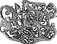
Figure 22. Adenocarcinoma.
The microscopic picture of such cancer is characterized by the following signs: the extreme hyperplasia of the glands and the epithelium contained within them already in itself testifies to the enhanced growth of the epithelium. Upon a more detailed study of the picture, one can become convinced that the epithelium in the glands no longer lies in a single row, but is layered in several layers, often filling the entire lumen of the gland; at the same time, it loses its cylindrical shape and takes on a various appearance: cubic, round, polygonal. This polymorphism is further enhanced by the diversity in the size of cells and nuclei. A significant accumulation of chromatin substance—hyperchromatosis—and a large number of mitoses are noted in the nuclei. But the most important sign characterizing malignancy is the violation of the physiological border of the epithelium, which is constituted by the finest fibrous membrane—membrana propria—separating the epithelium of the gland from the stroma and representing, as has already been precisely established, a plexus of collagen fibers. This destruction of the membrana propria indicates a weakening of the resistance of the vascular-connective tissue (reticulo-endothelium). As a result, there is a fusion of neighboring glands, giving conglomerates of such fused glands characteristic of adenocarcinoma. Sometimes the process of growth of the glandular epithelium proceeds so intensively that the pictures of previously existing glandular alveoli are completely lost, and those forms of cancer are obtained which are described under the name of carcinoma solidum. And only by the remnants of the glands, if they can be found, can one judge the genesis of a given cancer. Among the degenerative changes of adenocarcinomas, one should note the rather rarely encountered metaplasia of cylindrical epithelium into squamous with a tendency to keratinization. Thus, in glandular cancer, one can meet here and there nests of squamous cells, which, keratinizing, can give pictures of pearl cancer. Sometimes a pearl can be seen in the lumen of the gland. In very rare cases, such metaplasia can involve significant areas. As for the special form of glandular cancer described by some under the name of malignant adenoma (adenoma malignum), when supposedly the membrana propria is preserved and consequently the glands preserve the physiological border, then according to Ulezko-Stroganova this circumstance alone, i.e., the preservation of the physiological border by the gland, speaks against malignancy, and consequently against the admission of such a form. The appearance of glandular proliferations in the muscular wall of the uterus represents merely a picture of endometrioid heterotopias and is usually associated with the presence of inflammatory phenomena both in the endometrium and in the muscular wall. As for the gross anatomical appearance of glandular carcinomas, they usually give an exophytic growth, proliferating in the form of cauliflower and sometimes filling the entire uterine cavity. Such forms are called villous cancer (carcinoma villosum). The immersion of the process into the thickness of the muscular wall is observed less frequently, and if one takes into account the topography of the body of the uterus (the comparatively small amount of cellular tissue with its vessels surrounding the body of the uterus), cancers of the uterine cavity generally give a better prognosis than cancers of the cervix and especially squamous cell carcinoma of the vaginal portion, as stated above.
by Ulezko-Stroganova. Clinic of various forms of cancer. Cancer of the cervix of the uterus. Formerly, in the study of cervical cancer, it was customary to divide this form of the disease into two distinct groups: cancer of the vaginal portion of the uterus and cancer of the cervical canal. Extensive clinical material, processed especially by Kermauner, currently leads to the conclusion that such a division, both clinically and anatomically, cannot always be made. It can be assumed with a high degree of probability that cervical cancer in the vast majority of cases develops from one specific area, namely, near the external os of the cervical canal. It is hardly possible now to adhere to the old view according to which cancer of the vaginal portion, in its spread, remains a more local affliction, involving only the walls of the vagina, while cancer of the cervix spreads more in depth, heading toward the parametrium. More detailed observations teach that, arising in the region of the internal os, cancer can spread in any direction: either exophytically onto the vaginal portion and the vaginal wall, or endophytically into the tissue of the cervix along the course of the cervical canal. Only exceptionally rarely are forms of cervical cancer encountered whose site of origin is located deep within the cervical canal, or rather in the deeply situated layers of the cervical glands, or even originating from the rudiments of the Wolffian body, embedded perhaps even in the muscular layer of the cervix of the uterus. Clinically, in their course, spread, and symptoms, both forms of uterine cancer (cervical cancer and cancer of the vaginal portion) also do not provide sufficient data for their separation, and therefore below they are considered together. Symptoms. Cervical cancer is extremely poor in symptoms. They are expressed by discharges, bleedings, and—in advanced cases only—by pains. To these symptoms are added, in very far-advanced cases when the cancer spreads to neighboring organs, symptoms from these organs: the bladder, ureters, kidneys, intestines, peritoneum, and finally general phenomena—cachexia, sepsis, and uremia. Discharges in the form of purulent leukorrhea are one of the early symptoms of uterine cancer. However, it must be noted that these discharges cannot be observed in all cases, and that even frequently this important symptom may be absent. Bleeding in cervical cancer usually develops only gradually. Initially, it is limited to an insignificant amount of blood mixed with the discharge, which makes them bloody and imparts the appearance of the classic picture of "meat-water" appearance. Bleeding is undoubtedly a sign of the breakdown of the cancerous tumor, and its intensity is in direct dependence on the degree of this breakdown. Extensive breakdown is accompanied by stronger bleeding, which can reach the degree of the most severe blood loss. The process of breakdown of cancerous tissue proceeds extremely differently. Sometimes a carcinoma that does not break down for a long time gives an extensive breakdown all at once and within one or two days leads to the formation of a large crater penetrating the entire cervix; in other cases, voluminous cancerous infiltrates extending far beyond the boundaries of the cervix remain intact and give very little blood loss. Characteristic of cancer are so-called contact bleedings, i.e., bleedings resulting even from touching the cancerous tissue during examination, douches, sexual intercourse, defecation, urination, and sometimes even with simple slight straining of the abdominal press. If the woman is still menstruating, the menstrual phase usually does not affect the strength of the bleeding, and vice versa—the presence of cervical cancer does not stimulate ovarian activity and does not increase menstruation (Schroder). With long-lasting blood loss or heavy, even short-term bleedings, severe anemia and general cachexia naturally occur sometimes. The introduction of putrefactive bacteria into the breaking down cancerous masses imparts a putrid odor to the cancer discharges, which usually gradually increases and makes it extremely burdensome for those around the patient, although often almost unnoticeable by the patient herself. Pains usually appear only when the cancer has gone very far, when the parametrium is infiltrated and the lymph nodes are sharply enlarged. These pains, radiating to the extremities, become unbearable in the end, are difficult to treat even with large doses of narcotics, and are a real scourge not only for the patient herself, but also for the doctor and the patient's close relatives. The appearance of early pains may indicate either a rapid spread of the cancerous process or the development of inflammatory infiltrations of the parametrium accompanying the cancer. Some authors among the symptoms of even non-advanced forms point to general symptoms such as pain in the pit of the stomach, lack of appetite, nausea, and even vomiting. It is hardly possible, however, to attribute these symptoms exclusively to uterine cancer. The further development of symptoms of cervical cancer of the uterus depends on the spread of the cancerous process. Through careful study of both autopsy material, and especially preparations obtained from cancer operations, as well as during the performance of the operations themselves, it was possible to be convinced that cervical cancer, having captured the latter, can pass to neighboring organs and the paracervical tissue surrounding it, and generally to the entire parametrium. The spread of the cancerous process in this case proceeds initially per continuitatem, and then, when the cancer cells reach the lymphatic pathways, along these pathways and tissue clefts, vascular sheaths, and even in rare cases along the vessels. Thanks to such a spread, cervical cancer very often passes not downward—to neighboring areas of the vagina—but upward—to the body of the uterus. Schottländer and Kermauner on their operative material found the vagina already affected by cancer in 45% of all cases of cervical cancer. The uterus is captured by the cancerous process spreading here from the cervix very rarely. Usually, the cancer passes into the thickness of the muscular wall along the lymphatic pathways and less often per continuitatem through the region of the internal os. Rarely (however, such cases are observed) from the uterine cavity, cancer spreads into the cavity of the tubes. The frequency of metastasis of cervical cancers of the uterus to the ovaries is undoubtedly strongly exaggerated: in the vast majority of cases, metastases in the ovaries are absent. Somewhat more often such metastases are observed in cancers of the body of the uterus. Of practical importance is the spread of the cancerous process to the parametrium. The direction of its spread into the parametrium largely depends on the site of origin of the carcinoma. If the primary focus is formed, for example, on the posterior lip of the cervix, the cancer usually, spreading concentrically, passes to the posterior parametrium, into the region of the uterosacral ligaments. If the site of origin is the anterior lip of the cervix, the cancer has a tendency in its spread to go more anteriorly and laterally, frequently capturing the anterior parametrium as well as spreading to the bladder. Spreading laterally, it infiltrates the lateral parametrium up to the lateral surfaces of the pelvis, surrounding the ureters with a thick dense capsule, very rarely passing to the walls of the ureter itself, but compressing its lumen instead, which sometimes leads to its complete or partial obstruction. Of particular practical importance is the spread of cancer to the bladder. Usually, the urinary pathways resist this spread for a long time, but nevertheless, due to the intimate connection with the cervix, the cancer first captures the muscular layers of the bladder, and then, in more advanced cases, its mucosa. Upon breakdown in such cases, urinary fistulas are formed, giving urinary incontinence and all the consequences associated therewith. When the ureter is pinched by the cancerous infiltrate, a difficulty for the outflow of urine is created, which leads to hydronephrosis, and upon subsequent infection—to pyelitis and pyelonephritis. If the process captures only one ureter, the second kidney performs all the work necessary for the organism; if both ureters become impassable, a gradually increasing oliguria and even anuria develops, which already leads to uremia phenomena. However, pure cases of uremia are observed very rarely. In most cases, sepsis originating from the renal pelves leads to death sooner. When spreading posteriorly along the uterosacral ligaments, the infiltrate can reach the rectum and can give a constant retention of stool here, lead to the formation of a fecal fistula, incontinence of gas and stool with all the severe consequences associated therewith. The general condition of a cancer patient at the beginning of the disease apparently is not disturbed at all. Along with emaciated women at this stage, well-nourished and even blooming women are encountered. Only the psyche of the patients seems to be affected already very early. This probably depends on the experiences of the woman, fearing and thinking about the possibility of cancer. In many cancer patients, periodic, often short-term increases in temperature are observed (according to Fromme, in 9.2% of cases), which can be explained by protein breakdown and the formation of pyrogenic substances in the process (Koblank). In some cases, the only symptom of developing uterine cancer is the patient's complaints of emaciation and a drop in body weight. Metastases in distant organs in patients with uterine cancer are relatively rare and are usually determined only on autopsy material.
In contrast to the noted metastasis, metastatic involvement of the lymph nodes of the lesser pelvis and along the abdominal aorta is quite common and is of great importance in the treatment of cancer of the uterus. The arrangement of the lymphatic vessels and lymph nodes of the female genital organs therefore undoubtedly has important clinical significance. The lymphatic pathways of the cervix spread along the sides of the uterus into the parametrium towards the pelvic walls and along the uterosacral ligaments towards the rectum. The lymph nodes located here have a completely defined topography: the first group of nodes lies in the region where the uterine artery crosses the ureter; then come the nodes located in the triangle formed by the bifurcation of the external and internal iliac arteries; the sacral nodes are located on the posterior wall of the pelvis on the sides of the rectum. Finally, there are nodes along the course of the common iliac artery and higher up along the aorta up to the renal vessels. In most cases, initially only the nodes closest to the cervix are affected. However, the reverse is also frequently observed: the closest nodes, i.e., the nodes of the first stage, may remain free, whereas distant nodes may already be affected. Thus, there is no definite order in the spread of the cancerous process from the cervix to the lymph nodes; it is noted, however, that in cases of far-advanced cancer, the nodes are affected more often than in the initial stages of the disease. However, in 1/3 of inoperable, neglected cases, the nodes are found free of the cancerous process, and in 1/3 of non-neglected, just beginning cases, the nodes may already be affected. The total life expectancy of women who develop cervical cancer varies greatly, ranging from several months to several years. The average duration is considered to be 16-18 months. Apparently, in young women cancer proceeds faster, while in old women it is slower. Diagnosis of cervical cancer. When diagnosing cancer of the cervix of the uterus, it is also necessary to ascertain its spread to the adjacent tissue and adjacent organs. From this point of view, the diagnosis of uterine cancer sometimes presents insurmountable difficulties even for a specialist and often can only be fully revealed on the operating table. The need for the earliest possible diagnosis and the most accurate possible determination of the extent of the spread of the cancerous process is dictated by the important clinical significance that an accurate diagnosis has for the subsequent fate of the patient. Life teaches that procrastination in this matter is, more than anywhere else in another field, in the full sense of the word, "akin to death." It is necessary to divide all cases of cervical cancer in terms of diagnosis into 4 categories: 1) beginning cases, 2) pronounced cases, 3) widespread forms (standing on the border of operability), and 4) inoperable, neglected forms. All these forms are diagnosed mainly on the basis of determining, through inspection and palpation, local changes in the uterus, taking into account anamnestic data. The beginning forms of cervical cancer are of particular importance from the diagnostic point of view, since in these cases the physician's responsibility for the correctness of the diagnosis and its timeliness is particularly great. It should be noted that it is precisely in these forms that the most responsible and serious errors on the part of the observing physicians are often encountered, who frivolously overlook the disease and miss the precious time when the patient can be cured with almost complete certainty. The most necessary condition for the correctness of the diagnosis is not to be limited to a single routine gynecological bimanual examination of the patient, but unfailingly to subject her to examination with specula. It is necessary to examine the vaginal portion of the uterus most carefully, especially the region of the external os. The slightest ulcer, the most insignificant excoriation, and a change in the epithelial cover in the region of the internal os or on the vaginal portion of the cervix must be the subject of careful study. In those cases where the entire cervix is thickened, but no changes are detected on the surface of the vaginal portion, a probe should be used to go beyond the region of the external os and ascertain the condition of the cervical walls from the side of the cervical canal. It is necessary in every doubtful case to make it a rule to consider it suspicious for cancer and only then reject this diagnosis when this diagnosis is refuted with complete conviction. Frequently, the first examination by an experienced physician gives the decision that cancer is unquestionable, but never with just a cursory inspection, which gives an idea of the benign nature of an ulcer, can one say that alongside the benign nature in this case there is no cancerous process as well. A friable ulcer that bleeds easily even upon light touching, the bottom of which is covered with disintegrating tissue without infiltration along the periphery, speaks for cancer. A non-bleeding or slightly bleeding, sometimes even quite widespread ulcer, usually involving both lips of the cervix of the uterus, often nodular, covered as it were with small epithelial proliferations, speaks for an inflammatory process—papillary erosion. A large, thickened cervix that bleeds easily from the os when a probe is passed through it, devoid of Nabothian cysts, is more often an endophytic form of cervical cancer, whereas such a large, massive cervix with a mass of Nabothian cysts translucent on its surface is more often the result of follicular hypertrophy of the cervix of the uterus. However, the presence of even a large number of Nabothian cysts can in no way guarantee that in addition to this process there is not also a carcinoma developing endophytically in the cervix in this case. The view of the old doctors, according to which cervical polyps supposedly guarantee against cancer, is completely incorrect. Although a cervical polyp is benign in itself, its epithelium can often also give malignant growth. Small multiple polyps in the region of the internal os that bleed easily and are easily detached with tweezers or a curette are particularly dangerous in this regard. With respect to such malignantly degenerating polyps, the possibility of their complete removal during scrapings (e.g., diagnostic ones) should be kept in mind; in these cases, the laboratory diagnosis of cancer can be the reason for an already unnecessary operation of extirpation of the entire uterus. Great difficulties, even for the most experienced physician, are presented by the differential diagnosis between cancer and primary syphilitic chancre or tuberculous ulcer. These ulcers are extremely similar to one another. In some cases, the experienced physician notes only that non-cancerous ulcers are not as brittle, resist mechanical influences more, are of a more pink color, and are often covered with slightly yellowish, poorly removable deposits. A tuberculous ulcer is even more difficult to distinguish from a cancerous one. Usually, only the microscope decides the diagnosis here. A pronounced form of cancer, especially one that is disintegrating with the formation of a crater or growing exophytically in the form of cauliflower, presents no difficulties for diagnosis. This form is characterized by its easy bleeding (giving so-called contact hemorrhages), its extreme brittleness and consistency, which often allows, during examination with a probe, to introduce it deep into the tissue with almost no effort, like into "sand." The difficulties in diagnosing cervical cancer in these cases lie not in establishing the diagnosis of uterine cancer itself, but in determining the extent of the spread of the cancerous process. The determination of the extent of spread to the adjacent parametrium and organs is established by careful palpation of the uterus, the cervix, its ligamentous apparatus (broad ligament, uterosacral ligament) and the anterior fornix, behind which lies the urinary bladder. Each of the listed departments must be palpated in detail. It is necessary to make sure that there are no thickenings and indurations in them; it is necessary to try to accurately determine the mobility of the uterus in all directions, especially upwards and downwards, and only then draw definite conclusions. Examination per rectum or a double rectovaginal examination, in which the middle finger is inserted into the rectum and the index finger into the vagina, is very helpful for the accuracy of diagnosis in such cases. Accurate determination of the state of the parametrium is of paramount importance for assessing the severity of the case and for choosing the treatment method. A well-mobile uterus, even if it is increased in size, with a completely free parametrium, makes it possible to classify the case into the 2nd category of cases (mild, not neglected). Even a small uterus, but with a thickened cervix, with a thickened lateral parametrium or uterosacral ligaments, speaks for the difficulty of the case. The parametrial infiltrate can be palpated on both sides in the form of symmetrically located thickenings; but more often it affects one side more than the other. Cases are particularly unpleasant when, during examination, one observes as it were a shortening and induration of the anterior fornix. In such cases, the matter usually concerns the transition of the cancerous process to the bladder. For a more accurate diagnosis, the use of the cystoscopic method of examination is desirable, and in such cases, local vascular dilation and bullous edema are discovered. These cases usually have to be classified in the 3rd category, to cases standing on the border of operability. Infiltrates in the fornices, reaching the pelvic walls, making the uterus completely immobile, as if walled up in the thickness of the cancer masses, force one to classify the case into the last category—inoperable cancers.
When diagnostically evaluating each case, it is nonetheless always necessary to bear in mind that uterine cancer is very often combined with and proceeds alongside severe inflammatory processes in the appendages, peritoneum, and parametrium. This circumstance greatly complicates the diagnosis of the extent of the cancer, since the infiltrates determined upon palpation may be the consequence of an inflammatory disease rather than a cancerous process, which cannot fail to influence the assessment of the case. Therefore, very frequently, despite the thoroughness of the examination, it is impossible to fully assess the case prior to surgery, and this is done only upon opening the abdominal cavity and palpating all the pelvic organs from above. Among the special methods of examination without which the diagnosis of cancer can frequently not be established, it is necessary to point out the excision of a small piece of the cancerous tumor or the scraping of the cancerous mass with a sharp curette, followed by microscopic examination of the obtained material. This method, sometimes called trial excision, developed as early as 1882 by Ruge and Veit, is of particularly great importance in incipient forms of cancer when the nature of the ulcer cannot yet be clinically determined. It consists of exposing the cervix with specula, fixing it, if necessary, with bullet forceps, and excising from the cervix, wherever possible at the site of transition from the ulcer to healthy tissue, a wedge-shaped piece for its microscopic examination. After removing the piece, it is usually possible to stop the bleeding with a tampon; otherwise, a suture encompassing the entire thickness of the wound can be applied. In exophytic forms with the presence of decaying masses or with ulcers spreading across the surface, the manipulation can be significantly simplified by making a simple scraping of the superficial tissue with a sharp spoon, which heals easily and does not require the application of a suture. Trial excision undoubtedly has great significance in the diagnosis of cancer, but it is not devoid of its negative aspects. Even when performed with aseptic lege artis, in some cases it leads to suppuration and (more importantly) to severe tissue breakdown, and can apparently contribute not only to the infection of deeply situated sections of the genital system, but seemingly even give impetus to the rapid spread of the cancerous process itself. In this sense, trial scraping has advantages over excision. Unfortunately, both microscopic and clinical examinations are far from always providing a definitive solution: even the most experienced microscopist and specialist pathologist frequently encounters difficulties in interpreting the microscopic picture of the trial piece under investigation. In former years, many clinicians made it a rule to perform a microscopic examination of a tissue piece and establish a microscopic diagnosis, without which proceeding with operation was considered unacceptable. At the present time, many clinicians no longer adhere so strictly to this rule, but shift the center of gravity to a thorough micro- and macroscopic study of the preparation obtained during surgery, while the answer to the question of whether the uterus with its cellular tissue has been removed within the limits of healthy or diseased (cancerous) tissues acquires special importance. Of other special methods of examination, mention may be made of examination with a colposcope—a binocular loupe proposed by Hinselmann in Germany, which allows the vaginal mucosa to be examined at a significant magnification (up to 100 times) and the finest structural changes on it to be determined. Regarding general reactions proposed for the determination of cancer, see Cancer. Prognosis. In the old literature, and occasionally in the literature of recent years, reports can be found on the spontaneous cure of cancer or on cases of cancer cure by any local or general means. A critical evaluation of a number of such cases, repeatedly carried out (Strauss), nevertheless leads to the conclusion that not a single undoubted case of cure has been observed up to now, and that frequently it is either a matter of a diagnostic error or an incorrect illumination of the case. As a rule, a patient with cervical cancer who is not subjected to treatment usually dies rapidly, with the life expectancy of these patients calculated to average 16–18 months. Apparently, younger patients die sooner because the cancer proceeds more malignantly in them. Strong, lean women after 50 years of age can live, suffering from cancer, even for several years. Of great importance for predicting the further fate of a patient with cervical cancer is the degree of spread of the cancerous process. Everything depends on the period at which the patient is subjected to treatment. Since sufficient experience and indisputable conclusions already exist regarding operative treatment, its results will be taken into account in the future. Still comparatively recently (1902) at the international congress of gynecologists in Rome, voices were raised, chiefly by French gynecologists, to the effect that the operative treatment of cancer does not achieve its goal, since uterine cancer is a local manifestation of a general disease. Such pessimism can no longer have a place at the present time. Gynecologists possess thousands of operated cases, with hundreds of cases operated on by the same surgeon, and this extensive, often carefully studied and for many years verified material proves with irresistible conviction even to those who do not have their own many years of personal experience that cervical cancer is curable, although not always, but in a significant percentage of cases. This allows such an outstanding French gynecologist as J. L. Faure to say: "given good technique... I am sure that recovery will be a common occurrence." Nevertheless, patients subjected to operative treatment can unfortunately far from always count on a cure. The presence of pregnancy, in the opinion of the majority of doctors, sharply worsens the prognosis, since cancer spreads extremely rapidly in this condition. Of interest are attempts to find methods that facilitate the possibility of making more accurate predictions. Mention may be made here of attempts to determine local eosinophilia, especially in the marginal areas of the cancerous ulcer. In this eosinophilia, a certain defensive capacity of the organism against the invasion of cancerous masses was seen. However, these considerations have not justified themselves. The old idea that cancer of the vaginal part of the uterus proceeds more favorably than cancer of the cervix can be considered correct only in the sense that cancers of the cervix, frequently built of young, undifferentiated cells, proceed more malignantly than the more differentiated cancers of the vaginal part (squamous cell). Of undoubtedly great significance for predicting the outcome (in the event that a radical operation is performed) is the general state of health of the patient and the work of her individual organs, especially the heart and kidneys. Strong, dry women can achieve a cure in a much higher percentage than full, loose, sometimes edematous patients. One of the most important conditions for achieving good results with our modern methods of treating uterine cancer is the timely seeking of help by patients. This is why one of the most important prophylactic tasks at the present time is considered to be the widespread dissemination among women of knowledge about the necessity of consulting a doctor at the slightest suspicion of a disease, which, at the suggestion of Winter, should be carried out among the population through the general press. For the same purpose, numerous leaflets are published with appeals to middle-level medical personnel and practicing doctors about the necessity of the earliest possible recognition of uterine cancer. Special outpatient clinics and laboratories are established, and societies for combating cancerous diseases are organized. It can be thought that a widely mounted propaganda of this issue will provide the opportunity to timely attract women for treatment and will reduce the number of neglected, already incurable cases. Cancer of the body of the uterus. The diagnosis of this form of cancer, located in the depths of the uterine cavity inaccessible to the eye, presents greater difficulties than the diagnosis of cancer of the cervix, which is easily visible to the eye. To this is added the even slower course of this form and the insignificance of symptoms. The uterus is usually determined to be significantly enlarged, with its size in some cases reaching that of 3–4 months of pregnancy. Its soft consistency and sometimes slightly nodular surface can give reason to think of pregnancy or a myoma. However, the age of the patient, especially an unmarried woman, and above all symptoms, particularly bleeding that has appeared already in the climacteric period, can lead to the correct diagnosis. The case becomes especially clear if the uterine os is slightly open and if fragile, easily broken cancerous masses can be removed through it with a finger. In contrast to this, initial forms of cancer of the uterine cavity are determined with great difficulty and only after microscopic examination of the contents of the uterine cavity obtained by trial curettage. The latter must be performed with extreme caution in view of the existing danger of uterine perforation. However, it is precisely in the initial stages of cancer that it is necessary to obtain a scraping of the entire surface of the uterus so as not to overlook cases with an isolated focal lesion of the mucosa by cancer. Usually, the sheer abundance of masses obtained during scraping speaks in favor of cancer. However, abundant scrapings can also be obtained upon the breakdown of a myomatous submucous node or polyp.
At the same time, cases with scanty scraping are not uncommon in which the microscope nevertheless detects cancer. It must be taken as a rule to consider any bleeding in the menopause period suspicious of cancer and in every case to subject patients to a detailed examination. The prognosis for cancer of the body of the uterus is usually significantly more favorable than for cancer of the cervix. This is due to the fact that this form of cancer develops much more slowly. While the life expectancy of a woman with cervical cancer is estimated on average at 16-18 months, in cancer of the cavity the disease drags on for several (5-7) years. Cancer of the body of the uterus rarely produces infiltrates in the parametrium and metastases in the lymph nodes. The presence of lymphatic pathways between the body of the uterus and the ovaries makes metastasis to the latter quite possible; however, such metastases are observed quite infrequently. From all that has been said, it follows that operations for cancer of the body of the uterus yield better results. Unfortunately, patients with this form often seek medical help too late, when the case has already become inoperable. Treatment. Our time possesses essentially only two methods of treatment for cancer of the uterus—surgical and treatment with radiant energy. Both of these methods currently rightfully enjoy the reputation of being able to provide the patient with a radical cure. In the USSR, the surgical method of treating uterine cancer followed by X-ray irradiation has become the most widespread. Surgical treatment of cancer of the body of the uterus. At present, the main operation that enjoys the greatest prevalence in the treatment of uterine cancer both abroad and in the USSR is undoubtedly the extended abdominal operation for uterine cancer, usually justly called Wertheim's operation, although in performing it most operators have long since departed from the details of the technique of Wertheim's operation itself (see Wertheim's operation). The idea of this operation consists in exposing the parametrium, freeing the ureters, separating the bladder and rectum from their intimate attachment to the vaginal tube, and excising the entire uterus with its appendages, the surrounding parametrium, and a significant part of the vagina. Some surgeons pursue this same goal, striving to perform the operation through the vagina. Already Schauta, having abandoned simple vaginal extirpation, attempted by expanding this extirpation to create a method of the same expanded operation for cancer through the vagina. In recent years, the well-known Berlin gynecologist Stoeckel has pursued the same goal. Striving to lower the high operative mortality in the extended abdominal operation, Stoeckel replaces it with the safer vaginal extirpation, to enhance the insufficient radicalism of which he uses radiant energy in the form of preparing the patient with the preliminary introduction of radium and postoperative X-ray exposure. Such a combination, in Stoeckel's opinion, makes it possible to obtain the best results and to reduce the primary mortality of the operated patients by almost 2 times. The patient is prepared before the operation for at least 4 days. All these days she receives sublimate douches. The heart, even in cases where it works well, is strengthened with digalen; the day before the operation, a mild laxative. On the day of the operation, the abdominal wall is washed with soap and water, treated with alcohol, and iodized. The vagina, after the insertion of specula, is treated with a sublimate solution. After this, the patient receives ether anesthesia, under which the cancerous ulcer is scraped out and cauterized with a Paquelin cautery until completely dry. If the bleeding cannot be stopped by cauterization, the cavity of the ulcer is tightly tamponed with gauze. After this, infiltration anesthesia with a solution of novocaine with adrenaline is performed on the sides of the vagina at the sites of future vagino-perineal incisions (up to 120 cm3 of infiltration fluid). Stoeckel attaches special importance to this anesthesia as a method not only of pain relief, but mainly of ensuring a bloodless future operational field. The operation itself begins with making a circular incision in the upper section of the vagina and forming a cuff, onto which a large number of ligatures are applied to suture the anterior and posterior walls of the vagina and isolate the cancerous cavity. These ligatures serve subsequently as a holder. Only after wiping these ligatures and the resulting stump with alcohol for the purpose of disinfection are deep vagino-perineal incisions made from two sides, starting from the tubera ischii to the edge of the vaginal incision. After this, the operational field becomes extremely accessible, and without special difficulty one can proceed to separate the urinary bladder, expose the uterine artery and the ureter, which is done on both sides. When the ureters are well and widely exposed and the uterine artery is ligated on both sides, it is easy to open the vesicouterine pouch and bring the fundus of the uterus outward. Having ligated the appendages after applying clamps, with the preservation of the ovaries in typical cases, the posterior fornix is opened. If now, grasping the body of the uterus with firm forceps, one pulls it forward and upward, and by the holding ligatures forward and backward, the entire parametrium is strongly tensed and can be easily extirpated almost to the pelvic walls, and it is necessary to make sure not to injure the tensed bowel and ureters. It only remains to carefully and tightly connect the edges of the peritoneum with interrupted sutures and restore the paravaginal incisions. Usually Stoeckel, when suturing the paravaginal incision, leaves a small opening in the middle of the fornix, through which he inserts 1-2 small gauze drains between the bladder and rectum, and when suturing the walls of the vagina and perineal incisions in two layers—glass drainage tubes, which reach the parametrium. The gauze strips are removed on the 2nd day, the glass drains on the 3rd, and the Michel clips on the perineum on the 7th day. Stoeckel suggests prepending this operation with the introduction of 70 mg of radium into the cervix 2 times for 24 hours each. Shortly before discharge from the clinic, i.e., 3-4 weeks after the operation, the patient receives the first session of X-ray therapy and 6-8 weeks after discharge—the second session, each time for 6-8 fields of a carcinoma dose. Out of 206 cases of operations for uterine cancer, Stoeckel noted 19 deaths, which yields a mortality rate of only 4.8%. The excellent results obtained by Stoeckel should compel everyone who has radium or X-rays at their disposal to study the method recommended by Stoeckel. Not every patient can be subjected to surgical treatment. First of all, advanced cases with a poorly movable uterus, with the transition of the cancerous process to the bladder, must be recognized as inoperable; the operability percentage among different surgeons varies (50, 60, 70, and 80%). The more strictly patients are selected, operating only on cases that are beginning or not advanced, the better results the operation yields. At the same time, the results of the operation are evaluated by operative mortality, as well as by the duration of recovery. It is undoubtedly true (especially for uterine cancer) that a good outcome of the operation and the discharge of the patient as if healthy do not yet speak of a cure, since such patients often return after some time to the clinic where they were operated on, already with a relapse of the disease. As experience shows, relapses appear most often in the 1st and 2nd years after the operation; later they are observed comparatively rarely. On this basis, it was agreed (Winter) to consider a patient cured only if no relapse is observed in her for at least 5 years after the operation. Currently, hundreds of cases of such long-term recovery after operations for uterine cancer are cited in the literature. Skrobansky has almost up to 56% of such a complete recovery in his material, with several patients remaining healthy for more than 10 years. Approximately the same results have been obtained by a number of foreign and Russian surgeons. In order for a surgeon to more accurately evaluate the results of their operations for uterine cancer, in addition to the concept of long-term or complete cure, Winter introduced the following points: 1) the figure of absolute cure, by which he understands the percentage calculated as the number of patients remaining free of relapse for 5 years (cured), relative to the number of all patients with uterine cancer observed during the studied period; 2) the figure of relative cure, i.e., the number of patients who achieved a complete cure (5 years), relative to all patients operated on for cancer. In order to obtain an expression in figures of the combination of all factors having an influence in evaluating the operation, Winter proposed the following formula: A = ... With this formula, primary mortality is not taken into account. Waldstein, on the other hand, recognizing it necessary to take into account primary operational mortality as well, proposed a more complete formula, according to which A = ... In these formulas, A is understood as the absolute usefulness of the operation; O is the operability percentage; B is the percentage of complete recovery; L is the percentage of those remaining alive (100-M, where M is primary mortality). But the extended abdominal operation also has its downside—a high percentage of operative mortality, i.e., mortality directly related to the performance of the operation; this percentage is very large and generally ranges from 10 to 20.
To this are added further postoperative complications in the form of pneumonia, suppuration, phlegmon in the parametrium, and especially fistulas of the urinary bladder and ureters, combating which sometimes presents great difficulties. In the last two decades, dissatisfaction with surgical methods of treating cancer of the uterus and other organs has forced medical thought to seek new paths in combating this scourge of man. Huge hopes were pinned on radiant energy, initially in the form of the application of radium and mesothorium rays. Although expectations in this field were not fully justified, in recent years, after powerful X-ray apparatuses were constructed and thanks to profound persistent scientific elaboration of the question, especially in the direction of the dosage of the amount of rays given, it has been possible to develop a good methodology that makes it possible to obtain good results in this direction as well. However, despite all its seeming allure, even this path in its modern application cannot be considered satisfactory. It also forces one to limit the cases of treatment and gives, after apparent cure, relapses, fistulas, and even in some cases a complete lack of effect. Since for those treated with modern apparatuses and under the conditions of the newest technique, the five-year period has in most cases not yet expired, there is as yet no possibility of making a final assessment of the effectiveness of this method of treatment. At present, a number of doctors have begun to combine radiant energy treatment with surgical treatment. Most often, this combination is expressed in a preliminary operation with subsequent irradiation to destroy those cancer cells that might have been left behind during the operation. It is proposed, in order to reduce operative mortality, to perform even lighter surgical interventions relying on the effect of subsequent irradiation. Such a method of treatment can be especially recommended in the treatment of cancer of the uterine cavity. Some use irradiation before operation, especially in neglected forms of cancer of the cervix. But whatever method of treatment is used, each of them has its limits, and we remain helpless before at least one-third of all patients applying to us suffering from cancer of the uterus—patients who are inoperable. The table compiled by Stoeckel can approximately illustrate the achievements obtained at the present time with various methods of treatment of cancer of the uterus. Cancer of the cervix: 1) Treatment with X-rays alone. Küstner Clinic—212 cases; 3 years of observation; relative cure—29.2%, absolute cure—4.7%. Bumm—22 cases; 5-year standing; relative cure—13.8%. 2) Treatment with radium. Kehrer—129 cases; 5-year standing; absolute figure of recoveries—27.9%. 3) X-ray combined with small doses of radium. Vinz—415 cases; 5-year standing; relative figure of recoveries—52.7%. Radium with X-ray. Döderlein, on a large material, comes to the conclusion that if the patient does not prematurely abandon treatment, in operable cases 81% of recoveries can be achieved, in borderline cases 31%, and in inoperable cases 10.4%. Abdominal radical operation for cancer of the cervix. Collected statistics: 2,467 cases; primary mortality—20%; figure of relative recoveries—40-61.9%; figure of absolute recovery—27.4%. Vaginal radical operation for cancer of the cervix of the uterus. Primary mortality—4.8%; figure of relative recovery—38-50%; figure of absolute recovery—16-18%. The task of treating neglected forms consists in the removal of disintegrated masses, in the prevention of disintegration and the stench emanating from this disintegration, in the limitation of hemorrhage, in the maintenance of the general condition of the organism, and finally in the reduction of sometimes severe pains. All this is achieved by a number of measures consisting in the scraping of cancerous masses, the cauterization of the scraped surfaces with a hot iron or some chemical substances (zinc chloride, nitric acid, formalin, etc.), in various powders, douches with disinfectants, and finally in the prescribing of a number of narcotic drugs and nervines: aspirin, pyramidon, morphine, pantopon, etc. In the USSR, among some doctors, the method of treatment of neglected cancers proposed by Gruzdev—the cauterization of the ulcer with acetylene—and the treatment proposed by Tretyakov—the injection of ether—are widespread. The first method is very simple: after exposing the cervix with specula, a little sublimate or boric solution is introduced there, and a pinch of calcium carbide is added. However, it must be remembered that the addition of large pieces of calcium carbide leads to excessive gas formation and even an explosion, which of course must be avoided. Acetylene perfectly cleanses the cancerous ulcer and revitalizes healthy cells. Ether, according to Tretyakov, is injected with a Record syringe at 1-4 cm3 daily into the thickness of the uterine muscle. After 5-6 days, it is necessary to take a break for 1-2 days, after which injections into various parts of the uterine wall should be continued again. As the author states, improvement begins from the end of the 2nd week. In the middle of the second month, the cancerous tumor almost completely disappears. Here it is still necessary to say a few words about relapses of the disease. Very soon after the operation, in unsuccessful cases of treatment, nodules of various sizes can be felt in the scar of the vagina, in the parametrium, or in the region of the lymph glands. If it is not a question of local suppuration (inflammatory infiltrate), then there is usually a relapse here. The return of the disease is very severe; all symptoms of the disease develop with greater intensity, and the fate of such patients is especially difficult. Unfortunately, the treatment of relapses very rarely gives good results. Only isolated cases of mobile infiltrates, mainly of affected glands, can be operated on. Usually, however, operation only worsens the already grave condition of the patient. It must be said that both radium and X-rays do not bring a cure in relapses. The physician has to apply that conservative-narcotic therapy which is put into play in inoperable cancers. To cancers of the uterus is usually classified a disease that is peculiar both in its clinical course and origin and is relatively rare, which is given the name chorionepithelioma (see).
by Sckrobansky. Sarcomas and endotheliomas of the uterus are malignant tumors of a connective-tissue character. Sarcomas of the uterus (see separate table, Fig. 6) must undoubtedly be classified as very rare tumors. It is believed that there is 1 sarcoma for every 40-50 cases of uterine cancer. As for the frequency of sarcoma in relation to fibromyomas, on average there are 3 sarcomas per 100 fibromyomas. Such a ratio is obtained if one compares the total number of histologically precisely established cases of uterine sarcomas with the total number of operated fibromyomas. If, however, one takes into account all that remaining huge mass of fibromyomas which are treated with radiant energy or, due to the absence of symptoms, are not subjected to any therapy at all, then the frequency of sarcomas will drop sharply and yield less than 1 case per 100 fibromyomas. Sarcoma of the uterus is encountered at any age, even in 5-year-old children, but most frequently this disease is observed in the climacteric period (40-50 years). By localization, two forms are distinguished: sarcoma of the wall and sarcoma of the mucous membrane of the uterus. Sarcomas are located either in the body or in the cervix of the uterus, arising both in normal muscle tissue and at the site of a pre-existing myoma. The tumor usually develops in the form of a single node, which is located like myomas interstitially, in the submucosal and subserous tissue. Diffuse permeation of the entire thickness of the uterus is less frequently observed. In cases of the occurrence of sarcoma in a myomatous tumor, sometimes the sarcomatous tissue cannot be distinguished in any way from a myoma—neither in appearance nor in consistency—but more often a sarcoma to the touch appears softer and in appearance more matte than a myoma. Sarcomas of the uterine body in later stages, but generally still after a considerable time, grow through the uterine wall and involve the peritoneum and intestine. In this case, the parametral tissue can remain free for a long time. Metastases (via the circulatory system) are observed in the lungs, bones of the skeleton, liver, etc. Metastases in the ovaries and the walls of the vagina are also observed. Sarcomas of the uterine cervix are encountered much less frequently, having a diffuse character or the appearance of a polyp (see separate table, Figs. 5 and 9), sometimes branching (sarcoma arborescens), resembling a grapelike sarcoma of the vagina (see). Possessing great malignancy in general, these latter forms tend to rapidly spread to the vagina and disintegrate. By histological picture, round-cell, spindle-cell, polymorphous sarcoma, and myxosarcoma are distinguished (Fig. 23). Giant-cell forms, endotheliomas, and mixed tumors of the sarcocarcinoma and liposarcoma type are less common. Angiosarcomas, muscle-cell sarcomas (sarcoma myocellulare), as well as neurogenic ones of the ganglioma embryonale sympathicum type (see Ganglioneuroma) have been described. The clinical picture of uterine sarcoma is very indefinite and uncharacteristic. In many cases, symptoms may be completely absent. In other cases, wall sarcomas produce the same symptoms as myomas. It is generally accepted that the rapid increase and softening of a fibromyoma serves as an indicator of the sarcomatous "transformation" of the tumor. Albrecht, Béclère, and others consider this proposition correct only if these symptoms appear after the cessation of menses. Before the onset of menopause, the presence of these symptoms cannot under any circumstances be considered characteristic of sarcoma, since all these
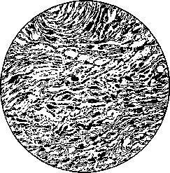
Figure 23. Myxosarcoma of the uterus.
phenomena are observed much more frequently with myomas than with sarcomas. Seitz and Wintz assert that the degree of rapidity of the effect of X-ray irradiation can serve as the basis for differential diagnosis between myoma and sarcoma: very rapid resorption of the tumor after irradiation serves as an indicator of the malignancy of the process, while slow reduction (over the course of several months) proves the presence of a benign myoma. This proposition is categorically disputed by Béclère and Gambarov. In some cases of uterine sarcomas, considerable weight loss of the patient and a general decline in strength attract attention. Cases have been described where, apart from rapid, seemingly causeless weight loss, there were no symptoms at all. Usually, however, sharp emaciation and phenomena of cachexia are observed in far-advanced cases—as a consequence of profuse hemorrhages, disintegration of the tumor, absorption of decay products, ascites, and metastases. In sarcoma of the mucosa, the same symptoms are usually observed as in cancer of the body of the uterus, i.e., hemorrhages, discharge, and pain. The discharge is watery, the color of meat washings, often foul-smelling. The pains are initially cramping, then take on a constant aching character. Treatment. In sarcoma of the uterus, as in cancer, two treatment methods can be used: surgical and radiant energy (X-ray and radium). According to the most complete cumulative statistics of Albrecht (314 cases), the results of surgical treatment are rather dismal. Primary operative mortality reaches 10-20%, the majority of operated patients die from recurrences and metastases already during the first year after the operation. Thus, the total mortality, including primary mortality, reaches enormous figures—30-75%. Cases of complete cure in world literature can be counted on the fingers. Thus, the results of surgical treatment of uterine sarcomas are immeasurably worse than the results of operations for cancer of the cervix and, in particular, the body of the uterus. Treatment with radiant energy (mainly X-ray therapy) likewise does not yield good, stable results in the vast majority of cases. X-ray therapy has two advantages compared to surgical intervention: 1) the absence of primary mortality; 2) the possibility of using X-ray therapy (in isolated cases even successfully) for inoperable forms of sarcomas. As for long-term results, although statistics of ray treatment look somewhat better than the statistics of operative methods, in view of the absence at present of sufficiently solid and verified material, the question of the advantages of surgical treatment or treatment with radiant energy still remains open. The primary results of X-ray therapy are sometimes striking in their staggering effect: cases of the resorption of a huge tumor within a few days after irradiation have been observed. But disappointment soon set in: still during the first year, and sometimes later, metastases appeared en masse, and the patients perished. Cases of complete cure are very rare. Regarding the choice of treatment method for uterine sarcomas, Gambarov, based on personal experience and literature data, finds it possible to put forward the following propositions: in operable cases with a relatively good general condition and in the absence of contraindications to surgical intervention, operate radically, and then subject to preventive irradiation with X-rays in order to prevent such frequent recurrences. In all other cases, including of course in inoperable forms, subject to energetic irradiation with X-rays. In some cases, combined radio-X-ray therapy is also indicated. As for the irradiation technique, Gambarov uses the single-stage Seitz-Wintz method: 70-80% HED.
G. Gambaro». IX. Functional diseases of the uterus. The majority of diseases of the uterus are associated with such path.-anat. changes in this organ which can be more or less easily determined by various methods of objective gynecological examination. At one time, namely during the heyday of Virchow's cellular pathology (Virchow), gynecologists tended to reduce the entire pathology of the uterus to such diseases. Thus, Slavyansky in his "Special Pathology and Therapy of Women's Diseases" (1888) divides all uterine diseases into four categories: 1) malformations, 2) incorrect positions and displacements of the uterus, 3) inflammation of the uterus, and 4) tumors of the uterus. However, even earlier, both foreign and Russian gynecologists, along with such diseases of the uterus, considered it necessary to distinguish others, the path.-anat. substrate of which was either determined outside the uterus or could not be determined at all by methods of objective examination and which manifested themselves only in purely functional disorders of this organ. Thus, the author of the first original manual on gynecology in Russian, Kiter (1858), having first described "local" or "organic" diseases of the uterus and other organs of the female genital sphere, in the second part of his work describes "general" women's diseases, which he divides into 3 groups: 1) irregularities of menstrual discharges, 2) diseases of the circulatory and nervous systems, and 3) general diseases of the puerperal period, and classifies amenorrhea, menorrhagia, and dysmenorrhea into the first group, which he nevertheless considers "more symptoms than independent diseases." The same is seen in the author of the next chronological complete manual of gynecology in Russian, Horwitz (1876); having devoted a separate chapter to the pathology of menstruation, he analyzes therein 1) absence of menstruation, 2) excessively heavy menstruation and uterine bleeding during menstruation, and 3) difficult or painful menstruation, while stipulating: "All these various disorders, whatever their character may be, constitute nothing other than the result of known pathological states of the uterus, and it would seem completely superfluous to devote a special chapter to this subject. Nevertheless, we consider it necessary to examine the pathology of the menstrual period separately, solely in view of the importance it has in practice." In modern times, authors of manuals on gynecology have again realized the need to single out functional diseases of the uterus from the complex of diseases of this organ into a special group, since precise studies have shown that these diseases are by no means inflammatory—chronic metritis and endometritis, to which they were attributed in Russia by Slavyansky, and in Germany by Scanzoni and his followers. To these purely functional diseases of the uterus, first Aschoff, then Pankow, Opitz, and others gave the name of metropathies, dividing them into two main groups: 1) disorders of menstruation and 2) disorders of uterine secretion. The first group of metropathies usually includes 1) amenorrhea and oligomenorrhea, i.e., complete absence of menstruation or lengthening of the intervals between them compared to the norm; 2) menorrhagia, polymenorrhea, and metrorrhagia, i.e., increased menstruation, frequent menstruation, and atypical uterine bleeding; 3) various varieties of dysmenorrhea, i.e., painful menstruation, including membranous dysmenorrhea. The second group includes uterine, or more precisely, cervical leukorrhea and that secreted by the mucosa of the body of the uterus. Among the functional diseases of the uterus can also be included the form first described by Snegirev, and then by Kozlenko, Chernyakhovsky, and other Russian authors under the name of endometritis dolorosa. This disease manifests itself mainly by severe pelvic pains, abdominal pains, and a number of nervous disorders up to and including epileptic seizures. The uterus in this disease in shape, size, position, etc., often appears normal. For recognition, Snegirev recommends paying attention to the following characteristic signs: 1) bimanual examination makes it possible to detect severe tenderness of the uterine fundus on the right, left, or both sides, which is especially noticeable during rectal-abdominal examination; 2) in patients, it is possible to detect five points, touching which proves to be very sensitive or painful; these points are located in the region of the pubic tubercle, an inch higher and outward from it, near the inner edge of the anterior superior iliac spine, on the outer surface of the iliac crest, and on the inner surface of the thigh, slightly below the inguinal fold; 3) touching the button of the uterine sound produces severe pain in the region of the internal os of the uterus, and especially painful is touching the button of the sound to the mucosa of the fundus, mainly in the region of the tubal openings. This disease, as can be seen from its very name, Snegirev attributes to inflammatory ones, ascribing the main role in its etiology to postpartum infection. Among therapeutic measures for it, he considers the best to be dilation of the cervical canal and leeches to the coccyx region. Kozlenko attributes a prominent role in the origin of this disease to a nervous-hysterical predisposition, and Dick observed a case of purely psychogenic origin of it. With the exception of this form, all other functional diseases are often purely symptomatic manifestations of various diseases of this organ having a completely definite path.-anat. physiognomy, e.g., malformations of the uterus, its inflammatory diseases, neoplasms, etc. Very often, however, they occur with a "completely normal"* uterus in anatomical terms, so that their cause has to be sought outside this organ. In such cases, these disorders must be regarded as independent nosological entities, especially since they themselves, independently of the causes provoking them, exert a harmful effect on the woman's health, sometimes even lead to death, and therefore again in themselves require therapeutic measures, up to and including the most serious types of surgical intervention.—In their clinical manifestations, essential disorders of the menstrual function and uterine secretion, or metropathies, are, as can be seen from their very definition, sharply different, sometimes even opposite, e.g., amenorrhea and menorrhagia, but etiologically they all have much in common. Thus, for example, constitutional factors can, according to many authors, play an important etiological role in the origin of both essential amenorrhea, and dysmenorrhea, and menorrhagia with metrorrhagia, which are especially often observed with an asthenic constitution, and uterine leukorrhea. General infectious diseases, both acute (typhoid, malaria, cholera, sepsis, etc.) and chronic, especially tuberculosis, can equally lead to various kinds of metropathies—amenorrhea, menorrhagia, metrorrhagia, etc. An equally important role in the etiology of various functional diseases of the uterus is played by poisoning, both acute and chronic, which must be taken into account when assessing occupational hazards; thus, for example, poisoning with lead, phosphorus, nicotine, and other poisons can cause amenorrhea in female workers, and sometimes these poisoning * Speaking of the "completely normal" anatomical picture of the uterus in metropathies, it must be stipulated that in all probability these diseases also have their anatomical substrate in the given organ; only this substrate consists of such changes that are inaccessible to conventional methods of gynecological examination. On the other hand, the causes causing metropathy can simultaneously cause such path.-anat. changes in the uterus that can be established by conventional gynecological examination. Thus, for example, increased blood supply to the uterus due to the persistence of follicles, leading to metropathia haemorrhagica (see below) together with its functional changes, namely, uterine bleeding of the menorrhagia and even metrorrhagia type, simultaneously causes its path.-anat. changes, e.g., an increase in volume, a change in configuration, consistency, etc. These path.-anat. changes in the uterus cannot, however, be regarded as the direct cause of bleeding, but only as its companion, as a consequence of the same cause that underlies the bleeding. lead to leukorrhea, and sometimes their consequences are severe uterine bleeding. Various metropathies, such as amenorrhea, dysmenorrhea, etc., can also develop on the basis of impaired nutrition, as well as general diseases leading to the weakening of the entire female organism. Among blood diseases, pernicious anemia and severe secondary anemia cause, as observations show, amenorrhea; leukemia, pernicious anemia, and chlorosis—meno- and metrorrhagia; chlorotic women often also suffer from leukorrhea.
Furthermore, a very prominent place in the etiology of metropathies belongs to the dysfunction of various endocrine glands; in hyper- and hypothyroidism, acromegaly, pituitary obesity, severe oophoritis, and ovarian neoplasms in women, amenorrhea often develops; ovarian hypofunction and other disorders of internal secretion frequently cause dysmenorrhea; on this same basis, leukorrhea often develops in women; the persistence of follicles leads to the development of that functional disease of the uterus which is known under the name of metropathia haemorrhagica; dysfunction of the thyroid gland, pituitary gland, and thymus also causes the development of meno- and metrorrhagia. A prominent place in the genesis of metropathies is also occupied by nervous disorders: nervous strokes can cause amenorrhea; disorders of the autonomic nervous system and vasomotor apparatus create the basis for the development of leukorrhea; dysmenorrhea is a frequent companion of hysteroneurasthenia; uterine bleeding can be the consequence of reflex irritations, especially painful ones. Recent observations of a number of authors in the USSR and abroad also testify to the important role played in the origin of various metropathies (amenorrhea, dysmenorrhea, leukorrhea, meno- and metrorrhagia) by purely psychogenic factors. Speaking about the etiology of functional diseases of the uterus, it should be noted in conclusion that in individual concrete cases it is often difficult (even impossible) to determine which etiological factors lie at the basis of the disease, and sometimes, apparently, metropathies are the results of the cumulative impact of a whole series of causal factors. This should be said especially about those cases where disorders of the endocrine glands lie at the basis of metropathies. If in an etiological respect various functional diseases of the uterus have much in common among themselves, then the same can be said regarding their prophylaxis and therapy. Reasonable hygienic measures applied starting from the early age of a woman, especially during her puberty, proper nutrition, rational clothing, fresh air, a correct alternation of labor and rest, physical culture within known limits, and so on—these are the preventive measures with the help of which one can best avoid the subsequent development of metropathies. And proper therapeutic measures in various metropathies are often the same, and first of all they must be directed toward the elimination of those causal factors which in the given case lie at the basis of metropathies. The improvement of labor and living conditions, general strengthening treatment (with sunlight, air, hydrotherapeutic procedures), the administration of appropriate organotherapeutic preparations, and similar therapeutic measures often turn out to be equally useful in amenorrhea, dysmenorrhea, meno- and metrorrhagia, and leukorrhea. In addition, since these disorders in themselves pose a danger to health and sometimes even to the life of a woman, along with causal treatment, purely symptomatic therapeutic measures deserve application here: in amenorrhea—the administration of emmenagogues, electrification, bloodletting from the vaginal portion, and so on; in leukorrhea—the use of vaginal douches ("wet method") and the introduction of powders into the vagina ("dry method"); in dysmenorrhea—the administration of narcotics, dilation of the cervical canal, swabbing of the "genital spots" in the nose with a 10% cocaine solution according to Fliess, in extreme cases—Cotte's operation; in meno- and metrorrhagia—the internal and subcutaneous application of various styptics, curettage of the uterine cavity, intrauterine injections according to Grammatikati, autohemotherapy, vaporization, and so on. At the same time, since it is far from always easy to determine the true cause of metropathies, it is not easy to establish the correct therapy for it either, and in individual cases of metropathies one sometimes has to try various therapeutic agents for a long time without success before the desired effect is obtained, and sometimes nothing remains except to resort to such radical measures as the surgical removal of the uterus. V. Gruzdev. X. Uterine hemorrhage. Of all the organs of the human body in general and the female body in particular, not a single one is so frequently a source of bleeding as the uterus. In sexually mature women, the mucosa of the uterine body already physiologically secretes blood every month (see Menstruation). Very frequently, however, the discharge of blood from the uterus is so significant that it far exceeds the limits of normal menstrual bleeding, affects the general condition of the woman, and in individual cases can even threaten life. If such bleedings repeat with the periodicity inherent to menstruation, they bear the name of menorrhagia, or hypermenorrhea; if they are disorderly in time and moreover take place in physiologically amenorrheic periods of a woman's life, then they are called metrorrhagia; besides, one also distinguishes as a special type of uterine bleeding—polymenorrhea, when these bleedings, either not differing in the amount of blood lost each time from normal menses or exceeding the specified amount, cyclically repeat more often than is characteristic of normal periods. Differing according to its type, uterine bleeding differs further according to the age of the woman in whom it appears. From this point of view, one usually distinguishes bleeding in newborn girls, bleeding in the period of a woman's puberty (pubertal or juvenile), bleeding in women of childbearing age (during pregnancy, labor, puerperium, or outside of them), preclimacteric, climacteric, and postclimacteric bleeding. Finally, uterine bleeding is divided into various forms depending on its etiology and pathogenesis. At the same time, it is necessary to make a reservation that although women suffer from uterine bleeding very frequently and already by virtue of this circumstance the causes of the latter have long served as a subject of attention on the part of numerous observers and researchers, the question of the etiology of uterine bleeding still remains extremely controversial; almost every author who has written on this issue adheres to his own views, sharply differing from the opinion of other authors; likewise, the question of the mechanism of the onset of bleeding from the uterine mucosa remains unclear. In general, as Freund justly remarks, if previously the causes of uterine bleeding were sought chiefly in somatic changes, then at the present time a tendency is manifested to seek them in functional or psychogenic disorders, as well as in pathological constitution; if previously these causes were seen in pathological changes of the uterus itself, then at the present time they are sought predominantly outside the uterus. Inasmuch as the etiology and pathogenesis of uterine bleeding remain unclear, confusion is likewise inevitably noticed in the question of its therapy. Below, in the review of various etiologically related forms of uterine bleeding, the scheme proposed by Halban, with minor modifications, has been adopted for their classification. This scheme, in comparison with other similar schemes, is both more coherent and at the same time sufficiently exhaustive. 1. According to Halban's scheme, the first group of uterine bleeding includes bleedings the causes of which lie in local changes of the uterus and especially of the endometrium. These changes are connected with various diseases of the uterus—developmental defects, inflammatory processes, neoplasms, position anomalies, and traumatic injuries. To this same group Halban assigns uterine bleedings developing in connection with pregnancy, the birth act, and the puerperium. It is necessary to note that in a very large number of cases of bleeding belonging to this group, it is difficult, and sometimes even completely impossible, to demarcate the influence of pathological changes localized in the uterus from the influence of concomitant diseases of the ovaries, general diseases of the woman's entire organism, and so on. Among developmental defects of the uterus, the main role in the origin of uterine bleeding is played by infantilism, resp. congenital hypoplasia of the uterus (see Infantilism). In Heyn's material, among cases of pure hypermenorrhea, the indicated developmental defect took place in 16.3%, whereas among cases of polymenorrhea combined with hypermenorrhea—in 19.7%. The origin of uterine bleeding in infantilism is linked by most authors to the insufficient contractility of the underdeveloped uterine musculature. The origin of atonic bleedings in the postpartum period in women with an infantile uterus is explained by this same cause. The significance of inflammatory diseases of the endometrium and myometrium in the origin of uterine bleeding is assessed at the present time much lower than it was previously, before the appearance of the work of Hitschmann and Adler, who proved that the changes in the uterine mucosa previously considered characteristic of hyperplastic endometritis are in fact inherent to certain phases of a completely normal menstrual cycle. Nevertheless, to deny all significance of inflammatory diseases of the uterus in the origin of uterine bleeding would be an extreme.
Undoubtedly acute endometritis, usually arising on the basis of an infection, can cause hemorrhages of the menorrhagia type, probably by virtue of the active hyperemia inherent in any inflammation, whereas chronic endometritis, usually accompanied by hyperplasia of the uterine mucosa, can produce hemorrhages of the menorrhagia type; some gynecologists (Winter) even consider menorrhagia to be the most prominent symptom of endometritis. It is also necessary to mention localized hyperplasias of the uterine mucosa, leading to the formation of so-called mucous polyps. Like other polypoid tumors of the uterus (submucous fibromyomas and sarcomas), mucous polyps cause hemorrhages, and these hemorrhages, usually in the form of metrorrhagia, are especially pronounced during the process of the "birth" of the polyps. Adler sees their cause in circulatory disorders within the uterine wall. From a practical standpoint, it is important to bear in mind that mucous polyps very frequently (in 13.1% of all cases according to Muret and in 30% according to Benthin) are the cause of uterine hemorrhages in postmenopausal women, in whom the appearance of such hemorrhages often points to the development of malignant lesions of the uterus. As for true neoplasms of the uterus, fibromyomas and cancers (see above) are most frequently accompanied by hemorrhages (in ⅓ to ¾ of all cases according to R. Schroeder). Among anomalies of the position of the uterus, hemorrhages are frequently observed in its retroflexion, prolapse, and especially in inversion of the uterus. Hemorrhages in retroflexion were formerly attributed by authors to venous congestion caused by the kinking or twisting of the uterine ligaments with the vessels passing through them; at present, R. Schroeder explains their occurrence by the insufficiency of the musculature of the retroflexed uterus, while Jaschke, agreeing with this explanation, thinks that both retroflexion and the concomitant weakness of the uterine musculature represent a manifestation of the same underlying anomaly, namely, general asthenia. In prolapse of the uterus (see Prolapse of the uterus, vagina), the presence of venous congestion is already more indisputable; in addition, the sources of uterine hemorrhages here are frequently ulcers of a decubitus character developing on the surface of the vaginal portion of the cervix. Inversion of the uterus (see) is usually accompanied by prolonged hemorrhages of the metrorrhagia type, in the origin of which a sharp venous congestion, caused by the strangulation of the broad uterine ligaments in the so-called funnel of inversion, also plays an important role. In addition, as with the birth of submucous fibromyomas, the inverted body of the uterus easily undergoes various traumatic influences, under the influence of which the delicate mucosa of the uterine body, poorly protected by a single-layered cylindrical epithelium, is easily eroded and bleeds. As already mentioned above, to the group of uterine hemorrhages whose cause lies in local changes in the uterus, Halban also assigns hemorrhages developing in connection with the reproductive function of the uterus (for details see Abortion, Pregnancy, Placenta, Puerperium, Labor, etc.). Among uterine hemorrhages occurring during pregnancy, one can distinguish, on the one hand, hemorrhages in the early stages of pregnancy, and on the other hand, in later stages. The most frequent cause of the former is the detachment of the ovum, i.e., miscarriage, further vesicular mole, detachment of the decidual membrane during the interruption of ectopic pregnancy, polyps, cancers of the cervix, and finally ruptures of varicose veins or aneurysms in the uterine wall. Uterine hemorrhages in the second half of pregnancy, besides polyps, cancer, and varicose veins (or aneurysms), may depend on placenta previa (pl. praevia) and on premature detachment of the placenta attached in a normal site. Among hemorrhages during the act of labor, two groups can also be distinguished: hemorrhages occurring in the periods of dilation and expulsion, and hemorrhages occurring in the third stage of labor. The causes of the former, besides polyps, cervical cancer, varicose veins, placenta previa, and its premature detachment if attached in a normal site, can be ruptures of the uterus and ruptures of umbilical vessels in velamentous insertion (see); in the third stage of labor, hemorrhages may depend, besides polyps, cancer, varicose veins, and uterine ruptures, on inversions of the uterus (see) and atony (see Labor, Puerperium). Finally, among uterine hemorrhages occurring in the puerperal period, from a practical point of view it is important to distinguish, on the one hand, hemorrhages occurring immediately after the expulsion of the afterbirth, and on the other hand, later ones. The most frequent causes of the former are ruptures of the uterus and its atony, while the latter may develop depending on placental polyps, chorioepithelioma, and puerperal endometritis. Kermauner, Frankl, and other authors consider their main cause to be insufficient involution of the vessels of the placental site, which Klaften, Weiss, and other authors associate with the retention in the uterus of the compact layer of the decidua. (Regarding the therapy of uterine hemorrhages observed during pregnancy, labor, and the puerperal period, see the corresponding articles.) As for the therapy of uterine hemorrhages developing outside of pregnancy in connection with local changes in the uterus, in hemorrhages depending on the insufficiency of the musculature of an hypoplastic uterus, various styptica administered per os or subcutaneously, causing contractions of the uterine muscle, can be used; such are various preparations of ergot and goldenseal (Hydrastis canadensis), liquid extracts of herbaceous cotton (Gossypium herbaceum) and Hamamelis virginica, extracts from the posterior lobe of the hypophysis (pituitrin, pituglandol, etc.). Remedies that increase blood clotting are also useful here, e.g., liquid extract of water pepper (Extr. fluid. Polygoni hydropiperis) and calcium salts. Of folk remedies, field horsetail (Equisetum arvense) recommended in its time by Snegirev and Zhdan-Pushkin can be used here in the form of a powder, 1 teaspoonful several times a day, or a decoction, infusion of stork's bill (Erodium cicutarium), infusion of common nettle (Urtica dioica), etc. To eliminate the primary anomaly—hypoplasia of the uterine musculature—the best remedy is pregnancy; since in an infantile uterus, impenetrability of the cervical canal for spermatozoa due to acute-angled anteflexion of the uterus (see Infantilism) apparently plays an important role in the etiology of sterility, to facilitate the onset of pregnancy it is necessary to dilate the latter with bougies. In hemorrhages accompanying acute endometritis, one can limit oneself to prescribing stypticin to patients (besides rest and ice on the abdomen) at 0.05 three to four times a day per os; in chronic endometritis, as well as in polypoid proliferations of the uterine mucosa, the best therapeutic measure is curettage. Regarding the treatment of hemorrhages in fibromyoma and cancer, see above. In hemorrhages accompanying anomalies of the position of the uterus, the latter must be eliminated either by orthopedic means or by surgical intervention. Finally, violations of the integrity of the uterine walls require their suturing with ligation of bleeding vessels if necessary. 2. The second group of uterine hemorrhages according to Halban's scheme includes hemorrhages arising on the basis of general diseases of the female organism. These primarily include various manifestations of hemorrhagic diathesis, especially Werlhof's disease and hemophilia (see). The question regarding the significance of hemophilia in the etiology of uterine hemorrhages remains highly doubtful to this present time. Some (Adler and Schroeder) believe that hemophilia plays no role here, and the very possibility of true hemophilia in women is doubtful (see Hemophilia). Of other constitutional anomalies, a particularly important role in the origin of uterine hemorrhages is attributed to asthenia. Hein could state asthenia in 29% of patients suffering from pure hypermenorrhea. According to Seligmann, so-called pubertal hemorrhages often depend on asthenia. Some authors explain these menorrhagias by the increased ability of the uterine mucosa to react to hormonal irritations from the ovaries, others by muscular incompetence of the uterus in asthenic women, and still others by a disorder of the sympathetic nervous system. Among therapeutic measures for this type of uterine hemorrhages, a strengthening diet, intravenous calcium, pituitary and ovarian preparations, etc., are recommended. Among diseases of the hematopoietic apparatus, authors put forward leukemia, pernicious anemia, and chlorosis as predisposing to uterine hemorrhages. According to Stockel, leukemia, like pernicious anemia, can lead to atonic hemorrhages. Kermauner became convinced that acute myeloid leukemia can cause fatal hemorrhage at the very first menstruation. Among therapeutic methods deserving application in this disease, Kermauner recommends rest, X-ray irradiation, and blood transfusion. Regarding chlorosis, Aschner asserts that it leads to menorrhagias in 15–20%, predominantly pubertal ones, while Guggisberg considers uterine hemorrhages in this disease to be rare.
For treatment, Guggisberg advises using styptics, large doses of iron, especially pituitrin, Roentgenization of the ovaries, and blood serum injections in bleedings in chlorotic women. Aschner in such cases saw a good effect from the use of repeated minor phlebotomies. From general diseases, all acute infectious diseases—sepsis, typhus, influenza, cholera, smallpox, scarlet fever, etc.—can also lead to uterine bleedings of the menorrhagia type, and the bleedings here, according to Halban, arise either on the basis of ovarian disorders or on the basis of endometrial changes. In postpartum sepsis, for example, according to Kermauner, atonic bleedings are so frequent that they are almost pathognomonic for this disease. As for chronic infectious diseases, they can also lead to uterine bleedings in the form of both menorrhagia and metrorrhagia. Among these diseases, tuberculosis and especially syphilis can be put in the first place. For a long style, many authors, among them Russian ones—Muratov, Markov, Yavorsky, and in modern times Levenson,—have noticed that syphilitic women may experience uterine bleedings that do not yield to any styptics, but easily yield to specific treatment. What the changes are in such cases that most directly lead to bleedings cannot yet be considered fully clarified. Like infections, intoxications, both acute and chronic, including those of an occupational nature, can cause uterine bleedings. It is precisely the dysfunction of the sex glands arising on the basis of poisoning that Gitelsohn uses, for example, to explain the menorrhagia he noted in women working in tobacco factories. 3. Halban refers to the next group of uterine bleedings those bleedings whose etiology lies in influences from the nervous system. These include primarily psychic strokes. Novak and Harnik recently published 45 cases of meno- and metrorrhagia of purely psychogenic origin, where bleedings arose under the influence of such moments as fear of catching a cold, marital conflicts in unhappy marriages, fear of coitus, disappointment after defloration, etc. Adler explains the occurrence of such bleedings by the influence from the center of the autonomic nervous system located in the hypothalamus. The best method of treatment here is psychotherapy. This also includes bleedings arising under the influence of somatic reflex irritations. Anufriev described, for example, a case where menorrhagia in a woman arose after tooth extraction. According to Halban, other pain irritations from the side of, for example, the intestines and other abdominal organs can reflexively cause dilatation of the capillaries of the endometrium and uterine bleedings on this basis. Meno- and metrorrhagia caused by thermal strokes, as well as uterine bleedings observed by some authors under the influence of masturbation, coitus interruptus, etc., can also be attributed to this group of uterine bleedings. 4. Halban limits the fourth group of uterine bleedings to bleedings depending on blood stasis due to heart defects and tumors in the vicinity of the uterus, but it is more correct to include here all bleedings depending on anatomical and functional diseases of the cardiovascular system. Among these diseases, heart defects, especially uncompensated ones, can be put in the first place. Although individual authors (such as Muret) speak out against the great importance of heart diseases in the etiology of uterine bleedings, in particular postclimacteric ones, the majority hold a different opinion on this matter. Danev thinks that menorrhagia and metrorrhagia develop on the basis of cardiopathies much more often than is commonly thought. Reznikov observed meno- and metrorrhagia in 46% of all his cases with heart defects. Heart diseases in general and heart defects in particular play an especially important role in the origin of certain forms of uterine bleedings; Keller, for example, thinks that postclimacteric bleedings most often arise on the basis of circulatory disorders depending on heart diseases; Lahm holds the same opinion; Duroziez and Landouzy believe that juvenile uterine bleedings are sometimes the first manifestation of mitral stenosis. Danev considers the use of cardiac remedies, in particular digitalis preparations and especially digalen, to be the best method of treatment for uterine bleedings depending on heart diseases. Among vascular diseases, arteriosclerosis has long been attributed the most important role in the etiology of uterine bleedings. This role is especially great, in the opinion of both Russian and foreign authors, in the origin of preclimacteric, climacteric, and postclimacteric menorrhagia and metrorrhagia. It is precisely in the arteriosclerotic changes of the uterine vessels that Halban and others see the cause of the so-called apoplexy of the uterus. Regarding treatment, bleedings of this kind are extremely persistent, they often do not yield at all to any styptics, and to eliminate them one has to resort to such measures as extirpation of the uterus or even better its vaporization. Among other vascular changes leading to uterine bleedings, varicose expansions of the veins of the uterine wall should be noted. Bleedings in this case are the result of ruptures of varicose nodes. The best means to stop them is circumferential suturing of the bleeding vessels. The same should be said regarding uterine bleedings arising on the basis of aneurysm ruptures. Such bleedings, however, are very rare in practice. Of functional disorders in the sphere of circulation, all authors unanimously attribute especially great importance in the origin of uterine bleedings, on the one hand, to venous stasis in the small pelvis, and on the other hand, to hypertension, no matter what these disorders depend on. According to Schröder, all conditions leading to blood stasis, especially in the lower part of the body, or causing hypertension, such as heart defects with compensation disorder, chronic nephritis and nephrosclerosis, liver cirrhosis, chronic lung induration, etc., lead first to menorrhagia and then to metrorrhagia. In the treatment of bleedings of this kind, main attention should be paid to the underlying affliction. 5. An extremely important role in the origin of uterine bleedings, especially bleedings of the menorrhagia type, is assigned by modern authors to causes to which Halban gives the name of ovariogenic. Halban subdivides this group into three subgroups, and classifies primary disorders of ovarian functions of an endocrine nature into the first of them (see Ovaries). Normally, the maturation of follicles, their rupture, the formation of corpora lutea, and their regression occur with a regular cyclicity (see Ovulation), in accordance with which the hormonal activity of the ovaries also varies, leading to cyclic changes in the mucosa of the body of the uterus and monthly bleedings from it (see Menstruation). If, under the influence of inflammatory processes in the ovaries or depending on the death of egg cells in maturing follicles (generative insufficiency of the ovary according to Schröder), the latter do not reach full maturity, do not undergo, as happens normally, rupture with the exit of mature eggs from them, and the formation of corpora lutea does not take place in the ovaries, then this of course cannot but affect the hormonal function of the ovaries and thereby the state of the uterus. Accumulating in the ovaries in an abnormally large number, "persistent" follicles stopped at various stages of maturation produce an abnormally large amount of hormone causing hyperemia of the uterus; on the other hand, due to the absence of the formation of corpora lutea, the ovaries cease to secrete the hormone that has a suppressing effect on the blood supply of the uterus. As a end result, the uterus turns out to be stationarily hyperemic, the mucosa of its body undergoes glandular-cystic hyperplasia, the myometrium of the body hypertrophies, and in connection with this, uterine bleedings develop in the form predominantly of menorrhagia. Such a pathological state of the uterus, depending on the pathological state of the ovaries, received the name metropathia haemorrhagica. This disease is encountered quite often; Schröder, for example, observed it in 0.8–1% of all gynecological patients of the Kiel and Rostock clinics. To this category probably belongs the majority of those cases which in former times were regarded as cases of chronic metritis of Scanzoni. What share of participation in the origin of metropathy falls on follicle hyperfunction and what on the absence of the hormonal activity of corpora lutea, on this score modern authors have a significant disagreement: some of them (like Schröder) are inclined to ascribe the main role here to follicles, others (like Adler)—to the absence of corpora lutea; it is most likely that both of these moments have an effect on the development of metropathy. The latter occupies a very prominent place among the causes of uterine bleedings in general and their individual forms in particular, for example, pubertal and climacteric bleedings. For the treatment of bleedings depending on metropathia haemorrhagica, various remedies have been tried by various authors.
Schroeder obtained good results from uterine curettage in 50% of these hemorrhages; Engl and other authors successfully used partial resection of the ovaries for them, whereas Sippel, on the contrary, used ovarian transplantation; Henkel, instead of ovarian resection, suggests using "Drosselung", i.e., the ligation of some of the ovarian arteries; Zondek recommends crushing persistent follicles with the fingers. Among non-operative methods of treatment, individual authors have successfully used protein therapy, autohemotherapy, injections of serum taken from women in the late stages of pregnancy, irradiation of the spleen, liver, hypophysis, and ovaries, various organotherapeutic preparations—adrenaline, extracts of the hypophysis, corpora lutea, and the like, in metropathia haemorrhagica. However, the very abundance of remedies proposed to combat hemorrhages dependent on metropathy indicates that there are no remedies among them that are both fully effective and at the same time safe for the life and health of the patients. Since the activity of all endocrine glands is in mutual connection and correlation, it is quite natural that dysfunction of various internal secretion organs can cause secondary disorders of the hormonal activity of the ovaries, the result of which can be, among other things, uterine hemorrhages. These disorders constitute the second subgroup of ovariogenic causes of uterine hemorrhages in Halban's scheme. Disorders of the hormonal activity of the thyroid gland and hypophysis particularly often lead to uterine hemorrhages, although other endocrine glands can also take part in their origin: according to Stöckel, for instance, these hemorrhages may occur depending on status thymico-lymphaticus. To this same category belong hemorrhages frequently (in 2.5% of all cases according to Zacharias) observed in newborn girls and attributed by most authors to the withdrawal of the hormonal functions of the placenta. When choosing therapeutic measures against hemorrhages belonging to this subgroup, one must keep in mind, on the one hand, which endocrine gland is involved in the given case, and on the other hand, whether hypofunction or hyperfunction of the given gland leads to hemorrhages. Thus, for hemorrhages depending on aplasia of the thyroid gland, Kermauner recommends administering preparations of the latter, and for hemorrhages observed in adenoma of the hypophysis, resorting to irradiation of the cerebral appendage. To the third subgroup of ovariogenic causes of uterine hemorrhages, Halban assigns functional disorders of the ovaries due to inflammatory irritations and tumors. It has already been pointed out above that inflammatory diseases of the ovaries can lead to the persistence of follicles, their increased hormonal function, and on this basis to metropathia haemorrhagica with uterine hemorrhages. Veit and many others think that the so-called microcystic degeneration of the ovaries can lead to the latter in a similar way, although Adler denies this. Aside from the hormonal pathway, inflammation of the uterine appendages can contribute to uterine hemorrhages by causing active hyperemia of the endometrium. By the aforementioned two ways, inflammations of other organs, not only belonging to the female genital sphere but also located in the vicinity, can lead to uterine hemorrhages: on the one hand, these inflammations can lead to hyperemia of the ovaries and on this basis to an increase in their hormonal action on the uterine mucosa; on the other hand, they can directly cause active hyperemia of the endometrium. Thus, for example, the origin of uterine hemorrhages in appendicitis can be explained. Hormonal action on the uterus in the sense of an increased influx of blood to it with subsequent hemorrhages can also be exerted by certain ovarian neoplasms. Schiffmann and R. Meyer established this for the so-called granulosa cell tumors; even earlier, Schiffmann noted the connection between postmenopausal hemorrhages and ovarian cancers; Lahm, confirming this connection, found that hemorrhages in women of climacteric age arising from cancerous diseases of the uterus are of a completely menstrual type; Kermauner observed uterine hemorrhages in teratomas of the ovary. Sometimes tumors developing in the lesser pelvis can lead to uterine hemorrhages due to the fact that they purely mechanically cause blood stasis in the uterus. The best method of therapy for all such hemorrhages is of course the surgical removal of the tumors. 6. The last group of causes of uterine hemorrhages in Halban's scheme consists of mechanical injuries to the uterus. That, for instance, tears of the cervix and generally of the walls of the uterus or their perforation inevitably lead to hemorrhage is self-evident. Mechanical trauma, as indicated above, plays a significant role in the origin of hemorrhages in cancer of the uterus. Bleeding can also arise in the same way in benign erosions of the vaginal portion of the uterus, as well as in various kinds of ulcers of this section of the cervix. Suturing tears and healing erosions and ulcers serve as the best methods of therapy for uterine hemorrhages of this kind. V. Gruzdev. XI. Occupational Diseases of the Uterus. Diseases of the uterus depending on occupational pursuits as such are essentially rare. They can develop as a result of gross violations of labor protection and its sanitary-hygienic norms, technical shortcomings of the environment, and improper placement of the female labor force. The hazards stemming from this in certain occupational pursuits can be one of the predisposing causes of uterine disease in combination with a whole series of etiological factors—occasional general illnesses, constitutional peculiarities of the organism, the woman's domestic burden, trauma after childbirth or abortion, inflammatory and other diseases of the female genital sphere, and a number of other conditions. Diseases of the uterus can develop as a result of the local action of harmful conditions of occupational work on the uterus and adjacent organs. In particular, diseases of the uterus under the influence of the indicated factors can be divided into the following groups: 1) changes in the position of the uterus, 2) lesions of the organ itself—the endometrium and myometrium, and 3) changes in functional activity, both reproductive and menstrual. The first are combined with lesions of the ligamentous apparatus, vagina, perineum, organs, and tissues of the lesser pelvis. The second can be independent and be connected with changes in the position of the uterus. The third can be dependent on the first two and as a result of direct trauma due to insufficient protection from the impact of the features of occupational work. Of the occupational hazards that can more or less directly affect the uterus, it is necessary to point out those that arise in connection with prolonged sitting or standing during an unnormalized working day. Usually, metritis and then a lesion of the uterine ligaments are observed in this regard. Hirsch and Levinsohn note a high percentage of displacement of the uterus, especially retroflexion, among seamstresses (23.3% compared to a general 16.8%). However, the backward inclinations and flexions of the uterus frequently observed in female workers occur due to the fact that they often delay urination, as a result of which the overfilled bladder displaces the uterus posteriorly, and intra-abdominal pressure is transferred from the posterior surface to the anterior. With an undamaged ligamentous apparatus and pelvic floor, temporary displacements are corrected; with insufficient ones, persistent deviations of the uterus develop. Constant sitting work, especially with the upper part of the torso tilted forward, causes blood stagnation in the abdominal organs and the genital system. The consequence is a congested uterus, chronic metritis, and endometritis with leukorrhea and menstrual dysfunction. On the other hand, a long standing position, according to some observations, also has an effect on menstrual disturbances, on the development of dysmenorrhea, and contributes to enteroptosis and retroflexion of the uterus. In women engaged in heavy physical labor, apart from deviations, descent and prolapse of the uterus develop (Shafranova, Spiegel, Anufriev). That strong physical exertions generally prepare the ground for prolapses is evident from the work of Okuneva, Steinbach, and Shcheglova, who, using a precise methodology (using a specially constructed double colpeurynter), experimentally investigated at the State Institute for Labor Protection the question of the maximum permissible norms for carrying loads by women, while performing a gynecological examination and noting changes in the position of the uterus under the influence of lifting various weights. It turned out that in women with normal anteflexion, significant descents of the uterine cervix occurred only when lifting and lowering a load of 30-40 kg, whereas for those with retroflexion, lifting a load of 10-20 kg was already sufficient for this. The authors in their experiments simultaneously recorded the pressure in the rectum and the movements of the uterine cervix, and became convinced that changes in the position of the latter during load lifting occurred under the influence of an increase in intra-abdominal pressure. On the basis of these preliminary data, a load of 20 kg was recognized as the limit norm for a single lift, which fully corresponds to other general physiological data (obtained in the very same experiments). Heavy physical labor has a particularly harmful effect on adolescent girls.
In connection with the displacement of the uterus, there may be disturbances in menstrual and reproductive functions, an increase in the number of miscarriages and premature births. With an intact pelvic floor and ligamentous apparatus of the uterus, moderate physical labor improves the functional activities of the uterus. Thus, labor proceeds faster and contractions are stronger among female physical laborers (Lurie), while the percentage of perineal tears leading sequentially to uterine displacements is smaller (Posner). Prolonged jarring of the trunk, according to some authors, predisposes to changes in menstrual function—to an increase and painfulness of menstruation in female tram conductors (Silin) and female workers in textile and other factories where the operation of machines causes significant body jarring. In women with lesions of the ligamentous apparatus (scarred parametritis) and the uterus (metritis), jarring of the trunk can be a trigger for miscarriage. Lesions of the uterus itself—the myometrium and endometrium—besides those that are a consequence of changes in the position of the uterus, can develop either as a result of intoxications acting on the entire organism and in particular on the vascular system of the uterus, or as a result of the direct action on the cervix of irritating agents, for example, tobacco dust, which can get into the vagina. Chronic occupational poisonings of the organism by metals and metalloids, causing damage to the vascular and nervous systems, often lead to exhaustion and anemia. Depending on this, they are also a cause of uterine diseases. A more pronounced effect on the sexual organs is exhibited by carbon disulfide, which is released during the vulcanization of rubber. As a result of carbon disulfide poisoning, women develop sexual indifference, inflammatory diseases of the mucous membrane and muscular layer of the uterus; irregular menstruation, amenorrhea. In those working with X-rays, it is necessary to keep in mind the harmful effect of X-rays on the egg cell and indirectly on menstrual function in the sense of temporary castration. Of essential importance in the prophylaxis of occupational diseases of the uterus is occupational selection taking into account the constitutional features of the organism and the state of the genital apparatus in general, as well as the state and position of the uterus. Furthermore, the correct placement of the female labor force is important. Measures to protect female workers from intoxication in harmful industries are at the same time measures for the prophylaxis of occupational diseases of the uterus (see Industrial poisons, Poisonings, Occupational poisonings). Physical culture and appropriate sports, widely deployed at present in industries, regulate blood circulation in the lesser pelvis and can to a sufficient degree paralyze occupational diseases of the uterus that might develop. For measures on the protection of female labor and motherhood, see Labor and Protection of motherhood and infancy.
L. Buyalichenko.
Related articles
Cite this page
“Uterus.” Soviet Medical Encyclopedia. English translation of Bolshaya Meditsinskaya Entsiklopediya, 1st ed. (Moscow, 1928–1936), ed. N. A. Semashko. https://sovietmedicalencyclopedia.pages.dev/article/uterus/