Ganglioneuroma
Historical document, translated for reference. It reflects medical knowledge of the 1920s–30s and is not medical advice.
Summary
Ganglioneuroma is a tumor composed of nervous tissue elements, including ganglion cells. It varies in structure based on the maturity of its cells and fibers. The article classifies these tumors by their location, such as the central nervous system, sympathetic nervous system, adrenal glands, and craniospinal nerves. It also discusses the clinical symptoms, which depend on the tumor's size and location, and notes that mature tumors are often asymptomatic.
Encyclopedia article (1928–1936)
GANGLIONEUROMA, neuroma ganglionare, neuroganglioma, or true neuroma, a tumor consisting of nervous tissue elements with the participation of nerve (ganglion-type) cells. Depending on the quantity and character (especially in the sense of maturity) of the ganglion cells and fibers making up the G., the structure of these tumors can be very different; usually the nerve fibers are amyelinic, the nuclei of neuroglia are present in insignificant or large quantities (ganglioglioneuroma), sometimes giant-type glial cells; layers of connective tissue are sometimes significant, sometimes absent; as for the ganglion cells, they are partly of the usual mature type, sometimes irregular, multinucleated (see separate table, fig. 6), often with sharp degenerative changes, or contain brown pigment; in immature G. the number of nerve cells can sharply predominate over other elements, the cells themselves representing great atypism. According to the place of development, G. are divided into: 1) G. of the central nervous system, for example, near the lateral ventricles; observed rarely; it is assumed that they develop here from undifferentiated neuroblasts (neurocytes) or are connected with the so-called hamartias and heterotopias; 2) G. of the sympathetic nervous system (more than half of all cases); 3) G. of the adrenal glands; 4) G. of the craniospinal nerves and ganglia (for example, Gasser's node, auditory nerve); 5) G. of other parts of the body (for example, in the area of the nasal skin, eyelids). And in relation to peripheral G., the view prevails pointing to the significance of primary irregularities in embryonic detachments and subsequent development in their origin. Mature G. are usually solitary, the size of a pinhead to a child's head; more often they lie in the left half of the body; in women (in young age) they occur more often than in men. Clinical symptoms vary depending on the localization and size of the tumor; often G. turn out to be an accidental finding at autopsy, since even very large of them can proceed without symptoms. Immature G. (synonyms: ganglioneuroblastoma, neurocytoma, neuroblastoma, parasympathoma, gangl. sympathicum embryonale, sympathoma embryonale, sympathogonioma) differ significantly, sometimes sharply in structure atypism, faster growth and sometimes abundant metastases. The greatest significance have tumors developing
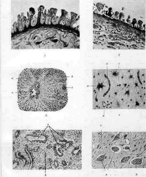
from undifferentiated elements of the sympathetic system, the so-called sympathogonia, which instead of normal maturation into sympathetic ganglion cells or chromaffin cells of the adrenal medulla give a lush growth. Typical sympathogoniomas are observed almost exclusively in early childhood and even in newborns; usually the adrenal glands are affected, sometimes both; the tumor nodes are white, juicy; often give abundant metastases to the liver, lymphatic glands. Microscopically sympathogoniomas [see separate table (st. st. 587-588), fig. 3] consist of a large amount of small lymphocyte-like cells, with nuclei very rich in chromatin; a tendency to form rosette-like figures and to early development of nerve-fibrous substances is also noted. In addition to the adrenal glands, these tumors are observed along the course of the sympathetic trunk. A more mature variety is represented by the so-called chromaffin tumors of sympathetic and parasympathetic tissue, described as paragangliomas, chromaffin suprarenal cords or pheochromocytomas; they are observed in elderly subjects, most often also in the adrenal glands (often on both sides), in the carotid body, in Zuckerkandl's organ; usually proceed without symptoms, do not give metastases; upon microscopy among the connective tissue stroma large cells containing chromaffin substance, pheochromocytes are found; in tumors of this kind the formation of adrenaline has been proven. (See also Adrenal glands, their development.) To immature G. also adjoin tumor-like processes in the central nervous system, known under the name of tuberous sclerosis, as well as some neurinomas (see.) and systemic diseases known as neurofibromatosis. Lit.: Vulfius G. G., On the question of true neuremmas, "Zhurn. nevropat. i psikhiatr.", t. I- IV, 1912; Wahl H., Neuroblastomata, Journal of med. research, t. XXX, 1914.
Related articles
Mentioned in
Cite this page
“Ganglioneuroma.” Soviet Medical Encyclopedia. English translation of Bolshaya Meditsinskaya Entsiklopediya, 1st ed. (Moscow, 1928–1936), ed. N. A. Semashko. https://sovietmedicalencyclopedia.pages.dev/article/ganglioneuroma/