Neurofibroma
Historical document, translated for reference. It reflects medical knowledge of the 1920s–30s and is not medical advice.
Summary
Neurofibroma is a tumor of peripheral nerves, typically appearing as a dense, whitish, rounded or spindle-shaped thickening. It can occur as a single tumor or multiple tumors in systemic disease known as Recklinghausen's disease or neurofibromatosis.
Encyclopedia article (1928–1936)
NEUROFIBROMA (neurofibroma), nerve fibroma; most commonly observed as a dense whitish, rounded or spindle-shaped, less frequently diffuse, cylindrical thickening of a peripheral nerve, and microscopic examination reveals that the tumor consists of fibrous connective tissue originating from the perineurium of the little-changed nerve. Less frequently the tumor originates from the endoneurium and pushes apart the nerve fibers. N. can occur as a single tumor node, for example on the optic nerve, on one of the nerves of the extremity, on a spinal nerve root, etc., or N. can affect the peripheral nervous system multiple times; the latter occurs in a systemic disease called "Recklinghausen's disease," or neurofibromatosis (see).- N. of cutaneous nerves sometimes have the appearance of soft, pendulous protrusions and are called "skin mollusks" (fibroma molluscum). Some N., diffusely spreading along the branches of some nerve, cause in addition to thickening and elongation of nerve trunks, which are located, winding like a snake, and form a bundle of twisted thick cords; this type of N. is designated as neurofibroma, s. neuroma plexiforme or German Rankenfibrom, Rankenneurom (from "Ranken" - plant tendrils); this type of N. most often occurs in the area of the upper eyelid, on the temple, neck, back and chest.-Regarding the histogenesis of the connective tissue proliferation in N., there is not yet complete unanimity. While most authors usually indicated that the basis of N. formation is the proliferation of connective tissue of the peri- and endoneurium and that N. belongs to false neuromas (see Neuroma), Verocay in 1910, based on his teaching about neurinomas (see), expressed the opinion that the proliferation in N. consists not of connective tissue, but of a special fibrous substance of neurogenic origin; although this view in general did not acquire many supporters, however many major experts on neuromas (Pick u. Bielschowsky, Herxheimer and Roth) on the basis that in some N. along with connective tissue there is a proliferation of neurinoma type, inclined to the thought that in the basis of every neurofibroma there is a proliferation of Schwann cells, i.e., elements of neurogenic origin, which is subsequently displaced by the accompanying proliferation of connective tissue of the peri- and endoneurium. With such an approach, neurofibroma cannot be unconditionally classified as false neuromas. Close to neurofibroma are those cases of single and multiple tumors of peripheral nerves, in which the proliferation consists not of fibrous, but of mucous, fatty or sarcomatous tissue (neuromyxomas, neurolipomas, neurosarcomas). It is very probable that the origin of neurofibroma is connected with a faulty development of nerves.
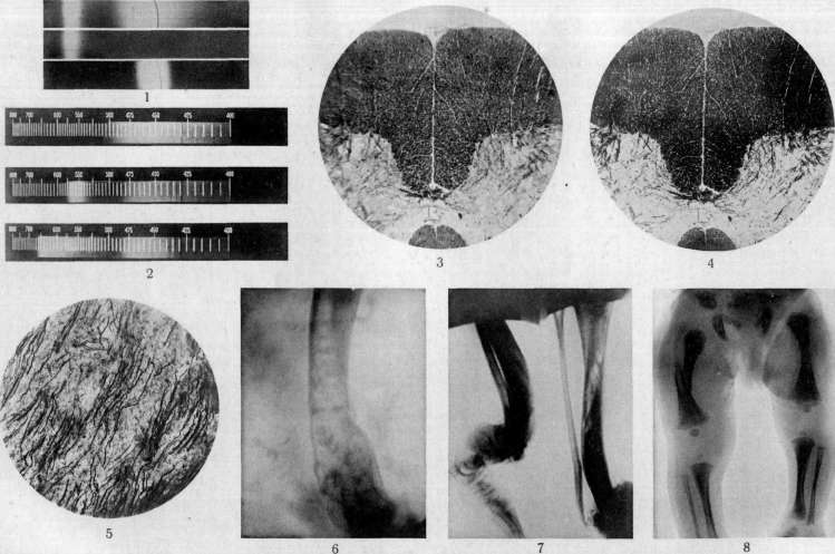
To the article Macrophotography, Neurofibromatosis, Newborn.
The numerous developmental defects noted in this disease, described by some authors (Verocay, Henneberg and Koch, etc.) as changes in the central nervous system in the form of gliomatous foci, isolated gliomas, etc., which are currently considered manifestations of embryonic developmental disorders, the frequent cases of congenital mental and physical retardation in such patients—all this provides sufficient grounds to assume that the basis of this disease is a congenital developmental defect or dystrophy. A significant number of authors currently recognize a congenital anomaly of the ectoderm as the basis of the disease. Verocay classifies this process as a systemic disease based on a congenital anomaly in the development of specific elements of the nervous system, which are the source of the formation of gangliogliomatous, gliomatous cells and elements of Schwann sheaths (Held's neurogliocytes or Kohn's neurocytes). Pathogenesis. At present, regarding the pathogenesis of tumors of nerve trunks in N., the most widely accepted viewpoint is that their source of formation is either the aforementioned embryonic cells (neurogliocytes) or the cells of the Schwann sheath, and as a result of their proliferation, a special neurogenic (Verocay) tissue is formed (see Neurofibroma). But in addition to this tissue, connective tissue also participates in such tumors, and sometimes to such an extent that it becomes the predominant tissue, while in some cases the tumor consists entirely of connective tissue. This connective tissue in these tumors is formed due to the proliferation of elements of the endo- and perineurium, which were considered before the works of Verocay (1908-10) the main sources of formation of such tumors; according to Herxheimer and Roth, the endothelium of lymphatic vessels also participates in the proliferation of connective tissue. The participation of connective tissue is explained by the fact that under the influence of improper development of the ectodermal part of the nervous system, developmental disorders also occur in the adjacent parts of the mesoderm, which can result in excessive development of mesodermal elements. Tumors of nerves in N. present a rather variegated picture upon histological examination. The main part of such tumors is a tissue that is special in terms of morphology and staining properties, so-called neuromatous tissue. The first author to give a detailed description of such tissue was Verocay, who called these tumors neuromas (see). Such a structure is characteristic of part of the tumors in N.; in another part of cases, they have a connective tissue structure and are of a fibromatous nature (neurofibromas). In a significant number of cases, both neuromatous and connective tissue are present in such tumors in different ratios, and then they are referred to as fibroneuromas or neuromatofibromas. In both neuromatous and collagenous connective tissue, processes of hyaline transformation are noted. In some cases, nerve fibers are found in neuromas and neurofibromas [see separate table (pp. 455-456), figure 5], sometimes in a state of degeneration (Brims, Marchand, Garre, etc.); in a number of cases where nerve fibers were not found in the tumors, authors explained this by degeneration and death of nerve fibers. There are a few reports of the formation of new nerve fibers in such tumors (Retren, Pick u. Bielschowsky, etc.). Symptoms in N. are divided into constant and possible. The constant ones include 1) tumors of nerve trunks, 2) tumors of the skin, and 3) pigmented spots of the skin; among the possible symptoms are developmental defects, mental disorders, changes in bones, etc. Tumors of nerves in Recklinghausen's disease can occur in any organ or tissue of the body. Based on the analysis (especially autopsy) of cases of Recklinghausen's disease, it can be noted that in the vast majority of cases there is a predominance of tumors of peripheral nerve trunks, and less frequently of visceral nerves. From these cases, some authors distinguish a group of so-called central N., when tumors are localized in the area of the central nervous system (roots of cranial, spinal nerves). In such cases, there may be no changes in the peripheral nervous system; sometimes only a few skin tumors are noted. Cases of isolated central N. are a great rarity. The localization of tumors of nerve trunks (branches) by organs and systems is of considerable interest. Based on the available literature, it can be said that there is no organ where neurofibromas have not been described. Naturally, the most frequent localization of tumors is the nerve trunks and their branches along their course. There are descriptions of a significant number of cases of tumors of the gastrointestinal tract. These tumors are mainly localized in the subserous or intermuscular layer; their starting point is taken as Meissner's or Auerbach's plexus. In tumors of gastrointestinal localization, tumors of the mesentery are often simultaneously present. Cases of neurofibromas of the tongue, tonsils, pharynx, liver, pancreas, gallbladder, heart, urinary bladder, etc., have been described. In some cases of general N., tumors of the cerebellopontine angle are noted, which by a large number of authors are attributed to tumors of the auditory nerve (Akustikustumoren); upon histological examination, they often show the structure of neuromas, sometimes fibromas or tumors mixed from both types of tissues. Tumors of nerves appear as limited nodes of rounded, spindle-shaped, less frequently irregular outlines or as clearly defined thickenings, often over a considerable length of the nerve trunk [see separate table (pp. 135-136), figure 4]. Limited tumors of nerves either involve the entire thickness of the nerve, so that nerve fibers are lost in the tumor tissue, or are as if located on it, with nerve fibers partly passing in the capsule of the tumor and partly isolated from it. Cases of diffuse thickening of nerves are noted, resembling in their external appearance the figures of earthworms, and these thickenings sometimes involve the main trunks and all branching nerves; in such cases, a network, a tangle of thickened, winding nerves is formed, which Verneuil called plexiform neuroma (neuroma plexiforme) [see separate table (pp. 135-136), figure 3]. Their most frequent localization: the temple area, occiput, eyelids, area of the ears. Here they can have the appearance of skin folds hanging from these areas and representing significant cosmetic inconvenience for patients. As one type of tumor-like growth in N., it is necessary to note a special type of elephantiasis, so-called elephantiasis neuromatodes Bruns', which, when localized in the skin and subcutaneous fat, has the appearance of a tumor that often passes without sharp boundaries into the surrounding parts and externally resembles an excess of skin hanging down. The skin over such tumors is usually wrinkled, often has a brown coloration, especially in those cases where a naevus pigmentosus preceded the development of the tumor at this site. In such cases, it is often possible to feel in the thickness of the tumor formations in the form of cords (cords), which are nerves thickened due to the proliferation of connective tissue. The basis of cutaneous neuromatous elephantiasis is the tumor-like thickening of nerve trunks of the plexiform neuroma type, as well as hypertrophy and hyperplasia of elements of the dermis. The most frequent localization of such tumors is the lateral parts of the head, occiput, back, the area of the thighs and shoulders. These tumors usually appear in early childhood or their rudiments are already present at birth, which gave Virchow the opportunity to designate them as elephantiasis congenita. In addition to cases of cutaneous elephantiasis in N., cases of elephantiasis of internal organs, e.g., intestines, vermiform appendix, etc., have been noted. Of the spinal nerves, the sciatic and intercostal nerves are most frequently affected by tumors; of the cranial nerves, the vagus and trigeminal are most frequently affected, and generally cranial nerves are more frequently affected extradurally. It is also necessary to note the more frequent involvement of sensory nerves compared to motor nerves. As an exception, tumors of the nn. optici and olfactorii are noted. The sympathetic nervous system is often affected by tumors, and not only nerve branches and trunks, but also sympathetic nodes are affected. Tumors of nerves can be single and multiple, and sometimes their number is so large that it is simpler to list the nerves without tumors than to speak of the affected nerves. As for the clinical manifestations in these nerve tumors, it must be noted that in some cases they give no symptoms and are discovered by patients accidentally or upon medical examination. In other cases, corresponding to the areas of branching of the affected nerves, anesthesia, paresthesia, less frequently hypesthesia or hyperalgesia are noted. Motor disorders in the form of pareses, paralyses, twitchings, etc., are rarely observed.
Such a discrepancy between anatomical and clinical data must obviously be explained by the fact that, on the one hand, the nerve fibers themselves in tumors undergo degeneration and atrophy late, and on the other hand, the tumor is sometimes connected only with a part of the nerve trunk, and the resulting phenomena of partial loss may be compensated by the unaffected parts of the nerve; and indeed, the neoplasm can reach significant size before clinical manifestations appear. On palpation, these tumors are usually slightly painful; in some cases, there are more severe painful sensations, and patients report widespread pains along the entire course of the nerve in the form of 'electric current'. A significant clinical interest is represented by plexiform neurofibromas, since patients often first seek medical help because of them, and because these tumors are a frequent object of surgical intervention. Skin tumors are a constant sign of Recklinghausen's disease. These tumors are located in the thickness of the skin or subcutaneous fatty tissue; in appearance, they can present as nodules, warty formations [see separate table (pp. 135-136), Figure 6] or tumors on a stalk (polyps). The skin over them is either unchanged or pigmented in a brown or blue-red color (blaue Flecken); sometimes excessive hair growth is noted at the top of the tumor. The consistency of skin tumors is predominantly soft, rarely hard. Histologically, these tumors appear as fibromas (most often) or neurinomas or mixed from both types of tissues. In some cases, nerve fibers can be detected in their composition with appropriate staining. According to the opinion of most authors, they develop from the sheaths (endoneurium, perineurium, Schwann's sheath) of the branches of cutaneous nerves. These tumors usually do not cause any subjective sensations, are painless on palpation or only slightly painful. Their sizes vary greatly - from a pinhead to giant ones (in individual cases from 12 to 18 kg). The number of skin tumors can vary and range from single ones (rarely) to a significant number, difficult to count; in such cases, the entire trunk and limbs are literally covered with them [hence the old designation of these skin tumors as multiple fibroma of the skin (fibroma multiplex)!]. In Subbotin's case there were 2,730 tumors, in Razumovsky's case 5,868, in Belkin's case 6,500-7,000, etc. Most often, the tumors are located on the trunk, neck, head. Limbs are affected less, feet and hands - as an exception. A constant phenomenon in N. is the presence of pigmented spots on the skin and less often on mucous membranes. These spots are usually brown in color of varying intensity (from yellow to dark brown), from pinpoint size to spots, often of irregular shape, reaching significant sizes. According to Soldan and others, the pigmented spots represent the initial manifestations of fibromatosis of the branches of cutaneous nerves. The favorite localization of pigmented spots is the trunk, neck, and face. According to Thomson's statistics, these spots occur in 25% of all cases of neurofibromatosis. In addition to these brown spots, blue spots or rarely naevi anaemici (pale spots) are sometimes noted. Among the possible symptoms are the frequently noted manifestations of physical and mental underdevelopment in patients. Thus, these patients usually have small stature, weak muscular development, late and insufficient hair growth, sometimes weakness of the ligamentous apparatus. In addition, a number of developmental defects are noted in the literature: underdevelopment of secondary sexual characteristics, cryptorchidism, facial asymmetry, prognathism, congenital strabismus, congenital defects of certain parts of the musculature, parts of the skeletal system > high position of the hard palate, epispadias, sometimes hypertrichosis of certain areas of the body, spina bifida, polydactyly, syndactyly, etc. The face and general appearance of patients often do not correspond to their age; on the one hand, their general infantilism is noted, on the other hand, cases are described where patients seemed significantly older than their years (had an aged appearance). Even old authors noted in patients a number of psychological phenomena, such as general dullness, lethargy, 'cretinism', apathy, sometimes decrease or absence of sexual desire. School-age children learn poorly, lag behind their peers, are lethargic, forgetful, sometimes speech disorders, stuttering are noted in them. Changes in the psychological sphere are so significant that they can serve for differential diagnosis with a number of other diseases. Particular interest are the changes in bones in N., described by a number of authors. It is necessary to note the atrophic processes in bones leading to thinning and local cessation of bone growth, to the formation of defects (e.g., defects of frontal, occipital, temporal bones, etc.); on the other hand, thickening and lengthening of bones are noted - e.g., of limbs or upper jaw. Bone deformations are not uncommon, especially of long tubular bones, which consist in thickening of the cortical layer, periosteal deposits, irregularities of bone surfaces, in the formation of exostoses; bone curvatures are noted, e.g., saber-shaped curvature of the tibia (not syphilitic!). Curvature of the spine is particularly often noted, of the type of scolioses or kyphoscolioses. Adrian observed these changes in 7% of all cases (out of 447 collective cases in 32); in reality, if one includes here also the mildly expressed degrees of curvature, the percentage of these cases will be significantly greater. Thus, Engmann considers these curvatures even a constant sign of N. Among other bone changes, it is necessary to note osteoporosis, flexibility of bones, which in their picture resemble osteomalacia. All the described atrophic processes in bones in N. gave French authors reason to speak of bone cachexia. A significant frequency of fractures of such changed bones is noted, with subsequent poor healing and tendency to form false joints [see separate table (pp. 455-450?), Figures 6 and 7]. It is interesting to note that changes in bones most often occur in those parts of the body where there are simultaneously changes from the skin and nerve trunks. Among other bone changes, it is necessary to note the asymmetry of individual parts of the skeleton, especially of the skull, sometimes of the chest. A number of authors describe changes in teeth in Recklinghausen's disease in the form of complete absence of some of them, their improper growth (obliquely, backward), etc. It is also necessary to note periosteal tumors, which on histological examination prove to be fibromas or neurinomas and which can cause erosion or atrophic processes in bones. In addition to the above-mentioned phenomena in patients with N., a number of tumors not directly related to this disease are sometimes noted, such as fibromas, fibroendotheliomas of the meninges, tumors of the brain substance itself (gliomas), etc. One cannot speak of any particular clinical picture in these cases, since the clinical phenomena depend on the relationship of the tumor to the nerves and adjacent organs (not to speak of the psychological phenomena); thus, in each individual case, these phenomena can differ from each other. In the presence of a tumor in the area of the central nervous system, we will have the same clinical picture as in tumors of the brain and spinal cord. In contrast to those forms of Recklinghausen's disease where all phenomena are present, cases are observed where only one basic symptom (e.g., skin tumors or nerve trunk tumors, pigmentation) is found together with psychological disorders or changes from the skeleton. Such cases have been given the name formes frustes or incompletes by French authors. The diagnosis of formes frustes is sometimes very difficult; these forms are quite often observed in families where there are already cases of expressed N. In N., a number of authors describe changes also from the eye. The most frequent changes are from the eyelids, on which soft tumors (folds) are formed, hanging down and presenting a picture of plexiform neurofibroma; the starting point of these tumors is the nerve elements of the eyelid. Then it was anatomically proven that the ciliary nerves of the eye are also affected by tumors, as well as the nerve elements of the eyelid. In addition, in patients with N., congenital or appearing in early age changes of the eyeball itself are described, such as its increase in size (elephantiasis), exophthalmos, leukomas, hydrophthalmos, intercalary staphylomas, glaucoma, etc. Decrease in visual acuity, narrowing of the visual field, dyschromatopsia, nystagmus are noted. In some cases of eye changes, chronic internal hydrocephalus obviously underlies them; there are a number of cases where visual impairment was congenital and without any visible cause. In a number of cases of Recklinghausen's disease, clinicians note atrophy of the optic nerves with corresponding visual disturbances.
Neurofibroma often appears in childhood (up to 10 years), and the onset of the disease is difficult to note, as patients usually do not pay attention to it; in most cases, patients report that in childhood they had the first spots or skin tumors, but since they did not give any subjective sensations, patients did not pay "attention" to them. At the same time, it is necessary to note a number of cases from the literature in which the first manifestations of the disease were noted in mature or old age. In collective statistics provided by a number of authors, there is a predominance of the male sex among these patients. Thus, Adrian, in 447 cases of Recklinghausen's disease, reports 289 (65%) men, 158 (35%) women; Tikhov, in 300 cases, had 196 (65%) men and 104 (35%) women; Wolf, in 124 cases, had 85 (68%) men and 39 (32%) women. The question of the familial, or hereditary, origin of Recklinghausen's disease is of considerable interest. Even in older authors, there are indications of multiple cases of the disease in the same family. Thus, in Virchow's case, the grandfather, father, brother, and sister were sick; in the case of Hecker and Cherny, the daughter, mother, grandfather on the mother's side, and some relatives on the mother's side had skin tumors; in Bazin's case, two sisters, father, and grandmother on the father's side were sick. Cases have been described where it was possible to note diseases in five generations. At the same time, there are cases of a visible break in inheritance, for example, a case was described where the child and his grandfather on the mother's side were sick, while the mother herself was healthy. In statistics regarding the familial occurrence of Recklinghausen's disease, the following data exist: Adrian notes clear heredity in 20% of literary cases; according to Tikhov, out of 69 collective cases, hereditary data existed in 36, i.e., 51%; on the other hand, some authors, such as Siemens, based on questioning and examination of relatives of a number of neurofibromatosis patients, conclude that familial inheritance of Recklinghausen's disease is very insignificant. Whatever the data of individual authors, it is clear that in a significant number of cases there is a familial origin of neurofibromatosis. Differential diagnosis. Typical cases of Recklinghausen's disease with tumors of nerve trunks, tumors, and pigment spots of the skin are hardly likely to be confused with any other disease. Differential diagnosis is needed only in cases where not all typical manifestations of the disease are present. First of all, differential diagnosis must be conducted with Dercum's disease and in particular with its nodular form (see Dercum's disease). In Dercum's disease, the tumors of the subcutaneous tissue have a lobular character, are usually of softer consistency than in Recklinghausen's disease (but not always), there are no deeply located tumors, and there are no pigment spots (a fairly constant sign of N.). It is also necessary to note the possibility of confusion with general cysticercosis, but the absence of skin spots in cysticercosis, painless and small size of tumors, absence of signs of physical and mental underdevelopment, etc., make it possible to differentiate these diseases. In particularly doubtful cases, the question is resolved by biopsy. As for the outcome of the process, it is necessary to note that a significant part of such patients live to a very old age; nevertheless, there are literary data that such patients are more susceptible to various intercurrent diseases, more often suffer from tuberculosis, tolerate surgical trauma worse, etc. In a certain percentage of cases, authors note malignant (sarcomatous) degeneration of tumors. Thus, Garre noted malignant degeneration in 12% of cases, Adrian in 11.8%, Thomson in 22%, etc. These figures must be treated with great caution, as this statistical material belongs to the period of time when neurinomas were not identified as a separate unit and when obviously a part of neurinomas with an abundant number of nuclei were classified as neurofibromas in a state of malignant degeneration. Treatment. To date, no real methods of treating this disease besides the surgical removal of individual tumors are known: radical surgical treatment is difficult due to the multiplicity of tumors and their often deep location. Injections of fibrinolysin into the tumor tissue have been proposed for treatment, with an effect observed in the form of disappearance of tumors and reduction of pain; but, on the other hand, cases of failure of such treatment, cases of recurrences after it, are described, and of course it does not prevent the new appearance of tumors. Supporters of the endocrine theory of the origin of the disease note improvement in clinical symptoms after treatment with preparations of the internal secretory glands: but all these data are of a random nature and have not received any systematic confirmation. In cases where the tumor reaches significant sizes, causes disruption of function either at its location (for example, the central nervous system) or due to pressure on neighboring organs or tissues, when it causes cosmetic inconvenience, or when there is suspicion of malignant degeneration of the tumor, it is necessary to resort to surgical intervention. If the tumor is well encapsulated and the nerve (its fibers) does not enter its entire mass into the tumor tissue, it is necessary to attempt its enucleation; in case of an intimate connection of the tumor with the nerve, it is necessary to resort to resection of the tumor and nerve with an attempt at subsequent nerve suture.
V. Shlapobersky. NEUROEPITHELIOMA (neuroepithelioma), a tumor from neuroepithelium. The term N. is applied primarily to rare tumors developing from neuroepithelium (ependymal epithelium), lining the brain ventricles and spinal canal, as well as covering the vascular plexuses of the brain. In addition, N. is called certain types of gliomas (see), containing formations from neuroepithelium. N. from ependymal epithelium of the ventricles and vascular plexuses can have the character of papillomas and papillary glandular tumors; sometimes, having the structure of adenocarcinomas and solid cancers, they show a malignant course. The most common type of N. is neuroepithelioma gliomatosum, a tumor with a basis of glial reticular tissue, in which there are epithelial formations either in the form of glandular cells or more often in the form of cylindrical cells arranged in a palisade around blood vessels; at the same time, from these epithelial elements originate thin processes, the interweaving of which forms the glial network. Neuroepithelioma gliomatosum most often develops near the lateral ventricles of the brain, the Sylvian aqueduct, IV ventricle, more rarely in the spinal cord. Cases of combination of N. with syringomyelia have been described. Most researchers believe that neuroepithelioma gliomatosum is a consequence of faulty development, hamartia (see Hamartoma, hamartia, hamartoblastoma) of the ependyma.
Related articles
Mentioned in
Cite this page
“Neurofibroma.” Soviet Medical Encyclopedia. English translation of Bolshaya Meditsinskaya Entsiklopediya, 1st ed. (Moscow, 1928–1936), ed. N. A. Semashko. https://sovietmedicalencyclopedia.pages.dev/article/neurofibroma/