Nerves
Historical document, translated for reference. It reflects medical knowledge of the 1920s–30s and is not medical advice.
Summary
The article describes the anatomy, classification, and histological structure of peripheral nerves, including their division into cerebrospinal and autonomic nerves, their formation from spinal roots, and their distribution throughout the body.
Encyclopedia article (1928–1936)
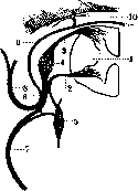
Nerves, the peripheral part of the nervous system, conducting impulses from the central nervous system to the periphery and back; they are located outside the craniovertebral canal and in the form of cords spread throughout all parts of the head, trunk, and extremities. They are divided into cerebrospinal and autonomic; the first have a white color due to the presence of myelin, while autonomic nerves lack myelin and have a gray color. The shape of nerves is in most cases cylindrical; however, large nerves are mostly flattened. The thickness of nerves varies, with a diameter ranging from 0.8 mm to 8 mm, depending not only on the number of nerve fibers making up the nerve but also on the thickness of the connective tissue sheaths. The density of nerves averages 1.037 (1.014-1.052); unmyelinated fibers are denser (1.040) than myelinated ones (1.028). The resistance of nerves to rupture during stretching is very great (in terms of resistance among tissues, they occupy third place). The coefficient of resistance (i.e., the force required to rupture the nerve) averages 1.351 kg for a nerve with a diameter of 1 mm. The coefficient of resistance for the sciatic nerve is 80 kg, for the median nerve 30 kg, and for the facial nerve 5-6 kg. Nerves are divided into cranial (12 pairs) and spinal (31 pairs). The first originate or end in the brainstem (see respective nerves), the second in the spinal cord. The latter are formed by two functionally different roots—the anterior root, containing centrifugal or motor fibers, and the posterior root, in which centripetal or sensory fibers pass (see Spinal cord); on it there is an enlargement—the spinal ganglion (ganglion spinale); lateral to the ganglion, both roots join together to form one common trunk—a mixed nerve, n. spinalis, French p. mixte (Figure 1). Sympathetic fibers from the trunk enter the mixed nerve; this nerve is very short and divides into two mixed branches—ramus anterior et ramus posterior. Rami posteriores are thinner branches and in turn divide into medial and lateral branches, which innervate the skin and muscles of the posterior surface of the trunk from the occiput to the coccyx. Rami anteriores of all cervical nerves, the two upper thoracic, the XII thoracic, all lumbar, sacral, and coccygeal nerves form plexuses, i.e., their trunks connect with each other in the form of triangular loops, and from these connections peripheral nerves are formed, which innervate the muscles and skin of the neck, chest, abdomen, upper and lower extremities, and perineum. Rr. anteriores of thoracic nerves or intercostal nerves do not participate in the formation of plexuses but end in the skin and in the muscles of the ribs and anterior surface of the trunk.
Figure 1. Formation of a mixed peripheral nerve: 1 - spinal cord; 2 - its anterior root; 3 - posterior root; 4 - spinal ganglion; 5 - sympathetic ganglion; 6 - mixed peripheral nerve; 7 - its anterior branch; 8 - its posterior branch; 9 - ramus meningeus; 10 - dura mater; 11 - vertebra. Nerves emerging from plexuses are located some deeply, others superficially; they follow a straight, shortest path between centers and corresponding organs, resembling the course of fibers in an embryo, where nerves from the medullary tube go to the nearest myotome. Superficial nerves go together with veins in the subcutaneous fatty tissue; deep nerves—between muscle layers together with blood vessels, forming a neurovascular bundle surrounded by loose connective tissue, from which partitions extend inward, separating nerves from blood vessels. In the neurovascular bundle, the nerve occupies a superficial position; on the extremities, it is located more externally relative to the axis of the extremity. On their course, nerves communicate through anastomoses; these anastomoses can be simple, single or complex, multiple; they can be reduced to three types: 1) simple anastomosis, when some fibers of one nerve pass into another; 2) double, when two nerves exchange part of their fibers; 3) complete anastomosis, or decussation. In addition, anastomoses can be recurrent, when fibers from one nerve, entering another, go in an ascending direction; finally, there are also false anastomoses, when fibers leave the nerve for a short distance and then return to it completely. Gradually toward the periphery, the nerve receives blood vessels from the epineurium, from there to the perineurium, and then to each fiber (vasa nervorum) (see Fas). The veins of the nerve have the same arrangement as the arteries, but for each artery there is only one vein. Lymphatic vessels in the form of separate channels are found only in the perifascicular tissue, never penetrate into the nerve and do not communicate with the subarachnoid space. Nervi nervorum in most cases belong to vasoconstrictor nerves (unmyelinated), they are distributed in the neurilemma; these nerves accompany the arteries of the nerve, forming plexuses around them; in addition to these fibers, there is a small number of myelinated nerve fibers which probably belong to sensory fibers and end in the endoneurium. Most nerves are mixed, their composition includes motor, sensory, and autonomic fibers; there are nerves exclusively sensory, but purely motor nerves are very few. The trophic center for motor fibers is the cells of the anterior horns of the spinal cord and the motor nuclei of the medulla oblongata; the trophic center for sensory fibers is the cells of the intervertebral ganglia. Each nerve always innervates the same skin territory and the same muscle groups; if the nerve is mixed, the skin surface it innervates covers the muscle group innervated by the same nerve. Due to numerous anastomoses between nerves, it is difficult to accurately delimit in the skin the area innervated by one or another nerve; in addition, it must be noted that many areas of the skin are innervated not only by one main trunk of some nerve but also by branches of neighboring nerves. This collateral innervation is revealed when innervation from the main trunk is lost—the insensitive area of skin is very small compared to what one would expect based on the distribution of the given nerve in the skin (see Sensitivity). Some muscles also receive innervation from many nerves, but within the muscle itself, some part is innervated by only one nerve (General physiology of the nerve fiber—see Nervous system, Nerve fibers, Nerve cells). Autonomic fibers enter into the composition of even the smallest cerebrospinal nerve and together with its branches penetrate all tissues and formations. To internal organs, autonomic nerves go isolated (see Autonomic nervous system).
Pathology. Among pathological processes affecting nerves, one can note: injuries, nerve damage, inflammations—neuritis (see) and tumors. Inflammatory processes affect either one nerve—neuritis (see) or simultaneously many—polyneuritis (see). New growths of peripheral nerves can be divided into 3 categories: new growths that have spread to the nerve from surrounding tissues; they can be of various origins—sarcomatous, cancerous; metastases from other organs into the nerve are very rare. New growths originating from the nerve itself are divided into neurinomas (neurilemmomas) and neurofibromas. New growths originating from the nerve sheaths are rare. Nerves are exhausted due to the fact that they give collaterals for the innervation of surrounding formations—skin, muscles, ligaments, tendons, bones, etc. At their termination, nerves divide into several branches, which after a very rich anastomosis with surrounding nerves end in the corresponding
Figure 2. Histological structure of a peripheral nerve: 1 - endoneurium; 2 - perineurium; 3 - epineurium; 4 - fatty tissue; 5 - blood vessels.
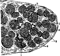
in the tissues with special endings (see Nerve endings). In the formation of nerve trunks, myelinated and non-myelinated fibers participate, connected into bundles. Usually, a nerve trunk consists of numerous individual nerve bundles and is covered by loose connective tissue-the epineurium (fig. 2), from which septa extend between individual nerve bundles (perineurium); from the perineurium, connective tissue partitions (endoneurium) extend into the thickness of the bundle, which divide the bundle into smaller bundles; from it, a thin collagen fiber extends to each nerve fiber, which runs along the nerve fiber and forms the Key-Retzius sheath. Very small N. have only one bundle of nerve fibers. When the trunk breaks down into bundles, the epineurium disappears, the perineurium thins out, and around the thinnest bundles a thin membrane remains, covered with flat endothelial cells, so-called Henle's sheath. Bundles of nerve fibers in N. do not remain isolated from each other, but exchange anastomoses; thus, plexuses form within the N. itself, thanks to which there is no isolated conduction along one bundle, but the entire N. participates in it. On a cross-section, it can be seen that the caliber of the fibers entering the nerve is not uniform, there are large fibers, and there are very small ones; along the length of the N., the distribution of these fibers is not uniform, it changes depending on the level of the section (histological structure of N.-see Nerve fibers). N. is very rich in blood vessels, which it receives from surrounding arteries; large vessels first lie in the epineurium, then divide several times and penetrate into the peri- * Tables "Human Nerves" (alphabetical list with indication of origin, topography, function, etc.)-see Volume XXI. are rare. To the second category belong tumors specific only to nervous tissue: neurinoma or neuroma, neuroglioma, neuromyxoma (see); finally, multiple tumors of skin N.-neurofibromas (see) occur, the source of which is the sheath of the nerve fiber and which, by a combination of clinical phenomena, form a special disease-multiple neurofibromatosis, or disease7 of Recklinghausen (see Neurofibromatosis). Sometimes neurofibromas form large local tumors, externally resembling plexiform neurofibroma or reticular neuroma. One of the first signs of N. involvement in a tumor is very severe pain along the course of the N., later anesthesia, pareses or paralyses with degenerative atrophy of the muscles in the area of the affected N. develop, and the same picture is obtained as in the case of N. severance. Treatment is only surgical. N. can also participate in a special general disease-lipomatosis dolorosa, or Dercum's disease (see). Anatomically, neuritis of the terminal branches is observed in this condition. Traumatic injuries to N. The mechanism of injury to N. can be very different: 1) one of the most common forms of N. injury is their compression (crushing of the trunk through the skin by a foreign body-during sleep, by a bandage during surgery, etc., in fractures by a displaced bone or its callus, by fibrous adhesions or a tumor growing from adjacent tissues toward the N., etc.); 2) contusion of N.; 3) excessive stretching or traction of N., observed with strong abduction of the limb both during surgery and under accidental circumstances; 4) dislocations of N., which are a very rare phenomenon (more often observed on two N.-ulnaris and peronaeus); 5) violation of the integrity of N.-incision, rupture. Depending on the injury, the changes in the nerve trunk are different. With compression of N., the continuity of the fibers is not disrupted, but more or less deep changes occur in them depending on the force and duration of compression-from slight edema to complete degeneration of the nerve fibers in the area of application of compression. Contusion of N. is accompanied by edema, hemorrhagic infiltrates with secondary fibrous proliferations inside the N. itself, which dissociate and compress the nerve fibers. With complete violation of the integrity of N., both its ends either remain close together with simple severance or diverge far from each other, making it difficult to bring them together (in severe injuries with loss of soft parts); microscopically, degenerative changes occur in both segments of N.-secondary degeneration in the peripheral segment and retrograde in the central one (see Wallerian degeneration, Nerve fibers, Neuritis). Regeneration phenomena are possible in N., which occur the faster the closer both segments are located (see Nerve fibers-regeneration). Disorders of the nervous system developing with injuries to peripheral N. encompass both motor and sensory and autonomic spheres, but not in all cases is there the same combination of symptoms; everything depends on which fibers are more damaged, as well as on the mechanism of injury. With complete violation of the integrity of a mixed nerve, complete and immediate paralysis of all muscles innervated by the damaged N. is observed, reaction of degeneration in the paralyzed muscles, their gradual atrophy, decreased tone, in general so-called flaccid paralysis (see Paralysis); complete loss of all types of sensitivity in the area innervated by this N., more extensive in the first days and gradually narrowing in the subsequent ones (due to collateral innervation and anastomoses); absence of pain on pressure on the N. and on the corresponding muscles/absence of paresthesia on pressure on the N. below the site of injury, fading of tendon reflexes. Autonomic phenomena are reduced to edema, cyanosis, dryness of the skin, sometimes necrosis of tissues is observed. With damage to only the sensory N., there are disorders of sensitivity and autonomic disorders; with trauma to the motor nerve, there are movement disorders. Compression of N. is expressed-by approximately the same symptoms, but only their development is more gradual, the symptoms are not so sharply expressed-there is relative preservation of muscle tone, partial degeneration reaction. Both with compression and with contusion of N., it is possible to damage only part of the fibers that make up the nerve trunk, and the damage itself is due not so much to the disruption of the continuity of the nerve fibers as to their traumatization, so a discrepancy can be observed in the degree of development of symptoms in the motor sphere and in the sensory sphere, just as paralysis and anesthesia can be incomplete. A disease of the nerves, accompanied by severe pain, is known under the name of neuralgia (see), and a special form of injury to peripheral N. with involvement of the autonomic sphere has been isolated under the name of causalgia (see). The further course of the disease in N. injuries depends on the condition of the nerve fibers. In those cases where the injury to N. did not cause disruption of the integrity of the nerve fibers or where regeneration of the fibers is not excluded, the symptoms gradually smooth out, paresthetic phenomena appear in the form of tingling, crawling of ants in the area of the diseased N.; objective sensitivity improves, first deep, then superficial, autonomic disorders disappear, muscle tone and movements appear in the paralyzed muscles. On the contrary, the picture of the disease remains persistent in those cases where, due to the nature of the injury, regeneration phenomena are excluded.--Treatment depends on the nature of the injury: in those cases where the nerve injury itself does not constitute an obstacle to the revival of the traumatized trunk, therapy is applied that promotes a more successful course of the regeneration process and maintains muscle tone: electrization, massage, warm baths are prescribed. In the same cases where there is a cause constantly injuring the N. (tumor of adjacent tissues, bony callus, bone fragments) and hindering regeneration of the fibers, or when the ends of the N. have diverged too far, surgical intervention is resorted to (see Nerve suture). Reduction of N. dislocations is performed either by bloodless means or surgically-by creating a canal from connective tissue OR MUSCLES.
E. Kononova. Injuries to N. According to the statistics of Foerster, which coincide with the data of other authors, gunshot wounds to peripheral nerves occurred significantly more often than traumatic lesions of other origin, which constituted only 1.4% of all injuries to peripheral nerves and were caused by tearing, piercing instruments, bruising by stone or earth, impact by a hoof, fracture or dislocation, pressure from a backpack, crutches, bone callus, etc. The total number of traumatic injuries to peripheral nerves during previous wars (Russo-Japanese, Balkan) was determined from 0.75% to 3.2% of all wounds. According to statistics from the first year of the imperialist war (Lewandowsky; 1914), their number is estimated at 1.5% of all wounds. Marburg (Marburg; 1916) already determines them at 4%. The increase in these injuries toward the end of the war is explained by the authors as an increase in the penetrating power of projectiles, which does not allow the nerve to deflect sideways and avoid injury. If at the beginning of the war the ratio of rifle wounds to artillery wounds was 7:1, then in 1918 it changed to 1:2.5. In addition to direct injury to the nerve, it can also be affected indirectly, being involved in a phlegmonous or purulent process. It can be injured by a fragment of developed bone or by bone callus. The nerve can rupture above and below the direct injury, it can be affected by hemorrhage, by ischemia when tying off an injured artery, by bone fractures, tissue inflammations, scars, etc. Some authors, even to the present day, attach particular importance to the moment of nerve concussion (commotio) and explain by it the changes in function and even the degeneration of the proximal segment of the nerve in cases of violation of its integrity. However, even with the visible integrity of the nerve, where so-called 'commotio' is assumed, adhesions and scars (Foerster) are often found inside it, depending on hemorrhages or other circulatory disorders. In some cases, disturbances of blood or lymph circulation in the nerve lead to its death. According to the materials of the vast majority of authors, injuries to the radial nerve are in first place. According to Foerster's statistics, out of 3,963 cases of peripheral nerve injuries, there were 3,907 gunshot wounds. Of these, the radial nerve was injured in 936 cases, the median in 800, the ulnar in 742, the sciatic in 523, the brachial plexus in 287, the peroneal nerve in 183, and the tibial nerve in 112 cases. The injury to the trigeminal (215 times) and facial (120 times) nerves is remarkably frequent. Injuries to other nerves are counted in tens or units. Often several nerves were wounded at the same time, among them relatively often the nerves of the base of the skull, for example, the last 3-4 nerves of one side (foramen lacerum syndrome). Surgical intervention in injuries to peripheral nerves consists in the removal of scars both around the nerves and inside the nerve fiber itself, in suturing the central segment of the nerve into its muscle, in connecting both segments of the nerve after freshening their edges and removing scar tissue. After such renervations, function is restored often only several years later and even then only on the condition of persistent treatment with electrization and mechanotherapy and the sooner the shorter the path from the interrupted nerve to the muscle. Thus, according to Foerster, after suturing the radial nerve, the long head of the triceps muscle is restored after 3 months, the middle and lateral heads after 4 months, the long supinator after 7, the long radial extensor of the wrist after 9, the short supinator and extensor after 13, the common extensor of the fingers after 14, the long abductor muscle of the thumb after 15, the ulnar extensor of the wrist after 16, the extensors of the fingers after 19 months, etc. In injuries to the nerve plexus, the functions of proximal nerves are restored relatively quickly and distal muscles extremely slowly - sometimes years later. In rare cases, however, significantly less time passes until the function of the injured nerve is restored. When the segments of the nerve are too far apart from each other, they have to be connected by the formation of a bridge (see Nerve suture).-What has been said about the restoration of peripheral nerves also applies to injuries to the roots, in particular to injuries to the cauda equina. With the complete destruction of the nerves supplying a muscle group and absolute hopelessness of restoring function by neurectomy, surgical measures remain either in the form of muscle or tendon transplantation, or in the form of fixing the joint in a more favorable position for function (tenodesis, arthrodesis, osteodesis). Sometimes in injuries to peripheral nerves, not paralysis of the corresponding muscles occurred, but contractures (see). The latter are of a tonic nature. After excision of the scar (neurolysis), in which a branch of a sensory nerve is compressed, the contracture disappears if prolonged fixation has not caused secondary shrinkage of tendons, muscles and joints. In some cases, especially in predisposed individuals, psychic fixation of the limb in a given position can also occur, which can continue even after surgical removal of the scar that causes irritation of the centripetal nerve. Contractures were particularly frequent in war injuries to the sciatic nerve.
m. kroll.
NERVES OF HUMAN
ciliare ^JJP Bulbus 0СцН Olandula lacrimal. »-da Glandula subma-2^.3-3 xillaris b-c Glandula sublin- ^-^ gualis -^Q^^ Glandula parotis G и_^~e*53Ш Glandula thyreoid. Ganglia trunci sympathiciT" G. cervicale stipe-/ rius G. cervicale medium G. cervicale info-rius G. stellatum G. thoracale II G. thoracale III G. thoracale IV G. thoracale Vr G. thoracale VI G. thoracale VII G. thoracale VIII < G. thoracale IX G. thoracale X G. thoracale XI G. thoracale XII 6. lumbale I G. lumbale II G. lumbale 111 G. lumbale IV G. lumbale V G. sac rale 1
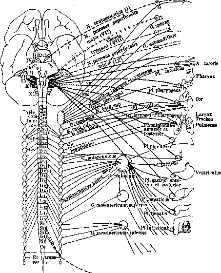
Diaphragma y Aorta abdominal. He par Pancreas. Intestinum \P1. suprarenalislirs^^ suprareualis Glandnla .supn Ren \ \ ^\iv~ ^h\ /Rectum, V..JV- ^nQ \ "W J sigmoide /-> -V PlNiolicus^ 4-/ G. pelvicum' ^PlLvesicalis^' deum Vesica urinaria PI. Ргапкеп^Д J ov Utenis, vagina, arium Рисунок 1. Схема симпатической и парасимпатической нервной системы. вв9 Для sinus transvereus я occipitalis N auricularis n faciahs Ушаад раковина (задняя поверхность) и наружный слуховой проход (задве-нвжвяя стенка) N ..glosso-pbaryngeus Ganglion cervical»; superius N acimisoriii!' Ganglion cervicale вирипч» N. hypoglossus M. levatiir veil paldtirn M. uvula»- Слизистая глотки ниже v«lum palatinum M. constrictor pharyngis superior M. constrictor pharyngis uiedius M. constrictor pharyngis inferior M. palato-glossus M. palato-pharyngeu* M. constrictor pharyngis inlerior M. crico-thyremdeue Слизистая гортани (задняя стенка)* Щитовидная железа Слизистая гортани выше голосовых связок (Слизистая надгортанник;! и корня языка Слизистая ниже голосовых евя: M. arytaenoideus M. crico-arytaenoideus posticus M, erico-aiyiaemiideus lateralis M. thyreo-arytaenoideus M. vocalis M. thyreo-epiglotticus, M. ary-epigioracu». Ganglion juguiarc R. interims n accessorii K lienalis R. raualis R. suprareiiali*
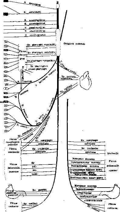
) oesophageus anterior Plexus Vagus dexter Vagus sinister Рисунок 2. Схема блуждающего нерва. Ниточки к pia mater R. anastomoticus cum n. vago R. anastomoticus cum ramo auriculari #ervl vagi A. anastomoticus cum nervo sympathico (c ganglion oervicale superius) R. anastomoticus cum nervo faciali (r. di- gastrlcus) Rr. pharyngei (sup.) nervl vagf^V Plexus \ >^ Pbaryngeus \ ,'Смещанное спле-> Rr- pharyngei J тение на лятер- л- Ramuli tympanici Слизистая барабанной полости и ячеек сосцевидного отростка Ramus tubae в Евстахиевой трубе N. petrosus superficial^ minor Ganglion oticum N carotieo-tympanicus superior (n. petrosus prorundus rumor) N. carotico-tympanlcus inferior Rr. pharyngei <tnf.) nervi va#i Rr. laryngo-pharyngei от ganglion cervica-le superius Слизистая миндалин и небных дужек' Слизистая задней трети языка
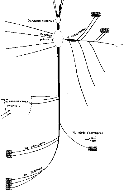
R. anastomoticus n. facialis cum plexu tympanico M. stylo-pharyngeus Слизистая вокруг m stylo-pharyngeus Рисунок 3. Схема п. glosso-pharyngei. «78 «74 Сл, *S«cra Сля Сл 1;аистая °б«-чочка верТ ней и средней раковин, носового прохода,eepXj ней части глотки и слуховой трубы Слизистая твердого неба и десен Мягкое нёбо и яаычок Слизистая нёбных дужек, миндалины и мягкого иёба
Fig. 4. Diagram of the trigeminal nerve. E. t. XX. 67ft Spheno-palatine ganglion Tympanic plexus Stapedius muscle Two-thirds of the tongue. Auricular branch of the vagus nerve Posterior auricular muscle Anterior auricular muscle Transverse auricular muscle. =^ Oblique auricular muscle Antitragic muscle Occipital muscle Posterior belly of the digastric muscle Stylo-hyoid muscle Orbicularis oculi muscle Corrugator supercilii muscle Frontal muscle Anterior auricular muscle Superior auricular muscle 5 Tragicus muscle Major helicis muscle Minor helicis muscle Orbicularis oculi muscle Zygomaticus muscle Superior quadratus labii muscle Zygomaticus muscle Risorius muscle Caninus muscle Buccinator muscle Incisivi muscles Orbicularis oris muscle Triangularis muscle Inferior quadratus labii muscle Triangularis muscle Risorius muscle Mentalis muscle Platysma Superficial petrosal nerve Anastomotic branch with the facial nerve Stapedius nerve
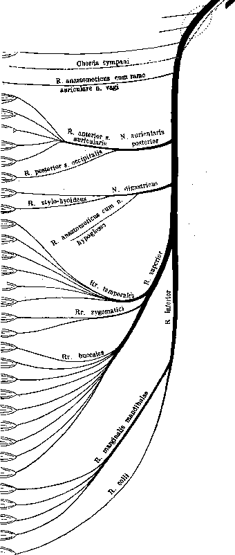
Fig. 5. Diagram of the facial nerve. Genio-glossus muscle Hyo-glossus muscle Stylo-glossus muscles Mm. linguae (longitudinal, vertical, transverse) Genio-hyoidus muscle Omo-hyoidus muscle Sterno-hyoidus muscle Sterno-thyreoj-=5 deus Thyreo-hyoi deus Skin of the occipital region to the temple Occipital nerve Skin of the parotid region Skin of the anterior surface of the auricle Skin behind the ear and posterior surface of the auricle Skin suprahyoid region Skin infrahyoid region Sterno-cleido-: mastoideus skin of the sternum and clavicle Skin of the upper lateral region of the chest to the IV rib Skin of the deltoid muscle and shoulder joint Trapezius muscle Pericardium Pleura Diaphragm Phrenic plexus
32 Scapula Latissimus dorsi Rhomboid major and minor Serratus anterior Subclavius Supraspinatus Infraspinatus Shoulder joint Pectoralis major Pectoralis minor Pectoralis major Deltoid Teres minor Shoulder joint
:.„>---------~p=- Back of the posterior half of the deltoid muscle Subscapularis Teres major Latissimus dorsi Brachialis muscle Brachii muscle Biceps brachii Brachio-radialis
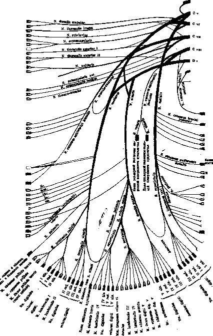
D i Intercostal-brachial nerve Skin of the medial surface of the arm Elbow joint Skin of the anterior lateral surface of the forearm Skin of the radial edge of the forearm Pronator teres Palmaris longus Flexor carpi radialis Flexor carpi ulnaris Flexor digitorum superficialis Flexor pollicis longus Flexor dig. profundus (caput radiale, caput ulnare) Periosteum and bones of the forearm Increase vessels Pronator quadratus Triceps (caput longum) Anconaeus Triceps (caput medium) Triceps (caput laterale) Arm and forearm Extensor carpi radialis brevis Supinator Extensor carpi 'ulnaris Extensor digitorum Extensor pollicis J^"--в ore vis -=Extensor pollicis ^ longus S-Abductor pollicis longus =Extensor indicis -propriae Interosseous membrane Wrist joint from the ' ...':dorsal side Carpal and carpometacarpal joints Fig. 7. Diagram of the brachial plexus. Skin of the outer surface of the pelvis above the gluteus medius muscle Skin above the inguinal ring M.
transversus abdominis M.
obliquusabdominis externus M.
obliquus abdominis externus M.' obliquus abdominis internus M. transversus abdominis Skin above the mons pubis and scrotum : (labia majora) Skin of the medial surface of the thigh in the upper part M. ere master Tunica dartos Skin of the anterior surface of the thigh Skin of the lateral surface of the thigh above the tensor fasciae latae Skin of the medial surface of the thigh Obturator externus M Adductor brevis Adductor longus Gracilis Hip joint Adductor magnus and minimus Pectineus Hip joint Iliacus Psoas major and minor Skin of the anterior surface of the thigh Pectineus Sartorius Rectus femoris Vastus medialis Vastus intermedius Vastus lateralis Articularis genu Skin of the medial side of the knee Skin of the medial surface of the shin to the skin above the tarsus tf.o«*a 1Л
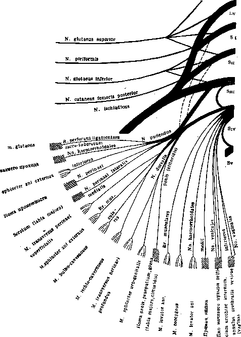
Fig. 9. Diagram of the pudendal plexus.
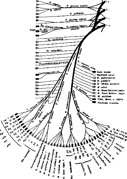
Gluteus minimus
Gluteus medius Gluteus minimus Gluteus medius Tensor fasciae latae Piriformis Gluteus maximus Hip joint Skin above the gluteus maximus Skin of the inner surface and scrotum (large labia) Skin of the thigh to the popliteal fossa Hip joint Obturator internus Quadratus femoris Gemelli muscles Semitendinosus Semimembranosus Biceps femoris (caput longum) Adductor magnus Knee joint Biceps femoris (caput breve) Skin of the lateral side of the shin to the ankle Anastomotic branch peroneus Peroneus longus Peroneus brevis Tibialis anterior Extensor digitorum longus Extensor hallucis longi Fig. 10. Diagram of the sacral plexus.
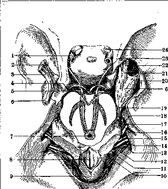
Fig. 11. Course of cranial nerves inside the skull: 1- internal carotid a.; 2- trigeminal nerve- motor branch; 3- trigeminal nerve- sensory branch; 4- posterior cerebral a.; 5- superior cerebellar a.; 6- Gasser's semilunar ganglion; 7- Sylvian aqueduct; 8- fourth ventricle; 9 - inferior posterior cerebellar a.; 10, 12 and 13- Willis' accessory nerve; 11- hypoglossal nerve; 14- vagus nerve; 15 - glossopharyngeal nerve; 16- acoustic nerve; 17- Wrisberg's intermediate nerve; 18- facial nerve; 19- trigeminal nerve; 20- mandibular nerve; 21- ophthalmic nerve; 22- maxillary nerve; 23- oculomotorius nerve; 24- optic nerve.
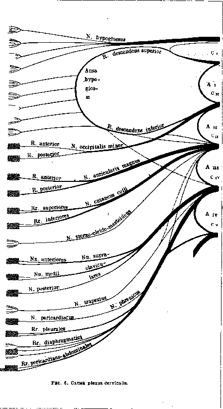
Fig. 12. Nerves of the orbit from the lateral side: 1- frontal nerve; 2- trochlear nerve; 3- lacrimal nerve; 4- internal carotid a.; 5- ophthalmic nerve; 6- oculomotorius nerve; 7- abdu-cens nerve; 8- Gasser's semilunar ganglion; 9- trigeminal nerve- sensory root; 10- trigeminal nerve- motor root; 11- ciliary ganglion; 12- maxillary nerve; 13- inferior rectus oculi muscle with its nerves; 14- ophthalmic a.; 15- optic nerve; 16- short ciliary nerves; 17- long ciliary nerves; 18- naso-ciliary nerve; 19- superior rectus oculi muscle with its nerves. Fig. 13. Diagram of the cervical plexus: 1- great auricular nerve; 2- lesser occipital nerve; 3- anastomotic branch for the sterno-cleido-mastoideus muscle; 4- anastomotic branch for the sterno-cleido-mastoideus muscle; 5 and 8- anastomotic branch for the trapezius muscle; 6- Willis' accessory nerve; 7- subclavian nerve; 9- descending branch of the cervical plexus; 10- phrenic nerve; 11- first branch of the brachial plexus; 12- descending branch of the hypoglossal nerve; 13- nerve for the thyreo-hyoidus muscle; 14- nerve for the genio-hyoidus muscle; 15 and 17- descending branch of the cervical plexus (root); 16- nerves for the scaleni muscles; 17^-descending branch; 18- anastomotic fibers of the cervical ansa, I; 19- nerves for the anterior neck muscles; 20- nerves for the anterior neck muscles; $a ч 21- meningeal nerve. ' «89
HUMAN NERVES
в«Ю
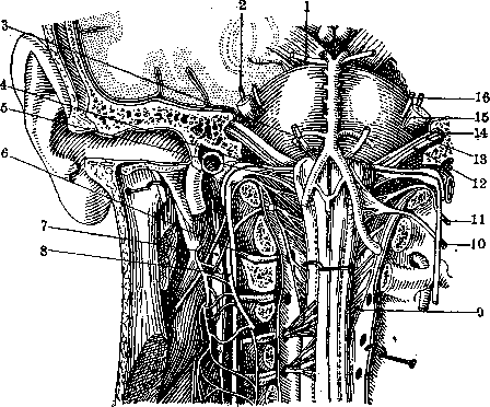
Fig. 14. Nerves of the base of the brain, medulla oblongata and cervical part of the spinal cord (ventral view): 1- oculomotorius nerve; 2- trochlear nerve; 3- abducens nerve; 4- glossopharyngeal nerve; 5- vagus nerve; 6- cervical nerve I; 7- superior cervical ganglion; 8- right hypoglossal nerve; 9- Willis' accessory nerve; 10- left hypoglossal nerve; 11- Willis' accessory nerve; 12- acoustic nerve; 13- Wrisberg's intermediate nerve; 14- facial nerve; 15- trigeminal nerve; 16- sensory root of the trigeminal nerve. (According to Bourgeret.)
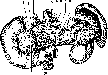
Fig. 15 Innervation of the pancreas: 1- gastro-duodenal a.; 2 and 7- right and left splanchnic nerves; 3- proper hepatic a.; 4 and 5- right and left vagus nerves; 6- left gastric a.; 8- splenic a.; 9- inferior pancreatico-duodenal a.; 10- superior mesenteric a.
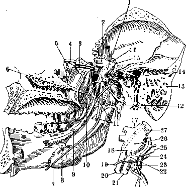
Fig. 16. Mandibular nerve with its branches:.*- semilunar ganglion (Gasser); 2- ophthalmic nerve; 3- nerve of the pterygoid canal (Vidian); 4- maxillary nerve; 6- spheno-palatine ganglion; 6 and 19- nerve of the tensor veli palatini; 7- mylo-hyoid branch; 8- submaxillary ganglion; 9- lingual nerve; 10- internal pterygoid nerve; 11- inferior alveolar nerve; 12- auriculo-temporal nerve; 13- facial nerve; 14- otic ganglion; 15- sensory root of the trigeminal nerve; .Z6 and J7- motor root of the trigeminal nerve; 18- sensory root of the otic ganglion; 19- nerve for the tensor tympani muscle; 20- root from the facial and glossopharyngeal nerve; 21- semilunar ganglion (Gasser); 22- sensory root of the trigeminal nerve.
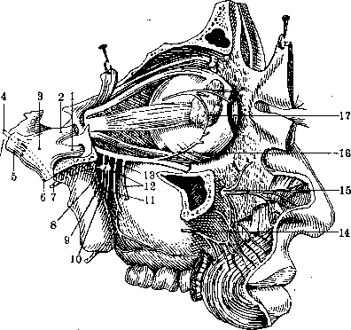
Fig. 17. Maxillary nerve with its branches (lateral view): /-maxillary nerve; 2- ophthalmic nerve; 3- Gasser's semilunar ganglion; 4- motor root of the trigeminal nerve; 5- sensory root of the trigeminal nerve; 6- mandibular nerve; 7- nerve of the pterygoid canal (Vidian); 8- spheno-palatine ganglion; 9- posterior palatine nerve; 10- middle palatine nerve; 11- anterior palatine nerve; 12- posterior superior alveolar nerves; 13- spheno-palatine nerves; 14- one of the branches of the facial 4 nerve, anastomosing with the branches of the infraorbital nerve; 15- minor pes anserina; 16- anastomotic branch of the zygomatic nerve; 17- lacrimal nerve.
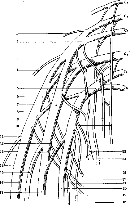
Figure 18. Schema of the brachial plexus: 1-n. dorsalis scapulae; 2-fasciculus primarius sup.; 3-fasciculus primarius medius; 4-n. suprascapularis; 5-fasciculus primarius inf.; 6-fasciculus post.; 7-fasciculus lateralis; 8-fasciculus medialis; 9-n. subclavius; 10-n. phrenicus; 11-n. axillaris; 12-radix medialis nervi mediani; 13-radix lateralis nervi mediani; 14 and 17-n. medianus; 15-n. musculo-cutaneus; 16-n. radialis; 18-n. cutaneus antibrachii medialis; 19-n. cutaneus brachii int.; 20-n. ulnaris; 21-n. thoraco-dorsalis; 22-n. subscapularis inf.; 23-n. subscapularis sup.; 24-n. thoracalis longus; 25-nn. thoracalesant. primus et secundus; C4-C8-IV-VIII cervical nerves; Th1-first thoracic nerve.
Figure 19. One of the unusual forms of the brachial plexus. In this case, the ventral branch of the fifth cervical nerve does not directly connect with the sixth, and the upper primary bundle consists only of fibers from the fifth nerve; subsequently, after giving off n. dorsalis scapulae, this nerve divides, as in normal cases, into two secondary bundles - posterior and lateral; the fibers of the sixth nerve also divide into two bundles and merge into the mass of the secondary bundles mentioned above. In all other respects, all other nerves do not differ from normal. 1-n. dorsalis scapulae; 2-n. suprascapularis; 3-fasciculus post.; 4-n. axillaris; 5-n. radialis; 6-n. ulnaris; 7-radix lat. nervi mediani; 8-radix medialis nervi mediani; 9-fasciculus medialis; 10-fasciculus lat.; 11-fasciculus primarius inf.; 12-fasciculus primarius medius. Figure 20. One of the unusual forms of the brachial plexus, sharply differing from normal. The plexus has, as always, three primary bundles, with the upper primary showing no deviations from normal and consisting of fibers from the fourth, fifth, and sixth cervical nerves; the middle primary bundle, however, is formed from fibers of the seventh and eighth cervical nerves and part of the fibers of the first thoracic, and finally the lower primary bundle consists only of the remaining fibers of the first thoracic nerve. As for the secondary bundles, they show the greatest deviations from normal, with the proper posterior secondary bundle being normal, the lateral one beginning, as in normal cases, from both primary (upper and middle), but subsequently it also takes in all fibers from the lower primary bundle; thus, there is no medial secondary bundle as such in the plexus; from the lateral secondary bundle all the nerves of the medial bundle also originate. The entire plexus clearly shows a primitive division into dorsal and ventral layers. 1-n. dorsalis scapulae; 2-fasciculus primarius sup.; 3-n. suprascapularis; 4-fasciculus post.; 5-n. axillaris; 6-n. radialis; 7-n. medianus; 8-n. ulnaris; 9-fasciculus lat.; 10-fasciculus primarius inf.; 11-fasciculus primarius medius. Figure 21. One of the unusual forms of the brachial plexus. All three primary bundles and the posterior secondary bundle are formed normally, but in the formation of the medial and lateral secondary bundles there is a deviation from normal. The lateral secondary bundle consists only of fibers emerging from the upper primary bundle; the middle primary bundle takes no part in its formation; instead, the medial secondary bundle sends part of its fibers in the form of two bundles that merge into the median nerve; thus the latter has only one lateral root from the upper primary bundle, while the medial root from the middle primary bundle as such is absent; the middle primary bundle divides into two bundles, one of which, as in normal cases, passes into the dorsal secondary, and the other, as a deviation from normal, passes into the medial secondary bundle; however, the latter, as mentioned above, sends an anastomosis to the median nerve. 1-n. dorsalis scapulae; 2-fasciculus primarius sup.; 3-fasciculus post.; 4-n. suprascapularis; 5-n. axillaris; 6-n. radialis; 7-n. medianus; 8-n. ulnaris; 9-radix medialis nervi mediani; 10-radix lat. nervi mediani; 11-fasciculus medialis; 12-fasciculus primarius inf.; 13-fasciculus primarius medius. (K and llyustr, art. Nerves of man.)
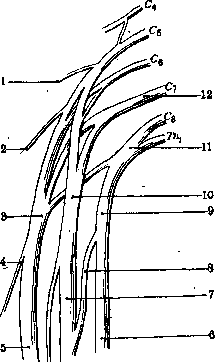
Figure 19.
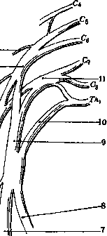
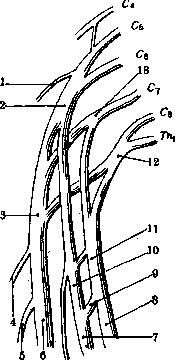
Figure 20.
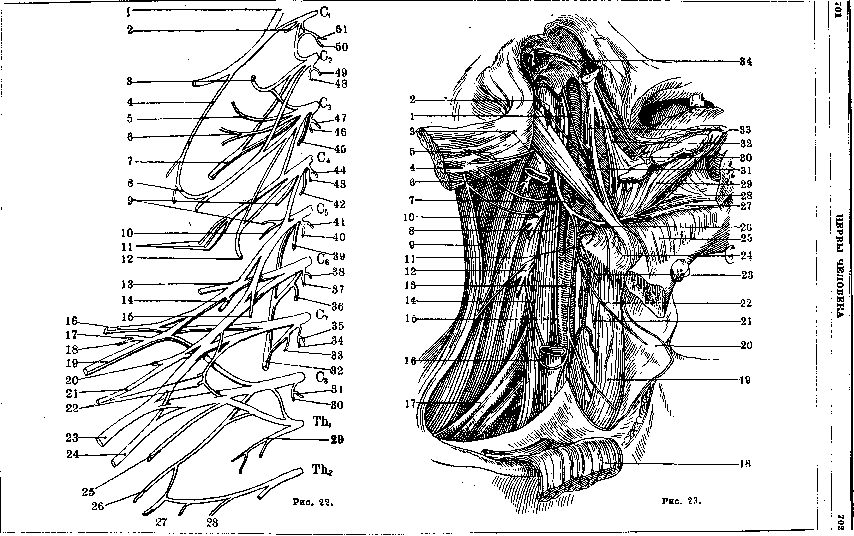
Fig. 21. Figure 22. Schema of the right cervical and brachial plexus from the front: 1-n. hypoglossus; 2-branch for m. rectus capitis lat.; 3-n. occipitalis minor; 4-ramus descendens n. hypoglossi; 5-n. auricularis magnus; 6-n. cutaneus colli; 7-branch for m. trapezius; 8-ansa hypoglossi; 9-branches for m. levator scapulae from n. dorsalis scapulae; 10-branches for mm. rhomboidei from n. dorsalis scapulae; 11-nn. supraclaviculares; 12-n. phrenicus; 13-n. suprascapularis; 14-n. subscapularis; 15-n. subclavius; 16-n. axillaris; 17-n. subscapularis; 18-n. thoraco-dorsalis; 19-n. radialis; 20-branch for m. coraco-brachialis; 21-n. musculo-cutaneus; 22-nn. thoracales ant.; 23-n. medianus; 24-n. ulnaris; 25-n. cutaneus antibrachii medialis; 26-n. cutaneus brachii medialis; 27-nn. intercosto-brachiales; 28-n. intercostalis II; 29-n. intercostalis I; 30-branch for m. scalenus medius; 31, 34, 43, 46, 48-branches for mm. intertransversarii; 32-n. thoracalis longus; 33-branch for m. scalenus medius; 35-branch for m. scalenus ant.; 36 and 39-branches for m. scalenus medius; 37 and 40-branches for m. scalenus ant. and intertransversarius; 38-branch for m. longus colli; 41-branch for m. longus colli; 42-branch for m. scalenus medius; 44-branch for m. longus colli; 45-branch for m. levator scapulae; 47-branch for mm. longi capitis et colli; 49 and 50-branches for m. longus capitis; 51-branch for m. rectus capitis ant.; C1-C7-plexus cervicalis; C8-Th1-plexus brachialis. Figure 23. The lower cranial nerves on the neck: 1-n. glosso-pharyngeus; 2-n. facialis; 3-v. jugularis interna; 4-m. sterno-cleido-mastoideus; 5-n. accessorius Willisi; 6-n. cervicalis secundus; 7-a. occipitalis; 8-a. carotis int.; 9-m. trapezius; 10-n. cervicalis tertius; 11-n. laryngeus superior; 12-n. cervicalis quartus; 13-ramus desc. n. hypoglossi; 14-ramus desc. plexus cervicalis; 15-n. vagus; 16-ansa hypoglossi; 17-plexus brachialis; 18-m. sterno-cleido-mastoideus; 19-m. sterno-thyreoideus; 20-m. omo-hyoideus (venter ant.); 21-n. laryngeus superior (ramus externus); 22-n. musculi omo-hyoidei (venter ant.); 23-n. laryngeus superior (ramus superior); 24-m. hyo-glossus; 25-m. mylo-hyoideus; 26-n. hypoglossus; 27-m. genio-hyoideus; 28-m. genio-glossus; 29-m. hyo-glossus; 30-n. lingualis; 31-m. stylo-hyoideus; 32-m. stylo-glossus; 33-m. stylo-pharyngeus; 34-chorda tympani. (To illustr. art. Nerves of man.)
705 M. sterno-cleido-mastoideus (n. access. +C1+C2) Mm. colli and nuchae (C1-4) M. trapezius (n. access.+C1-3) M. rhomboideus (C4-5) Diaphragma (C3-5) Mm. supra- and infraspinatus (C5-6) M. deltoideus (C5-6) M. biceps brachii (C5-6) M. serratus (C5-6) M. pectoralis (C5-Th1) M. triceps brachii (C6-8) M. brachialis (C5-6) Mm. extensores carpi and digitorum (C6-8) Centrum cilio-spinale (C8-Th1) Centrum sympathicum cordis (C8-Th1) M. latissimus dorsi (C6-8) Mm. flexores carpi and digitorum (C6-Th1) Mm. interossei (C8-Th1) Thenar (C8-Th1) Mm. intercostales and dorsi (Th1-XII) Mm. abdominis (Th1-L1) Centrum sympathicum ventriculi, duodeni and jejunum (Th1-L2) Centrum sympathicum vesicae felleae (Th10-X) Centrum sympathicum colonis (Th11-XII) Centrum sympathicum vesicae urinariae (L1) M. iliopsoas (L1-3) M. cremaster (L1-2) M. adductor femoris (L2-4) M. quadratus femoris (L4-5) M. tibialis anterior (L4-5) Mm. gastrocnemius and soleus (L5-S1) M. tensor fasciae latae (L4-S1) Mm. glutaei (L4-S1) M. semitendinosus (L5-S1) M. semimembranosus (L5-S1) M. biceps femoris (L5-S2) Mm. extensores hallucis and digitorum (L5-S1) Mm. peronaei (L5-S1) Mm. flexores digitorum and hallucis (L5-S1) Mm. interossei (L5-S1) Centrum erectionis, ejaculationis and defecationis (S2-4)
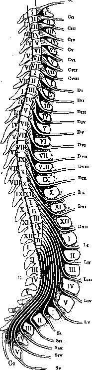
Figure 24. Schema of the origin of nerves from the spinal cord with indication of their place of origin, place of exit through the intervertebral foramen, height of their exit from the spinal cord in relation to the spinous processes of the vertebrae, and segmental innervation of muscles. (View from the right side)
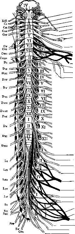
N. accessorius (XI) N. suboccipitalis (Ci) N. occipitalis minor (Ci-sh) N. auricularis magnus (Cm) N. cutaneus colli (Cm) N. accessorius (Ci-sh) Nn. supraclaviculares (Csh-iv) N. phrenicus (Csh-iv) N. musculo-cutaneus (Cv vn) N. medianus (Cv-Di) N. radialis (Cv-Di) N. axillaris (Cv-vn) N. thoracalis (Cv-vn) N. ulnaris (Cvin-Di) Nn. cutanei brachii et antibrachii (Cvin-Di) Reflex of the biceps brachii muscle (Cv-vi) Reflex of the triceps brachii muscle (Cvi-vn) Reflex of the radius (Cn-vm) Upper abdominal reflex (DVm-ix) Lower abdominal reflex (Dx xn) Nn. intercostales (Di-xn) Reflex of the cremaster (Ssh) Reflex of the knee (LiV) Reflex of the Achilles tendon (LV-Sn) Reflex of the anal sphincter (Sv) N. splanchnicus hypogastricus (Dvn-Li) N. ilio-inguinalis (Li) N. genito-femoralis (Lin) N. cutaneus femoris lateralis (Lum) N. femoralis (Li-iV) N. obturatorius (Ln-iv) N. glutaeus superior (Liv-Si) - N. glutaeus inferior (LV-Sn) N. peronaeus (LIV- Sn) N. tibialis (LiV- Sm) N. ischiadicus (Liv-Sin) N. cutaneus femoris posterior (Si-m) Plexus pudendus Plexus coccygeus . 25. Diagram of the origin of nerves from the spinal cord with indication of the level of their origin, place of exit through the intervertebral foramen, and formation of peripheral nerves. (View from the ventral surface.)
.7 18 1
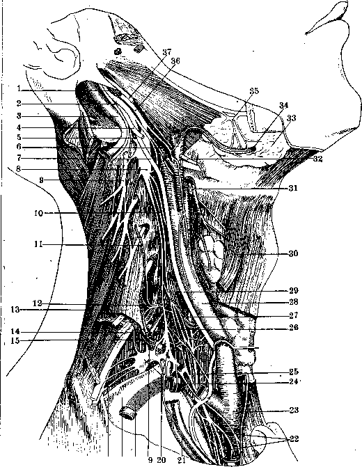
Figure 26. Sympathetic nervous system of the cervical region on the right: 1-a. carotis int.; 2-g. spinalis nervi accessorii Willisi; 3-v. jugularis int.; 4-n. hypoglossus; 5-ramus anastomoticus nervi cervicalis I cum ganglio cervicali superior!; 6-n. laryngeus sup.; 7-n. vagus; 8- gangl. cervicale sup.; 9- n. cervicalis sup.; 10-n. cardiacus sup.; 11-a.cervicalis ascendens; 12-gangl. cervicale medium; 13-m. scalenus; 14- truncus thyreo-cervicalis; 15-a. cervicalis prof.; 16-a., intercostalis suprema; 17-pleura; 18-a. subclavia; 19- n. vertebralis; 20-gangl. cervicale inf.; 21- a. и v. mammaria int.; 22-n. cardiacus inf.; 23-a. anonvma; 24-a. subclavia; 25- n. laryngeus inf.; 26-n. cardiacus medius; 27-a. tnyreoidea inf.; 28-a. carotis communis; 29-n. vagus; 30-m. omo-hyoideus; 31- a. tnyreoidea sup.; 32-n. hypoglossus; 33-glandula submaxillaris; 34- a. maxillaris externa; 35- gangl. submaxillare; 36-truncus sympathicus; 37-n. glosso-pharyngeus. 7 09
NERVES OF HUMAN
?|И»
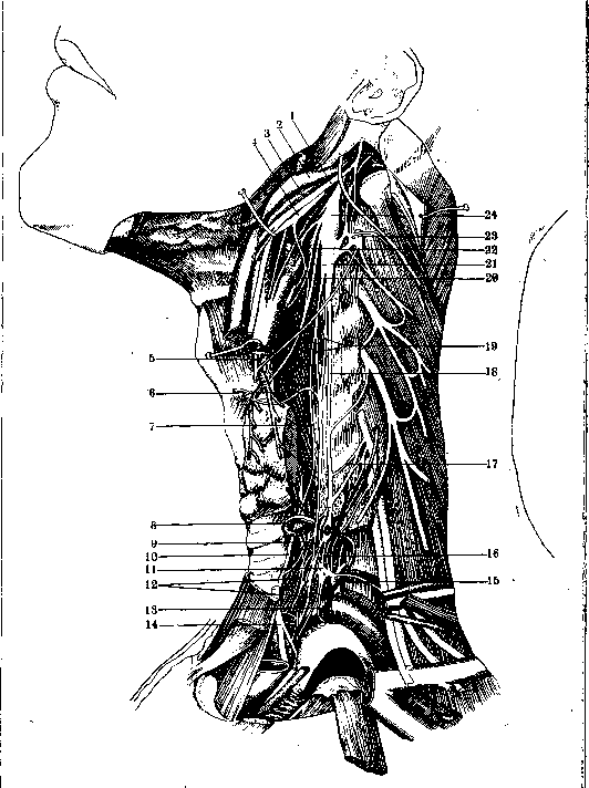
!fig. 27. Sympathetic nervous system of the cervical region on the left (another form): 1-п. hypoglossus;
\ 2-v. jugularis int.; 3-a. carotis int.; 4-п. vagus; 5-т. descendens nervi hypoglossi; б-п. thyreoideus
\ sympathicus; 7 and 9-r. anastomoticus sympathicus cum n. recur.; 8-a. thyreoidea inferior и п. thyreoideus
.4 sympathicus; 9-r. anastomoticus sympathicus recurrens; 10-n. recurrens; 1J-gangl. -eervicaJe inf»
; * praearticulare; 12- nn. cardiaci; 13-ansa Vieussenii; 14-n.vagus; 15-gangl. cervicale inf. retroarticuiaws
i IS-n. vertebralis; 17- r. anastomoticus V cervicalis sympathicus; is-truncus sympathicus; 19-й*
]: cardiacus sup.; 20-n. laryngeus sup.; 21-r. sympathicus ad n. laryngeum sup.; 22-r. sympathicus
j plexus carotici; 23-n. cervicalis secundus; 24-gangl. sympathicum sup.
1
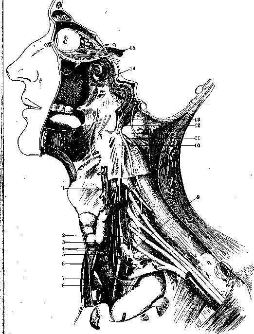
Figure 28. Sympathetic nervous system of the cervical region on the left (another form): 1-a. и v. thyreoidea ^superior; 2-gangl. cervicale medium; 3-a. thyreoidea inf.; 4-n. cardiacus; 5-gangl. cervicale inf.; 6-gangl. thoracale I; 7-n. vagus; 8-n. phrenicus; 9-n. vertebralis; 10-gangl. cervicale sup.; 11- n. i; hypoglossus; 12-n. vagus; 13-n. accessorius Willisi; 14-ramus sympathicus ganglii spheno-palatini; 15-ramus sympathicus ganglii ciliaris.
.
Related articles
Cite this page
“Nerves.” Soviet Medical Encyclopedia. English translation of Bolshaya Meditsinskaya Entsiklopediya, 1st ed. (Moscow, 1928–1936), ed. N. A. Semashko. https://sovietmedicalencyclopedia.pages.dev/article/nerves/