Median Nerve
Historical document, translated for reference. It reflects medical knowledge of the 1920s–30s and is not medical advice.
Summary
The median nerve is one of the main nerves of the upper limb, classified as a mixed nerve. This article describes its anatomical course, branches, functions, and various pathological conditions affecting it, including injuries, inflammations, and associated symptoms.
Encyclopedia article (1928–1936)
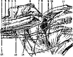
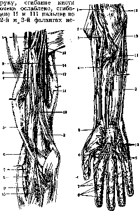
MEDIANUS NERVUS, median nerve, one of the main nerves of the upper limb; by its function, it belongs to the group of mixed nerves. The median nerve is formed in the axillary fossa from the brachial plexus, from which it begins with two roots: the outer, lateral root, which originates from the V and VI cervical spinal nerves, and the inner, medial root, which originates from the VIII cervical and I thoracic spinal nerves; thus the median nerve corresponds to the VI, VII, VIII cervical, and I thoracic segments. The roots of the median nerve surround the axillary artery—one from above, the other from below—and join at an acute angle on its anterior surface; after joining, the nerve lies on the outer side of the artery and descends along with it along the inner edge of the biceps muscle (fig. 1). Gradually curving around the artery from outside to inside and crossing it in the middle of the arm, in the lower half of the arm the median nerve (fig. 2) already lies on the inner side of the artery, and at the bend of the elbow it is located one finger's breadth medial to the artery. These relationships, however, are not constant (see Axillary Fossa). Along the course of the arm, the median nerve gives off no branches. In the depth of the elbow bend, the median nerve approaches the edge of the pronator teres muscle and extends to the wrist—along the midline of the forearm between the flexor digitorum sublimis and
Figure 1. Position of the median nerve in the axillary fossa: 1- median nerve; 2- brachial artery; 3- coraco-brachialis muscle; 4- biceps muscle (short head); 5- pectoralis major muscle; 6- musculocutaneous nerve; 7- axillary artery; 8- pectoralis minor muscle; 9- anterior thoracic nerves; 10- intercostobrachial nerves; 11- clavicle; 12- ulnar nerve; 13- medial cutaneous nerve of forearm; 14- medial cutaneous nerve of arm; 15- deep flexor digitorum muscle; on the forearm it gives numerous branches to all flexors except the head of the flexor digitorum profundus closest to the ulna and the flexor carpi ulnaris (see below). After passing (together with the tendons of the flexor digitorum muscles) under the transverse carpal ligament, the median nerve divides into 4 branches: the outer branch innervates the muscles of the thenar eminence (except for the deep head of the flexor pollicis brevis and the adductor pollicis) and the skin of the radial border of the thumb; the other three—common volar digital nerves—supply the skin of the radial half of the palm and two lumbrical muscles, go to the web spaces of the first three fingers, at the base of each of which they divide into two branches (proper volar digital nerves), located along the edges of the fingers and innervating the skin of the opposing surfaces of the I, II, III and IV fingers (fig. 3). The median nerve gives branches to joints, bones, periosteum, blood vessels; along its course it anastomoses with the musculocutaneous nerve in the region of the arm, and on the palm with the ulnar and radial nerves. For the function of the median nerve, see Human Nerves; for the function of the muscles it innervates, see Human Muscles. Pathology. The etiological factors causing disease of the median nerve are diverse: injuries (bruises, various wounds, ruptures), pressure (e.g., tumors), other mechanical causes causing muscle strain, or frequent and repeated pressure on the nerve (most often in the palm area) in certain professions (locksmiths, turners, leatherworkers, tailors, ironers, lacemakers, dentists, etc.). Infections and intoxications can cause inflammation of the median nerve, neuralgia; neuritis can be isolated or occur simultaneously with damage to other nerves. For pathological anatomy and pathogenesis, see Neuritis, Polyneuritis.-Diseases of the median nerve are accompanied by flaccid paralysis or paresis (depending on the intensity of the process) of the muscles it innervates (see Human Nerves) with changes in electrical excitability, disturbances of sensitivity, and autonomic phenomena (trophic and vasomotor). Paralysis of the muscles innervated by the median nerve causes disturbances of movement and a characteristic position of the hand. Of the movements, pronation, flexion of the wrist, movement of the thumb, and flexion of the 2nd and 3rd phalanges of the II and III fingers are disturbed. The intensity and extent of the paralysis depend on the location of the process. With damage to the median nerve in the region of the arm, all the above-mentioned movements are disturbed: pronation is impossible
Figure 2.
Figure 3.
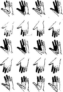
Figure 2. Position of the median nerve at the level of the arm: 1 and 4- musculocutaneous nerves; 2- median nerve; 3- biceps muscle; 5- cutaneous nerve of forearm; 6 and 13- radial nerve; 7- supinator muscle; 8- deep branch of radial nerve; 9- superficial branch of radial nerve; 10 and 12- ulnar nerve; 14- brachialis muscle; 15- radial artery. Figure 3. Position of the median nerve on the forearm and hand: 1- median nerve; 2- radial nerve; 3- brachial artery; 4- radial artery; 5- superficial branch of radial nerve; 6- common volar digital artery; 7- proper volar digital nerves of median nerve; 8- nerve endings (Vater-Pacini corpuscles); 9- common volar digital nerves of ulnar nerve; 10- common volar digital nerves of median nerve; 11- ulnar artery; 12- ulnar nerve; 13- brachialis muscle. movements of the thumb—flexion, abduction, opposition to other fingers—are impossible; flattening of the thenar is observed due to atrophy of its muscles; the hand takes on a characteristic appearance—main du singe (ape hand) (fig. 4). With damage to the median nerve in the lower parts of the forearm or on the hand, only the muscles of the thenar are paralyzed—its wasting is observed, limitation of movement of the I finger, mainly opposition to other fingers. medianus nervus Sensory disturbances are observed in the form of anesthesia, hypesthesia, or hyperesthesia on the outer two-thirds of the palm, the palmar surface of the first three fingers, and the outer half of the IV finger, on the dorsal surface of the last two phalanges of the II and III fingers (fig. 5).-Trophic disorders are expressed in the formation of pemphigus-like blisters on the palm and the thumb, in the appearance of shiny atrophic skin, in deformation or loss of nails, in pigmentation, disturbances of sweating, cyanosis. With damage and inflammation of the median nerve, joint disorders are also observed, more often in the form of limited mobility, sometimes ankylosis.-N euritis of the median nerve is characterized by very intense spontaneous pains, pain on pressure on the nerve trunk, on the corresponding muscle masses: extension of the elbow, supination are very painful; there is cutaneous hyperesthesia or hypesthesia, trophic disorders, especially of the nails; paralysis of the corresponding muscles, with flexion contractures («griffe du median»).
Fig 4. Variations in the area of impaired painful sensitivity with complete damage to the median nerve. Black shading indicates anesthesia, dots indicate hypesthesia. (According to Head.)
Very often in diseases of the median nerve, causalgias are observed (see), which is explained by its richness in sympathetic fibers and connections with the arterial system and perivascular sheaths. Often paralysis of the median nerve is combined with paralysis of the ulnar nerve. The diagnosis of diseases of the median nerve presents no difficulties; it is necessary to exclude mainly progressive spinal muscular atrophy (damage to both hands without sensory disturbances), syringomyelia and amyotrophic lateral sclerosis. Loss of working capacity with damage to the median nerve at the shoulder equals 45% (according to Tinel), 15-20% with injury to the forearm or hand; the right side increases disability, as does the presence of causalgia.-P revention is mainly concerned with preventing diseases of the median nerve from occupational hazards. Depending on the severity of the case or the conditions of work affecting the median nerve, a change of profession or temporary cessation of work is recommended; in other cases, it may be possible to change work methods or introduce new production techniques.-T reatment is mainly causal and restful; electrization, ionization, warm hand baths, massage with passive and active exercises are used, later—gymnastics with resistance. With strains, fractures, nerve damage—treatment is surgical and orthopedic-surgical. Operative technique. The median nerve can be exposed in the axillary fossa, the middle third of the arm, the elbow bend, the upper third of the forearm, and at the wrist level. In the first case, the upper limb is abducted at an obtuse angle; an incision is made from the apex of the axilla to the angle formed by the pectoralis major muscle with the inner surface of the arm along the edge of the pectoralis major muscle; after incising the skin and subcutaneous tissue, the coracobrachial sheath is incised; pulling the muscle upward, the aponeurosis is opened; the sheath of the exposed neurovascular bundle is opened bluntly: the nerve exposed in front of the others is the median nerve. In the middle third of the arm, the median nerve is exposed along a line extending from the apex of the axillary fossa to the middle of the elbow bend, with a 5 cm incision of skin and subcutaneous tissue; then the aponeurosis of the biceps muscle is incised, pulling it outward with a blunt hook; the second aponeurosis under the biceps muscle is opened and the median nerve is found. Exposure of the median nerve in the elbow bend is done in the middle of it with a vertical incision perpendicular to it 4.J 4J 4~\ 4-\ t w m m
Figure 4. Variations in the area of impaired painful sensitivity with complete damage to the median nerve. Black shading indicates anesthesia, dots indicate hypesthesia. (According to Head.)
with an incision of 5-8 cm: after incising the skin and subcutaneous tissue, the skin veins are ligated and the aponeurosis is incised with a grooved probe, beneath which the vessels are exposed: 1 cm medial to them is the median nerve. In the upper third of the forearm after an incision of the skin and subcutaneous tissue 5-8 cm long from the middle of the elbow crease along the middle of the anterior surface of the forearm, the aponeurosis of m. pronator teres and m. palmaris longus is incised; the latter is retracted medially - in the anterior angle of the wound the median nerve is found, lying deeply. When exposing the median nerve at the wrist, a vertical incision of the skin and subcutaneous tissue 3-4 cm is made medial to the tendon of m. palmaris longus for several cm, incising the aponeurosis of the latter; then another aponeurosis is incised medially as well and the median nerve is exposed
MEDK.4
Related articles
Mentioned in
Cite this page
“Median Nerve.” Soviet Medical Encyclopedia. English translation of Bolshaya Meditsinskaya Entsiklopediya, 1st ed. (Moscow, 1928–1936), ed. N. A. Semashko. https://sovietmedicalencyclopedia.pages.dev/article/median-nerve/