Scapula Alata
Historical document, translated for reference. It reflects medical knowledge of the 1920s–30s and is not medical advice.
Summary
Scapula alata, or winged scapula, is a condition indicating weakness in the muscles that fix the scapula to the thoracic cage, characterized by the scapula assuming a wing-like position. This condition results from various causes including myopathies, amyotrophies, spinal cord lesions, nerve injuries, and rickets, and can limit shoulder movement.
Encyclopedia article (1928–1936)
SCAPULA ALATA, winged scapula, a sign indicating weakness of the muscles fixing the scapula relative to the thoracic cage; it consists in the scapula assuming a wing-like position, with the medial edge and especially its inferior angle lagging behind the thoracic wall; the scapula glides upward when the patient attempts to abduct the arm and passively follows the movements of the shoulder (see figure), as a result of which movements in the shoulder joint are limited and the patient cannot sufficiently elevate or abduct the arm. S. a. occurs due to weakness of the serratus anterior muscle (m. serratus ant.), which presses the scapula to the thoracic wall and pulls it forward and outward (innervation - n. thoracalis ant., spinal center in C v - vn), and the major and minor rhomboid muscles, which pull the scapula inward and upward and bring its inferior angle closer to the spine (innervation - n. dorsalis scapulae; spinal center in Cv). S. a. are a characteristic sign of various forms of myopathies (juvenile form of Erb, scapulohumeroperoneal type, etc.) and amyotrophies (type Werdnig Hoffmann), when the muscles of the shoulder girdle are involved in the disease process. S. a. may occur in lesions of spinal centers (syringomyelia), in diseases of the corresponding nerves, in rickets. The literature contains isolated descriptions of S. a. in healthy individuals, which in this case are explained by the ability to voluntarily significantly relax the musculature. S. a. in progressive muscle disease may be the subject of orthopedic measures. In cases of S. a. due to paralysis of the serratus anterior muscle of traumatic origin, Willich applied the operation of fixing the scapula to the lig. supraspinale and to the IX rib (by means of a strip cut from fascia lata) with good functional effect.
S. Blinkov.

SCARLET FEVER. Contents: I.
608 II.
610 III. Statistics and geographical distribution . .
612 IV.
Pathological anatomy and pathogenesis ....
620 V.
630 VI.
652 IX.
657 Scarlet fever, an acute infectious disease, characterized by elevated temperature, lesions of the throat, a fine-point rash with subsequent peeling, and a series of typically occurring complications. History. Even in ancient times and the Middle Ages (Hippocrates, Galen), a disease with throat lesions and redness of the entire body was known, but it was not distinguished from measles. The first precise description and modern name (scarlet fever - purple fever) was given by the English physician Sydenham (1661). In the 18th and 19th centuries, epidemics were described in all countries of Europe, and the clinical picture of the disease was thoroughly developed (Trousseau). From the end of the 19th century to the present time, along with the clinical picture, particular attention of researchers has been attracted to the study of etiology - the search for the causative agent, the development of methods of specific prophylaxis and therapy (Babes, Gabrichevsky, Dick, Caronia and others). I. Etiology. In the search for the causative agent of S., a large number of microbes (bacteria, spirochetes, protozoa, filterable virus) were discovered, to which an etiological role in the origin of S. was attempted to be attributed. Such were Cyclasterion scarlatinae-Mallory, Chlamydozoa scarlatinae-Gamaleya, spirochetes-Pospischill, diphtheroid rods-Mandelbaum's, streptococci-Loftier's, Babes's, Moser's, Gabrichevsky and many others. Control studies did not confirm the etiological role of the discovered microbes; some of them turned out to be accidental parasites, while others (e.g., Dohle bodies) were a product of the breakdown of cell nuclei. At present, there are three main views on the nature of the causative agent of S. I. The true and only causative agent of S. is the hemolytic streptococcus, possessing specific properties. The streptococcal theory of S. existed for a long time (Babes, Loffler, 1882; Moser, Gabrichevsky, 1902-07), but it received more or less firm substantiation in 1923 after the work of the American scientists, the Dick couple. In favor of the streptococcal theory, the Dicks bring a number of proofs. The hemolytic streptococcus was found by them in all cases of S. They obtained the toxin of the scarlet fever streptococcus, which is neutralized by the serum of convalescents from S.; in this the Dicks see the main feature of the scarlet fever streptococcus, which distinguishes it from hemolytic streptococci of other origin. The skin reaction with the toxin of this streptococcus (Dick reaction) is as specific for S. as the Schick reaction for diphtheria: in the acute period of the disease it is positive, in the period of convalescence it becomes negative in all. Decisive proof for the streptococcal theory, its supporters see in the experiment of the Dicks with the toxin of the scarlet fever streptococcus on humans. To young healthy people who had not had S., the Dicks injected a sufficient amount of toxin (0.1) and in some of them obtained a disease resembling S.: elevated temperature, malaise and fine-point rash. Finally, the antitoxic serum obtained by the Dicks gave a very good therapeutic effect in scarlet fever patients. Thus, the Dicks consider S. similar to diphtheria as a toxemia with a local infectious focus in the throat, from where toxins enter the blood and cause the clinical picture of S. Immunity in S. is purely antitoxic, which does not prevent streptococci in certain cases to give purulent complications. At present, after numerous studies with the scarlet fever streptococcus - bacteriological, experimental, clinical - it is impossible to fully accept the teaching of the Americans about the exclusive and specific role of the hemolytic streptococcus and its toxin in the etiology and genesis of S. The specificity of the scarlet fever streptococcus cannot be considered proven, since the serum of convalescents also neutralizes the toxin of streptococci of other origin. The Dick skin reaction, as shown by the observations of many authors, including in the children's clinic of the 1st MMI, in 25% of cases remains positive after recovery from S. Similarly to the Dick reaction, in scarlet fever patients the skin reaction with the toxin of the septic streptococcus (Ag-rachev) behaves. For this reason alone, it is difficult to consider the Dick reaction as such an indicator of immunity with respect to S. as the Schick reaction is for diphtheria. Experiments with the injection of toxin to humans have been few in number, the resulting syndrome was not studied with sufficient completeness. According to other authors (Burgers, To-wada), a scarlet fever-like disease can be obtained with filtrates of various streptococci and even staphylococci and coli bacilli. The pathological process in S. is more complex than in diphtheria, and it cannot be reduced exclusively to a toxemia. Thus, despite the importance of the research of the Dicks and other supporters of the streptococcal theory, these studies do not completely solve the question of the causative agent of S. II. The causative agent of S. is a filterable virus. Some supporters of this theory believe that it is in symbiosis with the streptococcus. As early as 1911, Bernhardt, on the basis of experiments with monkeys, expressed the assumption that the microbe of S. should be classified in the group of filterable ones. In 1921-23, Italian scientists Di Cristina and Caronia from the organs of children who died from S. and from the filtrate of nasopharyngeal mucus by the anaerobic method on media with tissue fragments obtained a microbe that resembled a diplococcus. On the basis of serological reactions, experiments with rabbits, and mainly on the basis of experiments with children, they came to the conclusion that the microbe they found - the causative agent of S.: after injecting a culture of this microbe to children who had not had S., they in some cases observed diseases which they considered S. However, control studies (S. Meyer, Burgers) did not confirm this view. Most modern authors consider the microbe of the Italian scientists a product of cell breakdown. Among Soviet bacteriologists, the theory of symbiosis of a filterable virus with a streptococcus was supported and developed by Zlatogorov. This theory cannot be considered proven, but it cannot be considered refuted either. The clinical picture of S. gives some grounds for such a dualistic view of the etiology of S. (see below - clinical picture of S.). III. Anaphylactic theory. According to this theory, there is no need to look for a specific causative agent of S.; it does not exist. S. is an anaphylactic reaction of a sensitized organism to any streptococcus. According to the opinion of the extreme supporters of this theory (Szontagh), increased sensitivity to streptococcus can develop not only as a result of repeated streptococcal infections, but even under the influence of intestinal auto-intoxication, physical fatigue and a whole series of other non-infectious moments. Szontagh has long considered S. a non-contagious disease. The anaphylactic theory is held to a greater or lesser extent mainly by German pediatricians (Szontagh, Czerny, S. Meyer, Schlossmann, Fanconi); among Americans, in recent times, Dochez inclines to it. The anaphylactic theory at first glance is attractive for its simplicity, the possibility of explaining some facts from the epidemiology and clinical picture of S. (in some individuals in a collective S., in others - angina), and especially that it assigns the main role in the origin of S. not to the microbe, but to the macroorganism, which corresponds to the modern understanding of the etiology of infectious diseases. However, if one compares it with the main facts of the epidemiology of S. (the occurrence of epidemics in secluded places at large intervals after the arrival of a scarlet fever patient there, the proven contagiousness of S.), one cannot agree with it. Thus, the question of the causative agent of S. must at present be considered open, further research is necessary; in any case, it should be noted that all modern theories - some to a greater, others to a lesser extent - assign a significant role to the streptococcus in the etiology of S.
fc, II. Epidemiology. In large cities and densely populated areas, scarlet fever exists endemically; however, at certain intervals (5-10 years), more or less severe epidemics break out. The causes of the wave-like course of scarlet fever epidemics are not precisely known; the more probable cause is indicated as periodic changes in the population's susceptibility to S. Data on the influence of meteorological factors are contradictory. Brownlee for London found that in rainy years with heavy precipitation, the incidence of S. is higher than in dry years. Benda for Berlin gives the opposite relationship.-Methods of spread. The source of scarlet fever infection is primarily the scarlet fever patient. Obviously ill patients, especially hospitalized ones, are less dangerous than those in whom S. takes a rudimentary form; the disease often goes unrecognized and patients are not isolated. Such carriers of scarlet fever infection are usually adults in whom scarlet fever often manifests as angina. Apparently, healthy individuals can also be carriers if they are in close contact with a patient for a prolonged period. The scarlet fever virus is contained in mucus from the throat and nasopharynx and is transmitted to healthy individuals by droplet infection as well as through direct contact. The infectivity of throat secretions has been proven by Stickler in his experiments on healthy children. Urine and feces are not infectious. The question of the infectivity of the desquamation, which remained controversial for a long time, is resolved in the negative by most modern authors. The desquamation may become infectious under special conditions when the patient is not given baths and the desquamation becomes contaminated with throat secretions. The patient is infectious from the first day of the disease. It is impossible to give a precise answer as to how long the infectious period lasts. Examination of the throat for hemolytic streptococci cannot serve as a reliable criterion. The condition of the throat and the presence of purulent complications (otitis, lymphadenitis) are of greater importance (see below, prevention of S.). S. can be transmitted through objects and items used by the patient, but how long the scarlet fever virus remains on them is a controversial question. Reports of the infectivity of such items for many years (Golliner) should be treated critically. S. can also be transmitted through food products, for example, through milk. The portal of entry for scarlet fever infection is the throat and nasopharynx. According to Schlossmann, a necessary condition for the penetration of the microbe must be damage to the integrity of the mucous membrane. In rare cases, infection can penetrate the body through damaged skin and the mucous membrane of the genital organs in women (extrabuccal S., puerperal fever). Susceptibility to S. in humans is generally low, significantly weaker than in relation to measles. The coefficient of infectivity of S. according to Gottstein is about 40%, while for measles it is 95%. It is generally accepted that susceptibility depends on 1) age, 2) constitution, and 3) the condition of the body at the time of infection. Children aged 3 to 10 years are most susceptible (see below, statistics). In the first year of life, S. is relatively rare; during the first 3 months, S. is a very rare exception. True, cases in newborns have been described, but not all of them are impeccable in terms of diagnosis. The low susceptibility of infants depends mainly on the passive immunity received from the mother, but other little-known causes are possible (insufficient differentiation of cells, lack of sensitization, etc.). The non-susceptibility of adults is explained by active immunity resulting not only from obvious but also from rudimentary or even silent infection. The course of scarlet fever in infants and adults has significant features (see below, clinical picture of S.).-The role of the constitutional factor is far from being studied. According to Czerny, children with exudative diathesis and lymphatic constitution are particularly prone to S., and it runs a severe course in them. In neuropathic children, S. also gives a severe course. Attempts have been made to clarify the role of the constitutional factor in susceptibility to S. by studying blood groups in children and their parents (Hirszfeld and Brokman, Rubasheva and Yakobi). It turned out that all groups are equally susceptible to S.; neither the form nor the complications depend on the blood group. However, there is a regular relationship between the Dick reaction in children and their parents. Children with the same blood group as a parent with a positive Dick reaction retain a positive reaction after S., while children with the same blood group as a parent with a negative Dick reaction give a negative reaction after S. The condition of the body at the time of infection undoubtedly affects susceptibility, but it is difficult to say to what this influence should be most closely attributed. According to Czerny, overfeeding, especially with milk and eggs, increases susceptibility to S., as it intensifies the phenomena of exudative diathesis and leads to hypertrophy of the tonsils. Kobrak found that in families with insufficient nutrition, S. occurs less frequently than in families with adequate and excessive nutrition. The role of socio-domestic factors is differently assessed by various authors in capitalist countries. All agree that the mortality rate among proletarian children is higher, while the incidence of S. among them, according to some authors (Flügge, Benda), is higher, and according to others (Reiche), lower than among the well-off segments of the population. Studies by Soviet physicians showed that poor socio-domestic conditions lead to both increased incidence and a more severe course of S. Kaminsky found for Petersburg (1900-1910) that the incidence of S. among the wealthy population was 185.8 (per 100,000), the well-to-do-215.9, the insufficient-248.6, and the poor-261.8; the mortality coefficient among the wealthy was 11.8, the well-to-do-13.2, the insufficient-16.4, and the poor-19.9. The housing-domestic factor has a particularly great influence. On the one hand, overcrowding and close contact with sick individuals and carriers of the scarlet fever virus create favorable conditions for infection. The role of overcrowding was proven by Dudley through epidemiological investigation of a naval school in Greenwich (1912-1924) and some children's groups; the incidence of S. was higher in bedrooms with a large number of beds and with minimal distance between beds. On the other hand, the lack of light and air adversely affects the child's body and its immunobiological state, making it less resistant to scarlet fever infection. There are indications that some races are little susceptible to S., for example, the population of Central Asia in the USSR. However, the influence of racial, as well as family factors, cannot be considered established, and here one must primarily consider the cultural-domestic conditions. Immunity after S. is not absolute. Undoubted repeated diseases are observed more often than repeated cases of measles.
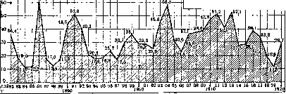
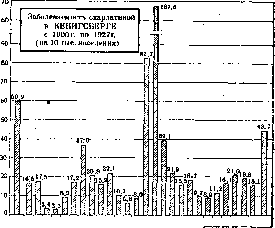
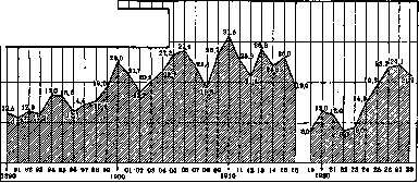
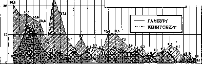
V. Molchanov. III. Statistics and geographical distribution. S. is distributed throughout the entire globe. The morbidity rate shows large fluctuations in different countries and in different periods of the year. In Western Europe in recent years, over 400,000 cases of S. are registered annually and about 12,000 deaths from it; in the USA - about 200,000 cases and about 2,500 deaths. Tables 1 and 2 present morbidity and mortality rates for 1921-1930 in different countries. The highest morbidity rates during all these years are observed in England and Scotland, the lowest in North Africa (Egypt, Tunisia, Algeria), in Japan and Korea. In the second half of the decade mentioned, a noticeable increase in morbidity is noted in a number of countries (USSR, Germany, Holland, etc.). Mortality rates in individual countries do not correspond to morbidity: in England and Scotland, for example, where morbidity is highest, mortality is significantly lower than in Romania and Yugoslavia, where morbidity rates are highest, and lower than in Italy, Poland and Czechoslovakia. This discrepancy between rates can perhaps be explained by incomplete registration of morbidity in the latter countries or by varying lethality (see below). The lowest mortality rates are observed among European countries in Switzerland, and among non-European countries in North African and Japanese countries. The most complete data on the distribution of S. can be obtained from materials of large cities (completeness of registration and accuracy of diagnosis). The highest mortality rates in the last 5 years in European countries were observed in cities of Hungary, Greece, Spain, Italy, Lithuania, Poland, Romania, Estonia and Yugoslavia (Fig. 1); in non-European countries - in cities of Turkey, Canada and Argentina. The lowest rates are observed in cities of North Africa and South America (Brazil, Peru). In the tables 1 and 2 presented, mortality rates were calculated for the entire population. Table 3 presents corresponding rates for some countries, calculated over a long period of years (from 1861 to 1925) for the child population (under 15 years of age). These figures indicate a significant decrease in mortality from S. in the listed countries. To study the patterns in the movement of S., a number of authors have carefully collected materials on mortality from it in individual cities over long periods of years. For example, corresponding materials for Konigsberg have been collected since 1779 (Kisskalt), for Hamburg - since 1820 (Burgers), for Berlin - since 1861 (Nesemann, Kaiser). Several such curves for large cities are presented in Figs. 2-4. Their study indicates the presence of rises and falls of S. over a number of years. It is not possible to establish regularity in the intervals between these rises. Burger (Burgers) calculates them for German cities as 8-10-16 years, Robertson for Chicago - as 3-6 years. The causes of these fluctuations in S. morbidity have not been studied. Among these causes, social factors probably play the main role. In particular, there is no satisfactory explanation in the world literature for the decrease in S. in a number of countries after the world war. The theory put forward by Cantacuzen about the antagonism between S. and typhus contradicts the facts and is of no significance in large cities of Western Europe in 1926-30 (on average per year per 100,000 population).
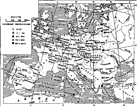
Figure 1.
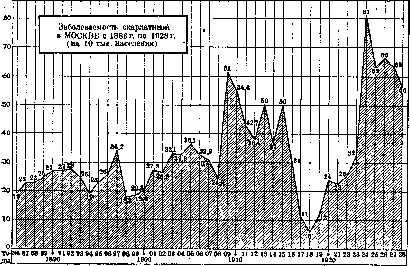
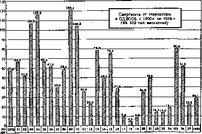
is not substantiated. Also does not withstand criticism is the theory, supported by a number of authors (Czerny, Kobrak, Schlossmann and others), about the connection between insufficient protein nutrition (in particular milk and eggs) and the decrease in S. We tend to connect this decrease in S. with the reduction in the child population as a result of the world war. Table 3. Mortality from scarlet fever among children under 15 years of age (per 100,000 children of corresponding age). Years Sweden Scotland 1861-70 ..... 1881-90..... 1891-1900 .... 1901-10..... 1911-20..... 1921-25..... 271 197 94 47 38 16 11 121 28 13 15 7 3 250 210 79 54 26 35 23 Scarlet fever in the USSR. In Russia before the world war, from 400 to 500 thousand cases of S were registered annually, per 10,000 inhabitants - from 21 to 32 cases. In terms of morbidity from S, pre-war Russia occupied an extremely high position compared with most Western European countries. After the world war, a noticeable decrease in it is noted. A new rise in S ended in 1930 and in the future a sharp decrease in it is noted (table 1). Morbidity in individual republics of the Union is not the same: it is highest in the RSFSR, Ukraine and Belarus, significantly lower S is registered in the Transcaucasian and Central Asian republics (table 4). Curves of morbidity and mortality from S in Moscow, Leningrad and Odessa give an idea of its movement in large cities of the USSR over a long period of years (fig. 6, 7). Morbidity from S in cities and rural areas. A number of researchers note the uneven spread of S. Table 4. Morbidity from scarlet fever in individual republics of the USSR (per 100,000 population). Republics 1924 1925 1926 1927 1928 RSFSR . . Ukraine .... Belarus . . Azerbaijan . Armenia .... TurkSSR . . . Uzbekistan . . 14.5 14.6 15.0 2.1 0.06 0.8 18.5 18.8 18.5 3.3 0.3 0.6 0.2 0.5 20.6 37.1 29.8 5.1 4.4 1.6 0.09 1.6 21.7 36.5 22.6 . 9.8 3.7 3.7 1.3 1.5 20.8 24.1 28.5 19.5 1.1 1.1 in cities and rural areas: in the former it is higher than in the latter. According to Reiter in Mecklenburg (Germany) for the period from 1903 to 1927, morbidity per 10,000 inhabitants was 13.7 in the city and 4.1 in rural areas. According to Vaughan, mortality in the US per 100,000 inhabitants in cities is approximately 2 times higher than in rural areas. Similar ratios (higher morbidity in cities) can also be seen in the materials on Moscow and the Moscow province (table 5). The uneven morbidity in cities and rural areas should be explained by social causes: the great overcrowding in cities, and consequently more favorable conditions for contact infections.- Morbidity and mortality by
Morbidity from scarlet fever
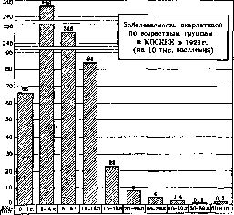
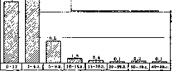
in BUDAPEST from 1882 to 1920 (per 10,000 inhabitants) Figure 2. age and sex. Corresponding materials are given in tables 5 and 6 (see figure 8, 9). Table 5. Morbidity from scarlet fever by age groups and sex in Moscow, Moscow province and Berlin (per 10,000 population of corresponding age). Moscow, 1926. Moscow Prov., 1926. Berlin, 1912-14, both sexes m. m. Under 1 year 1-4 years 5-9 years 10-14 years 15-19 years 20-29 years 30-39 years 40-49 years 50-59 years 60 years and over 46 99.1 102.7 144.6 78.7 19.2 Table 6. Morbidity and mortality from scarlet fever by age groups in Leningrad from 1/VII 1926 to 1/VII 1927 (per 100,000 population). Morb- Age Mortality Age Morbidity Mortality Under 1 year 1
»> 2 years 3 » 4 » 5 years 6 » 7 » 8 » 9 » 10 » 75.4 265.4 321.8 381.3 415.3 402.7 390.8 308.8 289.5 282.0 206.3 1 19.9 54.0 41.0 33.7 26.2 11.1 11.1 10.1 6.7 9.6 4.1 11 years . . . 12 » ... 13 » ... 14 » ... 15-19 years . 20-29 » . 30-39 » . 40-49 » . 50-59 » . 60 years and over 158.7 140.9 99.3 67.5 29.0 8.1 3.8 1.4 0.7 0.3 1/' 3.4 1.3 0.4 0.7 0.4 0.1 0.1 0.1 The most frequent morbidity is observed in the age group from 1 to 9 years (in children under 1 year it is significantly less frequent), the highest mortality in the age group from 1 to 4 years. According to detailed Leningrad data (table 6), morbidity is highest in the age group from 3 to 6 years, mortality from 1 to 4 years (this is consistent with data from foreign countries). On the susceptibility to S of individual age groups, one can judge from the epidemic of it in 1873-1875 on the Faroe Islands, where S had not been present for 57 years. A total of 38% of the population contracted scarlet fever (measles--99%). Hoff calculated morbidity on these islands per number of residents of certain ages, taking into account only persons exposed to the danger of infection (living in houses where there were scarlet fever cases). He obtained the following indicators (table 7). It should also be mentioned about the relatively high morbidity from scarlet fever in a number of armies (i.e. among adults) of those countries where morbidity from it is generally quite low. An example can be the French army, where in the last decade morbidity per 1000 people ranged from 1.56 to 2.64, and mortality from g iQQfi
°'009 to °'07- From 1886 to 1895, according to Doppler, mortality from S in the troops of the Paris garrison (20.4 per 100,000) was 3 times higher than that among men in Paris aged 20 to 24 years (6.5). Table 7. Age 0-1 1-5 5-15 15-20 Percentage of diseases 66.6 67.1 63.9 75.0 Age 26-40 years.-.. . . . 40-60 » . .' . 60 years and over Percentage of dis- eases 23.4 2.9 1.8
Case fatality from S shows significant fluctuations both in different countries and in the same country (city) in different years depending on the nature of the epidemic. If we start from the figures of registered cases of diseases and deaths, then e.g. in 1925 it in different countries amounted to the following percentage: in Belgium--23.0, in Italy-16.4, in Japan-11.0, in Czechoslovakia-4.0, in Germany-2.1, in England
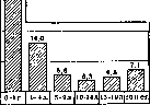
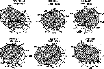
Figure 3. 2.1, in the USA-1.5, in Sweden-0.6. In Königsberg for the period from 1900 to 1915, it fluctuated in individual years from 1.4% to 23%, in hospitals of Moscow for the period from 1900 to 1928.-from 5.1% to 21%. In recent years in a number of countries (England, Germany, Denmark, Sweden, Switzerland, USA) S proceeds easily with little detail- nosti (1% and below). An idea of the fluctuations in case fatality by individual years is given in fig. 10. The height of case fatality is greatly influenced by the age factor: the younger the patient, the greater the danger of a fatal outcome. According to data from Moscow hospitals for ,210 '200 | 1 1 - ч in HAMBURG from 1851 to 1899 (per 100,000 population) ,1 M II 1 1 1 1 1 1 1 1 0 years 1m l |__jL_L )2 г в ) 7" \ / ! 50 1 /|| \ 35 1 / 37 \ / Г' \ / 2{ f 1 23 \ \ (. ' 16 \ / 1 6^ \ 1851 53 55 57 59 i 18 61 63 65 67 69 60 18 71 79 70 5 77 79 1 188 81 83 85 87 89 0 18 til 93 95 97 1899 90 Figure 4. 1900-28 years, case fatality in individual age groups amounted to the following percentage (table 8, fig. 11). Table 8. Age Case fatality Age Case fatality 5-9 years .... 39.2 16.0 5.6 10-14 years . . . 15-19 years . . . 20 years and over 3.3 4.5 7Д I I I I I I TT Morbidity from scarlet fever in RUSSIA and the USSR, for 1890-1928. (per 10,000 population) Similar data has also been noted in hospitals of Leningrad and Western European countries. Seasonality of morbidity. The main mass of scarlet fever cases occurs in the autumn-winter period, in summer their number is greatly reduced. This distribution of diseases is explained mainly by living conditions - greater overcrowding in autumn-winter months and, in connection with this, an increase in contact infections; according to some authors - meteorological. factors influencing the increase in these months of 'cold' diseases, which should facilitate easier perception of the scarlet fever infection. In fig. 12 are given monthly curves of S in some countries .(everywhere calculated average-daily monthly to average-daily annual). In the RSFSR and Ukraine the rise begins in September, the high level of scarlet fever in the RSFSR as a whole continues until March, in Ukraine-until January. In individual cities the maximum of the curve occupies a shorter period, for example in Moscow- € October to December.
I. Dobreytser. IV. Pathological anatomy and pathogenesis. The most characteristic reaction of tissue at the site of primary fixation of the scarlet fever virus is justifiably considered necrotizing inflammation, which in acute cases is most often identified on the autopsy table. More rarely, this process takes on a catarrhal character, and very rarely a fibrinous one (so-called scarlatinal diphtheria or croup). In this connection, it must be borne in mind that although mild cases of S. almost always proceed with catarrhal angina (with the usual localization of the primary scarlatinal affection), however, no reverse conclusion can be drawn from this, since the catarrhal reaction in S. (in contrast to diphtheritic catarrh) in no way ensures either a mild course or a favorable outcome of the disease. On the contrary, it is a well-known fact that the same reaction is to no lesser degree characteristic of the most severe, so-called toxic forms. Death from scarlet fever, with the exception of the rare toxic forms just mentioned, in the vast majority of cases is death from septicemia. Among other factors that may cause a fatal outcome, one should mention kidney lesions, erosions and perforations of the neck vessels due to the spread of purulent-necrotic processes from surrounding neck tissues, and finally pneumonia. Returning to the toxic forms, it should first be noted that the path.-anat. picture observed here turns out to be rather poor in characteristic features. The most striking in
Figure 5.
it, besides the rash, is the lesion of the upper part of the digestive tract, but even this does not represent anything specific. Usually this is a very sharp catarrh, which not only affects the fauces and pharynx but almost always spreads to a greater or lesser degree also to the esophagus. The mucous membrane of these parts swells, takes on a saturated dark red color, often with a cyanotic fi21 tint, and has a velvety appearance. The latter is explained by the necrosis of the superficial layers of the epithelium, which deprives the mucous membrane of its normal luster. If the necrosis of the epithelium goes somewhat deeper, then the color of the mucous membrane in such places accordingly acquires a more gray
FIG. 6.
ish g or yellowish tint. The tonsils usually swell more or less strongly, but this symptom never acquires predominant significance here. The surface of the tonsils retains the same appearance as the surrounding mucous membrane of the fauces, but on their section, necrotic areas are often found, standing out with their pale whitish color and located 6. ch. along the walls of the crypts, thanks to which they most often have the appearance of strips directed perpendicular to the surface. The swelling of the cervical lymph, glands in this form is also usually moderate, sometimes quite insignificant. The anat. substrate of it here, as in all other forms of S., is inflammatory edema and hyperplasia of both the follicular, and mainly reticulo-endothelial apparatus, occasionally with a rather leniently expressed myeloid metaplasia of the lymphadenoid tissue along the periphery of the follicles. The glands are tense, succulent, gray-pink or gray-red in color; necroses in them are rarely observed. B. ch. to this is added a reaction from the more distant parts of the lymph, system in the form of a moderate increase in the mediastinal, mesenterial and retroperitoneal glands, as well as the follicles and Peyer's patches of the intestines. The spleen in toxic forms reacts not strongly and mainly from the side of the lymph, apparatus, and in the most acute cases (so-called scarlatina fulminans) its increase may almost not occur at all. To the hyperplasia of follicles, partial necrosis of them may be added, thanks to which they stand out particularly distinctly with their whitish color against the dark red background of the congested pulp. In some cases, as in the pulp, so along the periphery of the follicles, myeloid metaplasia is clearly expressed. Even more often than in the follicles of the spleen, one can observe pinpoint necroses in the liver, where they are best seen from the surface in the form of pale-yellowish points, showing through the capsule. As microscopy shows, they are sometimes present in the liver tissue in very large numbers, being scattered without any particular order inside the liver lobules. It is also necessary to note the changes in the sympath. and parasympath. ganglia (mainly in the cervical sympath. nodes and in gangl. nodosum et jugulare n. vagi), indicating a significant lesion of the vegetative nervous system. They are mainly of a destructive nature and affect most of the ganglion cells, in k-rykh pictures of karyolysis and cytolysis of various degrees are determined, as well as
Figure 7.
degenerative fatty changes and necrosis. Inflammatory changes in the interstitial tissue here are almost absent. In other respects, the lesion of the body in this form amounts to a disorder of blood circulation in the form of general venous congestion and to protein and fatty degeneration of parenchymatous organs. T. o., on the one hand, superficial local changes, moderate reaction from the side of regional lymph, glands, absence of significant, and sometimes any, swelling of the spleen, as well as inflammatory changes in the stroma of internal organs, on the other hand, a sharply expressed fall in the tone of the vascular system - all this fully justifies the attribution of such forms of S. to the toxic group. The same is said by the bacteriol. data, since not only blood cultures from the heart give negative results in these cases, but sometimes streptococcus cannot be detected even in the fauces. Where death occurs in later periods, always, if not in the clinical symptom-complex, then in the anat. and hist. investigation, one or other septic features clearly emerge. The main signs of the septic form are:
[_DY
Figure 8. 1. Significantly greater intensity of local changes. Necroses here are always so deep that the underlying vascular network ceases to be visible, due to which the corresponding areas of the mucous membrane acquire a dull grayish or yellowish color. At first the mucous membrane is only dull, but soon after the separation of small particles of necrotized tissue it becomes rough. The deeper the necrosis, the larger the size of the separating particles, k-rye either form a layer of crumbly decay on the surface or, mixing with the mucopurulent exudate, turn into a mushy mass, sometimes very reminiscent of softening fibrinous films. The tonsils in this form can increase particularly strongly; at the same time they are usually necrotized on the surface and penetrated by necrotic foci in the depth. 2. A sharp reaction is observed from the side of the regional lymph, glands. The latter are greatly enlarged, very succulent, with a tense capsule, of a characteristic yellowish color, and always contain necrotic foci. Under the microscope, vessels in them are little noticeable; inflammatory edema, proliferation of reticulo- endothelium and myeloid metaplasia prevail. Often* in the glands, as well as in the tonsils, small foci of beginning purulent melting are found. Other parts of the lymph, apparatus
Mortality from scarlet fever by age groups in MOSCOW in 1926 (per 10,000 population)
Figure 9.
react in a similar way, although to a lesser extent than in the neck. 3. The spleen is significantly enlarged (2-3-5 or more times compared to normal), giving a typical picture of acute hyperplasia, also usually with a clearly expressed myeloid metaplasia of the pulp and the peripheral zone of the follicles. An exception to this rule are only very small children (up to 1'2 year) and rachitics, in k-rykh the spleen b. ch. reacts weakly. 4. Extremely strong degenerative changes (mainly in the form of dull swelling) of parenchymatous organs (liver, kidneys, myocardium, adrenal glands), in k-rykh under the microscope one can very often also discover an acute interstitial inflammatory process (so-called septic hepatitis, nephritis, etc.). Toxic and septic S. are, of course, images of extreme types, between which there exist countless transitions, and besides they concern only particularly severe cases, ending in death during the first week of the disease. As for the majority of patients dying from S, they end up on the autopsy table in
Mortality from scarlet fever in HAMBURG and KÖNIGSBERG from 1900 to 1927. (per 100 cases) 07 08 09 ,' 11 12 18 14 15 16 17 18 19 * 21 22 23 24 25 26 27 Figure 10. later periods, due to which the changes observed here differ significantly in greater number, variety and depth. This first of all applies to the primary necrotic process in the fauces and pharynx. The latter by no means always spreads over the entire surface of the organs mentioned, but very often is limited to individual 62<S Mortality from scarlet fever by age groups in hospitals of MOSCOW from 1900 to 1928 (per 100 patients)
Figure 11.

of very diverse size and shape. In some cases, as has already been indicated, the necrotic changes affect only the tonsils, in others they are found almost exclusively on the posterior surface of the soft palate and in the nasopharyngeal cavity, thereby escaping observation during life and complicating the clinical diagnosis; sometimes they reach their greatest development in the deep parts of the pharynx at the edges of the entrance to the larynx, causing disturbance of swallowing, etc. At the same time, the main and most important feature of the scarlatinal necrotic process is its tendency to penetrate deeply. Often from the very beginning the necrosis crosses the boundary of the epithelium, involving a larger or smaller part of the basal layer of the mucous membrane. However, in many cases this does not end there. Due to the fact that after the primary necrosis a true demarcation line often does not form, the necrotic process, even after the separation of the dead tissues, continues further, mainly in depth, but sometimes also on the surface, with varying intensity in different places. Thus, the resulting ulcers for a very long time do not clean up, but, on the contrary, continue to deepen, and their bottom, while retaining its necrotic character, at the same time acquires an extremely uneven, as if eroded appearance. The described progression of the process can continue for 2-3 weeks or longer, leading to extensive destruction of soft tissues, perforations of the soft palate, exposure of individual areas of the underlying bony and cartilaginous skeleton (the horns of the hyoid bone, the horns and edges of the thyroid cartilage, the posterior edge of the bony hard palate), to damage to the vessels passing here with subsequent bleeding, etc. The spread of the primary necrotic process to the esophagus occurs in approximately 10-12% of all autopsy cases of S. The changes are distributed here either without any particular order in the form of separate elongated spots along the length of the organ or more often occupy certain places, namely the upper part corresponding to the level of the larynx and the beginning of the trachea and the very lower segment on a length of 2-4 cm before the cardia. Necroses and ulcers in the esophagus are usually not deep, which is why their healing mostly occurs without noticeable deformities of the organ. However, as an exception, the scars that develop here can turn out to be so powerful that they cause narrowing, all the more severe the deeper and more widespread the original process was.
The stomach is affected significantly less frequently (in 3-4% of autopsy cases). The changes in it usually do not differ either in depth or in extent and most often manifest as the appearance of grayish or gray-red cloudy dry necrotic strips, located here and there on the tops of the folds. We never see the formation of chronic ulcers or perforations as a result of them, but occasionally they can give rise to the development of phlegmonous gastritis, which always ends fatally.-As for the respiratory tract, the nasal cavity and nasopharynx are most affected in it. Anatomically, the process is expressed here in a sharp catarrh with superficial necrotic changes and abundant purulent discharge. Occasionally this process also extends to the adjacent sinuses-the maxillary, ethmoidal, and in older children-the sphenoidal. As for the larynx and trachea, they usually remain free; however, this rule allows not so few exceptions, especially in certain epidemics, and the percentage of combined lesions of the respiratory tube (mainly the upper part of the larynx up to the vocal cords) with the pharynx can sometimes reach 6 or more, and in a small number of cases the larynx and trachea can even be the main site of lesion, in which the initial changes reach maximum intensity (the initial clinical symptoms are usually 128,*
with this, phenomena of false croup). As a rare exception, cases of scarlet fever are described in which the central point of primary changes becomes the mucous membrane of the bronchi or the pulmonary parenchyma, but such forms must, of course, always give rise to strong suspicion of one or another mixed infection and be treated with great caution. The lymph glands of the neck, in a prolonged process in the pharynx and throat, increase particularly strongly. In addition to those changes that have already been described, purulent foci often develop in them as well, which, increasing, may give rise to the transition of purulent inflammation to the capsule of the gland and further to the adjacent cellular tissue, i.e., to the manifestation of the so-called adenophlegmon. It should be noted, however, that purulent processes of various kinds (retropharyngeal and lateral pharyngeal abscesses, phlegmonas) can arise in different areas of the neck and quite independently of the glands due to the penetration of streptococcus from necrotic areas into the depth of tissues along lymphatic vessels. It is noteworthy that the tendency to early or progressively prolonged necrosis and in purulent processes sometimes manifests itself very clearly. This is expressed primarily in the development of the so-called 'hard phlegmonas,' when a certain area of tissue (cellular tissue, muscles, skin), after a sharp infiltration with leukocytes, does not undergo purulent melting as usual, but, while maintaining its density, completely dies and subsequently separates by demarcation inflammation, immediately exposing the organs beneath it. On the other hand, pharyngeal abscesses, even after their incision or rupture and evacuation of pus, sometimes do not granulate for a long time, but, on the contrary, continue to increase due to progressive necrosis and purulent inflammation in their walls. This feature leads to the fact that both in the primary necrotic process going from the mucous membrane of the pharynx, and in secondary purulent processes in the surrounding tissues, the blood vessels passing near the tonsils, arches, and lateral parts of the pharynx are comparatively often subjected to destructive changes due to the transition of the necrotic or purulent process to their walls. Of course, damage to arterial trunks is especially dangerous, as they almost always end in fatal bleeding. Most often, the arteries affected are the external maxillary artery, the ascending palatine artery, and the ascending pharyngeal artery. When the process spreads to the middle ear, it shows the same tendency to tissue necrosis and penetration into the depth; therefore, with no other infection does otitis give such a large number of successive lesions of the adjacent bone tissue as in S., not to mention its spread to the antrum. Of course, the mastoid process is most often affected due to the direct transition of the process from the antrum to the mastoid cells. However, purulent inflammation often spreads directly to the bony walls of the tympanic cavity. Of special importance among them are: 1) the bony plate separating the middle ear from the Fallopian canal, because from it the process can spread to the sheath of the facial nerve, and through it reach the internal auditory foramen and cause meningitis, and 2) the posterior wall, to which from the side of the posterior cranial fossa lies the sigmoid sinus. Necrosis of this wall always leads to inflammation or necrosis of the adjacent part of the sinus with subsequent purulent thrombophlebitis of it and with possible further development of multiple septic infarcts in the lungs and other organs. The most common localization of purulent foci are the joints and serous cavities, and among the latter—the peritoneum. Thus, purulent arthritis and purulent peritonitis most often figure among the complications of S. of a septicopyemic nature.-As for the later changes in organs not directly related to the area of the primary affection, among them deserve mention lesions of the cardiovascular system, liver, and kidneys.-In the heart, the most affected part, especially in septic cases, is the muscle, the changes of which are usually expressed in the form of interstitial myocarditis of moderate intensity. This myocarditis began to attract increased attention after some authors (Schmorl) pointed out that its histological picture in some cases can in places be very similar to the picture of Aschoff's rheumatic nodules. However, a number of authors (Aschoff and Tawara, Frenkel, Fahr, Talalaev) believe that this similarity is far from complete and that if one can speak of it, it is only in relation to atypical forms of rheumatic granuloma. The endocardium in S. is affected very little and very rarely despite the frequent presence of streptococcus in the bloodstream. Septic endocarditis of the valves, warty or ulcerative, occur in approximately 0.1-0.15% of autopsy cases and then mainly, apparently, where the endocardium has been altered by some previous pathological process (for example in rheumatics). The vessels are affected much more frequently. True, their changes are usually limited to individual small areas in the walls of small arteries and veins of various organs and are determined only histologically; however, in some cases where the process becomes more widespread and severe, their effect can also be clinically very serious, leading in the nearest months after the acute period of the disease to gangrene of the extremities, to renal insufficiency, cerebral hemorrhage and similar manifestations of arteriosclerosis. The morphology of vascular lesions consists of degenerative-necrotic and infiltrative-proliferative changes in the walls, and the size and intensity of each reaction can be extremely different. Apparently, most often here it is a matter of limited damage to the elastic membrane with a rapid subsequent proliferation of subendothelial connective tissue, to which thrombosis with subsequent organization sometimes joins. All this leads to more or less marked thickening of the inner membrane and narrowing of the lumen. However, in some rare cases, one can see either significant infiltrates penetrating the vascular wall over a certain space with very small alterative changes, or, conversely, necrotic areas involving a large part or even the entire thickness of the wall (in small vessels throughout the circumference, in large ones—usually within the limits of one or another segment). The pictures encountered in cases of the latter kind often very much resemble those characterized by the so-called nodular periarteritis (see). In the kidneys, in addition to glomerulonephritis in S., nephroses are also observed, especially in the first week in severely ill patients, and interstitial nephritides, as one of the expressions of septicemia (and both of these to a greater or lesser degree almost always join the glomerulitis as well). The interstitial nephritis, as well as the interstitial hepatitis, often observed in septic forms of the disease, do not differ from the corresponding lesions of these organs in any other kind of sepsis, except that in S. the infiltrates arising in their stroma often contain a fairly significant number of eosinophils. This sign, as well as the eosinophilia of the blood, constantly observed in S., constitutes a very constant distinctive feature of this disease. Scarlet fever septicemia, like any other, in some cases can take on a hemorrhagic character, causing the appearance of hemorrhages in various organs and cavities (skin, pleura, lungs, mucous membranes, cavity of the stomach and intestines, etc.), and can sometimes be accompanied by jaundice. The scarlet rash histologically does not present anything specific. Under the microscope in the skin, numerous foci of hyperemia, perivascular infiltrates, and small inflammatory edema are determined in it. The exudate, both liquid and partly cellular, usually penetrates the epidermis throughout its entire extent, giving it a cloudy appearance and sometimes accumulating between the cells in the form of very small vesicles (the so-called scarlatina miliaris). Subsequently, the exudate-impregnated epidermis gradually undergoes keratinization, which differs from normal in that the nuclei in the keratinizing cells do not disappear, and the cells themselves do not separate from each other, but remain in rather firm connection, forming on the surface a compact layer (the so-called parakeratosis). Due to this, the separation of keratinized cells under the influence of the increasing new cell mass from below does not occur in small, unnoticeable scales, as usual, but in much larger areas, which in places where the epidermal covering is generally thick (e.g., on the palms), reach especially significant sizes (T. H. LAMINAR desquamation).
M. Skvortsov. Pathogenesis, just like etiology, is still far from being studied. Each of the three theories mentioned above tries in its own way to explain the mechanism of the clinical picture of scarlet fever. Pathogenesis from the point of view of the streptococcal theory of the Americans is set forth above. According to the second theory (filterable virus in symbiosis with streptococcus), the pathogenesis of S. is more complex. The initial stage of the disease (fever, sore throat, rash) depends on the primary causative agent of the disease. Often the entire course of S. is limited to the indicated symptoms, and no signs of streptococcal infection are observed ("pure S." of French authors - Lesage). But apparently part of the complications is caused by the scarlet fever microbe ("scarlet fever heart", synovitis). Nevertheless, all purulent complications, septicopyemic phenomena depend on the streptococcus. The anaphylactic theory considers the initial stage as an anaphylactic shock, similar to the sudden onset of an attack of serum sickness. It is more difficult to explain from the point of view of this theory the second period of S. with late complications. It is assumed (S. Meyer, Schlossmann) that in the acute period anti-anaphylaxis occurs only partially, the remaining unused antibodies again combine with the antigen and give a picture of relapse and late complications. Clinical study of S, regardless of the acceptance of one or another causative agent, forces one to recognize that the pathogenesis of this disease is very complex; it consists of three moments, which either coexist simultaneously or follow one another: 1) toxemia, 2) septicemia, 3) allergy. The initial stage, according to the entire clinical picture, according to the blood picture (leukocytosis, neutrophilia), should be considered as an infectious toxicosis. Elements of toxicosis to a greater or lesser degree are present in all forms of S, even the mildest, but they are especially sharply expressed in the severe form, in its toxic and lightning-fast varieties. In this form, toxicosis manifests itself both from the side of the animal nervous system (disorders of consciousness, convulsions) and especially from the side of the vegetative system. Septicemia, or rather streptococcemia, according to some authors (Schleissner), can be detected even in cases of moderate severity; otitis and other purulent complications are often observed in these cases. But as a constant and prolonged state determining the entire picture of the disease, streptococcemia with purulent metastases in many organs constitutes a characteristic feature of the septic form. Finally, allergy since the research of Pirquet and Schick is almost universally recognized as a typical feature of the second period of S. Late lymphadenitis, nephritis, relapse of sore throat, complete relapse of S, late rashes, late polyarthritis, endocarditis with outcome in valve defects - all so-called late complications should more correctly be considered as a reaction of increased sensitivity of the scarlet fever patient to streptococcus or to the unknown virus of S. A typical time for allergic processes is the 3rd and 4th weeks of the disease. These processes in some cases can begin earlier, in others later, sometimes after 6 weeks depending on a number of conditions, mainly on the degree of sensitization of the body. V. Clinic. Incubation period - of indeterminate duration - from 1 to 11 days, more often 3-7 days. Clinical course is extremely diverse. Three forms are distinguished: 1) mild, 2) moderate in severity, or typical, and 3) severe, which is divided into a) toxic, b) septic and c) toxic-septic. Typical, or moderately severe, S. Prodromal phenomena are absent, in rare cases for 1-2 days general malaise is observed. It begins suddenly with complete health with an increase in t°, vomiting, headache, older children complain of pain when swallowing. On examination during the first 12 hours, the following are found: t° around 39°, severe weakness, repeated vomiting, pulse very frequent, disproportionately to t°, skin is clean, cervical glands are enlarged, painful, tongue is coated, in the pharynx there is bright hyperemia of the tonsils, uvula and soft palate, in the lacunae or on the surface of the tonsils small deposits. At the end of the first or beginning of the second day, a rash appears in the form of delicate pink hyperemia with small dots on the chest, neck and quickly spreads to the whole body; cheeks are red, but chin, lips and nose are pale. During the second day, the picture of the disease is fully expressed. 3-5th days - the period of the height of the disease, the highest development of all symptoms: t° 38-40° and above, sharply expressed headache and general weakness, mild delirium at night, but no disturbances of consciousness, vomiting stops by the 2nd day, no appetite, stool is delayed, rash is bright all over the body, pain and swelling of the cervical glands increase, deposits on the tonsils become membranous, take on the character of necrosis, pulse 140-160, but no signs of depression of cardiac activity. Starting from the 5th day, slow improvement and reverse development of all symptoms begins: t° drops daily by 0.5°-1.0°, rash fades, pharynx clears up, appetite appears, general condition improves and by the 8-10th day the patient seems to have recovered (fig. 13). Symptoms of the initial stage of S. Temperature. A characteristic feature of the temperature curve in S. is the rapid rise, constant t° for the first 4-5 days and lysis-like drop by the 8-10th day of the disease. If the drop in t° does not occur or during lysis a new rise is observed, this indicates some complication. In general t° in S. serves as a good indicator of the course of the disease. In rare cases t° on the 3-5th day drops critically (abortive S). Only in mild forms is t° subfebrile or even normal. Rash. The main element is small spots or dots against the background of general hyperemia of the skin (see separate table, fig. G). These dots are not due to hemorrhages, but correspond to places in the deep layers of the skin where hyperemia is more pronounced. The punctate nature of the rash can be more clearly revealed if the skin is stretched with the fingers of both hands and thus the general hyperemia is eliminated. With strong pressure of the finger on the skin, both the dots and the general hyperemia disappear. The distribution of the rash is typical. On the face, the nose, lips and chin are free from rash (scarlet fever triangle); on the limbs and trunk the rash is present everywhere, but it is more sharply expressed on the inner surfaces and in the flexures of the limbs, on the lower half of the abdomen. In these places the rash can be detected even in cases when it is generally very weakly expressed. Often at the height of the rash the skin takes a slightly yellowish tint; this tint appears more distinctly if the skin is strongly pressed with a finger. During the rash, a number of characteristic symptoms can be detected on the skin. White dermographism, or "white line" of French authors, is obtained if the back surface of the nail is drawn over the skin of the patient. It appears on the 3-4th day of the disease and lasts for some time after the disappearance of the rash; it is observed in almost all cases of S. (98% according to Molchanov). Its diagnostic value is small, since it is also found in other exanthematous diseases, but it has some significance for prognosis: absence, late appearance, sluggish character should be considered an unfavorable symptom. The mechanism of origin is not fully studied; white dermographism should be considered as a direct reaction to mechanical irritation from the skin vessels, which are in a state of moderate hyperemia (Molchanov, L. Mueller).- Rumpel-Leede symptom - pinpoint hemorrhages, which are obtained if the skin is pinched between the fingers or a rubber bandage is applied to the shoulder. The symptom is constant, but its diagnostic value is also small: it is observed in measles and some other rashes.-Phenomenon of fading of the rash (Schultz-Charlton): if 1/1-1 cm³ of serum from convalescents from S. or normal human serum is injected intracutaneously, blanching of the rash occurs at the injection site (see below - diagnosis). Anomalies of the rash are very common: late appearance, on the 3-4th day; scanty rash on limited, above-mentioned places; S. without rash in children is rare, more often in adults; sometimes the rash lasts only 1-2 days, even several hours; such cases present great difficulty in diagnosis. The form of the rash is also diverse. Along with the typical fine-pointed, miliary in the form of small vesicles is observed; papular or large-spotted - usually on the extensor surfaces of the limbs (Sc. variegata or "double rash" - Doppelexanthem - of German authors); petechial - in the form of small petechiae on the elbow flexures, in the groin; all these forms have no special significance for prognosis.
After scarlatinal rash, there is no pigmentation. Desquamation usually begins 2 weeks after the disappearance of the rash, but in the miliary form it appears even during the rash. Desquamation on the trunk is bran-like, fine; on the palms and soles it is large, plate-like (see separate table, fig. 2). Angina is the most constant symptom; it is absent only in extrabuccal or wound S. Its character and intensity are very diverse: catarrhal, lacunar, follicular, membranous (greatest difficulty in differentiation from diphtheria), necrotic. Regardless of the character of the angina typical for S., there is an intensely bright redness of the entire pharynx, uvula, soft palate, sharply demarcated at the edge of the hard palate. In the first hours of the disease, it is sometimes possible to see a pinpoint (and in serious cases with subsequent development of necroses-petechial) rash on the soft palate, which quickly turns into confluent redness (prodromal enanthema). Angina is accompanied by pain on swallowing, as well as more or less significant swelling and tenderness of the submaxillary lymph glands (lymphadenitis).-Cavity of the mouth. Hyperemia of the oral mucosa and coated tongue- '* л> Fig. I. Scarlatinous rash on the chest and abdomen, Fig. 2. Desquamation of the skin of the hands after scarlatina. Hyperpigmentation of the skin. (Fig. 3 after Grützner-Zeissler, Fig. 4. Scarlatina (desquamation). Fig. 5. Schwabach's reaction (reaction to iron). Fig. 6. Scarlatina. Stains from iron deposit. "38 also constant phenomena of S. Characteristic changes of the tongue: from the 3rd-4th day it cleans and the enlarged papillae become clearly visible on the surface (raspberry tongue).-Stomach-intestinal canal. Vomiting, single or repeated, is observed in the first hours of the disease in 70-80%. Vomiting lasting into the second day usually indicates severe intoxication. Stool is delayed, diarrhea is observed in infants and in toxic cases.--Liver and spleen are slightly enlarged.-Urinary organs. The amount of urine is reduced. Protein and cylinders (toxic nephrosis) only with severe intoxication. Diazoreaction is absent. The amount of urobilin is increased.-Respiratory organs. Cataracts of the respiratory tract are usually absent in the initial stage of S. With cough and runny nose, all the more with tracheo-bronchitis and pneumonia, one should think of mixed infection, most often influenza.--Heart-frequent pulse, not corresponding to t°, slightly elevated blood pressure, muffled tones, sometimes functional nature murmur. After the fall of t° the so-called scarlatinal heart (see below). Blood. In the initial stage, the typical blood picture: leukocytosis b. or m. significant depending on the strength of the infection and the body's reaction (10-30 thousand), neutrophilia, sharp shift to the left (up to 30 and above instead of normal 8%). Starting from the 2nd-3rd day of b-ny-increase of eosinophils (up to 15% and above). Changes in other elements are less typical and insufficiently studied. The so-called inclusions or Dohle bodies in the protoplasm of leukocytes-constant phenomenon in the initial stage of S. In cases of moderate severity, if there are no complications, the blood picture returns to normal by the end of the 2nd week. Fluctuations in severity and the appearance of complications are sharply reflected in the blood picture.-Nervous system. From the side of the central nervous system in cases of moderate severity-indistinct phenomena of excitation or depression (delirium, drowsiness); convulsions at the beginning of the disease only in neuropaths and infants of breast age (spasmophilia). From the side of the autonomic nervous system in the initial stage, symptoms of sympathicotonia are characteristic (rapid pulse, elevated blood pressure), which after the fall of t° are replaced by vagotonia (see genesis of "scarlatinal heart").-Deviations from the typical picture in the general course of the b-ny are extremely diverse: from the mildest forms, when the symptoms of S. are barely expressed, to those in which the patient dies within 1-2 days. Mild S. Phenomena of intoxication are negligible or absent, local changes in the pharynx are insignificant. The onset is the same as in moderate severity, sudden, often with vomiting. Temperature does not exceed 38.0-38.5°, subfebrile or even normal. Rash is either clear typical, with all the features of scarlatinal rash, or weakly expressed. Angina according to b', ch. catarrhal or with insignificant rapidly disappearing superficial coatings, no necroses. For the mild form, rapid reverse development of all symptoms is typical-by the 5th-7th day of b-ny. Frequency varies depending on the nature of the epidemic; in the last epidemic of 1924-30 in the children's clinic of I Moscow University it was 30-40% of all cases of S. The practical significance of this form is great. It sometimes remains undiagnosed and for the surroundings becomes a source of epidemic, while for the b-ny himself complications (lymphadenitis, nephritis) are unexpected. Mortality is insignificant, from complications; does not exceed 1-2%. Severe S. The toxic form is characterized by a sharply expressed toxicosis from the side of the central nervous system and cardiovascular apparatus, while the local lesions of the pharynx and lymphatic glands of the neck are expressed relatively moderately. The onset is stormy, intractable vomiting, continuing also on the second day, diarrhea. Consciousness is clouded, the patient is either in a state of lethargy close to coma, or excited, tosses in bed, there are convulsions. Vessels of the sclera are hyperemic, lips are cyanotic. Rash is not abundant, appears sluggishly, with a cyanotic tint; white dermographism is absent; if obtained, it is weakly expressed with an elongated latent period. Extremities are cool. Pulse is not only extremely frequent (up to 160 and more) but it is very weak, blood pressure is lowered. Tones are muffled, sometimes embryocardia. At the same time from the side of the pharynx-not too extensive necroses, neck glands are enlarged, painful, but do not show a tendency to fuse with each other and with the skin. In rare cases-death within 1-2 days (hypertoxic, so-called fulminant S.) (fig. 14). Usually the patient dies on the 3rd-5th day with signs of cardiac failure (fig. 15). In some cases with a violently begun picture of toxicosis around the 5th day, detoxication occurs, and the patient recovers. Such a favorable outcome is most often observed after timely and sufficient introduction of serum (see treatment).
Thanks to deep and extensive necroses, streptococci penetrate into the blood, flood the body (streptococcemia in blood cultures) and lead to a picture of sepsis and septicopyemia. In some cases, the patient dies on the 10-12th day, before the lymphadenitis reaches suppuration, and on autopsy, in addition to necroses in the pharynx and glands, one finds purulent inflammation of the ears, paranasal sinuses, sometimes joints, and purulent sinusitis and arthritis, especially of small joints, may remain unrecognized during life (fig. 16). In other cases, the disease drags on, suppuration develops in the glands, ears, mastoid process, in joints, mostly in large ones (hip), sometimes pneumonia with purulent pleurisy, purulent meningitis, etc. The patient also dies after a more or less prolonged time from the onset of the disease. In rare cases, despite multiple purulent foci and prolonged course (2-6 months), the patient recovers, but more or less serious defects may remain (deafness, immobility of joints). The frequency of septic S. is about 4-6%, mortality is high: 50-80%, especially so in children under 2 years. Toxico-septic S. (mixed form) begins as toxic, but around the 3-5th day, severe lesions of the pharynx and glands develop. In some cases, the picture of the disease is dominated by toxicosis phenomena, in others - local changes and septicopyemia. Frequency about 5%, mortality average between toxic and septic (about 60%). The frequency of all varieties of severe S., as well as mild, varies mainly depending on the nature of the epidemic; in Moscow, during the 1920-30 epidemic, about 15%. Extrabuccal (rh surgical) S. The site of infection entry is not the pharynx, but damaged skin (after burns, abscesses, etc.). A characteristic feature is the absence of angina (enanthema may be present) with the presence of typical scarlatiniform rash on the skin. Some are reluctant to consider the scarlatiniform rash, e.g., after a burn, as true S; indeed scarlatiniform rashes can have various origins. However, epidemiological data, the appearance of complications characteristic of S, leave no doubt in the existence of extrabuccal, including burn, S. The course is usually mild; in exceptional cases, a severe course, even with fatal outcome, has been observed. According to some (Escherich and Schick, Sokolov, etc.), the site of entry of scarlatinal infection can also be the lung. In such cases, with rapidly developing pneumonia, a scarlatiniform rash appears; changes in the pharynx are insignificant. On autopsy, one finds foci of necrosis in the lungs, no necroses in the pharynx (pneumo-scarlatina). The existence of such S. cannot be considered proven. Complications. By time of appearance, one can distinguish early (within the 1-2nd week) complications and late (on the 3-4th week and later). This division is partly justified by the difference in pathogenesis. Early complications develop from the symptoms of the initial stage of S. or directly follow it. Their origin is based on scarlatinal toxemia or streptococcal infection. Late ones are often separated from the initial stage by an afebrile period, during which no manifestations of scarlatinal infection may be present. According to modern understanding, they represent an allergic reaction of a sensitized organism to the scarlatinal microbe. According to the view of Pirquet, Schick, etc., the 3rd and 4th weeks are a period of increased sensitivity to this microbe; German authors call this period with a series of late complications 'the second scarlatina,' because many complications of this period repeat the symptoms, and sometimes the entire picture of the initial S. A special position is occupied by complications from the heart; they occur both in the early and in the second period of the disease. The clinical picture and pathogenesis differ depending on the stage and severity of S. Early complications. Cervical lymphadenitis is the most frequent complication (40% according to the material of the pediatric clinic of 1st Moscow University). Swelling of the cervical glands is essentially one of the constant symptoms of the initial stage of S. It is considered a complication when the swelling reaches significant size and is very painful. In general, a parallelism is observed between the intensity of the pharyngeal lesion and the size of the lymphadenitis; however, sometimes there is no parallelism, and the lymphadenitis proceeds as if independently, regardless of the condition of the pharynx. The usual outcome is regression, much more rarely - suppuration (3%). Signs of purulent infiltration: rise in t°, the glands become immobile, adhere to each other and to the skin, the skin reddens, softens, fluctuation appears. Sometimes (in septic S.) the glands and surrounding connective tissue, undergoing inflammatory infiltration, form a dense tumor, which does not show signs of softening; on section - necrosis of the glands and connective tissue; necrotic masses separate spontaneously, exposing the deeply lying organs of the neck ('dry necrosis'). Such forms of lymphadenitis have a poor prognosis. The serious significance of this complication is due, firstly, to the possibility of streptococci penetrating into the blood; secondly, to the sometimes fatal bleeding from the vessels of the neck, which are destroyed by the necrotic process. - Necrotic angina (scarlatinal diphtheroid, 30%) (fig. 17). In moderately severe S., very frequent, in septic - constant symptom of the initial stage. As a complication, it develops after the 4-5th day of the disease; due to it, the fall in t° is delayed for a long time. The deposits have a cream-like appearance, do not rise or rise only slightly above the level of the mucous membrane. Although the external resemblance to diphtheritic deposits can be great, but in essence these are two different processes: diphtheritic bacilli are absent, fibrinous exudation is weakly expressed, unlike diphtheria; the essence of the process comes down to deeply penetrating necrosis of the mucous and submucous (see separate table, fig. 3); after the deposits separate, deep ulcers, perforations and defects remain in the tonsils, soft palate, uvula. One of the most serious complications; the danger here also lies in the possibility of sepsis, fatal bleeding from the vessels; more often the inflammatory process directly penetrates into the middle ear and paranasal sinuses. - Necrotic (aphthous) stomatitis (20%). Superficial necroses of the mucous membrane of the tongue and oral cavity, sometimes small in size, sometimes more extensive. After the necroses heal, painful ulcers remain. The significance of this complication lies in the difficult feeding of the patient. Otitis (purulent inflammation of the middle earg 15%). In septic S., it appears already in the 1st week as a result of the direct transition of the necrotic process from the pharynx. In moderately severe and mild form, usually in the 2nd or at the beginning of the 3rd week, more rarely - later; with late otitis, an exacerbation of angina often precedes it. Symptoms: rise in t°, weakening of hearing, pain on pressure on the tragus; children often do not complain of independent pain, which distinguishes scarlatinal otitis from influenza or measles otitis. Another difference is the rapid formation of purulent exudate and spontaneous perforation of the eardrum. With favorable course, the process ends in 2-3 weeks. Unfavorable outcomes: 1) often transition to the mastoid process (see Mastoiditis), more rarely thrombosis of the sinuses and purulent meningitis, 2) chronic purulent discharge with significant weakening of hearing. When the inflammatory process spreads to the inner ear - complete deafness, which in small children leads to deaf-mutism. According to statistics of German otologists 1!3-7g of the deaf-mute became so after scarlet fever, contracted in early childhood (Manasse). Sinusitis (ethmoiditis, frontal sinusitis, maxillary sinusitis, 3%). More often ethmoiditis and frontal sinusitis. In septic S., they appear in the 1st week, but clinically do not manifest; suspicious are profuse purulent discharges from the nose. The usual outcome is death from general sepsis. In moderately severe - on the 2-3rd week. According to Killian, late sinusitis begin already in the early period of S. Symptoms: rise in t°, abundant discharges from the nose, swelling and pain in the area of the sinuses; with ethmoiditis - at the upper-inner angle of the eye and on the upper eyelid, sometimes protrusion of the eye, with frontal sinusitis - in the area of the eyebrows, with maxillary sinusitis - on the cheek and on the lower eyelid.
Outcome - most often complete recovery, in rare cases - empyema of the sinus, osteomyelitis, purulent meningitis. - Synovitis (scarlatinal rheumatoid, 8-10%). Appears at the end of the 1st or on the 2nd week. Primarily small joints (handsg fingers, etc.) are affected. Symptoms: elevated temperature, all signs of inflammation of the synovial membrane of the joint: pain, redness, swelling. Exudate is sterile. The genesis is not precisely clarified, probably of a toxic nature. One of the most favorable complications. Outcome - spontaneous and complete reversal. It should be distinguished, first, from purulent arthritis as one of the manifestations of septicopyemia in septic S., second, from late polyarthritis (see below) (fig. 18).-Laryngitis, tracheo-bronchitis, pneumonia, pleurisy. Complications from the respiratory organs are considered rare in S. Laryngitis in the form of necrotic ulcerative is observed exclusively rarely in septic S. with extensive necroses. It can lead to stenosis and require tracheotomy. In the pediatric clinic of the 1st Moscow State University due to a recurrence of stenosis on the 5th week, a tracheotomy was performed, and the child recovered. Pneumonia is more common (5-6%), but their time of appearance, etiology, and outcome differ. In septic S., pneumonia develops during the 1st week (streptococcus) and gives an extremely poor prognosis. In the recovery period, pneumonia depends on secondary infection, usually influenza (pneumococcus), prognosis is not poor. Some complications, e.g., nephritis, predispose to the development of pneumonia and worsen the prognosis. In infants, pneumonia occurs especially often, from which the high mortality of these children during S. mainly depends. All of the above also applies to purulent pleurisy, which are a frequent companion of pneumonias. Complications from the liver are observed not infrequently. Clinically they manifest as enlargement of the liver and jaundice. Enlargement of the liver often appears in the first days of the disease and may remain for an indefinitely long time. It depends, on the one hand, on parenchymatous and interstitial hepatitis; hepatitis can be detected long after S., cases of its transition into cirrhosis have been described. On the other hand - enlargement of the liver of a congestive nature due to circulatory disorders. The time of appearance and pathogenesis of jaundice differ. With a pronounced rash, already in the first days, an iceric tone of the rash can sometimes be noted. The origin is partly hematogenous due to increased breakdown of erythrocytes, partly hepatogenous as a result of damage to the liver parenchyma by scarlatinal poison (urobilinogen in urine, increased bilirubin content in the blood). Jaundice observed in septic S. has the same clinical features (colored stool) and the same genesis as septic jaundice of other origins. Finally, in the recovery period, jaundice of apparently congestive nature (discolored feces) is observed. As a cause of such jaundice, cholecystitis, enlargement of the glands in the area of the liver gates (Pospi-schill) are indicated. In some cases, jaundice appears as a combination of S. with infectious or so-called catarrhal jaundice. Late complications. Lymphadenitis - the most common of late complications. According to Pospishil, 50% of all manifestations of the second S. It differs from early lymphadenitis, besides the time of appearance, by localization: most often the glands near the sternoclavicular muscle swell; moreover, the outcome in suppuration is observed less often (less than 1%). It is observed both as an isolated complication and in combination with other late complications - nephritis, exacerbation of angina and catarrh of the nasopharynx. It is accompanied by elevated temperature, which sometimes takes an intermittent character. Nephritis. Kidney damage occurs in the initial [stage of S., but almost exclusively in the severe form, or in the form of toxic nephrosis ('fever albuminuria') in toxic S. or in the form of septic interstitial nephritis in septic S. The clinical picture of early damage is not prominent against the background of the severe general disease and does not represent anything characteristic of S. When speaking of scarlatinal nephritis, they mean late kidney damage, which appears on the 3-4th week, sometimes even later (5-6th week), very rarely on the 2nd week, most often around the 20th day of the disease. Patho-anatomically, this is acute glomerulonephritis or nephrosonephritis (see Nephritis). The frequency according to literary data ranges from 2% to 20%; in most authors around 10%. In the pediatric clinic of the 1st Moscow State University out of 1,054 scarlatinal patients, nephritis was observed in 128, i.e., in 12.1%. The onset in some cases is acute: elevated temperature, headache, vomiting; simultaneously - changes in urine: it is scanty, macro- and microscopic hematuria, protein in a moderate amount (0.1-2%), cylinders. In other cases - without elevated temperature, if there are no other complications; the first symptom is weight gain, change in urine and edema, first on the face. With regular measurement of blood pressure 2-3 days before the change in urine, a significant increase (115-130 mm) can be detected, and even earlier with capillaroscopy - spasm of skin capillaries (O. Muller, Bossé). Duration - on average 3-6 weeks. The usual outcome with proper care - complete recovery. Unfavorable course and outcome are very rare; they are caused by: 1) uremia of eclamptic type (see Uremia) (about 5-10% of all cases of nephritis), 2) decompensation of the heart (myocarditis), 3) transition to chronic form and 4) accompanying other complications of S., e.g., pneumonia. Mortality varies among authors, in general it is small. According to Pospishil - 8%, according to the material of the pediatric clinic of the 2nd Moscow State University - 4.5% (Girschberg and Sukhareva). Mortality is higher in cases where patients were already admitted to the scarlatinal department with nephritis; mortality of such cases according to Pospishil = 21.8%. This fact is probably explained by improper care in the first days of the development of nephritis. Mortality from uremia also varies greatly; according to the material of the pediatric clinic of the 1st Moscow State University it = 0%, in the pediatric clinic of the 2nd Moscow State University out of 11 patients with uremia 1 died (Girschberg and Sukhareva); according to other authors mortality is significantly higher (up to 40%). How often scarlatinal nephritis transitions into chronic form is not precisely known; according to Rosenfeld out of 95 patients who left the clinic of Escherich with remnants of scarlatinal nephritis, in none was a transition to chronic form observed, although the condition of the patients remained chronic lordotic albuminuria. Patients were observed in whom for 3-5 years after S. hemorrhagic nephritis with repeated exacerbations remained; patients nevertheless recovered with complete restoration of the functional capacity of the kidneys (Molchanov). Such observations were reported by Escherich, Heubner and others. Very mild, rudimentary forms of nephritis with slight albuminuria and hematuria lasting for a short time are encountered. In these cases, both weight gain and increased blood pressure, as well as the spastic state of skin capillaries can be detected. The study of scarlatinal nephritis and skin vessels in various periods of S. (histological, capillaroscopy) confirms the modern view (Volhard, Kylin and others) on the pathogenesis of glomerulonephritis, according to which glomerulonephritis represents a partial manifestation of general vascular damage (capillaropathia universalis), primarily of skin capillaries, which on the 3-4th week of S. show a constant tendency to spastic state (Bossé) (fig. 19).-On the 3-4th week, edemas sometimes appear, either obvious on the face, legs or hidden, which can be detected with daily weighing of the patient (weight gain of 500-1,000 g). The urine shows no pathological changes, blood pressure is not elevated, kidney function is not impaired, almost always there are symptoms of scarlatinal heart. In some cases, the appearance of edemas is accompanied by lymphadenitis and elevated temperature, more often the temperature is normal. These are the so-called protein-free edemas. They disappear after 1-2 weeks. The mechanism of their origin (see Edema) in S. is complex.
First place must be given to the change in capillaries, then to the disturbance of metabolism and violation of the equilibrium of colloid osmotic pressure of blood and tissues; less importance is attached to heart damage. Late polyarthritis is a little-known to physicians and insufficiently studied complication. It is observed in the 3rd-6th weeks of S. and even later. Unlike early synovitis, the large joints are mainly affected: knee, ankle, shoulder, elbow, etc. Duration-2-3 weeks and more; it shows a tendency to relapses; despite prolonged existence and relapses of functional disorders and persistent changes, it does not remain. It is often observed in combination with other late complications: with nephritis (in Szontagh's 15 cases), relapsing angina, lymphadenitis, etc. Simultaneously with polyarthritis or directly following it, a benign endocarditis often develops with an outcome in valve defects (in Molchanov's 14 cases). See below on the relationship to rheumatism. Relapsing angina. The character of angina is varied - from catarrhal without temperature elevation to necrotic with severe and prolonged course and high temperature. In some cases-outcome in abscess of the tonsils (phlegmonous angina) or in retropharyngeal abscess. In most cases, angina in relapse is milder than the initial angina, but the opposite also happens; it appears, like other late complications, most often in the 3rd-4th week. Its frequency in reality is higher than that given by the statistics of various authors, since mild forms are usually not taken into account. According to Pospischill, relapsing angina constitutes 18% of all manifestations of the second S. According to the materials of the children's clinic of I Moscow State University, it is observed as often as late lymphadenitis.--Complete relapse of S., i.e., return of S. with all its cardinal symptoms: elevation of temperature, angina, and typical rash. A necessary condition for recognizing the new appearance of these symptoms as a relapse of S. is an unquestionable diagnosis of S. in the initial stage. The earliest time of relapse of S. according to the material of the children's clinic of I Moscow State University is the 11th day of the disease, the latest is the 7th week, in most cases according to the observations of all authors-the 3rd and 4th weeks. In some cases it can run more severely than the initial S, even with a fatal outcome. It is observed more often in mild S. than in severe. It often appears in combination with other late complications: nephritis, polyarthritis, etc. A relapse is probably favored by the addition of a new infection to S., e.g., influenza, measles, chickenpox. There are indications of familial predisposition (in several children in families; in twins in the children's clinic of I Moscow State University simultaneously). After a relapse, new peeling and complications characteristic of the initial stage may appear (e.g., otitis). Frequency, according to Pospischill-in 2,605 cases, 46 relapses, i.e., 1.76%. Frequency according to other authors ranges from 0.3% to 4%.-Pathogenesis is insufficiently studied: some (Henoch, Comby) consider it as a flare-up of the unextinguished scarlatinal infection, others (English authors, Pospischill, Rauchfus, etc.) as a result of a new infection with the scarlatinal virus; the theory of Schick about the allergic origin of the relapse (fig. 20) should be considered more probable. temperature 1 5 d n i !1 b l e 16 ! 17 3 ! N 1 V 18 |19 |2 D 21 i 22 I 28 ... | 28 Y v Y v yv YV3 . v. y. v Y v Y-v Y v Y v Y1 Y v Y. v. Y v- Y 1 Jj'-B-iyi* Y v.1, y. v. y. v. Y v- |y. v. ye. Y v Y v Y v Y v Y v Y v Y v 42° 41.8 41.4 41.2 40.6 39.6 39.4 39.2 38.8 37.8 87.v 87.4 87.2 86.8 36.8 36.6 85.4 гг 1 I I 1 1 я1 n iff | :5 lu _she ni e. 'г лпь- :_, i -t jt , ч \, \ ? \ /' / Q-, t ' V \ \ / ,.' '. I \ ,, "Ч >\ E5: / i~ <" ' / y ч \ ' гХ _ 1 , Figure 20. Mild form with relapse. Late rashes (Fanconi), post-scarlatinal erythema (Schick) or false relapse (Thomas). On the 2nd-3rd-4th week and later a rash appears, differing in appearance from the fine-pointed scarlatinal one. Fanconi distinguishes four main varieties: 1) maculo-papular, 2) reticulate-streaked, 3) large-papular, and 4) pityriasis-like. Sometimes it has the appearance of urticaria. It is often observed simultaneously with elevation of temperature, angina, nephritis, and other complications. Localization-buttocks, extensor surfaces of the limbs, trunk, sometimes face. Duration from 3-5 days to 2 weeks. The basis of pathogenesis is the same allergy as for other late complications. According to Fanconi, other factors also play a role: mechanical irritation and local changes in the skin under the influence of S. Complications from the cardiovascular system. Initial period. In moderate forms-increased pulse rate, not corresponding to temperature, elevated blood pressure, transient functional murmurs. Pathogenesis-irritation of the sympathetic system under the influence of the scarlatinal poison. In toxic S-pulse is extremely frequent and weak, lowered blood pressure, heart dilation, dull tones, decline in cardiac activity up to collapse. Pathogenesis-severe damage to the sympathetic system (Abricosov, Mogilnitsky) and adrenal glands (Hutinel, Molchanov), i.e., the entire sympathico-adrenal system. In septic S. simultaneously with this, organic changes in the heart muscle itself are observed, and the longer the patient lives, the more sharply these changes appear both in the clinical picture (weak pulse, dull tones, heart dilation, congestive liver) and at autopsy (interstitial myocarditis). In septicopyemia, septic endo- and pericarditis is sometimes added to this, which is sometimes discovered only on the dissecting table. Period of recovery. After the fall of temperature at the end of the 1st week, more often on the 2nd-4th week, regardless of the severity of the initial period, disturbances of cardiac activity appear, which are called 'scarlatinal heart'. The pulse, which was normal or rapid, becomes slow and arrhythmic. The tones become prolonged, dull, and impure; later the impurity can turn into a real murmur, simultaneously heart dilation appears. A characteristic feature is the variability and changeability of these symptoms. The general condition does not suffer or is disturbed to a slight degree (paleness, lethargy). Duration of existence-from several days to 2-4 weeks and more. The prognosis is completely favorable. Frequency according to the observations of various authors is very diverse: according to Schick-5%, according to Koltypin-almost 100%. According to the material of the children's clinic of I Moscow State University-about 35%, if only definitely expressed cases are counted. Pathogenesis-disturbance of the equilibrium between the sympathetic and parasympathetic systems; the excitation observed in the initial stage of S. is replaced in the recovery period by its weakening, as a result of which the vagus takes over, i.e., 'scarlatinal heart' is considered as a symptom of vagotonia (Molchanov, Koltypin, etc.); it is impossible to deny, at least for some cases, the possible participation of myocarditis as well (Romberg, Filatov). The existence in S. of the so-called benign (non-septic) endocarditis with an outcome in valve defects is completely denied by some (Pospischill), others (Heubner, Feer, Schick) recognize it. Its pathogenesis is also understood differently: Schick, Hallez, Comby consider it a complication of S. itself. Zischinsky-result of a random combination of S. with rheumatism. According to Molchanov, endocarditis with transition to valve defects is observed in S. more often than is generally thought, but its connection with S. is not always established, since it sometimes appears in the 5th-6th week and even later. Often endocarditis develops simultaneously with other late complications of S.-nephritis, relapsing angina and S., most often with late polyarthritis (fig. 18). Genetically it is closely connected with the scarlatinal process and it can be considered as one of the late complications of S, at the same time according to the clinical picture and path-anatomical changes (Aschoff's granulomas) it is a rheumatic endocarditis. 64B More rare complications of S. Nervous system. In addition to toxic and septicopyemic damage to the brain and its membranes in the initial stage of severe S. and the characteristic of all forms of S. damage to the sympathico-adrenal system, in rare cases in the late period of S., hemiplegias of cerebral origin appear, sometimes with aphasia and hearing loss.
In most cases, other late complications are observed simultaneously with them - nephritis, endocarditis. The anatomical cause is encephalitis or circulatory disorders (embolism, thrombosis, hemorrhage). Even rarer is the involvement of the striopallidal system with symptoms of parkinsonism. Older clinicians (Trousseau) often observed chorea as a complication of S. At present time, it occurs less frequently as one of the manifestations of the rheumatic syndrome developing after S. (Molchanov). In rare cases, polyneuritis with flaccid paralyses, loss of tendon reflexes must be observed. Endocrine glands. In addition to the above-mentioned lesion of the adrenal glands, S. can apparently lead to the manifestation of constitutional inferiority of various glands with internal secretion: the thyroid gland with symptoms of either hyperthyroidism (Basedow's disease in girls) or hypothyroidism, the pituitary gland (acromegalic gigantism), the pancreas (diabetes) and others. Cases of gangrene of the extremities of the Raynaud disease type as a result of vascular lesions, cases of appendicitis and others have been described. S. in infancy. Age has a great influence not only on the incidence of the disease (see above - predisposition), but also on the course, frequency and nature of complications, as well as the outcome of S. The infancy age presents the most deviations from the typical course of the disease. The main feature is that the symptoms of toxemia are less pronounced, while septicopyemia is significantly sharper than in older children. The temperature, if there are no early complications, is not high, falls faster, the rash is less bright, atypical, and lasts relatively short; the white dermographism is often absent regardless of the severity of the course; desquamation also very often (40% according to Sokolova) does not occur; angina often has only a catarrhal character. Among the complications - synovitis, nephritis, 'scarlatinal heart' occur much less frequently. Purulent complications - lymphadenitis, otitis, sinusitis - occur more often and proceed more severely. But what is especially characteristic for S. in infancy is the frequency of pneumonias and their dangerous nature. Mortality is very high: 20-40%; it depends primarily on pneumonias, then on septic complications. S. and other infectious diseases. If other infectious diseases are added to S., they as a rule influence each other and this influence is the more pronounced the closer the new infection is added to the initial stage of S. S. + measles is the most frequent combination. When both rashes appear simultaneously, the diagnosis is especially difficult. If measles is added after the disappearance of the scarlatinal rash, a delayed appearance of the measles rash and a flare-up of the scarlatinal rash are observed. In the late period of S., the mutual influence is less pronounced, however, recurrences of angina and recurrences of S. itself have been observed (Pospischill, Molchanov). The number of complications under the influence of measles increases, this especially applies to pneumonias. Mortality with mixed infection is generally higher (Volovin). The widespread use of preventive vaccinations, according to Degkwitz, in recent years has sharply reduced the danger of mixed infection. S. + chickenpox. Sometimes a severe course of chickenpox with abundant and deep pustules is observed; an outcome in septicopyemia is possible even with moderate scarlet fever. S. + diphtheria. If diphtheria is added in the initial stage of S., the diagnosis presents great difficulties. Although in typical cases scarlatinal necroses and diphtheritic membranes differ from each other, however, in septic cases of S., widespread necroses having the appearance of fibrinous membranes are sometimes observed. Bacteriological research also does not always resolve the recognition, since it has long been known (Baginsky, Shabad) that in the initial stage of S., rods morphologically similar to Löffler's rods can be quite often isolated from the pharynx. Depending on the prevalence of the diphtheria epidemic, the frequency of this combination can reach 6% or more (Shabad, Roily). In any case, with the slightest suspicion of mixed infection, anti-diphtheritic serum must be injected. Diphtheria, occurring during the period of convalescence, differs little from the usual course and does not present great difficulties in recognition. Mortality, especially in small children, is significantly increased (croup, pneumonias). S. and rheumatism. It was said above that in the 2nd allergic period of S., polyarthritis and endocarditis are sometimes observed as late complications. According to the clinical picture, course, outcome and patho-anatomical changes, they should be considered rheumatic. The appearance of the rheumatic syndrome in S. cannot be regarded as a coincidence of two infections or as an exacerbation of rheumatism under the influence of S., although S. really has the ability to exacerbate the rheumatic process. According to Molchanov, the connection between S. and rheumatism is genetic. The allergic theory of rheumatism best allows to understand this connection. During S., an allergic restructuring of the organism in relation to the streptococcus occurs, as a result of which, in the presence of predisposing conditions (rheumatic heredity, previous sensitization under the influence of anginas, living conditions), a certain category of children by the end of the disease can turn into typical rheumatics. The latest patho-anatomical research speaks in favor of this view. Fahr, Siegmund, Pashchin and Krich found Aschoff granulomas in the heart muscle during autopsies of scarlet fever patients. The problem of the connection between S. and rheumatism requires careful collective study by patho-anatomists, clinicians and dispensary doctors. VI. Diagnosis and prognosis. In typical cases, the diagnosis of scarlet fever does not present difficulties. The main points in the initial stage are the sudden onset with high temperature, vomiting, pain in the throat; angina with bright redness of the entire pharynx; enlargement of the neck glands; a fine-pointed rash on a hyperemic background with a triangle on the face; from the 3rd day, a raspberry tongue. In the 2nd (late) period: fine desquamation on the trunk and plate-like on the palms and soles, 'scarlatinal heart' and other late complications (lymphadenitis, nephritis, etc.). In doubtful cases, a number of auxiliary methods have been proposed. Blood - neutrophilic leukocytosis and eosinophilia; inclusions in leukocytes described by Dohle - constant phenomena in S, but they are not specific, as they occur in other diseases as well. Skin reactions. White dermographism - a constant symptom of S, but it is also observed with other rashes (measles, erythema from the sun, etc.). The same must be said about the fragility of skin capillaries - the Rumpel-Leede symptom. The phenomenon of rash extinction (Schultz-Charlton). The direct method consists in injecting 0.5-1.0 cm³ of serum from convalescents from S. or healthy people intracutaneously into a patient with doubtful rash. The disappearance of the rash at the injection site indicates its scarlatinal nature. However, the rash in other diseases (measles, rubella) also gives a positive phenomenon of extinction. Therefore, an indirect method has been proposed, which is based on the fact that the serum of S. patients during the rash does not possess the ability to extinguish it. It consists in injecting the serum of a patient suspected of having scarlet fever into a patient with undoubted scarlatinal rash. A positive result speaks against S. However, this method cannot be considered absolutely reliable, since in some cases the serum of patients, e.g., with rubella, behaves in the same way as the serum of S. patients (Neumann). The mechanism of the Schultz-Charlton phenomenon cannot be considered clarified. It is assumed that the ability to extinguish the rash depends on the presence of specific scarlatinal antitoxin in the serum. However, it is difficult to reconcile this view with the often observed ability to extinguish the rash in the serum of such convalescents who give a positive Dick reaction, as well as with the spontaneous extinction of scarlatinal rash at the sites of furuncles, impetiginous rash. Bacteriological examination of mucus from the pharynx for the presence of hemolytic streptococcus cannot serve as a reliable criterion in the diagnosis of scarlet fever, since on the one hand it is often found in the pharynx outside of scarlet fever, on the other hand it may be absent with undoubted scarlet fever. Differential diagnosis. One must first of all keep in mind various rashes of infectious and toxic origin. Difficulties may be presented by cases with anomalies of rash in measles - confluent measles on the one hand, s. papulosa, variegata - on the other; the diagnosis should be based on cardinal symptoms: angina in S., Filatov-Koplik spots and catarrhs of the respiratory tract in measles. Great difficulties may be presented by the recognition of mild S. from scarlatiniform rubella (4th disease, or Filatov-Dukes' disease). Markedly expressed angina, raspberry tongue, desquamation, appearance of complications speak for S, but all this may also be absent in mild S. The epidemiological moment (the simultaneous existence of undoubted cases of S) has greater significance. Doubtful cases should more correctly be considered as mild S.
Other scarlet fever-like rashes at the 5th and 6th days of the disease, and in influenza (see), occur less frequently and present fewer difficulties. The prodromal rash (so-called Rash) in smallpox and chickenpox is quickly diagnosed with the appearance of the typical rash of these diseases. Serum disease - scarlet fever-like rash occurs rarely, but at the same time other rashes are also observed: urticarial, polymorphous erythema. Erythema scarlatiniforme recidivans occurs very rarely, there is no angina, no raspberry tongue. In small children, one must remember about miliaria, erythema from crying. In S. without rash, necrotic angina is not easily distinguished from angina of another origin - from diphtheria, Vincent's angina. In addition to bacteriological research, one must remember the typical features of each of these diseases. In general, the diagnosis of S. should not be based on any one symptom, but on the entire clinical picture of scarlet fever - on a thorough examination of the patient (e.g., when there seems to be no rash, one must look for it in certain places), on the history, the epidemiological factor, etc. Prognosis. It has long been known that the prognosis in S. must be made with great caution due to its tendency to unexpected changes in the course of the disease. In the moderate form, even regarding the near future, one cannot speak definitively until the 5th day, as around the 4-5th day a transition to the septic form is possible. The same caution is necessary until the end of the 4th week due to the possibility of nephritis and other late complications. The same must be said regarding the mild form. Complications and relapses in it sometimes give a more severe course than the initial period of the disease. In severe S., the basis for prognosis should be the degree of general intoxication and the severity of the local process. Very high temperature (41°), unconscious state, excitement or comatose state, lesions of the cardiovascular system (not only frequent but also weak pulse, dull tones, weakly expressed rash with a cyanotic tint, absence of white dermographism) give a poor prognosis. The prognosis is even worse if necrosis spreads to the nasopharynx and nasal cavity, into the accessory cavities and middle ear, especially if adenophlegmon of the neck appears early. Among the purulent complications, the most favorable prognosis is given by purulent lymphadenitis; much more serious - otitis and mastoiditis. Poor prognosis is given by purulent lesions of the serous membranes (multiple purulent arthritis, pericarditis, pleuritis, especially in small children). In nephritis, the prognosis depends not on the amount of protein and formed elements, not even on the degree of edema, but mainly on the amount of urine and the functional ability of the kidneys; in the further course, the condition of the heart and respiratory organs is very important; an accompanying pneumonia sharply worsens the prognosis. In general, for the prognosis of S., the age and condition of the child before the disease, the external conditions in which he lives (see above) are of great importance. The role of constitution and constitutional anomalies cannot be considered sufficiently clarified. The overall mortality rate varies sharply depending on the nature of the epidemic, the age composition of the patients, the country, living conditions of the population, and a number of other factors (see statistics). VII. Treatment. Treatment is divided into non-specific and specific; the former in turn can be general and symptomatic. In each case of S., regardless of form, general treatment should be in the forefront. It is conducted on the same principles as in other acute infectious diseases of childhood, but in relation to S., certain special measures are applied. Bed rest for 3-4 weeks even in mild cases and with normal temperature. It is true that this does not prevent nephritis, but observations show that nephritis runs worse if the patient in the initial stage of this complication is out of bed. Changes in the kidneys in S. occur before the appearance of edema and protein in the urine and are not detected by ordinary clinical examination (see above). Fresh air - in summer the patient can remain outdoors all day; in winter - thorough ventilation of the room. Baths. In the acute period, daily rubdowns, perineal washes, in general strict observation of body cleanliness, baths are not contraindicated; they are mandatory in the period of recovery (after 1-2 days). Drinking. Introduction of a sufficient amount of fluid is a necessary condition for the normal course of the disease in children (up to 800 cm3 - 1 liter per day). In necrotic angina, as well as in small children, frequent and regular (every 15 min.) introduction through the mouth of small amounts (1 teaspoon - 1 tablespoon) of fluid (water, tea, lemonade, etc.) is necessary. Food in the initial febrile period, with angina with difficult swallowing should be liquid and semi-liquid (milk with tea, coffee, broths, cereals, jelly, purees, fruit and vegetable juices); solid food and meat should not be given. But after the temperature drops and the local process in the pharynx weakens, the food should be sufficient in calories and full-fledged, i.e., should also contain meat. Exclusively milk and milk-vegetable food is not advisable. Observations in many clinics and hospitals, including the children's clinic of I Moscow State University, showed that with mixed food, nephritis is observed no more frequently, whereas nutrition with milk-vegetable food suffers. Great importance is attached to care of the oral and pharyngeal cavities. Frequent drinking already helps to eliminate dryness and maintain cleanliness in the mouth and pharynx. In addition, regular rinsing with weak antiseptic solutions (2% boric acid, hydrogen peroxide, rivanol, etc.), physiological solution or boiled water is necessary; in small children, mouth douching; for the pharynx - cold or atomized spraying (the same solutions, as well as with a large amount of mucus, alkalis, e.g., 2% soda, lime water - 2 tablespoons per glass of water). To avoid cracks on the lips - lubrication with 20% bismuth ointment or vegetable oils; for the nose - lubrication with the same ointments or 1% Ung. pp. flavi. Treatment of individual symptoms and complications. High temperature does not require special measures; if nervous phenomena are also observed - cold on the head, rubdowns and cool wraps, better slightly warm baths with gradual decrease in water temperature. - Angina - lubrications should not be done; rinsing and spraying (see above). - Lymphadenitis - heat in the form of a warming compress, hot water bottle or electric lamps. With fluctuation - incision. Early incision is not advisable. - Otitis - 5% carbolic glycerin, warming compress. With obvious signs of purulent exudate (bulging of the eardrum, high temperature) - paracentesis; later frequent changing of turunds; as a rule, we do not do washings. - In suspected mastoiditis (pain on pressure, temperature) - ice on the mastoid process. Indications for operation: pain, swelling behind the ear, narrowing of the external auditory canal, temperature. One has to resort to operation also for hidden mastoiditis with intermittent temperature, abundant purulent discharge from the ear, but without swelling in the area of the mastoid process. - Sinusitis - heat, drops with adrenaline solution. - Serous synovitis - heat, in purulent arthritis - surgical intervention. - Disorders of circulation in toxic and toxic-septic S. - a formidable phenomenon. Means that increase the tone of the heart and vascular system are indicated: camphor, caffeine, infusion of physiological solution with adrenaline. - 'Scarlet fever heart' - rest, drug treatment is excessive. Nephritis. The principles of treatment are as follows. 1) Bed, and moreover, warm rest. Avoid cooling the body, with uniform hyperemia of the skin, try to weaken the spasm of skin capillaries. The vessels of the skin functionally behave in concert with the vessels of the kidneys, therefore spasm of skin vessels when it is cooled causes spasm of vessels in the kidneys and thus leads to worsening of the process. When ventilating the room, it is especially necessary to take care of warming the body (hot water bottles, hot water bags). One can get out of bed after the acute process in the kidneys ceases, and one should be guided not only by the examination of urine, but also by the examination of blood pressure and the examination of the functional ability of the kidneys. 2) The diet should be composed on the principle of sparing the diseased kidney, but at the same time be such as to minimize starvation. The amount of fluid should be limited to 600-800 cm3 per day. All food without salt; if there is no edema, table salt in an amount not exceeding 1-2 g. Animal proteins (meat, eggs) are excluded. Exclusively milk food, besides its one-sidedness, is not advisable already because to cover the caloric value one would have to introduce a large amount of milk^ consequently a significant amount of fluid, protein and salts. Therefore, in the acute stage, the food should consist of a moderate amount of milk (200-400 g) with tea, cereals, soups, mainly rice, fresh butter, purees of vegetables and fruits, bread and a large amount of sugar.
In recent years, we have begun treatment with 1-2 sugar days (200-400 g of sugar in 600-800 cm3 of tea or liquid coffee, calculated at 40-50 calories per 1 kg of weight) and then proceed to the specified diet. In prolonged nephritis, such a diet bores the child, so one should not refuse to add a certain amount of salt (in the absence of edema), fresh fish and meat. 3) Regular bowel movements are of great importance; in cases of a tendency to constipation—enemas, in addition to mild laxatives every 2-3 days, e.g., Pulv. liq. corr. 4) We do not give baths in the acute period; later, hot baths followed by sequential warm wrapping are advisable. In the pre-uremic stage (vomiting, headache)—1-2 sugar days; for continuing uremic phenomena, especially in cases of convulsions—bloodletting (about 100 cm3—leeches, venipuncture or better venesection), good results are also obtained from lumbar puncture; enemas of chloral hydrate (0.5-1.0). Diuretics are inappropriate in the acute period. In disorders of cardiac activity, which nephritis is often accompanied by, cardiac remedies (camphor, caffeine) are indicated. Specific treatment. First of all—anti-streptococcal serum. Of all microbes, the role of the streptococcus in the etiology and pathogenesis of S. can be considered the most well-founded. As early as the end of the 19th century (1895), Marmorek prepared an anti-scarlatinal serum by immunizing horses with killed cultures of streptococcus isolated from scarlatinal patients. But Marmorek's serum, as well as Aronson's, was soon abandoned due to its ineffectiveness. The serum of Moser (1902) gained relatively wider circulation. Unlike Marmorek and Aronson, Moser used freshly isolated cultures of scarlatinal streptococcus for immunization, without first passing them through animals. Moser's serum, if applied early (no later than the 4th day of the illness) in sufficiently large quantities (200 cm3) and in appropriate cases (toxic form), as observations on a large material mainly by Russian physicians (Fedynsky, Aksenov, Egiz, Langovoy, Molchanov) showed, exhibited noticeable antitoxic action; the mortality rate of severe forms of S. decreased. The drawbacks of Moser's serum: lack of effect on the local process in the pharynx and septic phenomena, inconsistent effectiveness of sera obtained from different institutes, lack of a standardization method, the need for large doses, severe serum reactions. For this reason, Moser's serum did not receive general recognition, and almost everywhere, with the exception of Austria and Russia, it ceased to be used. A new era in serum treatment of S. began after Dick obtained an antitoxic serum in 1924 by immunizing horses with toxin from scarlatinal hemolytic streptococcus. At present, after numerous observations in America, Germany, and other countries, including the USSR (Egiz, Danilevich, Sokolov, and others), almost all agree that Dick's serum shows obvious antitoxic action in the initial period of S., which is expressed in a drop, sometimes critical, in temperature, improvement of nervous phenomena and circulatory disorders (pulse, blood pressure), rapid fading of the rash, a persistent change in white blood cells in the direction of reducing leukocytosis and decreasing the shift to the left. However, there are significant disagreements regarding the effect on the local process in the pharynx and on complications; according to the opinion of most authors, such an effect, if it exists, is inconsistent and barely noticeable. No uniform method for titrating serum has been developed. Some (for example, the Institute named after Mechnikov in Moscow) use the American method—by neutralizing the skin reaction with toxin on white goats, others (Leningrad)—the German method by means of the reaction of fading rash. Indications for serum: it should be used in severe and serious cases with toxic phenomena; its use in mild and septic cases is inappropriate; it must be injected as early as possible, in the first 4 days of the illness. Dosage: 15-20 thousand units of titrated serum (1 unit neutralizes 100 skin doses of toxin), in hypertoxic cases 20-30 thousand; non-titrated—50 cm3, in severe cases 100 cm3; if no noticeable effect is observed—repeat the injection after a day. Serum reactions, although often observed, are not of dangerous significance and should not serve as a contraindication for the use of serum. The serum of convalescents from S. is much less studied. In general, its action differs little from antitoxic anti-streptococcal serum, i.e., it amounts to a favorable effect on the toxic phenomena of the initial stage of S.; on complications and the septic process, according to most authors, it shows no noticeable effect. Danilevich obtained good results in small children with septic complications and considers the serum of convalescents indicated mainly in early childhood, but these observations have not yet been confirmed. The difficulty in obtaining convalescent serum limits the possibility of its use. Dosage: 1-2 cm3 per 1 kg of weight (Hottinger, Schlossmann). Soviet physicians (Danilevich, Prigozhy and others) inject intramuscularly 20.0-50.0 cm3 or more. For septic cases, a vaccine of scarlatinal hemolytic streptococcus (1 billion bacterial bodies in 1 cm3) was proposed in doses of 0.1-0.2-0.5 or more every other day under the skin. Despite favorable reports from some authors, we in the children's clinic of the 1st Moscow State University could not verify the effectiveness of this method, as well as a number of others recommended for the treatment of septic S.: Strepto-jatren, milk preparations, colloidal silver in the form of electragol intramuscularly and collyargol in the form of enemas, blood transfusions, etc. A relatively more favorable impression is obtained from hemotherapy (5.0-10.0-20.0 cm3) in terms of its effect on necroses and other complications. If for the treatment of toxic S. we have a more or less effective means in the form of antitoxic serum, then for septic S. we have not a single more or less reliable means. The question of the treatment of septic S. is closely connected with the general problem of the treatment of sepsis (see Sepsis). The more attention the physician should pay to general non-specific treatment—nutrition, careful care, as well as the treatment of complications—in septic forms of S. We have observed very severe cases of septic S. with numerous purulent foci (in glands, joints, pleura, ears), with prolonged streptococcemia, which after many (6-8) months recovered without the use of any of the above-mentioned 'specific' means (O. Molchanova). VIII. Prevention. Two conditions are necessary for contracting scarlet fever: 1) penetration of the microbe into the human body and 2) the body's susceptibility to the microbe. Accordingly, prevention should be carried out in two directions: 1) in relation to the microbe—try to prevent its penetration into the human body, and if possible, destroy it, 2) in relation to the human body—make him non-susceptible, immune to the scarlatinal virus. 1. In relation to the microbe—the first place is occupied by general sanitary-hygienic measures: rational organization of housing and children's collectives (schools, kindergartens, hospitals, outpatient clinics, etc.), elimination of overcrowding and over-density, strict cleanliness in caring for children, introduction of cultural habits into daily life, sanitary-educational propaganda, etc. Upon detection of S.—immediate isolation of the patient. For as rapid isolation as possible, early diagnosis, immediate notification, and close cooperation in the work of dispensary, school, and other physicians with sanitary-epidemiological health authorities are of great importance. Isolation can best be carried out in a hospital; it is difficult to implement it at home. The patient may be left at home in exceptional cases with the permission of the sanitary physician after examination of the apartment; all children and those adults who work in children's collectives, are involved in the production or trade in food products must be removed from it. The person caring for the patient should not have contact with healthy individuals. It is very difficult to establish the duration of isolation of a scarlatinal patient. According to the rules of the People's Commissariat of Health, he is considered non-infectious 40 days after the onset of the disease and in the absence of acute purulent complications. Desquamation and examination for hemolytic streptococcus cannot serve as a sufficient criterion, although some authors (Friedemann and Deicher, Yudalevich and Leshkov, etc.) recommend to be guided by the presence of streptococcus in the pharynx. The condition of the pharynx and purulent complications are of greater importance; in exacerbation of angina, even catarrhal, and with purulent discharge, the patient remains infectious for more than 40 days. On the other hand, in mild cases without complications with a calm pharynx, infectivity apparently ceases earlier than the specified period. Infection by patients discharged from the scarlatina ward even after 40 days (retourn cases) is undoubtedly observed, but it is difficult to establish how often this happens.
According to Pospischill, such cases constitute 3.15% of all diseases, according to Schlossmann 2-3%. Since the scarlet fever virus from fresh patients can reach recovering patients before their discharge, in London hospitals it has long been customary to separate fresh patients with severe complications from recovering patients, and the latter before discharge are to pass through special departments for disinfection (baths, fresh linen, etc.). This system ('fractional disinfection') for many years was widely applied by Pospischill in the children's hospital of the city of Vienna and in recent years has been applied in Berlin (Friedemann and Deicher-'air stations') and in the Botkin hospital in Moscow. Although the results of this experience turned out to be contradictory (Pospischill did not observe a noticeable decrease in the number of return cases), however, such a system should be considered expedient. Even greater guarantees should be provided by the so-called individual isolation. Patients suspicious of S. (S. without rash, very mild, 'erased' forms) should be isolated in special departments of hospitals. For children who have been in contact with scarlatinal patients and have not had this disease, separation from healthy ones according to the rules of the People's Commissariat of Health continues for 12 days, and for children who have had S., as well as for adults in the absence of acute inflammatory phenomena from the throat and nasopharynx, separation ceases on the next day after the patient's isolation. Along with isolation, disinfection should also be carried out—both current throughout the disease, and final (see Disinfection). 2. In relation to humans—first of all, general preventive measures, which consist in creating conditions for the child's physiologically normal development, therefore for the greatest possible resistance to infections in general, and in particular to S. These measures: nutrition, light, air, physical education, etc., which are widely carried out in the USSR by the organs of the People's Commissariat of Health and the departments of public health. Some of these measures coincide with the general sanitary-hygienic measures aimed at the microbe, for example, creating the best living conditions. As for nutrition, it should be full and sufficient; one cannot agree with the view (Czerny, Belonovsky) according to which underfeeding contributes to a decrease in the incidence of S. Along with general preventive measures stands specific immunization. In passive immunization, antiscarlatinal antitoxic serum is injected intramuscularly in an amount of 10-20 cm³. Its preventive action is less reliable than that of antidiphtheritic; the shortcomings of this method of immunization are the same: short duration of immunity (2 weeks.), the need for repeated injections and the associated possibility of anaphylaxis. It cannot be recommended as a broad preventive measure; it is applied to children who have been in contact with scarlatinal patients. Deggwitz proposed for such cases the serum of convalescents (5-10 cm³) or healthy adults (20-40 cm³). Danilevich recommends the serum of adults as a broad measure for combating intrahospital infections of S, however, the effectiveness of such vaccinations against S. cannot be considered proven; the difficulty of obtaining the serum limits the possibility of its widespread application. Of greater importance is active immunization, the founder of which was Gabrichevsky (1905); his vaccine represents a broth culture of streptococci isolated from the blood of those who died from S. and killed by heating to 56°. With Gabrichevsky's vaccine, a large number (several hundred thousand children in Russia, Poland, Romania) were immunized, but this material was heterogeneous, was often processed without sufficient regard for age, epidemiological and other factors; therefore, despite the favorable reviews of many doctors who worked with Gabrichevsky's vaccine, it was soon abandoned all the more so since the streptococcal theory of S. at that time had few supporters. Active immunization became widely spread after the work of Dikov and their discovery of the scarlatinal toxin. At present, there are several methods of active immunization: 1) Gabrichevsky's vaccine, 2) combined vaccine according to Korshun-Spirina (killed culture of scarlatinal streptococcus + scarlatinal toxin), 3) Zlatogorov's vaccine (killed culture of a freshly isolated generation of scarlatinal streptococcus), 4) pure scarlatinal toxin (American method), 5) scarlatinal anatoxin (toxin treated with formalin), 6) double vaccine for simultaneous immunization against S. and diphtheria (scarlatinal toxin + neutral diphtherial mixture according to Korshun), 7) local immunization according to Belonovsky-Miller (spraying the throat with virus-toxin, which represents a filtrate of the broth culture of the first generations of scarlatinal streptococcus). In addition, in Italy experiments were conducted with the Di Cristina-Caronia vaccine, in Japan with immunization with the blood of scarlatinal patients (Takahashi) in the acute stage, etc. The XI Congress of Bacteriologists placed all the above-mentioned methods in the first place: immunization with combined vaccine (Moscow), pure toxin (Leningrad) and anatoxin. Active immunization against S. is scientifically substantiated, tested on a large material and at present in the USSR and other countries is carried out as a mass preventive measure. In the major cities of the USSR (Moscow, Leningrad), antiscarlatinal vaccinations (as well as antidiphtheritic) by decisions of local health departments are recognized as mandatory first of all for organized children's departments (nurseries, kindergartens, schools, etc.). Immunization is subject to children from 6 months to 14 years of age. Up to 7 years, immunization is carried out without regard to the Dick reaction; after 7 years only to those who have a positive Dick reaction. The dosage and technique of vaccinations vary depending on the vaccination material and the scheme adopted in one or another institute. In Moscow, with the combined vaccine (in 1 cm³ 1 billion streptococci + 2,000 skin doses of toxin), vaccinations are given 4 times, of which 3 times with the vaccine in increasing doses and the last time (4th or 5th) with pure toxin (2,000-9,000 skin doses depending on age). The interval between individual vaccinations is 7-10 days. Contraindications: febrile conditions, acute gastrointestinal diseases, hypo- and atrophy, active tbc, kidney diseases, blood diseases, etc. at the doctor's discretion. Vaccinations with proper dosage are safe. In rare cases, toxicosis is observed, which is expressed in the appearance first of all of a strong reaction at the injection site—redness, pinpoint rash, which quickly spreads to the trunk and limbs; t° can reach 39°, sometimes vomiting, white dermographism, hyperemia of the throat, plaques and enlargement of the submaxillary glands does not occur; the general condition is good, all phenomena disappear without a trace in 1-2 days. The duration of immunity is 1-2 years, after local immunization, according to Belonovsky, 4 Mt s. Thus, repeated vaccinations after a year are necessary (especially in preschool age). The Dick reaction (see Dick reaction) serves as the main criterion for the effectiveness of vaccinations. According to Korshun and Spirina (Moscow), the positive Dick reaction turned negative after threefold injection of the combined vaccine in 83.9%, after an additional (fourth) injection of toxin—in 85.4%, after threefold injection of pure toxin—in 88% and after fourfold injection of toxin—in 89.2%. In Leningrad, those who received a single injection of toxin showed a transition of the positive Dick reaction to negative in 55%, threefold—in 70.8%, fourfold—in 85.1% (Besedin). Many do not consider the Dick reaction a true indicator of immunity to S., therefore observations on the vaccinated in terms of the incidence of S. and the course of this disease in them are more convincing. In Moscow, the incidence among the vaccinated decreased compared to control groups of children by 10-15 times, in Leningrad by 6-7 times. A decrease in incidence was also obtained in America and other countries. As for the course of S. in the vaccinated, according to the data of the Botkin hospital in Moscow (Mikhailov), it proceeds in them much more mildly and the percentage of complications is lower. At the same time, in the Model Children's Hospital of Moscow, no significant difference was obtained (Skvortsov). However, the number of cases in both hospitals is small and they were investigated, especially the deceased, not sufficiently thoroughly, without indications of the Dick reaction, constitutional, socio-domestic and other conditions. The question of the course of S. in the vaccinated remains the least studied. The general result of active immunization at the present time should be recognized as positive, but an exhaustive scheme of active immunization.
Standard B 1 CJU3 Age Dosage Duration of intervals between vaccinations Duration of immunity Vaccination material combined toxin Combined vaccine 1 billion streptococci + 2,000 skin doses 6 mo.-2 yr. 3 yr.-5 yr. 6 yr.-10 yr. 10 yr.-14 yr. 0.15 0.25 0.3 0.4 0.3 0.5 0.7 1.0 0.8 M 1.5 2.0 2,000 3,000 6,000 9,000 4,000 6,000 7-10 days 1-2 years Scarlet fever streptococcus toxin B 0.1-300 - 600 k.d. 6 mo.-3 yr. 3 yr.-7 yr. 7 yr.-14 yr. 300 400 600 1,600 2,400 3,000 4,000 6,000 6,000 10,000 15,000 8,000 7-14 days 1-2 years Divaccine 1 yr.-4 yr. 4 yr.-7 yr. 7 yr.-14 yr. 0.15 0.25 0.3 0.4 0.6 0.8 0.8 1.0 1.5 6,000 8-10 th. 12-15 th. 7-14 days 1-2 years Virus-toxin by Belonovsky Pulverization of the tonsils with Belonovsky virus-toxin for 4 consecutive days 4 months evaluation can be given only later; not only mass observations over a sufficiently long period are necessary, but also, what is especially important, careful and objective processing of the material taking into account epidemiological, biological, socio-domestic conditions. The very organization of vaccination must be set up correctly, besides bacteriologists and epidemiologists, dispensary doctors, pediatricians and Soviet public must be involved in it. It should be pointed out once again that despite the importance of active immunization as one of the methods of mass prevention of S., the above-mentioned general preventive measures and sanitary-hygienic measures must be placed in the forefront in the fight against scarlatina epidemics. All these measures on a broad state scale covering the entire working population can be carried out only in a socialist structure of society. There are every grounds to expect that in the USSR on the basis of social construction S. will be eliminated along with other infections.
IX. Postpartum scarlet fever.
The first description of postpartum S. belongs to Malfatti (1799). However, the observed cases of S. in puerperal authors are inclined to consider as a manifestation of ordinary puerperal infection caused by streptococcus (Helm, Schroeder, Winkel, Ahlfeld, Doleris, Lebedev and others). The causative agent of postpartum S. is most often the hemolytic streptococcus, to which the body at the moment of infection has increased sensitivity, due to previously suffered streptococcal diseases. From this point of view, postpartum S. can be called "scarlatinal sepsis" and infection with it occurs more often at the moment of childbirth. At the same time, the gateway for streptococcus can be not only the area of the pharynx, but also the area of the genital organs (scarlatinae extrabuccalis). The clinical picture of postpartum S. can be varied. It depends on the nature of the body's reaction to the introduced streptococcus. If the body's reaction goes in the direction of anaphylaxis, then in its maximum manifestation, in anaphylactic shock, the entire syndrome characteristic of the disease is revealed: fever, exanthema, general severe condition ("pure" form of scarlet fever). With the usual normergic reaction of the body, the disease may be limited only to angina or endometritis (formes frustes) or the disease may go along the type of septicopyemia (septic form). In puerperal women, there is also a "pure" form of S., which often proceeds toxically with a fatal outcome in the first days of the disease or its septic form (mortality up to 50%). The frequency of postpartum S. is not constant. In addition to single sporadic cases, small epidemics of it were observed (in Lyon in 1909 and 1921, in Toulouse in 2012, in Paris in 1919-1920, etc.), and usually they were simultaneously with epidemics of sepsis. The fight against this disease should be directed towards prevention. Since in the pathogenesis of postpartum S. widespread diseases of the nose and angina among the population play a major role, it is necessary to examine the condition of the pharynx in incoming parturients, as well as in puerperal women. Appropriate control must also be carried out over the caring personnel. In addition, it is necessary to once again emphasize the need to observe strict asepsis during childbirth and in the postpartum period, taking into account that streptococci persistently maintain their virulence, despite even drying (Burger). The therapy of postpartum S. should be directed against the streptococcus, however, it is necessary to take into account the peculiarities of each case, as well as the nature of the body's reactions to the pathogen. A. Lebedev. Psychoses in S.-see Infectious psychoses.
Related articles
Mentioned in
Cite this page
“Scapula Alata.” Soviet Medical Encyclopedia. English translation of Bolshaya Meditsinskaya Entsiklopediya, 1st ed. (Moscow, 1928–1936), ed. N. A. Semashko. https://sovietmedicalencyclopedia.pages.dev/article/scapula-alata/