Deltoid Muscle
Historical document, translated for reference. It reflects medical knowledge of the 1920s–30s and is not medical advice.
Summary
This article from the 1928–1936 Great Medical Encyclopedia details the anatomy, innervation, vascular supply, and functional significance of the deltoid muscle. It also discusses clinical implications of its paralysis, associated muscle atrophy, and surgical or conservative treatments such as arthrodesis and abduction splints.
Encyclopedia article (1928–1936)
DELTOIDEUS MUSCULUS (from the Greek delta, the name of the 4th letter of the Greek alphabet Δ, which has the shape of a triangle, and eidos—form), the deltoid muscle. The term "deltoideus" designates both the muscle covering the shoulder joint and truly resembling in shape an inverted Δ (in this case, it is recommended to place the stress on the third syllable from the end), as well as the nerve and vascular branches related to said muscle (in this case, Triepel suggests shifting the stress to the second syllable).
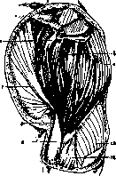
Musculus deltoideus is a coarsely fibrous, fan-shaped, multipennate muscle in which, especially during contraction and through the skin, 3 parts can easily be distinguished: the scapular part, originating from the spine of the scapula; the acromial part, from the acromion of the same bone; and the clavicular part, from the lateral third of the clavicle (see figure). The strong combined tendon of these portions, separated from the greater tubercle of the humerus by a mucous bursa (bursa mucosa subdeltoidea), terminates on the tuberosity of the lateral surface of the humerus.
The muscle can contract
with individual parts,
overall (abducting the shoulder) or with
individual parts. 7—m. pectoralis major;
8 and 9—m. biceps (short and long heads);
10—m. brachialis.
11—m. triceps. (After Fr. Frohse and M. Fraenkel.)
The clavicular part moves the arm forward, the scapular part—backward. The muscle is innervated by the axillary nerve (from the brachial plexus), and vascularized by the thoracoacromial and brachial arteries. Between the deltoid and pectoralis major muscles (in Mohrenheim's fossa) runs the cephalic vein.
In paralysis of the axillary nerve supplying this muscle, the activity of the entire shoulder is sharply disrupted due to the loss of function of the deltoid muscle. The supraspinatus muscle and the long head of the biceps partially compensate for the action of this muscle. The loss of the dynamic action of the deltoid muscle is also partially compensated by the movement of the entire shoulder girdle together with the scapula. The muscle also undergoes severe atrophy in certain inflammatory processes in the region of the shoulder joint, for example, in caries sicca. Replacement of the lost function of the muscle is partially achieved by transplantation of the pectoralis major or trapezius (cucullaris) muscle. A good practical result is also obtained by arthrodesis in the shoulder joint at a position of marked abduction of the arm. Movements are performed together with the scapula. In so-called birth palsy of the deltoid muscle in children, the timely use of an abduction splint prevents the development of pseudoparalysis due to muscle stretching and promotes the subsequent restoration of its function.
Related articles
Mentioned in
Cite this page
“Deltoid Muscle.” Soviet Medical Encyclopedia. English translation of Bolshaya Meditsinskaya Entsiklopediya, 1st ed. (Moscow, 1928–1936), ed. N. A. Semashko. https://sovietmedicalencyclopedia.pages.dev/article/deltoideus-muscle/