Mucous Bursa
Historical document, translated for reference. It reflects medical knowledge of the 1920s–30s and is not medical advice.
Summary
The mucous bursa is a fluid-filled sac that develops in areas of friction between tissues and bone, facilitating movement. This article describes their anatomy, development, location throughout the body, and distinguishes them from joint capsule herniations.
Encyclopedia article (1928–1936)
Mucous Bursa, bursa mucosa, s. serosa, (from the Greek bursa - skin, in the Middle Ages this term came to denote a bag, pouch). The systematic study of M.B. begins in 1784 (Camper). M.B. appear where skin, fascia, muscles, tendons are subjected to continuous pressure over bones in a certain direction, as a result of which they shift, therefore Langermok, for M.B. proposed the name 'sliding bursae' (Gleitbeutel). From the above it is understood that the number of M.B. is not constant, that in newborns, with the exception of a few so-called congenital M.B., they are absent, and later appear in various places depending on the characteristics of a particular profession, constitution, pathological condition, etc., for example, on the sternum in cobblers, under the skin of the vault of the skull in porters carrying loads on the head, over the projecting spinous process in kyphosis, at the head of the first metatarsal bone in hallux valgus. It is generally accepted that the friction of skin coverings, tendons and muscles against deeper-lying bony prominences, ligaments and tendons of other muscles is the moment that causes the formation of M.B.; when muscles work, the connective tissue fibers located between, for example, skin and tendon, are subjected to tension, become loose, and gradually thinning completely disappear, a slit is obtained, which is the cavity of the M.B., but bursae can also develop where there is no friction. Thus, the cause of bursa formation may be the periodically occurring divergence of contacting surfaces. An example can be bursa Achillis, where at the moment of extension of the foot the tendon moves away from the calcaneus, and the connective tissue located between the tendon and bone becomes loose, which leads to the formation of a cavity. The cavity is initially irregular in shape, in places penetrated by atrophied connective tissue fibers, then gradually forms into a real pouch, the walls of which are very similar to the synovial membrane of joints. The inner surface of the pouch is smooth, sometimes uneven, lined, like any other connective tissue cavity, with a single layer of flat epithelium and contains a small amount of fluid of synovial joint character. Presenting in its natural state as a collapsed pouch, compressed by a tendon and bone or skin, the M.B. is fused on its outer surface with the tissue of the organs among which it is located and imperceptibly passes into the surrounding connective tissue, for which reason during dissection M.B. are found with great difficulty, and they are noticed only when the wall is opened and air penetrates the cavity; in some cases due to the hardening of connective tissue fibers the bursa acquires considerable resistance, and then it is possible to clearly establish the boundaries of the bursa cavity. In anatomy for studying the shape and position of M.B. use is made of injection of colored masses into the bursa cavity, for example, wax, gelatin, fat. Bundles of connective tissue in some cases take the form of plates, which divide the cavity into several chambers, separated from each other or communicating with each other, these are so-called multilocular bursae (bursae mucosae multiloculares) in contrast to simple, unilocular (bursae mucosae simplices). The same bursa can be both multilocular and unilocular, for example, bursa trochanterica m. glutei maximi. An example of a multilocular bursa can be bursa subdeltoidea. The size and shape of M.B. show considerable variability. Their shape, elliptical or round, is determined by the nature of the structures between which they arise. Their size ranges from fractions of a centimeter to several centimeters. M.B. located near joints often communicate with their cavity. Such relations are characteristic of adults, while in the fetus they have a different character. Thus for example b. suprapatellaris in the fetus is always separated from the joint cavity, while in adults this occurs only in 1/5 of all cases (Fick). M.B. should be distinguished from herniations of the synovial capsule of joints. While the former communicate with the joint under the influence of muscle activity, the latter are a direct continuation of the synovial bursae of joints and are formed as follows: when muscles contract, a tendon closely fused with the joint capsule, in its movement, pulls it and forms a protrusion (for example, the tendon of m. biceps in the shoulder joint). The moment causing the appearance of joints and bursae is muscle movement; if one takes into account the similarity in the anatomical structure of the walls, it turns out to be possible to consider M.B. as a special (very simple) type of connection (Tonkov). For example, the joint connecting os triquetrum with os pisiforme - articulatio ossis pisiformis - originated from a simple M.B. The above circumstances are of great importance in understanding the pathological processes occurring in M.B. and allowing in some cases to draw a parallel with similar joint diseases. Located in areas of tendons, M.B.
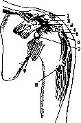
Figure 1. 1-bursa subcutanea acrom.; 2-b. lig. coraco-clavicularis; 3-b. coraco-brachialis; 4-b.subacromialis; 5- b. supraspinata; 6-b. subdeltoidea; 7-b. sub-coracoidea; 8-b. pectinei majoris (Merkel); 9-b. subscapularis.
in areas of greatest friction facilitate movement and free up part of the muscle energy for useful work; friction occurs not directly between contacting surfaces, but between the walls of the bursa, lubricated with synovial fluid. The synovial fluid of M.B. according to modern views is not a product of the secretory activity of the endothelium of the wall, but represents tissue breakdown, mucous degeneration of the cells of the wall with the formation of a fine-grained, homogeneous mass of alkaline reaction, slightly yellowish in color, viscous, containing mucin.
Figure 2.
Figure 3.
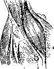
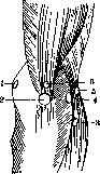
Figure 2. 1-bursa extensoris carpi radialis brevis; 2-b. bicipito-radialis (Merkel); 3-b. cubitalis interossea. Figure 3. 1-bursa subcutanea epicondyli med.; 2- b. subcutanea olecrani (Merkel); 3-b. m. extensoris radialis brevis; 4-b. m. anconaei; 5-b. subcutanea epicondyli lat.; 6-b. subtendinea olecrani.
Location of M.B.: according to R. Redi M.B., located near joints, can be constant and inconstant. In the shoulder joint the following constant M.B. are distinguished: 1) b. acromialis, 2) b. subacromialis, 3) b. subdeltoidea, 4) b. subcoracoidea, 5) b. subscapularis, 6) b. coraco-brachialis, 7) b. intertubercularis and 11 inconstant bursae (fig. 1).-In the elbow joint-constant M.B.: 1) b. bicipito-radialis, 2) b. subcutanea olecrani and 14 inconstant
5 i
Figure 4.
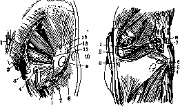
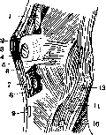
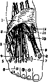
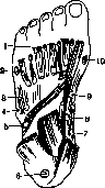
Figure 5. Figure 4. 1-bursa subcutanea sacralis; 2-b. obturatoria inf.; 3-b. tuberis ischii; 4-b. flexorum; 5-b. tendinea m. obturatoris ext.; 6-b. tendinea m. obturatoris int.; 7-b. glutaeo-femoralis; 8-b. trochanterica glutaei max.; 9-b. trochanterica superf.; 10-b. subcutanea troch.; 11-b. m. piriformis; 12-b. glutaei medii ant.; 13-b. glutaei min. Figure 5. 1-bursa gastrocnemia med.; 2-b. semimembranosa; 3-b. semimembranosa propria; 4-b. poplitaei (Merkel); 5-b. bicipitalis inf.; 6-b. poplitaea post.; 7-b. gastrocnemia lat.; 8-b. bicipito-gastrocnemia. M.B. (fig. 2 and 3).--The hand has no constant M.B., two inconstant ones are observed.-In the gluteal region constant bursae: 1) b. trochanterica superficialis, 2) b. m. bicipitis femoris super., 3) b. trochanterica profunda, 4) b. ischiadica profunda, 5) b. trochanterica m. glutaei medii anter., 6) b. trochanterica m. glutaei minimi, 7) b. glutaei medii post., 8) b. m. obturatoris int. and 5 inconstant M.B. (fig. 4 and 5). In the hip joint region constant M.B.: 1) b. ilio-pectinea, in 10% of cases communicates with the joint cavity, 2) b. iliaca subtendinea, 3) b. m. recti.-In the knee joint and popliteal fossa the following constant bursae: 1) b. praepatellaris subfascialis, 2) b. subcutanea tuberos. tibiae, 3) b. infrapatellaris profunda, Figure 6. 1-bursa suprapatellaris subfasc; 2-b. praepatellaris subfasc; 3-b. patella; 4-b. infrapatellaris profunda; 5-b. tendinea m. semimembranosi propria, 6-b. lig. collateralis tibiae; 7-b. infrapatellaris subcut.; 8-b. infrapatellaris subcut.; 9-b. subcutanea tuberosit. tibiae; 10-b. bicipitalis inf.; 11-b.
4) b. suprapatellaris (communicates with the joint), 5) b. gastrocnemia media, 6) b. m. semimembranosi lat.; 7-b. poplitaei, 8) b. anserina, 9) b. m. bicipitis femoris inf. and 14 inconstant (fig. 6).--In the foot region constant M.B.: 1) b. tendinea calcanei Achillis, 2) on the plantar surface-b. subcutanea digitorum, 3) b. infratendinea m. tibialis post. Inconstant M.B. number up to 20 (fig. 7 and 8). Traumatic injuries of M.B. Open injuries of superficial M.B. usually escape the attention of the patient and
8-b. infrapatellaris subcut.; 9-b. subcutanea tuberosit. tibiae; 10-b. bicipitalis inf.; 11-b.
by the physician, as they do not give any symptoms that distinguish this injury from the frequently encountered wounds of the subcutaneous tissue, while injury to the deep bursae is usually overlooked because it recedes to the background before the easily detectable damage to other important structures (vessels, nerves, etc.). The danger of injury to the mucous bursae is especially great because, in the presence of direct connection with the joint or through lymphatic vessels, infection easily penetrates into the latter. Treatment of injuries to the mucous bursae should be carried out according to the general principle of wound treatment, but in doubtful cases, when there is no certainty that the walls of the bursa are not infected, it is better to limit oneself to excising the edges of the wound without resorting to sutures. If the inflammatory process drags on, accompanied by the formation of strands, scars and thickening of the wall of the M. b., then these secondary changes, after the complete cessation of inflammatory phenomena, can give an unpleasant complication in the form of limitation of movement of the joints, muscles, tendons, and then, if conservative treatment (heat and gymnastics) is unsuccessful, it is necessary to resort to excision of the altered M. b.-Injury to the M. b. without violation of the integrity of the skin (contusion) is determined only when it is accompanied by serous, sero-hemorrhagic effusion or hemorrhage into the cavity of the M. b., which usually resolves spontaneously or, if delayed in the bursa, passes into a chronic form. Treatment consists of rest, application of pressure bandages, heat; only sometimes with slow resorption, puncture is indicated. Inflammations of the M. b. are divided into acute and chronic. Acute bursitis (bursitis acuta) arises as a result of damage to the wall of the bursa or penetration of bacteria along lymphatic pathways from the nearest foci. It is also possible for the wall of the bursa to be destroyed by the proteolytic enzyme of an adjacent purulent focus, or finally, in very rare cases, infection enters the M. b. by the hematogenous route. Inflammation of the M. b. begins acutely, is accompanied by a local rise in temperature, a febrile state with the formation in the cavity of the bursa of a serous, sero-hemorrhagic or sero-fibrinous exudate (Aschoff). Subsequently, the exudate may completely resolve or, the process acquiring a productive character, passes into a chronic form, or the exudate becomes purulent (bursitis purulenta). The causative agents of the inflammatory process may be staphylococci, streptococci, pneumococci, gonococci, bacillus coli (Lexer) and dysentery bacillus (R. Redi). Pneumococcal bursitis most often occurs in the area of b. subdeltoidaea, b. bicipito-femoralis, b. semimembranosae, b. gastrocnemii, b. praepatellaris, b. ischiadicae; gonococcal-in b. tend. Achillis and in b. iliaca subtendinea. The dysentery bacillus affects the M. b. of the lower extremity. The male sex is affected more often than the female, the most affected age group is children under 10 years and adults from 35 to 50 years. The spread and clinical manifestations of acute bursitis depend on the nature of the infection and the anatomical location of the bursa. Usually the patient shows all signs of inflammation: redness of the skin, fluctuation, swelling, pain and disturbance of function of the given area of the extremity, but it must be borne in mind that in deep bursitis some symptoms may be absent or weakly expressed, for example, in bursitis iliaca there may be no redness of the skin. In such cases, swelling is also not detected for a long time due to the large capacity of the bursa and its deep location. With low virulence of bacteria, there may be no rise in temperature, but instead a number of symptoms appear, consisting in a forced position of the extremity-for example, in bursitis iliaca the thigh is flexed, abducted outward and rotated, i.e., it assumes a position that minimizes painful sensations; in bursitis subdeltoidea flexion and extension are possible, but rotation along the longitudinal axis, abduction and adduction are impossible, which never happens in arthritis. In the knee area, extension is difficult, in the elbow area in bursitis bicipito-radialis flexion and extension are preserved, but pronation and supination are very painful. If acute bursitis has a tendency to localize, an abscess forms, lined inside with granulation tissue. If the virulence of the bacteria is especially great, the inflammation may end in necrosis of the bursa with penetration of infection into the surrounding tissues and cellular tissue and development of phlegmonous inflammation. Sometimes bursitis, left to its own devices, eventually ruptures outward, the consequence of which are non-healing fistulas. In acute bursitis, accompanying phenomena in the form of lymphangitis, lymphadenitis and erysipelas are often observed. The danger of purulent bursitis, besides that inherent in any purulent focus, lies in the possibility of spreading infection to the adjacent joint. In this case, bacteria penetrate either through lymphatic vessels or through the existing direct anatomical connections of one or another M. b. (for example, b. poplitaea, b. semimembranosa) with the cavity of the neighboring joint. Among the bacterial forms encountered, acute gonorrheal inflammation of the M. b. deserves attention, which often develops together with arthritis and is usually of serous, more rarely purulent, nature. Of the M. b., b. calcanea Achillis and b. iliaca subtendinea are affected most often. The disease usually has a benign character and resolves spontaneously. Swelling in bursitis calcanea is observed on both sides of the Achilles tendon. When walking, pain appears radiating to the calf-the so-called heel pain, 'achillodynia' (it also occurs in gout and rheumatism); the patient, in order to reduce compression of the bursa, places the foot in the position of pes equinus. The resting position of the extremity, puncture in case of abundant effusion and timely resumption of movements quickly lead to recovery with preservation of function. Treatment of purulent bursitis consists of timely incision followed by tamponade-free management of the patient. For serous bursitis-rest and heat in all forms. Some have seen good results from the application of hyperemia according to Virchow. In persistent retention of exudate, puncture is indicated. After the acute inflammatory phenomena subside, it is necessary to begin timely active and passive movements to prevent contractures. If the process passes into a chronic form with the formation of free bodies and thickening of the wall of the bursa, the latter is completely excised. Chronic bursites are divided into non-specific and specific; the latter include tuberculous and syphilitic bursitis. Under the influence of prolonged, often repeated or single mechanical injuries, chronic non-specific bursitis, so-called hygroma (hygroma), may develop. The latter is especially often observed in places subject to frequent trauma, for example, in the knee area bursitis praepatellaris, in the area of the elbow process-bursitis ole
Figure 9. Bursitis ole
Figure 10. Bursitis prae- rani chronica.
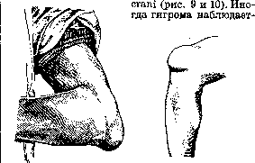
patellaris. It is found where bone is located directly under the skin, e.g., the inner surfaces of the base of the big toe in hallux valgus. The bursa praepatellaris is most frequently affected, followed by the subdeltoidea, then the subacromialis, semimembranosa; very rarely the praetibialae, ischiadica. Chronic bursitis develops both in the early years of life and in mature age. The frequency of bursitis, according to Redi (in %): up to 20 years--12, 20-40 years-33, 40-60 years-52, 60-90 years-3. Men are affected more often than women: according to R. Redi, for every 45 men, 21 women were ill, apparently this is connected with the peculiarities of male labor. The cause of bursitis, authors (Tetit) consider trauma in 53%, rheumatism-15%, jaundice, puerperal fever, cooling-2%; in 30% the cause is not determined. Other authors also confirm that the most frequent cause is trauma, understanding by this not only direct injuries but also indirect (improper position, wearing various belts, prostheses, repeated irritation from poorly fitted instruments). Pathoanatomically chronic bursitis may be serous, proliferative and hemorrhagic (according to Bianchetti). Serous bursitis can reach the size of an adult's head, the form varies from a simple tumor to two-, three-lobed and more. The wall of the M. b. in this case shows all signs of chronic inflammation, is infiltrated, has necrotic areas and hemorrhages, in the thickness of the wall a considerable number of vessels is found. Sometimes the wall is infiltrated with cartilage cells and lime deposits, the fluid located in the bursa cavity is usually transparent or slightly turbid, of alkaline reaction, contains mucin. Proliferative bursitis is characterized by significant thickening of the wall, the inner surface appears warty, villous, full of papillary growths. The endothelium disappears in these cases. The fibers of connective tissue thicken (fig. 11). Hemorrhagic bursitis is similar to proliferative. The size of the affected mucous bursa varies, reaching the size of an adult's head. The walls consist mostly of connective tissue. Necrotic areas and hemorrhagic infiltrates deep in the wall are characteristic of it. Free bodies (corpora libera), found in bursae, develop most often in proliferative bursitis, rarely in serous and very rarely in hemorrhagic. Their number varies from one to 200, their size from 2 mm to the size of a rice grain. Their formation occurs due to disturbance of nutrition of the papillary growths located on the wall of the bursa on a stalk. The subjective sensations of patients with chronic bursitis are insignificant, and only during exacerbation pains may appear; besides, the swelling, reaching large sizes, can limit movement. In differential diagnosis it is necessary to consider the possibility of a hematoma of the M. b., cold abscess, aneurysm, joint cysts, soft tumors (myxomas and lipomas). For the treatment of chronic bursitis old authors proposed the method of crushing with subsequent application of pressure bandage and heat; but this method cannot be recommended because of the danger of awakening the dormant infection in the cavity and wall of the capsule; this is especially dangerous if there is an unrecognized tuberculous bursitis. Incision of the capsule and scraping out the contents sometimes lead to adhesion and obliteration of the bursa cavity. The most effective method is complete or partial, in case of adhesion with bone, removal of the M. b.
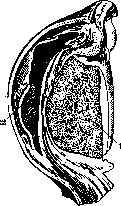
Figure 11. Bursitis praepatellaris: 1-the patella; 2-the expanded bursa praepatellaris with thickened wall.
Besides the described changes in the M. b. lime, uric acid, bone tissue have been found. Thus, three more forms of diseases of the M. b. can be distinguished: bursitis calcarea, urica and ossificans; trauma and preceding infectious diseases can give calcification of the M. b. (bursitis calcarea), which must be considered as a secondary reaction, rarely such bursitis is an expression of diathesis. Most frequently affected are the subacromialis and subdeltoidea. The age of patients is around and above 40 years, men get sick more often. The symptoms of bursitis calcarea are peculiar: the swelling sharply protrudes from the bursa cavity, of dense consistency and small size. Pains are not constant, but very sharp, often appear several years after the cause that caused the anatomical damage. Bursitis calcarea is usually well diagnosed by X-ray, but it must be differentiated from detachment of tuberculum majus, free joint bodies, calcified tuberculous foci. Treatment: heat, massage, iodotherapy, diathermy; especially effective is irradiation with X-rays--after 3-4 sessions the shadow of the M. b. disappears and with it the subjective phenomena. In some cases--removal by operative means. Bursitis ossificans exists as a special form and differs from bursitis calcarea, in which the remaining lacunae among the lime deposits can create a false impression of bone tissue. The development process of bursitis ossificans generally resembles bone development. Vessels of the wall and metaplastic cells penetrate into the lime deposits, from which as the lime is absorbed bone tissue is formed, resembling spongy substance. Bursitis urica occurs in gout, when as a result of metabolic disturbance retention of uric acid and salts can be observed. These salts are deposited mainly in cartilages and joint capsules. In serous bursae and para-articular tissues the deposition of uric acid salts is often accompanied by inflammatory phenomena and sharp pains. Macroscopically in the M. b. white spots and accumulation of white, like chalk, substance are noticeable. Microscopically bundles of crystals in the form of bouquets are noticeable. The part of the wall which they line, after their removal, appears necrotic; necrosis of the wall occurs as a secondary phenomenon as a result of salt deposition. Treatment--anti-gout, locally the same as in bursitis calcarea. Tuberculous bursitis--see Bursitis. Syphilitic bursitis--see Bursitis, syphilitic bursitis.
Tumors of the M. b. are very rare, occur mainly in the form of myxomas and sarcomas. Ombredanne counts in the literature only 6 cases of sarcoma of the M. b. Adrian up to 1909 found in the literature only 19 cases of all tumors of the M. b. Most frequently the praepatellaris is affected by sarcoma, second place is taken by the subdeltoidea, extremely rarely a tumor can develop in other bursae. Usually men of middle age get sick. According to the histological picture the tumors belong to the group of round- and spindle-celled sarcomas and have the usual malignancy characteristic of sarcomas, i.e., they quickly grow into underlying tissues and give metastases. At first the tumor is movable on the underlying tissues, but mobility is quickly lost. As the tumor grows the usual dilation of the superficial venous network develops. The tumor often has the character of myxosarcoma. For recognition it is necessary to consider the following circumstances: the anatomical position of the tumor at the site of a serous bursa, mobility on the underlying tissues at the beginning of development, rapid growth and dilation of the superficial venous network. Confusion is possible with chronic bursitis, non-specific, specific (tbc, lues), with bone and periosteal tumors. Treatment: early and wide excision. With large spread and recurrences--amputation. Primary sarcomas are very rare. In them a bloody exudate accumulates in the joint, movements in the joint are preserved. X-ray examination does not reveal a bone focus.
n. makhov.
Related articles
Mentioned in
Cite this page
“Mucous Bursa.” Soviet Medical Encyclopedia. English translation of Bolshaya Meditsinskaya Entsiklopediya, 1st ed. (Moscow, 1928–1936), ed. N. A. Semashko. https://sovietmedicalencyclopedia.pages.dev/article/mucous-bursa/