Hallux Valgus
Historical document, translated for reference. It reflects medical knowledge of the 1920s–30s and is not medical advice.
Summary
An overview of hallux valgus (bunion) from the 1930s Soviet medical literature, detailing its anatomical and radiological features, etiology involving flatfoot and occupational overload, and various conservative and surgical treatment methods.
Encyclopedia article (1928–1936)
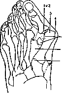
HALLUX VALGUS, outward deviation of the big toe, is a very widespread foot deformity; for example, according to Payr (Rauth), hallux valgus occurs in 20-25% of all adults, while Weinert considers an unbent foot in an adult to be a rarity. Furthermore, hallux valgus also occurs in children, in which case it is a congenital, often hereditary, deformity. This foot curvature leads to two main issues that prompt patients to consult a physician: 1) pain and 2) disfigurement of the foot. In mild cases of this deformity, the gait changes and a greater foot fatigue is observed. In more pronounced cases of hallux valgus, pain develops to such a degree that not only walking, but even standing becomes agonizing. Patients are completely unable to find suitable footwear for themselves. In cases of sharply pronounced hallux valgus, besides the outward deviation of the big toe, which frequently lies under the second and third toes rather than above them, the protruding inward head of the first metatarsal bone catches the eye. It is usually covered with calloused skin, and sometimes a multilocular mucous bursa develops right here, which easily becomes inflamed and frequently leads to inflammation of the first metatarsophalangeal articulation and severe pain. The second, third, and fourth toes are often slightly mobile, remaining in a claw-like position; corns form on the bent phalangeal joints. The fifth toe is in a supinated position (Schede). The transverse size of the foot at the level of the metatarsophalangeal joints is significantly wider than usual: 1) due to the medial departure of the first metatarsal bone from the second and 2) due to the lowering of the transverse arch of the foot, in place of which there is usually a calloused corn, often very painful. If X-ray examination is added to these main data of external foot inspection in hallux valgus, the picture becomes completely clear. On the X-ray image of a normal foot, the phalanges of the toes, metatarsal bones, and cuneiform bones are projected as 5 straight rays diverging somewhat fanwise. In hallux valgus, all rays, especially the first, represent a broken line because the phalanges of the toes are deviated outward, the metatarsal bones inward, and the cuneiform bones outward. In addition, all bones, especially the metatarsals, are projected wider, and at their heads, except for the fifth metatarsal, so-called "exostoses" appear on the inner side, especially large on the first metatarsal. Weinert explains the appearance of the widening of the metatarsal bone heads on X-raygrams in hallux valgus by the rotation of these bones around their longitudinal axis inward. Besides these general changes, the following almost constant features in the first ray in hallux valgus can be noted on the X-raygram (see Figure 1): 1) subluxation of the proximal phalanx outward, 2) formation of a steeper joint arch on it, 3) continuous or patchy atrophy of the inner part of the head of the first metatarsal bone, 4) development of an "exostosis," 5) widening of the intermetatarsal space between the first and second bones, 6) displacement of the sesamoid bones outward, 7) change in bone relationships in the metatarsocuneiform joint (Weinert) and formation of angles, with the angle formed by the phalanx and metatarsal bone (a) being open outward, and that of the metatarsal and cuneiform (b) inward. Thus, on the X-ray image we see that the first metatarsal bone undergoes the main changes. Pathologico-anatomical changes in hallux valgus in the first metatarsophalangeal articulation consist of the following: the joint capsule of the metatarsophalangeal articulation is stretched on the inner side and shortened on the outer; the articular cartilage covering the head of the metatarsal bone, and the bone itself on the inner side, are atrophic because the phalanx, having shifted laterally, no longer presses on it. On the outer part of the head, a new articular surface forms, which has strong articular cartilage and is separated from the former one by a groove (Heubach). Beneath the newly formed joint, the bone has a thick structure. In almost every instan
Figure 1.
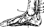
Figure 2. 1 - tendo m. tibialis anterior; 2 - tendo calcaneus (Achillis); 3 - m. flexor hallucis longus; 4 - m. flexor digitorum longus; 5 - m. tibialis posterior; 6 - m. abductor hallucis.
ce of hallux valgus, there is a thickening of the periosteum on the inner side of the head of the first metatarsal bone, representing a tubercle, the so-called "exostosis," which serves as the site of attachment of the collateral ligament and hypertrophies as a result of rupture and pressure (Keszli). The equilibrium of muscle action in hallux valgus is disrupted. The abductor of the big toe (as Hohmann points out) is displaced plantarly and strongly stretched (see Figure 2). The extensors, both long and especially short, are in a shortened state. Their action, especially the short one, rather amounts to abduction (Nélaton) than to extension. Thus, the tense extensors not only fix the abduction of the big toe, but also contribute to its progression. Hallux valgus can be congenital or arise on the basis of trauma. These are two clear etiological moments. In those cases where these moments are absent, the emergence of hallux valgus is interpreted by many authors in various ways, but the occupational factor is usually not indicated; meanwhile, it plays a major role in the development of hallux valgus, since all conditions causing foot overload (long standing, heavy strain on the feet at work) and leading to flatfoot are the cause of hallux valgus. Regarding the origin of hallux valgus due to wearing improperly tailored footwear with narrow toes and high heels (see Figure 3), narrow and tightly stretched stockings (Hoffa's vestimentary theory), a swaying gait with spread-out feet during which overload of the head of the first metatarsal bone occurs, and others, all these can be considered as factors that contribute to and accelerate the painful condition, but do not cause it. Weinert and, chiefly, Hohmann, who devoted much of their labor to the study of foot and leg
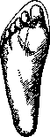
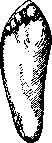
Figure 3. On the left, correct position; on the right, displacement of the toes by narrow footwear.
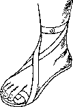
diseases, point out that the cause of hallux valgus is pes plano-valgus, which develops as a consequence of overstretching and weakening of the ligamentous apparatus. Hohmann finds that "disturbance of muscular equilibrium due to overstretching of the abductor and plantar displacement of the abductor leads to hallux valgus." Schede illuminates this issue in the following manner: "It is generally accepted that the entire complex of painful transformations of the toes and forefoot is not the result of wearing overly narrow footwear, but is in the closest connection with all the processes that we designate by the general term — foot drop." During foot drop, a displacement of the articular surfaces occurs, naturally sharper on the inner side (above the longitudinal arch). The descending cuneiform bone displaces the first metatarsal forward, and the flattening transverse arch pushes its head inward, which carries along the base of the big toe, while its distal end is held and then pulled outward by the extensors. The more strongly the big toe deviates, the more the extensors shorten (retraction), and the displaced abductor cannot oppose them. Thus, all occupational factors leading to pes plano-valgus play a role in the development of hallux valgus as well — for example, peasants, among whom hallux valgus is observed so frequently, mostly do not wear narrow shoes, but walk either barefoot or in wide felt boots, yet they engage in heavy labor. Treatment for hallux valgus can be preventive, at the very beginning of its development, and operative, when the deformity is clearly expressed. With barely noticeable signs of the appearance of hallux valgus, wearing conventional arch supports or a "varus" shoe is sufficient; in more pronounced initial stages, wearing arch supports with a special device for abducting the big toe (see Figure 4) is used. Patients rarely consult a physician in these stages. Of the bloodless operative methods, Schede recommends forcible correction (redressement) of hallux valgus, but it
must be accompanied by the correction of flatfoot as well, which is unachievable in major deformations, especially in adults. Regarding operative treatment, specifically the methods and technique of operations, there is no unity. Up to the present time, about 30 independent methods have been proposed. All these methods, despite their number, can basically be divided into three types of operations: 1) resection of the head of the first metatarsal bone with plas
Fig. 6.
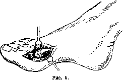

1) tenoplasty or without tenoplasty of tendons; 2) various forms of osteotomy of the first metatarsal bone with or without tenoplasty of tendons; 3) tenoplasty of tendons and removal of "exostoses". The most common method is the one proposed by Hueter, which is used by orthopedic surgeons in America, France, Germany, the USSR and other countries (see figure 5). This method is particularly successful in patients with severe flatfoot. Its advantage is that patients can walk on the 10th day after the operation without experiencing significant pain. The direction of skin incisions over the head of the first metatarsal bone can be arcuate or linear. Attention must be paid to the good smoothing of the end of the first metatarsal bone to preserve good joint mobility. For this purpose, some authors perform interposition of soft tissues; for example, Mayo places a bursa. Not limiting themselves to only resection, some authors add tendon plastic surgery; Gocht lengthens the tendon of the extensor hallucis longus; Lorenz completely severs the extensor hallucis brevis. - Osteotomies for the treatment of H. valgus are performed on the first metatarsal bone in all directions: transverse, wedge-shaped, oblique, ladder-shaped, etc. (Hohmann, Ludloff). All these osteotomies are performed with the aim of shortening the first metatarsal bone, rotating the head of the first metatarsal bone outward, and placing it so that it can again serve as a point of support, i.e., the head is placed in the plantar direction, which creates an angle between the fragments of the metatarsal bone open to the sole, forming an arch. Some authors accompany the osteotomy with muscle plastic surgery; for example, Homan transplants the anterior head of the displaced m. abductoris hallucis brevis distally, and the belly of this muscle is sutured dorsally. After these operations, 3 weeks of bed rest and a splint are required (see figure 6), but after that, the foot receives support on the head of the first metatarsal bone, and an arch is formed. Operations involving only the removal of "exostoses" are usually accompanied by the transplantation of the tendon of the extensor hallucis longus to the medial side of the proximal phalanx of the big toe.
Related articles
Mentioned in
Cite this page
“Hallux Valgus.” Soviet Medical Encyclopedia. English translation of Bolshaya Meditsinskaya Entsiklopediya, 1st ed. (Moscow, 1928–1936), ed. N. A. Semashko. https://sovietmedicalencyclopedia.pages.dev/article/hallux-valgus/