Sesamoid Bone
Historical document, translated for reference. It reflects medical knowledge of the 1920s–30s and is not medical advice.
Summary
This 1930s encyclopedia article describes sesamoid bones, small inconstant bones found in joint regions such as the fingers and toes, their development, comparative anatomy, and pathological ossifications. It details their biomechanical role in altering the direction of tendon pull and increasing muscle leverage, noting that they can be visualized via roentgenography.
Encyclopedia article (1928–1936)
SESAMOID BONE, os sesamoideum. Galen used this term to designate small (inconstant) bones found by him in the joints of animal digits due to their resemblance to the seeds of the plant that the Greeks called "sesamos" (sesame). These bones, the size of a pea or small bean and somewhat similar in shape to
them or to a triangular pyramid, are located on the palmar and plantar side of the digital joints (in numbers of 1–7, Pfitzner) (Figs. 1 and 2), into the cavity of which faces a small articular surface covered with cartilage, sliding either in a groove on the surface of the articular head of the phalanx or on one of its side projections (Figs. 3 and 4). Being woven into the terminal tendon of the flexor muscles of the digits, the sesamoid bone, depending on its thickness, changes its direction to an oblique or almost perpendicular one relative to the axis of the terminal phalanx, which significantly increases the "moment" of the muscle and the force of its traction. In the fetus and in childhood, sesamoid bones are connective-tissue or cartilaginous formations, which in further development become calcified or ossified. They are of diverse origins: some arise from primordial cartilage, others from bone-forming cells of the periosteum that separate from it under significant irritations and become enclosed in nearby tendons; a -- among birds, especially those rich in sesamoid bones -- also at a considerable distance; in carnivorous mammals, sesamoid bones are also present on the dorsal surface of the thumb joint. Isolated ossifications are found in the thigh muscles ("Reitknochen"), in the capsule of the knee joint, in the triceps muscle of the arm in animals with a short olecranon process (amphibians, reptiles, birds, bats, hares). Pathological ossifications can also occur in other muscles and even in internal organs (spleen, kidneys, parotid gland) that are not, of course, connected with the bony or cartilaginous skeleton. In the living, the presence of sesamoid bones is easily detected on roentgenograms, as well as sometimes by careful palpation. Many anatomists regard the pisiform bone and the patella as sesamoid bones.
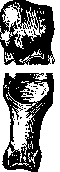
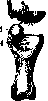
Figure 3.
Figure 4. Head of the first metatarsal bone of the right thumb and the sesamoid-phalangeal apparatus (joint opened from the dorsal side and articular surfaces removed): 1 - inner sesamoid bone of the thumb (scaphoideum); 2 - outer sesamoid bone of the thumb; 3 - head of the metatarsal bone; 4 - articular surface of the phalanx.
Related articles
Mentioned in
Cite this page
“Sesamoid Bone.” Soviet Medical Encyclopedia. English translation of Bolshaya Meditsinskaya Entsiklopediya, 1st ed. (Moscow, 1928–1936), ed. N. A. Semashko. https://sovietmedicalencyclopedia.pages.dev/article/sesamoid-bone/