Crus
Historical document, translated for reference. It reflects medical knowledge of the 1920s–30s and is not medical advice.
Summary
This article from the 1928–1936 Soviet Great Medical Encyclopedia provides a detailed anatomical description of the crus (the leg or shin), including its embryological development, osteology, musculature, and topographical boundaries. It covers the structure of the tibia and fibula, the articulations of the lower leg, and the arrangement of muscle groups.
Encyclopedia article (1928–1936)
CRUS (shin), the middle (central) section of the free lower, or posterior pelvic limb, appears for the first time in amphibians. In some reptiles, there are only rudimentary pelvic limbs (Python, Boa), while in most snakes and legless lizards (Amphisbaena), there are no limbs at all. In mammals, the crus varies in relative length; it is especially long in bats and insectivores. The crus, homodynamic to the forearm, is very constant; only in some animals (e.g., horses) does one of the bones, failing to develop, fuse with the other. In humans, the crus is 1/6 shorter than the thigh. The first primordium of the lower limb with vessel-containing mesenchyme appears in the third week of intrauterine life. The tibia and fibula each have three ossification points—in their diaphyses and both epiphyses.
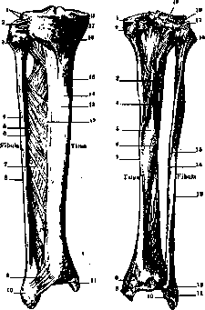
Figure 1.
Figure 2.
Figure 1. Right tibia and fibula from the front: 1-condylus lat.; 2-lig. capituli fibulae; 3-capitulum fib.; 4-facies lat.; 5-fac. med.; 6-crista ant.; 7-membrana interossea cruris; 8-crista lat.; 9-lig. malleoli lat. ant.; 10-malleolus lat.; 11-malleolus med.; 12-crista interossea; 13-facies lat.; 14-crista ant.; 15-facies med.; 16-tuberositas tibiae; 17-cond. med.; 18-margo infraglenoidalis. Figure 1. Tibia and fibula from the back: 1-margo infraglen.; 2-condyl. med.; 3-linea poplitea; 4-for. nutritium; 5-facies post.; 6-crista interossea; 7-margo med.; 8-sulcus malleolaris; 9-malleolus med.; 10-facies artic. malleoli lat.; 11-malleolus lat.; 12-sulc. tend. mm. peronaei; 13-crista lat.; 14-crista med.; 15-facies post.; 16-capitulum fibulae; 17-apex; 18-condyl. lat.; 19-for. nutrit. (According to Spalteholz.)
Ossification in the diaphyses appears in the 7th–8th week of intrauterine life. By the time of birth, ossification centers appear in the upper epiphysis of the tibia; in the second year of life, the lower epiphyses of the tibia and fibula begin to ossify; and in the third, the upper end of the fibula. The musculature of the crus begins to differentiate in an embryo 11 mm long. In an embryo of approximately 15 mm, the terminal links of the primary arterial network atrophy, and the arteries, with their main trunk (a. femoralis), differ little from the vessels of an adult. The crus borders the thigh at the level of the upper recess of the knee joint capsule or 3–4 fingers above the kneecap. The lower boundary of the crus is considered to be a circular line at the level of the base of the malleoli.
The basis of the crus is the tibia and fibula. The tibia (shinbone) (see figures 1 and 2), homodynamic to the radius and thicker, is located medially. Its body is approximately triangular; the surface facing median-ventrally is located directly under the skin, while the lateral and posterior surfaces are covered by muscles. Above the anterior crest (crista anterior) protrudes a roughness (tuberositas tibiae), to which the tendon of the quadriceps muscle (lig. patellae proprium) attaches; the interosseous crest (crista interossea) faces laterally; on the dorsal surface, the linea poplitea is visible at the border of the attachments of the muscle of the same name and the soleus muscle (m. soleus).
On the upper surface are two somewhat depressed condyli tibiae medialis and lateralis, separated by the eminentia intercondyloidea with fossae located in front and behind (fossae intercondyloideae); under the lateral condyle is the facies articularis fibularis; at the lower end of the bone is the incisura fibularis, where the syndesmosis tibiofibularis is formed. On the medial side is the inner malleolus (malleolus medialis) with an articular surface (a continuation of the same on the lower end of the bone) and a groove on the posterior surface, in which the tendons of the deep flexors slide. On the dorsal surface, below the linea poplitea, is located the foramen nutricium. A flattening of the tibia from the sides is often noted, especially sharply expressed in prehistoric humans and lower races.
The fibula (see figures 1 and 2) is homodynamic to the ulna; its body is also triangular-prismatic; the head (capitulum fibulae) bears an articular surface for the formation of the articulatio tibiofibularis; the tendon of the biceps femoris muscle ends on the head, and the m. peronaeus longus and m. soleus begin there. The lower end extends into a longer lateral malleolus with an articular surface and a groove similar to those present on the medial malleolus; the peroneal muscles slide in the groove. The foramen nutricium is located on the medial surface of the bone in its upper section.
The articulatio tibiofibularis is a flat joint with surfaces of approximately equal extent, sometimes slightly concave, in approximately 20% of cases communicating with the cavity of the knee joint; its capsular ligament is strengthened by the thick lig. capituli fibulae anterius; the posterior ligament is weak and often absent. The membrana interossea cruris is stretched between the corresponding crests of both bones of the crus; in its upper section, there is a slit for the passage of vessels. The syndesmosis tibiofibularis has a small cavity communicating with the cavity of the ankle joint; it is strengthened by the lig. malleoli lateralis anterior and posterior.
The axes of the crus on both sides form an angle of varying magnitude between them. In some people (and most often in women), the knees touch each other and the crura diverge, forming an angle open downwards (genu valgum, X-shaped legs); in others, on the contrary, the knees do not meet, while the heels touch each other (genu varum, O-shaped legs).
The muscles of the crus are divided into three groups: 1) the anterior group—extensors of the foot, located between the tibia and fibula, in front of the membrana interossea; 2) the posterior group—flexors of the crus and foot; 3) the lateral group of muscles of the crus, flexors of the foot, consists of two muscles—m. peronaeus longus (s. primus) and m. peronaeus brevis (s. secundus). The anterior group of muscles is placed in a sheath formed posteriorly by the inner surface of the tibia, the membrana interossea, and the outer surface of the fibula, and anteriorly by the aponeurosis cruris ant. The same posterior aponeurosis of the crus, together with the corresponding surfaces of the bones and ligaments, form a more voluminous posterior sheath. Between the muscles of the crus is the canalis cruro-popliteus of Gruber. Its upper opening is limited by the head of the fibula.
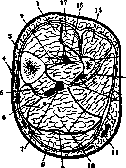
Figure 3. Cross-section
of the crus: 1-m. tibialis ant.; 2-membrana interossea; 3-crista tibiae; 4-v. saphena magna et n. saphenus; 5-m. flex. digit. long.; 6-m. tibialis post.; 7-n. et a. tibialis post.; 8-m. gastrocnemius; 9-tendo m. plantaris; 10-m. soleus; 11-v. saphena parva; 12-m. tibialis post. et a. peronea; 13-m. flex. halluc. long.; 14-tibia; 15-m. extensor digitorum longus; 16-m. extensor halluc. long.; 17-m. extensor digitorum longus. (According to Corning.)
In the canal pass the m. soleus, m. tibialis post., m. flexor digitorum long., m. flexor hallucis long., m. gastrocnemius, and the vessels. The lower opening is located in the lower part of the crus.
vasa tibialia and nerves (see figure 3). M. tibialis anterior (anterior tibial muscle) begins at the upper end of the tibia from its lateral surface, interosseous ligament, and fascia of the leg; its terminal tendon passes under the medial section of the cruciferous ligament, is enclosed by a sheath, and attaches with two teeth to the internal cuneiform and first metatarsal bones. The tendon may split and attach in an unusual place. M. extensor hallucis longus (long extensor of the great toe) begins from the middle part of the fibula and interosseous ligament, is covered at the beginning by the anterior tibial muscle and the toe extensor; its terminal tendon passes under the middle section of the cruciferous ligament to the base of the terminal phalanx of the first toe. Not infrequently it gives off a narrow tendinous bundle to the main phalanx of the first toe. M. extensor digitorum longus (long extensor of the toes) takes its origin on the proximal section of the tibia, between mm. tibialis ant. and peronaeus longus, from the anterior edge of the fibula, interosseous ligament, and fascia of the leg; its tendon, splitting twice, attaches to the dorsal tendinous expansion of the second and fifth toes of the foot. M. peronaeus longus and m. peronaeus brevis are closely adjacent to the extensors. M. peronaeus longus (long peroneal muscle) begins with two heads: the anterior head from the proximal end of the tibia, as well as the head and body of the fibula; the posterior from the body of the latter; the terminal tendon runs along the groove of the cuboid bone, reaches the medial edge of the sole, and attaches to the base of the first metatarsal, sometimes to the second and internal cuneiform bone. M. peron. brevis s. secundus (short peroneal muscle) begins beneath the preceding, from the outer surface of the fibula, ends at the tuberosity of the fifth metatarsal bone, may give additional attachments to various bones, and almost constantly sends a thin tendon into the composition of the tendon to the fifth toe from m. extensor digitorum longus. Quite not infrequently there is m. peronaeus parvus s. peronaeus quartus (in most monkeys as a norm), separating from m. peronaeus brevis. Both peroneal muscles are strengthened by means of retinaculum mm. peronaeorum. Posterior flexor muscles are arranged in two layers and separated by a fascial leaf. All muscles of this region are supplied by branches of the tibial nerve. Triceps surae consists of 3 heads: 2 superficial (m. gastrocnemius) attach to the medial and lateral condyles of the femur and demarcate the popliteal fossa; their flat tendon merges with the tendon of the 3rd head (deep)—the soleus muscle (m. soleus), beginning from the head of the fibula, its lateral edge, and linea poplitea; and together with it forms the so-called Achilles tendon (tendo Achillis s. calcaneus), which ends on the calcaneal tuberosity (tuber calcanei). Mucous bursae are located under the initial tendons of the gastrocnemius muscle and the Achilles tendon. Sometimes three heads are observed in the gastrocnemius muscle; a sesamoid bone is found in its lateral head initial tendon in 25%. M. plantaris (plantar muscle). The short flat belly of this rudimentary muscle, corresponding to the long palmar, begins from the lateral condyle of the femur, immediately above the gastrocnemius muscle; its long narrow tendon passes between it and the soleus muscle to the medial edge of the Achilles tendon and merges with it or attaches near it to the calcaneus or is lost in the fascia of the deep flexor muscles. The muscle is frequently absent or, conversely, appears developed stronger than usual. In many monkeys it is developed extremely strongly and connects with the plantar aponeurosis. M. popliteus is located in the same layer as m. soleus, begins from the lateral condyle and arcuate ligament of the knee joint, runs obliquely, and attaches above the popliteal line of the tibia. It is covered by a dense fascia. It tensions the joint capsule and helps rotate the leg medially. M. flexor digitorum longus (long flexor of the toes) begins from the posterior surface of the tibia, below the attachment of m. soleus and the tendinous arch thrown over the next muscle; its cylindrical tendon crosses the tendon of m. tibialis post., being located more superficially, stretches along the groove of the medial malleolus, strengthened here together with the tendons of the following muscles by means of lig. laciniatum, and attaches to the base
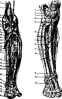
Figure 4.
Figure 5.
Figure 4. Arteries of the right leg from behind: 1 - hiatus tendineus (adductorius); 2 - a. poplitea; 3 - a. genu sup. lat.; 4 - a. genu sup. med.; 5 - aa. surales; 6 - a. genu med.; 7 - a. genu int. lat.; 8 - a. genu inf. med.; 9 - m. popliteus; 10 - a. recurrens tib. post.; 11 - a. tibialis ant.; 12 - ramus fibul.; 13 - a. tib. post.; 14 - a. nutritia tibiae; 15 - m. soleus; 16 - a. peronaea; 17 - a. nutritia fib.; 18 - m. flexor halluc. long.; 19 - m. tibial. post.; 20 - m. flexor digit. long.; 21 - ramus perforans; 22 - a. malleol. post. lat.; 23 - a. malleol. post. med.; 24 - ramus communicans; 25 - ramus calcan. med.; 26 - ram. calcan. lat.; 27 - rete calcaneum. Figure 5. Arteries of the right leg from the front: 1 - a. genu sup. lat.; 2 - rete art. genu; 3 - rete patellae; 4 - a. genu inf. lat.; 5 - a. genu inf. med.; 6 - a. recurrens tib. ant.; 7 - a. tib. ant.; 8 - m. tib. ant.; 9 - m. extens. hallucis long.; 10 - m. extens. digit. long.; 11 - ramus perfor. a. peron.; 12 - a. dorsalis pedis. (After Spalteholz.) of the nail phalanges of the 4 lateral toes. Accessory bundles are frequently observed. - M. tibialis posterior (posterior tibial muscle), pennate, originates from the interosseous ligament, tibia, and fibula in their upper section; the terminal tendon attaches to the tuberosity of the navicular bone, as well as to the second and third cuneiform bones. - M. flexor hallucis longus (long flexor of the first toe) is the strongest of the deep layer muscles, originating from the posterior and medial surfaces of the fibula and the interosseous ligament; the tendon passes beneath the sustentaculum tali and attaches to the distal phalanx of the first toe.
Fascia cruris (fascia of the leg) receives accessory fibers from the fasciae of the mm. gracilis, sartorius, semitendinosus, and the edge of the tibia; it connects to the fibula via the septum intermusculare anterius, which separates the long extensor of the toes from the peroneal muscles, and the septum intermusculare posterius, which separates the latter from the calf muscles. Beneath these lies a deep layer stretched between both bones of the leg and enveloping the deep flexors as well as vessels and nerves. It extends to the malleoli, where on both sides of the Achilles tendon it merges with the superficial layer. The united fasciae attach to the malleoli and form the lig. laciniatum and retinaculum peronaeorum. On the anterior surface of the dorsum of the foot, transverse fibers woven into the fascia form the lig. transversum and lig. cruciatum.
Arteries. A. poplitea (popliteal artery) is the direct continuation of the femoral artery (a. femoralis) and extends from the lower end of the canalis adductorius Hunteri to the lower edge of the popliteal muscle, lying on the planum popliteum tibiae, capsula articularis, and on the muscle of the same name; in the fat of the popliteal fossa (fossa poplitea), dorsally from the artery, the vein and nerve are located sequentially. The popliteal artery gives off muscular branches (lower) — aa. surales — to the muscles of the calf, fascia, and skin; articular branches (2 upper, 2 lower, and 1 middle) that form the rete articulare and rete patellare. Then it pierces the tendinous arch of the m. soleus, receiving the name a. tibialis anterior, while the main trunk remains behind it and receives the name a. tibialis posterior (see Figure 4).
- A. tibialis anterior, having arched over the upper edge of the interosseous membrane to the anterior surface of the leg, runs with accompanying veins and covered by a fibrous layer along the lateral edge of the anterior tibial muscle to the ankle joint, and here, passing under the lateral band of the cruciate ligament and taking the name a. dorsalis pedis, heads onto the foot (see Figure 5). In addition to short muscular branches (some of which pierce the interosseous ligament), the anterior tibial artery gives off the a. recurrens tibialis posterior and anterior to the rete articulare of the knee joint and to the articulation between the tibia and fibula, further the ramus fibularis, which wraps around the head of the fibula into the mm. peronaei, and the rami malleolares anteriores lateralis and medialis.
- A. tibialis posterior runs between the superficial and deep layers of the posterior muscles of the leg, first along the middle of the leg, then along the lateral edge of the common digital flexor, and in the lower third of the leg directly beneath the fascia of the deep flexors. Beneath the lig. laciniatum, behind the malleolus medialis, it turns forward and divides into plantar branches. In addition to numerous muscular branches, which partly anastomose through the interosseous ligament with branches of the anterior tibial artery, it gives off the a. peronaea (peroneal artery), descending to the lateral malleolus and along the way giving off, besides muscular branches, the a. nutritia fibulae, the ram. perforans, which pierces the interosseous ligament above the articulation tibiofibularis inferior to enter the rete dorsale pedis, and the aa. malleolares posteriores. Further — a. nutricia tibiae, rami calcanei.
Variations of the leg arteries present similarities to relations found in various animals. The aa. tibialis anterior, posterior, and peronaea, thanks to existing anastomoses between them, can completely or partially replace each other, with the initial part of one or the other artery obliterating (becoming obliterated). Veins. The cutaneous veins of the leg collect into two trunks, of which the v. saphena parva usually empties into the popliteal vein, sometimes into the vena saphena magna, and originates at the lateral edge of the foot from the rete dorsale plantare, then turns toward the furrow between the heads of the gastrocnemius muscle and before emptying receives the v. femoro-poplitea (see Figure 6). Vena saphena magna (great saphena vein) throughout its extent on the thigh is imperceptible beneath the skin, and is visible only at the malleolus, where the Arabs used to open it, whence its name originates (Greek saphenus — ... Fig. 6. Veins and nerves of the leg: 1 — n. peronaeus ...; 2 — n. cutaneus surae lat.; 3 — n. cutaneus surae med.; 4 — ... malleoli lat.; 5 — ... malleoli med.; 6 — ...; 7 — ramus calcan[ea] ...
manifest); it begins at the medial malleolus and extends onto the thigh. Deep veins generally follow the arteries. Lymphatic vessels. Superficial lymphatic vessels originate from the dorsal and plantar networks, with the vessels of the medial side of the leg following the course of the great saphenous vein, while those of the lateral side cross the popliteal fossa and pass between the heads of the gastrocnemius muscle to the deep lymph nodes. On the thigh, lymph is received by the inguinal nodes. The methodology of investigating deep lymphatic vessels has been developed recently by Iosifov and his school. Deep lymphatic vessels accompany the blood vessels, approach the upper third of the leg, and partly the anterior tibial lymph node. Proceeding further, they pass through the interosseous membrane and empty into the deep popliteal nodes. Nerves. The sciatic nerve divides into two branches approximately in the middle of the thigh: 1. The common peroneal nerve, on its way to the long peroneal muscle, divides into the superficial peroneal nerve, which innervates both muscles of the same name, then pierces the fascia and heads to the foot, and the deep peroneal nerve, which pierces the origin of the long peroneal muscle and extends along the interosseous ligament, ventral to the tibial vessels, between the long extensor of the digits and the anterior tibial muscle (lower down—the extensor of the first digit), which it supplies with motor branches. 2. The tibial nerve, positioned initially dorsally and laterally to the popliteal vessels, then lies between the posterior tibial muscle and the soleus muscle and gives off branches to the posterior muscles of the leg; the medial sural cutaneous nerve with the small saphenous vein extends to the lateral margin of the Achilles tendon, receives an anastomotic branch from the peroneal nerve, thereby giving rise to the sural nerve, which gives cutaneous branches to the dorsum of the foot. Pathology of the leg. Developmental defects. With improper folding and compression of the fetal limbs, underdevelopment of the tibia as well as the fibula occurs, the so-called congenital fracture. In this condition, the defect of the tibia, giving the impression of a pseudarthrosis, is usually localized at the border of the middle and lower thirds of the leg. Rickets causes curvature of the bones of the leg both from the action of body weight on poorly calcified bone and from muscle pull; at the border of the middle and lower third, the bones are convex outward and anteriorly and flattened from the sides (" saber sheath"). Injuries of the leg are common to other parts of the body as well (bruises, traumatic emphysemas, burns, frostbite). Wounds of the leg occur with damage to arterial or venous trunks and subsequent formation of aneurysms. Traumatic injuries of nerve trunks result in the formation of neuromas and impairment of sensitivity or movement in the underlying parts of the leg or foot. Fractures of the bones of the leg are frequent, yielding displacements of fragments of various characters. Usually in peacetime practice, the so-called Dupuytren's fracture of the fibula above the lower malleolus with simultaneous avulsion of the tibial condyle is observed. Injuries of the leg of varying severity associated with occupation and sports (football players) are noted. In syphilis (syphilitic osteochondritis), osteomyelitis, and sarcoma, pathological fractures occur, arising with relatively minor violence. Diseases of the skin and subcutaneous tissue are frequent on the leg, such as pyodermas, erysipelas, boil, carbuncle, phlegmon, etc. Symptomatic ulcers are especially noted: tuberculous, eczematous, varicose, lupus, syphilitic, etc. In addition, independent ulcers (ulcus simplex) are observed as a consequence of disorders of the so-called trophic innervation. Treatment of ulcers associated with disorders of "trophic" innervation consists in the transection of the corresponding nerve trunks. In so-called spontaneous gangrene (obliterating endarteritis), thrombosis most frequently affects the arteries of the leg. In addition to conservative treatment, surgical treatment is used (Leriche, Oppel, and others). Varicose veins (see), which more frequently affect the left leg, sometimes yield extensive nodules. Thrombophlebitis is most frequent in women in the postpartum period. With virulent infection of the distal parts of the limb, the lymphatic vessels are involved in the process (lymphangitis). A fairly frequent disease of the bones of the leg, mainly in the prime of life, is osteomyelitis, leading to necrosis of more or less significant areas of bone substance (sequestration). Neoplasms encountered on the leg: osteosarcomas, cancer metastases, exostoses, enchondromas, lipomas, fibromas, lymphangiomas, and others.
Related articles
Mentioned in
Cite this page
“Crus.” Soviet Medical Encyclopedia. English translation of Bolshaya Meditsinskaya Entsiklopediya, 1st ed. (Moscow, 1928–1936), ed. N. A. Semashko. https://sovietmedicalencyclopedia.pages.dev/article/leg-2/