Varicose Veins
Historical document, translated for reference. It reflects medical knowledge of the 1920s–30s and is not medical advice.
Summary
This article from the 1928–1936 Soviet Great Medical Encyclopedia discusses the pathology, theories of origin, predisposing factors, and clinical manifestations of varicose veins, particularly in the lower extremities.
Encyclopedia article (1928–1936)
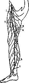
VARICOSE VEINS (varices venarum), an independent, peculiar form of disease of the peripheral veins, chiefly of the lower extremities, the spermatic cord (see Varicocele), and the rectum (see Hemorrhoids). This disease is encountered frequently and has been known since ancient times; many valuable works have been devoted to it. The pathogenesis of this condition is still insufficiently clarified. Proponents of the mechanical theory explain the changes in the veins by prolonged stagnation of blood caused by compression of the outflow veins of the lower extremities (frequent pregnancies, abdominal tumors, hernia trusses, scars). Weber, Cornil, Fischer, and Schmaus consider the primary cause of varicose veins to be pathological changes in the venous walls of the chronic phlebitis type. Thoma and Epstein see the cause in the reduced resistance of the venous walls on the basis of diseases previously suffered by the organism. The possibility of changes in the venous walls as a result of various infectious diseases (especially typhus), various intoxications (alcoholism, lead poisoning, nicotine), and diatheses (rheumatism and gout) has been pointed out by a number of authors. Schambacher points to a congenital weakness of the venous walls, which manifests as an irregular formation and distribution of the muscular and elastic elements of the wall. Mechanical factors only contribute to the appearance of phlebectasia. Kocher also speaks of a congenital predisposition—status praevaricosus. Kraemer, Trendelenburg, Remy, Delbet, Hesse, and Schaack attach importance to the insufficiency of the venous valves and prove beyond doubt the importance of the latter in the development of venous dilations. Thus, Kraemer found a complete absence of valves in the vena saphena magna in 50 operated patients. Mechanical factors that impede blood outflow and abdominal pressure are, in his opinion, within the limits of normal physiological conditions, but veins devoid of valves are unable to meet physiological requirements and dilate. Here, too, the hereditary or acquired abnormality of the wall of the great saphenous vein apparently plays a role. Trendelenburg and Delbet consider the insufficient function of the valves of the system of the great saphenous vein to be the cause of varicose veins. Hesse and Schaack also found an insufficient number of valves in removed dilated veins; in some branches and sections of veins where the valves were normal, there was no dilation; the impression was obtained that the valves as it were protected these veins from varicose dilation. Leonardi looks upon varices as a manifestation of a neuropathic state of the organism. Finally, there are authors who see in the dilation of veins features of a true neoplasm (Jessel, P. Gauld). Considering all these theories, it must be said that mechanical causes (intravascular pressure, stagnation) by themselves cannot cause varicose veins, since veins possess great compensatory properties and, thanks to their elasticity, cope with them quite easily. Only the loss of elasticity and the insufficiency of the valve apparatus can help mechanical causes produce varicose veins. Therefore, the congenital weakness of the venous walls and the insufficiency of the valves, as well as chronic endophlebosclerosis causing a decrease in the elasticity of the venous walls, must undoubtedly be the main primary causes of varicose veins. As predisposing moments, mention is made of sex, age, heredity, constitutional features (Schwartz's varicose diathesis), occupational hazards, and the anatomical features of the affected veins. Persons aged 30–45 who by nature of their occupation have to stand a lot and who show a lack of muscular movements fall ill most often, followed by multiparous women and young people with a congenital weakness of the venous walls. Men and women fall ill in almost equal numbers. Tall people with long, muscular legs, according to Billroth, fall ill more often. Bier points to the frequent occurrence of varicocele, hemorrhoids, and flat feet in individuals suffering from varicose veins of the lower extremities; in his opinion, this is a manifestation of a special constitution, a general weakness of the connective tissue. There are indications that some races contract varicose veins more easily than others. As for heredity, it must be understood here in the sense of transmitting a special weakness of the venous walls; undoubtedly, cases of such transmission have been observed. The frequent affection of superficial veins by the varicose process is explained by some as being located above the aponeurosis, outside the direct influence of muscular contractions. The importance of occupational activities and hazards as a factor predisposing to the disease of varicose dilation of veins has been pointed out for a long time. It has been noted that occupations associated with the vertical position of the trunk and bending in the lower back contribute to stagnation in the veins of the lower extremities. Occupational activities associated with heavy physical labor and prolonged standing on the feet (porters, bakers, blacksmiths, printers, stonemasons, streetcar conductors, etc.) yield the highest percentage of diseases. Occupational hazards—lead and copper dust, anesthesia—being the cause of pathological changes in veins, can also contribute to the development of the varicose process. Long-term practice of a certain profession affects diseases: thus, according to Khesin, there was a sharp increase in the number of varicose dilations of veins in porters with long professional experience. Dilation affects, chiefly, the superficial veins of the system of the great saphenous vein (see plate, Figs. 2 and 3). The process begins most often from the oblique and transverse anastomoses of the superficial network, and then passes to the venous trunks. The small saphenous vein is rarely affected. On the thigh, in addition to the great saphenous vein, the systems of the minor saphenous vein (a branch of the great saphenous vein located more superficially than the latter), the anterior saphenous vein, and the posterior saphenous vein dilate, if they exist; on the leg—collateral channels of the posterior crural saphenous vein and the external crural saphenous vein (see figure). The latter is extremely rare. The deep veins of the leg can also undergo dilation, but rarely (Verneuil, Remy).
Three main types of dilations are distinguished: 1) cylindrical, 2) serpiginous, and 3) varicose saccular protrusions sitting on a broad or narrow base. As a variety of the latter form, saccular dilations of the entire vessel are encountered, as well as so-called "multichambered varices," formed from the fusion of small protrusions. The walls of dilated veins are sometimes thick, sometimes thin (especially saccular protrusions), the lumen of the veins is dilated, and the veins gape on cross-section. The valves are altered, insufficient, or completely absent. Histological changes are noted in all layers of the vessel wall. Characteristic proliferations proper to endophlebitis are observed in the intima; the number of muscular and elastic elements is increased. In the middle tunic, in the initial stages of development, there is hypertrophy of the muscular elements, which is then replaced by their atrophy with the excessive development of fibrous connective tissue (phlebosclerosis). In the outer tunic, there is an enhanced development of elastic elements and proliferation of vessels; the latter penetrate into the intima itself; round-cell infiltration is found around them (see plate, Fig. 4). Thus, "endophlebosclerosis" or, according to Orth, "fibrous panphlebitis" is observed here. Dilated veins, in the form of thin or thick tortuous cords, show through and protrude through the skin. The skin over the varicose nodes becomes thinned and often fuses with their wall; frequently it becomes pigmented as a result of past minor hemorrhages. Edema of the extremities is very frequently observed, especially in the area of the foot and lower leg. The skin in such patients possesses a high propensity for chronic eczemas and ulcerations, thanks to constant nutritional disorder (its thinning), chronic edema, and the harmful effect of chronic stagnation, which is accompanied by a significant disorder of tissue metabolism (Klapp). Because of prolonged edema, the skin and subcutaneous tissue...
cutaneous tissue gradually thickens, and the development of elephantiasis sets in. The complaints of patients boil down to severe fatigue, heaviness, and weakness in the legs, at times to cramps and pains; all these phenomena are explained by circulatory disorders and myositis. Neuralgic pains apparently depend on expansions of the veins of the nerves themselves. Complications of varicose veins are thrombophlebitis, hemorrhages, and ulcers (see Varicose ulcers). Thrombophlebitis easily develops in dilated vessels as a result of intima proliferation and slowed blood flow. Depending on the size and width of the thrombosed vessel, huge tortuous tumors sometimes result, occupying a considerable extent along the limb. These thrombophlebitis cases, in view of their tendency to progress, require special attention; usually they become organized and not infrequently calcify (phlebolith-vein stone), but in rare cases they can turn into purulent ones with all the severe consequences characteristic of this process. As a result of thrombus mobilization, pulmonary embolism (infarct) and even a fatal outcome can be observed. Hemorrhage from dilated veins occurs from the most insignificant injuries to the thinned skin fused with the varix. The blood, being under the pressure of the blood column, spirts in a stream from the ruptured node. Hemorrhage is sometimes significant, bringing patients to anemia. Ulcers can also bleed; bleeding is easily stopped by a pressure bandage. Diagnosis of varicose veins presents no difficulties. To determine valve insufficiency, the Trendelenburg and Hesse symptoms are used. A sharply localized area of small dilated veins can give rise to confusing them with a cavernous angioma, and a separate saccular node on the great saphenous vein in the groin can simulate a femoral hernia. The prognosis for varicose veins is always serious, since the disease almost never undergoes regression, but usually progresses steadily. Preventive measures must first of all take into account the early recognition of the initial forms of the disease and correct, timely, systematic treatment. Professions associated with prolonged standing and strain are contraindicated for carriers of varicose veins. The treatment of varicose veins cannot yet be considered radical, despite a number of proposed methods. Treatment can be divided into three groups: 1) conservative, 2) surgical, and 3) conservative-surgical. Conservative methods of treatment are constant bandaging of the affected limb with a flannel, knit, or flannelette bandage or wearing elastic rubber stockings. The latter are less practical because they easily stretch out, lose their elastic properties, and are expensive. Flannel bandages completely replace them, eliminating venous stasis and edema in the limb. A great many methods have been proposed for the surgical treatment of varicose veins.
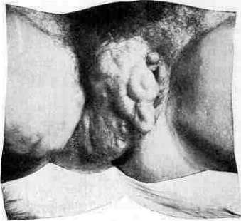
Fig. 1. Diagram of the structure of the venous wall: a - intima, b - inner elastic layer, c - middle muscular layer, d - adventitia. Fig. 2. Diagram of the venous valves: a - valve pocket, b - sinus of the vein, c - base of the valve, d - wall of the vein above the valve, e - area of thinning of the wall above the valve, f - inner elastic layer of the wall, g - adventitia.
methods, but few of them deserve attention. For the first time, ligation of the great saphenous vein as a method of treating varices was proposed by the Italian surgeon Rima (Rima, 1836), but it did not meet with sympathy. In 1888, this operation was performed in our country by A. A. Troyanov, and in 1889 Trendelenburg, unaware of Troyanov's proposal, published this operation, substantiating it with anatomical and physiological data. It consists in the resection of a piece of the great saphenous vein, several centimeters long, at the border of the upper and middle thirds of the thigh. This operation, quite well-founded, nevertheless did not yield stable results (no more than 15%), since the removal of a small piece of the great saphenous vein does not disconnect the superficial venous network from the deep one and does not relieve it from the action of increased blood pressure. Thanks to a number of superficial collateral channels on the thigh and the abundance of anastomoses with the deep network, the reverse flow of venous blood through the great saphenous vein is quickly restored, and the beneficial effect of the operation is reduced to zero. Resection of the great saphenous vein in several sections, with the ligation of a number of collateral channels encountered here (18%), with additional incisions on the leg, gives better results (Jeanne 1, 56%). A number of subsequent operations pursue the goal of "disconnecting the subcutaneous venous network from the deep one, either as far as possible along the entire length of the great saphenous vein (Madelung, Babcock, Helferich) or at numerous points of the subcutaneous network of the thigh and leg (Schede, Wenzel, Rindfleisch). Madelung's operation is performed by a longitudinal incision along the course of the great saphenous vein, from the place of its entry into the femoral vein to the internal malleolus; the saphenous vein is separated and removed entirely, and all lateral and anastomotic branches flowing into it are ligated. This achieves the complete separation of the saphenous veins from the influence of the deep veins. Resection of the great saphenous vein should be performed at the very place of its entry into the femoral vein, so as not to leave a long segment of the vein in which thrombi easily form, sometimes resulting in a fatal outcome (Oppel, a case of pulmonary artery embolism). This method, due to its validity and good long-term results, can be considered one of the best. Its negative side is a lengthy and bloody operation. Therefore, some surgeons prefer to remove the saphenous vein in parts, through a series of small transverse incisions (Helferich), or resort to removing the entire vein by a special extraction method (according to Babcock's method). For this purpose, after the usual resection of the vein (according to Trendelenburg), a small incision is made in the lower part of the dilated vein, through which a long wire is passed upward into the lumen of the vein, brought out through the upper end of the resected vein, which is then tied to the end of the wire. After this, the end of the wire protruding in the lower wound is slowly pulled out, together with the vein tied to its upper end. Thus, it is possible to pull out the entire main trunk of the dilated vein, which at the same time is torn off from its branches, sometimes producing significant subcutaneous bleeding. Both of the latter methods should be used, mainly, when the saphenous vein itself is affected. Circular incisions (Wenzel on the thigh, Schede on the leg) do not give lasting results, and therefore almost no one uses them now. Spiral incisions (according to Rindfleisch) give significantly better results, since they ligate a significant number of anastomoses of the superficial network with the deep one, in addition to repeated incisions of the great saphenous vein itself. This method gives good long-term results, and is used, mainly, in cases of extensive dilations covering a significant extent. The negative property of this operation is the prolonged healing of wounds, lymphorrhea, and subsequent edema. Excision of the saphenous vein can be replaced by bloodless circumvallation according to Schede and Klapp (Klapp). Standing completely apart is the operation of the femorosaphenal anastomosis (see) of Delbet, which aims to restore the normal relationship in the blood circulation of the system of the great saphenous vein by moving the latter somewhat lower into the femoral vein, under the protection of the valves of the latter. Treatment of venous dilations by introducing various medicinal substances into the nodes was proposed a long time ago, and recently there have again appeared many supporters of this method of treatment, for example, the recent proposal of Unger. They use sublimate 1:3,000-5,000 (Scharf), Lugol's iodine solution (Schiassi), hypertonic solution (15-20%) of sodium chloride (binder), 20-40% solution of sodium salicylate, the same solution of sodium citrate, and others. Application technique: with a thin needle, an injection is made into the dilated vein; having made sure by the appearance of blood in the syringe that the needle is in the lumen of the vein, 2-5-10 cubic centimeters of the solution are slowly injected in the distal direction. The needle is removed from the vein after a few seconds to allow the blood flow to carry away the liquid. Then the injection site is pressed with a finger for 2-3 minutes so that the injected liquid does not flow out into the surrounding tissue (necrosis). Injections are made in the standing, sitting, or lying position of the patient. Injections are repeated about twice a week. It is recommended to perform this treatment on an outpatient basis, and patients can go to work immediately. According to Pisareva's research, the thrombus formed in the veins is unstable for 24 hours, and therefore embolism is possible during this time after such injections (cases of death from embolism have been described); the thrombus becomes stable in 2-3 days. Complete obliteration of the nodes is not observed in view of the shrinkage of the thrombus and its canalization during organization. These studies make one treat with great doubt the suitability of this method for the outpatient treatment of varicose veins. After the injection, as after any other operation on the veins, it is necessary to lie in bed for the first time. Postoperative care should consist of bed rest (10 days), in order to avoid complications up to embolism of the lung and pulmonary artery, about which there are a number of reports. In view of this, some (Steblin-Kaminsky) consider early getting up after operations on the veins to be contraindicated. In the future, to reduce the edema developing in the first time after the operation, it is recommended to bandage the leg with a flannel bandage. G. Steblin-Kaminsky. Varicose veins in pregnancy. During pregnancy, conditions are created for the overfilling of the veins of the pelvis and lower extremities with blood. In the pelvic cavity, there is a rich venous network with numerous anastomoses, but without valves. Arteries are accompanied by two or three veins. During pregnancy, the vessels nourishing the uterus and the fetus developing in it become much larger, and new vessels necessary for such enhanced nutrition are added to them. The work of the veins also increases; more blood passes through them, and they have to stretch more strongly than in the non-pregnant state. After childbirth, all of them gradually contract and decrease, but in individual cases, veins that have lost their elasticity remain dilated, and their nodes turn into tumors near the uterus. Blood stasis here becomes habitual, and vein dilations persist and usually even increase. The accumulation of blood and the stretching of the venous plexuses near the ovaries and in the broad ligament also arise from their compression by uterine tumors and tumors located near it; they are also explained by difficulties in bowel function, weakening of the abdominal press, and other similar mechanical influences. The same can happen from the action of the nervous system and from sexual arousal. All these causes exert their influence and lead to chronic stretching of the veins in those who have a congenital predisposition to this (in connection with the patient's constitution). Weakness and distensibility of the venous system, leading to such dilatation of stretched and thinned vessels, are observed in large maternity institutions in 5% of births. Vein dilatation affects vessels not only in the pelvic cavity, but also of the external genitalia (see table, Fig. 1), the anus, and the lower extremities, where varicose nodes are also observed, swelling during pregnancy and accompanied by edema in the tissue. Large size of the fetus, polyhydramnios, and everything that gives a significant increase in the uterus, as well as its tumors (fibromas), compressing the deep venous trunks and increasing the stretching of the veins, prevent the free flow of blood—the tissue becomes edematous, a feeling of heaviness and sensitivity is obtained, urination is difficult and stops, and the stretched venous nodes become painful and inflamed. On the skin of the lower extremities, edematous and erythematous, redness and eczema appear, painful and weeping, and ulceration is obtained, which is difficult to treat. - Treatment. Recently, diathermy has been successfully applied. Itching and sensitivity disappear from electrical warming, but this treatment has to be repeated several times a year, and it does not relieve from the usual surgical treatment, treatment with rest, bandaging, elastic stockings, etc. - Prevention of this disease comes down to hygienic measures, to the reduction and elimination of physical labor, lifting weights, standing on the legs, etc. All this turns out to be effective when there is no constitutional predisposition.
The dangers and complications of pregnancy in varicose veins arise from their injury and from hemorrhages and extravasations into the cellular tissue, with the formation of a hematoma in the pelvis and in the external parts, to which it sometimes spreads from the pelvic cavity. The blood is subsequently gradually reabsorbed, and recovery ensues. Any injuries and incisions to remove the hematoma are accompanied by great danger due to the possible infection that can easily be transmitted here and via the blood as a site of least resistance. The distension of varicose nodes affects the mechanism of labor only by delaying the act of labor, but nature usually copes with this by itself, and operative obstetric assistance is generally not required. Subsequent therapy consists of baths, rest, and sometimes spa mud treatment.
Related articles
Mentioned in
Cite this page
“Varicose Veins.” Soviet Medical Encyclopedia. English translation of Bolshaya Meditsinskaya Entsiklopediya, 1st ed. (Moscow, 1928–1936), ed. N. A. Semashko. https://sovietmedicalencyclopedia.pages.dev/article/varicose-veins/