Varicose Ulcers
Historical document, translated for reference. It reflects medical knowledge of the 1920s–30s and is not medical advice.
Summary
This 1930s encyclopedia article discusses varicose ulcers as a complication of venous dilations in the lower extremities, detailing their pathology, clinical appearance, and treatment methods ranging from conservative bandaging and moist compresses to surgical intervention and skin grafting.
Encyclopedia article (1928–1936)
VARICOSE ULCERS, a complication of venous dilations in the lower extremity, belong to the class of idiopathic ulcers in the genesis of which local causes play a part. The occasion for the formation of ulcers is provided by disorders of blood and lymph circulation in the limb, depending on the underlying disease. Ulcers can develop directly from a ruptured varicose node (true varicose ulcers) or form through thrombosis of the varix, with the penetration of infection into it via the slightest skin lesions (scratch marks), followed by breakdown. At the site of the damaged skin, an ulcer arises, the bottom of which may lie directly above the thrombosed varicose node. Ulcers can also form near dilated veins. Most often they are located on the inner surface of the lower leg, in its lower third, and can be multiple and affect both extremities at once. The size of the ulcer ranges from a millet grain to enormous dimensions occupying a significant extent along the lower leg (girdling ulcers). Young ulcers most frequently have a rounded shape, thin smooth edges, a flat bottom covered with bluish, easily bleeding granulations; often the bottom is covered with a whitish-yellow coating. The discharge of the ulcer ranges in appearance from muco-purulent to ichorous. With the spread of the process and the long existence of the ulcer, in the surrounding tissues, thanks to prolonged edema and stagnation, an excessive proliferation of connective tissue occurs, by the type of elephantiasis, as a result of which the ulcer can sharply change its appearance. Its edges thicken, become callously indurated, unyielding, of irregular shape, the bottom is covered with scanty, sluggish granulations, painful, easily bleeding, the discharge becomes sharply ichorous. Such ulcers stubbornly fail to heal despite the adoption of the most energetic and rational therapeutic measures. The absence of a tendency to healing—a characteristic feature of any ulcer—is expressed here extremely sharply. The irrepressible spread of the process and the absence of a tendency of the granulation tissue to scar depend here, mainly, on the disorder of blood and lymph circulation. If scarring of the ulcer does occur, very often the scars are unstable, easily ulcerating anew, because the conditions disrupting tissue nutrition are not eliminated. Subjective complaints of patients usually boil down to heaviness and fatigue in the legs, itching, cramps, and insignificant pains in the ulcer. In other cases, pains are the main reason for complaints, hindering walking and work. They often appear upon the addition of fresh inflammatory phenomena to the ulcerative process—phlegmon and perithrombophlebitis, developing near the ulcer. As a severe complication, especially of old callous ulcers, one should point out their particular susceptibility to erysipelatous infection; carriers of such ulcers often suffer from recurrent erysipelas. The course of the disease is chronic, often for years, with insignificant, short-lived improvements. The prognosis in young ulcers is fair, in callous ones very often hopeless. In differential diagnosis, one must keep in mind syphilitic (gummous) and cancroid ulcers. In doubtful cases, when cancer is suspected, a biopsy is indicated. The treatment of varicose ulcers requires much patience on the part of the physician and the patient. This is one of the most ungrateful diseases in the sense of positive treatment results. It is necessary to distinguish the treatment of small fresh ulcers located on a comparatively healthy base, and old callous ones on a base sharply altered by chronic edema. The principle of treatment for both types of ulcers is to improve blood and lymph circulation in the limb, bring the ulcer into a "sanitary" state, lowering the virulence and, if possible, neutralizing the pathogens dwelling in it. The first condition can be implemented by rest, lying down with the affected leg elevated, and continuous elastic bandaging with a flannel or knitted bandage. By the outpatient method, i.e., bandaging alone, it is difficult to combat chronic edema, especially in neglected cases: only bed rest helps, albeit slowly, to cope with it. The second condition is achieved by warming compresses of weak, non-irritating antiseptic solutions—potassium permanganate, rivanol, ichthiol, soda, boric acid, Burow's solution, with the mandatory condition of smearing the skin surrounding the ulcer with an ointment to protect it from maceration, which easily leads to the formation of new ulcers (ointments: dermatol, xerform, boric). As soon as the inflammatory phenomena in the ulcer and surrounding skin subside and the ulcer discharge ceases to be ichorous, one can try treating the ulcer with Bayton's strapping (binding the ulcer with strips of adhesive plaster), which renders irreplaceable services in cases that might seem hopeless. In this case, the ulcer under the plaster is, as it were, in a "thermostat", under conditions similar to those necessary for the successful growth of tissue cultures. The granulation tissue, pale, painful, bleeding, and easily decaying before treatment, gradually revives, reddens, begins to grow intensively, ceases to hurt and bleed. Granulations acquire a healthy appearance and a tendency to scar. The ulcer discharge, ichorous-purulent before treatment, changes its character under the plaster, becoming more benign, muco-purulent, mucous, its quantity drops sharply with each dressing. The epidermis develops intensively from the edges of the ulcer and comparatively quickly, after 2–3 days, begins to narrow the ulcer; its white marginal rim grips the ulcer ever tighter and firmer with a strong ring, and soon its entire surface is covered with a strong skin. Removal of the dressing in this case does not disrupt the growing young skin ring, in contrast to treatment with dressings (with ointments, powders, etc.), upon the removal of which the integrity of the growing young skin is disrupted, which delays the healing of the ulcer for a long time. The first time during treatment, the adhesive plaster dressing should be changed more frequently, after 2 days, then less frequently, after 5–7 days. Sometimes during this treatment, especially in the first days, pains and inflammatory phenomena may intensify, which had ceased before this from warming compresses. This indicates the need to postpone adhesive plaster treatment somewhat and, after treating the ulcer again with warming compresses, resume it. If necessary, the adhesive plaster can be replaced by strips of rubber tissue (old glove), but it is still worse because it lacks one valuable property of adhesive plaster, which plays a significant role in healing—the property of reducing the edema of the ulcer edges through pressure that develops here upon tension of the strips of adhesive plaster at the moment of applying them to the ulcer surface (in a shingle-like manner). Treatment with ointments did not yield special results, and drying powders are capable only of intensifying the breakdown in the ulcer, rather than stopping it (under the scab, breakdown proceeds into the depths). A great help in the treatment of ulcers is the surgical treatment of venous dilations; in many cases, healing of ulcers occurs only after it. Stubbornly non-healing ulcers have to be cauterized, scraped, and even subjected to Thiersch skin grafting, with undoubted success in some cases. Dry-air electric baths and diathermy are also used. To improve healing conditions, some authors recommend making lateral crescentic incisions, stepping back 4–5 cm from the edges of the ulcer, to enhance mobility and reduce tension in the tissues surrounding the ulcer. Cases of successful treatment by this method have been described. Recourse is also had to operations on nerves innervating the ulcer region (stretching, section with subsequent suture), and even to amputation of the extremity in hopeless cases with a callous ulcer. Attention must be paid in the described disease to the general condition of the patient—often at the root of the tendency to the ulcerative process lie general constitutional diseases, in the form of, for example, gouty diathesis, cardiac weakness (debilitas cordis), diabetes, lues, intoxications, etc. Accordingly, appropriate treatment should be carried out—diet, regimen, treatment of the underlying disease, resort treatment. For literature, see the article Varicose Veins.
G. Steblin-Kaminsky. VARICOCELE (from Latin varix - knotty dilated veins, and Greek kele - tumor), dilation and elongation of the veins of the spermatic cord. A slight dilation of the cord veins, especially on the left, is encountered quite often and usually does not cause subjective symptoms. The disease develops most often at a young age, during puberty. According to Curling's statistics, at the conscription age among Englishmen varicocele is encountered in 2.3%, among Frenchmen - in 1%. It is predominantly observed on the left (in 92 cases out of 100). Factors contributing to the occurrence of varicocele are considered to be heavy physical labor and prolonged exhausting walking. The onset of the disease in youth is associated with an excessive influx of blood to the genital organs during sexual excitement. Varicocele can either be an independent disease or merely a symptom of impaired blood outflow due to compression along the spermatic vein, most often in the presence of a tumor in the renal region. The anatomical features of the veins of the spermatic cord that cause the occurrence of varicocele are as follows: these veins have very imperfectly constructed valves, and in the inguinal region they have no valves at all. The veins of the cord run in loose connective tissue and receive no help for their emptying from contracting muscles, as is the case in the veins of the extremities. On the left side, there are features that aggravate the situation, consisting in the fact that the left spermatic vein empties not directly into the inferior vena cava, but into the renal vein, flowing into it at a right angle. Moreover, the left testicle usually hangs lower than the right, so on the left side the pressure of the blood column must be higher than on the right. In addition, the left spermatic vein can be compressed during constipation by the sigmoid colon filled with fecal masses. The occurrence of varicocele can also be promoted by anything that leads to diseases of the vascular system - primarily infections and intoxications. The lesion usually begins with the anterior bundle of veins, but in far-advanced cases, the posterior bundle is also affected. Usually, the matter is limited to the lesion of the cord veins, but sometimes the painful changes spread to the veins of the skin of the scrotum and penis. Occasionally, the veins of the testicle itself are affected. The stretched, tortuous, blood-engorged veins of the cord under the influence of their own weight sag in the form of a loop below the testicle (see figure) and in pronounced cases change the position of the testicle, bringing it closer to a horizontal one. The circulatory disturbance caused by stagnation phenomena can lead to degenerative processes in the testicle itself, resulting in its atrophy. Usually, however, during the period of the disease, a flabby consistency of the testicle and its edematousness are observed; sometimes hydrocele is an accompanying phenomenon. Frequently, along with varicocele, a lesion of the veins in other areas of the body is observed - on the extremities, in the rectum. Pathomorphological changes in the cord veins are similar to those observed with the dilation of the veins of the extremities. In most cases, a small varicocele gives insignificant pain sensations. In far-advanced cases, patients complain of a feeling of heaviness in the scrotum, dragging pains in the groin, perineum, and lower back. These symptoms increase with prolonged walking, especially in hot weather. Sometimes sexual impotence and hypochondria are observed. Upon examination, a swelling of soft consistency is discovered along the course of the spermatic cord, consisting of a bundle of dilated tortuous veins. With gentle pressure, the blood from the bundle of dilated veins can be removed to a significant extent, and then the swelling becomes smaller. The swelling increases in a standing position. The corresponding half of the scrotum takes on a pear-like shape. There are cases of spontaneous regression of the disease and self-cure. This usually coincides with coming of age, after the end of the period of sexual development.
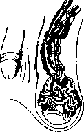
Sharp degree of dilation of the veins of the spermatic cord (after Lexer).
In significantly pronounced cases, one should not count on self-cure. In general, it is difficult to outline a prognosis for this disease. One should also keep in mind the possibility of severe consequences in the form of testicular atrophy, thrombophlebitis, and even bleeding from dilated veins with altered walls. As for treatment, gradations are possible here depending on the development of painful symptoms. First of all, it is necessary to eliminate everything that hinders the outflow of blood from the veins of the spermatic cord - eliminate constipation, prolonged physical fatigue. In the initial stages of the disease, it is necessary to add the wearing of a well-fitted suspensory, worn so that there is no pressure on the inguinal region. In case of failure of these measures, surgical intervention is necessary. The most conservative is resection of the lower half of the scrotum. After this simple operation, the testicles turn out to be placed in a small scrotum and lifted high. The meaning of this aid is the same as wearing a suspensory. This operation is not done very often, in view of frequent relapses after it. The most widespread method to the present time is the excision of the dilated veins of the cord. The operation consists in the fact that, upon exposing the cord, the bundle of dilated veins is isolated from the artery and vas deferens and excised between two ligatures; the lower segment is pulled upward; thereby the testicle, usually lowered in this disease, is also lifted upward. This surgical intervention usually brings benefit, but often leads to prolonged troubles in the postoperative period, which are expressed in edema of the cord tissue and pains along its course. Relapses of the disease are possible. In addition, the excision of veins with the inevitable trauma to other elements of the spermatic cord can adversely affect blood circulation in the testicle itself. These fears were confirmed by experiments on rabbits (Fiorini), in which degenerative changes in the testicle were stated after ligation of the cord veins. Hence the attempts to find other surgical methods for varicocele are understandable. Of these methods, one should point out the proposal of Ts. Manteuffel, which later gave rise to several modifications. The operation is as follows: after incision of the aponeurosis of the external oblique muscle in the inguinal region, a bundle of fibers of the internal oblique muscle is isolated, and the spermatic cord is thrown over this bundle. The veins are not crossed; the testicle turns out to be lifted high. There are reports of good results from this operation. The greatest difficulties in treatment are encountered when varicocele is accompanied by sexual impotence and related nervous diseases. If sexual impotence is caused by pathoanatomical changes in the testicles, one cannot surely count on the regression of these changes after the operation. In such cases, one must be especially careful with indications for surgical intervention.
Related articles
Mentioned in
Cite this page
“Varicose Ulcers.” Soviet Medical Encyclopedia. English translation of Bolshaya Meditsinskaya Entsiklopediya, 1st ed. (Moscow, 1928–1936), ed. N. A. Semashko. https://sovietmedicalencyclopedia.pages.dev/article/varicose-ulcers/