Cardiospasm
Historical document, translated for reference. It reflects medical knowledge of the 1920s–30s and is not medical advice.
Summary
A 1930s medical description of cardiospasm (now known as achalasia), detailing its etiology, clinical presentation, and diagnostic methods like X-ray and esophagoscopy. The article discusses the condition's pathology, including esophageal dilation and the associated risks of complications like aspiration pneumonia.
Encyclopedia article (1928–1936)
CARDIOSPASM (cardiospasmus), a pathological condition of the esophagus caused by a disturbance of its innervation, manifesting as a narrowing of the lumen of the cardia without organic changes to it. In this condition, dilation of the thoracic part of the esophagus is often observed, which bears the name 'idiopathic dilation' (dilatatio oesophagi idiopatica). The etiology and pathogenesis of cardiospasm, despite numerous studies, remain insufficiently clarified. In view of the fact that esophageal dilation has been observed in newborns and children, some authors consider the disease to be congenital. There are no scientific studies to date indicating a genotypic nature of this affliction. It should also be noted that in many cases the onset of cardiospasm is associated with psychic trauma, and the patients belong to the type of 'constitutional neuropath.' The dilation of the esophagus usually extends from the cricoid cartilage to the entrance into the hiatus oesophageus of the diaphragm, whereas the abdominal part of the esophagus remains undilated. The esophagus has its greatest dilation either in the middle part (spindle-shaped form) or in the lower two-thirds (bottle-shaped form). The diameter of the greatest dilation reaches 6-7 and even 16 cm (Gottstein), and its circumference can reach up to 30 cm (Luschka). The dilation may be combined with elongation of the esophagus, as a result of which it takes on an S-shape, with its bulging right wall resting on the right half of the diaphragm. The capacity of a normal esophagus is 50-150 cm3, that of a dilated one is 1,400-2,000 cm3 and more. The wall of the dilated esophagus is in most cases thickened to 5-6-7 mm, mainly due to hypertrophy of the circular muscle layer, which reaches a thickness of 3-4 mm. Only in rare cases is the wall atrophied or subjected to fatty degeneration. The mucous membrane may have a normal appearance; but usually, the stagnation and putrefaction of food in the esophagus lead to the development of inflammatory processes in its wall—from simple thickening of the mucosa to the formation of ulcers with infiltration of all its layers, sometimes to phlegmonous inflammation with secondary phenomena of periesophagitis. Inflammatory changes are most often observed in the lower parts of the dilated esophagus. Ulcers sometimes leave scars upon healing. In rare cases, diverticula and cancerous neoplasms are observed in the lower part. The cardia itself is either normal or appears thickened, hypertrophied, chronically inflamed, with adhesions along the circumference; sometimes there are scars in it. According to data by Bull, the cardia is normal in 50% of cases and changed in 50%. With the introduction of esophagoscopy and X-ray examination, it became clear that cardiospasm is not a great rarity. In 1900, Neumann collected 70 cases from the literature, Thieding in 1921 cites 455 cases, and Lotheissen in 1926 speaks of 600 cases. Cardiospasm is observed at any age. Observations have been described in newborns, in children, and in very old people (68, 74, and 84 years old), but most often cardiospasm affects people aged 30-40 years. The clinical course of cardiospasm is generally as follows. During eating or drinking, it is suddenly noted that food or liquid does not pass into the stomach but is retained in the esophagus, and a feeling of pressure in the chest appears. After 5-10 minutes, the food or liquid passes into the stomach, and the unpleasant sensation disappears. This happens without any apparent reason during a normal meal, more often when swallowing cold or hot liquid. After weeks or months, a second and third similar attack appear, and then they repeat at increasingly shorter intervals. Over time, the difficulty in swallowing becomes constant. In other cases, the development of the disease begins with the patient gradually starting to feel difficulty in swallowing solid food (meat, bread, potatoes), while liquid and semi-liquid food pass freely. The patient begins to take food more cautiously, chews well, and during or after a meal drinks a significant amount of liquid, thereby facilitating the passage of food into the stomach. Sooner or later, the esophagus begins to overflow with food retained in it; this causes extremely unpleasant sensations. The pains can intensify to sharp attacks with the appearance of cyanosis and suffocation, which gives reason to confuse them with asthmatic attacks. Some patients instinctively learn various techniques by which they try to push the food retained in the esophagus into the stomach. All these techniques may remain unsuccessful, and then the patients resort to vomiting or—more likely—to regurgitation, since the contents of the esophagus are expelled without any straining. To produce it, it is often enough for the patient just to assume a horizontal position. The discharge of food residues from the esophagus can occur at night, during sleep. The patient wakes up once or many times with a mouth full of food residues or mucus. In the vomited masses, residues of food eaten several days ago can be found. In some cases, patients suffer from constant regurgitation, chew the regurgitated food, and swallow it again (rumination). There are cases in which the symptoms are not so severe and the general condition of the patients remains little changed for a long time; but sometimes slowly, sometimes more quickly, weight loss and progressive decline in strength follow. Patients can die from exhaustion if they are not provided with help. Severe complications, such as aspiration bronchitis, pneumonia, and gangrene of the lungs, can also lead to death. In the presence of ulcers in the dilated esophagus, mediastinitis may develop or bleeding may occur, which also lead to death. Diagnosis can be made on the basis of the clinical picture; but objective examination methods decide the matter definitively. If a gastric tube is inserted 20-30 cm into a filled esophagus, food residues are discharged both through the tube and past it without vomiting movements. The discharged mass consists of undigested food, is of neutral reaction, and does not contain HCl. If the tube can be passed through the cardia into the stomach, the withdrawn mass contains HCl or at least is of acidic reaction. The tube sometimes passes into the stomach freely, sometimes it is delayed in the region of the cardia. Sometimes the tube simply abuts against the wall of the dilated esophagus. Only fluoroscopy can decide at what point the tube has stopped. X-ray examination of the esophagus with a contrast mass establishes the position and shape of the esophagus, determines the dimensions of its dilation and the duration of retention in it, its function, and the position and function of the cardia. Characteristic is the retention, which can reach 24 hours. In some cases, peristalsis is visible in the esophagus; in other cases, it appears as an atonic, flaccid sac. Sometimes one can see how a larger or smaller part of the contrast mass passes into the stomach. The lower end of the dilated esophagus is either limited in a sac-like manner or is wedge-shaped and pointed. Often, a narrow strip of contrast mass goes from its lower outline into the stomach. Unlike cancerous strictures, the contours of the sac are even and do not have ulceration. During esophagoscopy, the esophagoscope, having passed the cervical part of the esophagus, encounters no further obstacles and seems to dangle in the esophagus. Here, one can often see a widely gaping cavity, but often the walls of the esophagus form large folds that close its lumen. Respiratory movements are usually weak, but pulsatory ones are sharply expressed. The mucosa of the esophagus often appears thickened, dark red in color, sometimes covered with erosions, and bleeds easily. If the cardia allows the gastric tube to pass, then it also allows...
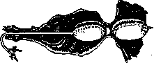
esophagoscope. In other cases, it appears contracted, has the appearance of a rosette (Fig. 1) or a vertical or horizontal slit with ridge-like edges (Fig. 2, on the right in the figure—a small white scar). In the case of the presence of a diverticulum here, one can see the entrance opening into it. The etiology of cardiospasm remains not yet clarified, although there are many theories. In view of the fact that dilatations of the esophagus have been observed in newborns and children, some authors consider the disease to be congenital. Hacker considers it analogous to Hirschsprung's disease and gives it the name "megaesophagus." Strümpell (1881) believed that the cause of cardiospasm is esophagitis. Mikulicz (1882), on the basis of his esophagoscopic examinations, came to the conclusion that the cause of the dilatation of the esophagus is an active spasm of the cardia and that the dilatation itself develops sequentially on the basis of the retention of food masses. One cannot deny the possibility of spasm of the cardia. But it is extremely doubtful whether such a spasm can lead to that sometimes colossal dilatation of the esophagus which is noted in this disease. According to Rosenheim, the primary factors are atony of the musculature and dilatation of the esophagus, and the spasm of the cardia occurs secondarily by reflex from the ulcerated and inflamed mucous membrane of the esophagus as a result of food stasis. Giesse, as early as 1860, expressed the thought that the cause of the dilatation of the esophagus lies in a disease of the vagus nerves. Meltzer believed that the cause of the dilatation of the esophagus lies in the loss of function of the vagus nerves, in particular their relaxing effect on the cardia, as a result of which such strong compression and closure of the latter occurs that the normal force of the esophagus cannot overcome them. Kraus, during a microscopic examination of the vagus nerves from the corpse of a patient who suffered from cardiospasm, found atrophy of them. However, with systematic observations in cardiospasm, no influence of atropine on the patency of the cardia has been noted. The most convincing seems to be the theory of Rieder, who, on the basis of experiments on animals, comes to the conclusion that the cardia in a resting state normally remains closed and opens only during swallowing due to a physiological reflex from the mucosa of the esophagus. The dilatation of the esophagus can therefore hardly be considered as a consequence of cardiospasm; most likely, it is a manifestation of the same disorder of innervation as the obstruction of the cardia. Deviations from the norm in cardiospasm thus boil down to a disturbance of the normal innervation of the esophagus and the reflex opening of the cardia. The disturbance of this reflex mechanism, which is under the influence of the psychic sphere (Boehm), leads to the emergence of cardiospasm.
Therapeutic treatment. The very widespread use of atropine should be recognized as inexpedient both on the basis of
Figure 3.
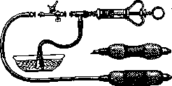
numerous observations and proceeding from physiological premises. Boehm showed that the injection of adrenaline leads to a short-term restoration of the patency of the cardia. However, the elimination of persistent cardiospasm by a non-bloody method is apparently possible only with the help of psychotherapeutic influence (suggestion, hypnosis). For the purpose of non-bloody dilatation of the cardia, elastic probes are used, an example of which can serve as the Geissler-Gottstein probe (Figs. 3 and 4). Its expanded cylindrical balloon is placed in the region of the cardia and is stretched by filling with water. Although Gottstein did obtain good results from such dilatation of the cardia, nevertheless this method, as well as the method of "endless bougienage," did not find widespread use. Better results are given by purely operative treatment of cardiospasm. Jaffe (1897) proposed, and Reisinger performed, a partial excision of the wall of the dilated esophagus. Lambert,
Figure 4.
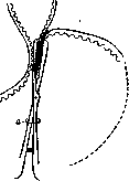
having previously connected the dilated part of the esophagus with the stomach by sutures, crushes the fold (Fig. 5) with a clamp inserted into the stomach. Meyer (1910) performed suturing of the wall of the thoracic part of the esophagus (oesophagoplicatio) three times. Previously, a gastrostomy was performed for nutrition. The success in all cases was disappointing. Sauerbruch in one case performed a transpleural isolation of the esophagus from the diaphragm and vagolysis, but the result was also doubtful, so that all these operations on the thoracic section of the esophagus have been abandoned, and operations aimed at dilating the cardia, which in many cases give quite good results, are enjoying greater success. Mikulicz introduced the method of forceful stretching of the cardia from below from the cavity of the stomach. He opened the stomach in a transverse direction, stretched the cardia first with two fingers, and then with a sponge forceps with branches covered with a rubber tube. The cardia was dilated up to 4 cm in diameter. Out of 6 cases, Mikulicz obtained complete success in 5 cases; in one, a perigastric abscess formed, which fortunately opened along the line of the sutures of the abdominal wall. Such blind dilatation of the cardia should be performed slowly and carefully, since Schmilinsky had one case of death from mediastinitis due to a rupture of the cardia. Wilms recommended performing the stretching of the cardia only with fingers. According to Hesse, this operation was performed in 48 cases; of these, in 39 cases the result was good, in 5 - improvement, in 2 - death followed. In 1900, Marwedel, intending to perform a cardioplasty, used the method of turning the left costal margin upward for access to the cardia. Cardioplasty was first successfully performed by Wendel in 1909. He dissected all layers of the cardia in a longitudinal direction and applied a two-layer catgut suture according to the type of pyloroplasty according to Heineke-Mikulicz. Wendel's case ended in recovery
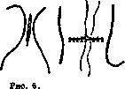
Figure 5. Scheme of crushing the cardial fold according to Lambert: a - incision of the stomach.
Figure 7.
and followed for 12 years. In 1901, Gottstein proposed to perform cardioplasty extramucosally, having previously isolated the esophagus from the hiatus oesophageus of the diaphragm (Figs. 6 and 7). This operation has been successfully performed by a number of surgeons. A patient (33 years old) who had suffered from cardiospasm for 4 years was completely rid of her suffering and followed for more than 3 years (Braitsev). The operation of Heller—extramucosal longitudinal cardiotomy (cardiotomia extramucosa)—has found the greatest spread among surgeons. After turning the left costal margin upward, the esophagus is isolated from the diaphragmatic opening and pulled downward by 10 cm. On the anterior surface of the cardia, a longitudinal incision of 8 cm is made through the serous and muscular membranes to the mucosa. The incision begins 2 cm in the region of the dilatation of the esophagus and ends on the fundal part of the stomach. The same incision is made on the posterior wall of the cardia. With these incisions, by which the sphincter is dissected in 2 places, the possibility of spasm is excluded completely. However, this operation is delicate and harbors certain dangers. Cases of rupture of the mucosa, rupture of the esophageal wall during its isolation from the diaphragmatic opening, peritonitis from the penetration of infection through the undamaged mucosa, and subsequent cicatricial stenosis have been described. With a long abdominal section of the esophagus and cicatricial narrowing of the cardia, some authors have proposed making an anastomosis between the esophagus and the stomach (oesophago-gastrostomia). This operation was first performed by Hoyrovsky. Among Russian surgeons, Hesse performed this operation twice. Herzberg collected 15 cases of this operation with 12 good results. Lit.: Teimanovich Z., On the etiology of dilatation of the esophagus, Vrach. delo, 1921, No. 1-6; Herzberg V., On the question of the operative treatment of cardiospasm, Zhurn. sovr. khir., vol. II, issue 1, 1927; Hesse E., On surgical interventions on the cardia and the abdominal section of the esophagus, Vestn. khir., vol. XV, book 43-44, 1928; Girshberg L., On idiopathic dilatation of the esophagus, Vrach. delo, 1923, No. 24-26; Dillon Ya., On cardiospasm, Trudy Gos. med. instituta v Moskve, vol. I, issue 1, Orel, 1923; Konchalovsky M. and Aizenshtein A., On the question of the symptomatology and treatment of cardiospasm, Ref. med. zhurn., vol. I, No. 1, 1920; Novodvorsky V., On the question of the so-called cardiospasm accompanied by dilatation of the stomach, Yekaterinosl. med. zhurn., 1923, No. 13-14; Private pathology and therapy of internal diseases, ed. by G. Lang and D. Pletnev, vol. II, issue 1, M.-L., 1927; Bull P., So-called idiopathic dilatation of the oesophagus, Ann. of surgery, v. LXXXI, 1925; Hacker V. u. Lotheissen G., Chirurgie der Speiserohre, Stuttgart, 1926 (lit.); Meyer H., Entstehung u. Behandlung der Speiseröhrenerweiterungen u. des Cardiospasmus, Mitteilungen a. d. Grenzgebieten d. Medizin u. Chirurgie, B. XXXIV, 1922; Mintz W., Operative Eingriffe bei Cardiospasmus, Deutsche med. Wochenschr., 1920, No. 47; Rieder W., Der sogenannte Cardiospasmus, Deutsche Zeitschr. f. Chirurgie, B. CCXVII, 1929; Sauerbruch F., Chirurgie der Brustorgane, B. II, Berlin, 1928; Thieding F., Über Cardiospasmus, Atonie und «idiopathische» Dilatation der Speiseröhre, Bruns Beitr. z. klin. Chir., B. CXXI, 1920. V. Braitsev. Figure 6 and 7. Scheme of extramucosal cardioplasty according to Gottstein: Fig. 6 - I moment; Fig. 7 - II moment.
Related articles
Mentioned in
Cite this page
“Cardiospasm.” Soviet Medical Encyclopedia. English translation of Bolshaya Meditsinskaya Entsiklopediya, 1st ed. (Moscow, 1928–1936), ed. N. A. Semashko. https://sovietmedicalencyclopedia.pages.dev/article/cardiospasm/