Esophagoscopy
Historical document, translated for reference. It reflects medical knowledge of the 1920s–30s and is not medical advice.
Summary
Esophagoscopy is the examination of the esophagus using long metal tubes illuminated electrically. This article describes the instruments, techniques, and complications of this medical procedure as practiced in the 1920s-1930s.
Encyclopedia article (1928–1936)
ESOPHAGOSCOPY, ESOPHAGOSCOPE. Esophagoscopy is the examination of the esophagus by introducing into it long metal tubes illuminated from the proximal or distal end by electric lamps-
Figure 1.
Fig. 2.
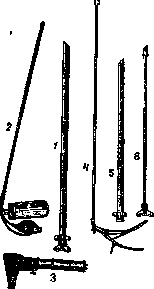
Figure 1. Set for esophagoscopy, containing: 1, 5 and 7 - straight tubes of various lengths; 2 - suction apparatus for removing mucus and secretions; 3 - Kasper's illuminating apparatus; 4 - holders for gauze packets and forceps for
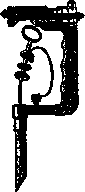
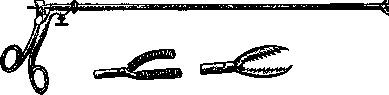
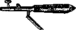
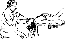
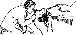
removal of foreign bodies. Fig. 2. Brüning's universal esophagoscope. The first attempts at such research were made in the early 19th century (Bozzini), but did not become widespread due to the imperfection of the instrumentation. In the 1880-1890s of the 19th century, thanks to the invention of incandescent lamps and the application of local anesthesia, endoscopic methods for examining and operating on hollow organs began to improve rapidly, and now on the basis of this methodology, a separate branch of medical science has arisen - broncho-esophagology. Modern bronchoscopic instrumentation, which also includes devices for direct examination of the esophagus, consists of metal extendable tubes of various calibers (diameter from 5 to 14 mm), an illuminating apparatus arranged either in the form of a spotlight throwing a parallel beam of rays along the axis of the tube (fig. 1, 2), or in the form of a long rod with a small electric bulb at the end; this rod is inserted bulb-first into a special groove in the wall of the tube; in this case, only the area of the respiratory tract or esophagus closest to the end of the tube is illuminated. (Both methods compete with each other, having certain advantages and disadvantages.) Esophagoscopes are those tubes of endoscopic sets that are specially intended for examination of the esophagus. In older types of these sets, special tubes of wide diameter and maximum length were placed for this purpose, so that it would be long enough for the entire length of the esophagus. In modern sets of Brüning's, C. Jackson's, Kaller's, Haslinger's, etc., the tubes differ not in their purpose, but only in caliber and length, and the difference in the design of the devices for esophago- and bronchoscopy consists only in that, for example, the insertion extension tube for the esophagus (in Brüning's set) does not have side openings for air passage, while bronchoscopic ones are equipped with them. Furthermore, the set includes operating instruments - forceps and hooks for extracting foreign bodies, nippers for trial excisions, knives, dilators, gauze holders, tubes for suctioning mucus, sprayers for anesthetic agents, etc. (fig. 3-8). Although the esophod can be examined with the same instruments intended for the respiratory tract, the complete set usually includes certain additional parts, for example special insertion tubes (for extending the main ones), special, more massive forceps for foreign bodies, and special probes for examination of esophageal diverticula. The examination is performed with the patient in a sitting or lying position (fig. 9, 10). The position on the abdomen on the operating table with the foot part slightly elevated gives great advantages; then it is much easier to prevent saliva from flowing into the respiratory tract and the stagnation of gastric juice and mucus in the lumen of the tube; in addition, orientation is facilitated when introducing the tube into the esophagus. For anesthesia, the entrance to the esophagus is previously smeared with the help of a curved gauze holder with a 10-20% solution of cocaine or another similar substance used for local anesthesia in otorhinolaryngology. There is, however, an opinion that it is preferable to perform esophagoscopy without anesthesia, since the nausea reflexes arising in the pharynx from the first touch of the instrument facilitate its deeper penetration, and on the other hand - the procedure of cocaine anesthesia itself is extremely unpleasant for the subject. Previously, to ensure the introduction of the esophagoscope, a flexible conductor (mand
Figure 3. Naskeg's forceps.
rene) was used and it was inserted blindly; now this method is completely abandoned, as it is not safe for the integrity of the esophageal wall, and moreover, under visual guidance - the esophagoscope can be introduced even more easily than with the mandrene method. The secret of success lies in the ability to find the upper opening of the esophagus, on the level of the posterior wall of the larynx closed in the form of a slit hardly distinguishable by the eye. To enter it with the beak of the instrument, it is first necessary to direct it exactly along the median line of the body,
Figure 4. Naskeg's curette.
and for this purpose, the lumen of the larynx is first examined and the line of closure of the true vocal cords is determined by eye. By passing the beak of the esophagoscope along the median line slightly posterior to the arytenoid cartilages of the larynx, one enters the cervical part of the esophagus.
Figure 5. Clamp for gauze packets Naskeg-Mikulicz's.
It is also closed and has a star-shaped slit-like lumen. Advancing the tube a few centimeters further, one enters the thoracic, gaping part of the esophagus. The gaping disappears again when the end
Figure 6. Mikulicz's forceps.
of the tube reaches the cardiac (gastric) opening of the esophagus; its lumen has the appearance of closed lips; if this obstacle is forced, the mucous membrane of the stomach will appear in the field of view, differing in a more saturated red color. Orientation is also possible according to
Figure 7. Auxiliary instruments of Hacker's (various instruments for grasping, scraping, etc.).
the depth of insertion of the tube: the distance from the edge of the upper teeth to the pharyngeal opening of the esophagus is 14-15 cm, and to the cardia - from 40 to 45 cm (in adults). By means of esophagoscopy, the presence of foreign bodies, scar strictures, tumors, dilatation and spasm of the esophagus can be recognized, and various therapeutic effects can be performed on it, for example extraction
Figure 8. Handle for forceps Hacker's.
of stuck objects, opening of abscesses in peri-esophagitis, excision of a trial piece, introduction of a radioactive capsule in esophageal carcinoma, bougienage of strictures, etc.
Difficulties and complications of esophagoscopy: anatomical conditions may favor or hinder its performance; in some it succeeds with surprising ease (this formed the basis of the possibility of the circus trick 'sword swallowing'); in others, especially in people with a short neck, curvature of the spine and strongly protruding front teeth, esophagoscopy may prove impossible. In children it is easier than in adults, but sometimes their resistance and agitation require general anesthesia to avoid undesirable traumatism when forcing the insertion of the tube. Since the wall of the esophagus bleeds and ruptures relatively easily, bleeding from superficial abrasions during esophagoscopy is rarely avoidable; however, careless advancement of the tube without visual control, especially when the esophageal wall is pathologically altered, can lead to a more serious complication - through perforation of it and subsequently - purulent mediastinitis. In varicose dilatation of vessels and even more so in aneurysms, there is a danger of their traumatization and life-threatening bleeding. During deep esophagoscopy, the touch of the instrument to the area of the cardia can cause shock, as well as the esophagoscopy procedure itself,
Figure 9. Moment of insertion of the esophagoscope.
which some tolerate very severely. The possibility of secondary infection necessitates preliminary sanitation of the mouth, tonsils, as well as the nose and para
Figure 10. Examination of the esophagus.
nasal sinuses, but often this cannot be done sufficiently radically, since for example in cases of foreign bodies, extraction has to be done more or less urgently. Where time permits, it is desirable before extraction of a foreign body to make an approximate X-ray (localization of opaque foreign bodies, constrictions, diverticula), to perform possible sanitation and to examine the patient on an empty stomach. On the contrary, blind probing of the esophagus, if for some reason it is required, should follow esophagoscopy, not precede it, since in the latter combination the chances of traumatizing the esophageal wall are increased.





Related articles
Mentioned in
Cite this page
“Esophagoscopy.” Soviet Medical Encyclopedia. English translation of Bolshaya Meditsinskaya Entsiklopediya, 1st ed. (Moscow, 1928–1936), ed. N. A. Semashko. https://sovietmedicalencyclopedia.pages.dev/article/esophagoscopy/