Gynecological Instrumentation
Historical document, translated for reference. It reflects medical knowledge of the 1920s–30s and is not medical advice.
Summary
This article provides an overview of the instruments used in gynecological examinations and surgeries during the early 20th century. It details various types of specula, dilators, curettes, and specialized forceps used for procedures ranging from routine examinations to complex abdominal surgeries.
Encyclopedia article (1928–1936)
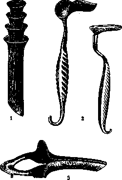
GYNECOLOGICAL INSTRUMENTATION can be divided into two groups: 1) for examination of the genital apparatus and 2) for gynecological operations. Instruments for the latter are, to a significant extent, common to surgery as well. Specifically, gynecological instruments for operations, depending on their purpose, can be subdivided into instruments for vaginal operations and for laparotomy (per abdomen). Instruments used in gynecological examination. For the inspection of the vagina and the vaginal portion of the cervix, cylindrical (or tubular) and gutter-shaped (or spoon-shaped) specula are used. The former are manufactured from milk or transparent glass, porcelain, or hard rubber (ebonite). They are usually available in four sizes, corresponding to the width of the vagina, up to 15 cm in length, with an oblique cut at the end, which allows for the quick and precise positioning of the vaginal portion into the opening of the speculum. Among cylindrical specula, preference is given to Mayer specula made of porcelain or milk glass (see Figure 1). Fergusson specula made of transparent glass, amalgamated on the outside and also covered with a layer of gutta-percha, are also widely used. Spoon-shaped (or gutter-shaped) specula include Simon specula (see Figure 2), consisting of two spoons: one gutter-shaped for retracting the posterior wall of the vagina (posterior speculum) and the other in the form of a plate for the anterior wall (anterior speculum). More practical are the same specula, but made from a single piece of metal and having spoons of different sizes at both ends, for example, Sims specula (see Figure 3). Gutter-shaped specula make it possible not only to examine the vagina and portio vaginalis but also to perform vaginal operations, for example, curettage, etc. In addition to the described specula, so-called bivalve Cusco specula (see Figure 4) and various modifications of them are used, for example, Collin specula (see Figure 5), which make it possible to manage without an assistant; with the help of a screw, the spoons of such a speculum are pushed apart, stretching the vaginal fornices and well exposing the vaginal portion of the cervix. For performing a biopsy, in the case of incipient cervical cancer or suspicious erosion, the Schubert instrument is very convenient—fenestrated forceps with triangular openings having sharp edges. For retracting and fixing the uterus, bullet (American) forceps (see Figure 6), Museux forceps (see Figure 7), and their various modifications are used. Uterine sounding is performed with a metal probe with a handle at one end and a head at the other, with a thickening located 7 cm from the head to indicate the normal size of the uterus. The probe is sometimes divided into centimeters to indicate the dimensions of the uterine cavity—the Martin probe (see Figure 8). Volkmann probes (see Figure 9) made of flexible metal are often used (the instrument can be given any curvature). Among the instruments used in vaginal operations to expose the surgical field, gutter-shaped and flat specula and various kinds of retractors are usually employed, with the help of which the surgical field can be sufficiently exposed [see the above-described Simon and Sims specula, as well as Fritsch (Fig. 10), Doyen (Fig. 11), Ott (Fig. 12) specula, and others]. For the inspection of the abdominal cavity during vaginal operations, Ott proposed spoon-shaped specula equipped with a lighting device—small electric bulbs, which are attached to the corresponding specula on a special rod (as can be seen in Figure 13). Ott's specula make it possible, when illuminating the opened abdominal cavity, to clearly examine various tissues, organs, bleeding sites, etc.
For the dilation of the cervical canal, many instruments have been proposed; of these, the most commonly used are Hegar metal bougies (see Figure 14), which have a conical shape, with the end of the bougie being 1 mm thinner than the bougie itself. The set usually consists of twenty-five to thirty dilators, from 1-2 mm to 25-26 mm in diameter; each subsequent number differs from the previous one by 1 mm. The advantage of the described dilators over others lies in the fact that dilation with them is performed with little effort and traumatizes the tissues less. Curettage of the uterine cavity is performed with sharp and blunt curettes. Of the large number of instruments proposed for this purpose, the Sims model is widely used, sometimes incorrectly called the Hegar curette. Several numbers of different sizes are available for sale, corresponding to the diameter (see Figure 15). When curetting a pregnant uterus, for the removal of large parts of the ovum and placenta, so-called abortion forceps, ending in two closing spoons (see Figure 16), were proposed by Winter; Martin proposed fenestrated forceps for this purpose (see Figure 17). These instruments are the most commonly used. Lubrication of the uterine cavity is performed with a Playfair probe (see Figure 18), at the end of which there are serrations that allow for good fixation of the cotton wool, which does not slip during manipulations. For intrauterine injections, a Braun syringe is used, which consists of a cannula having the shape of a uterine probe with a lateral opening at the end, and a Pravaz syringe with a piston and graduations for dosing the injected substance (see Figure 19). Intrauterine injections can also be performed with an ordinary Record syringe, onto which the corresponding cannula from the Braun syringe is attached. The opening and emptying of pelvic abscesses through the vagina are performed with various trocars; of these, the most convenient is the Landau trocar together with the Vogel dilator-forceps (see Figure 20). The Landau trocar is a puncture needle having a device for connection to an aspirator or an ordinary syringe. The Vogel dilator is a slightly curved, sharp-pointed dressing forceps, having a groove inside the branches for grasping the Landau trocar. The lateral surfaces of the dilator branches are sharpened. In operations for vesicovaginal fistulas, some use special hooks to expose the fistulas (see Figure 21), scissors curved on the flat and on the edge (see Figure 22), and spear-shaped scalpels (see Figure 23).
During laparotomy, gynecologists often use the Fritsch (see Figure 24) or Doyen (see Figure 25) abdominal speculum to retract the edges of the wound, which is inserted into the corner of the wound; to expand the edges of the wound to the sides, hooks (see Figure 26) and retractors with a blunt fork (see Figure 27) and others are used. To expand the abdominal wound, one can use the self-retaining Collin speculum (see Figure 28) with three automatically expanding spoons or the three-valved Stockel speculum, consisting of a large abdominal spoon-shaped speculum and two lateral retractors, which are pushed apart to the sides by means of a rack-and-pinion mechanism to the desired distance, or finally the Franz speculum (see Figure 29) with four specula attached to a quadrangular frame, in which one side is movable; by moving this side, the abdominal wound is expanded. Ovaries and cystic tumors are grasped with fenestrated Hegar (see Figure 30), Nélaton (see Figure 31) ovarian forceps, and others. Dense, voluminous fibromyomas are fixed with Segond (see Figure 32) and Doyen (see Figure 33) corkscrews, which are screwed into the thickness of the tumor. A flaccid, tearing uterus can be most conveniently grasped with Mainzer forceps; these are forceps similar in shape to ovarian forceps, but only larger in size. In operations for cervical cancer according to the Wertheim method, the clamp proposed by him, bent at an angle, is used to close the vaginal tube in order to isolate the cancerous cervix from the abdominal cavity during the amputation of the vagina and removal of the uterus. For the same purpose, the Siegwart clamp (see Figure 34) is used, which has a greater curvature and represents a modified Wertheim model. In gynecological operations, it is often necessary to work at great depth, for which it is necessary to have Kocher forceps, Pean forceps, and scissors of sufficient length, up to 20-24 cm. Lit.—see the literature for the articles Gynecological Examination, Gynecology.
Related articles
Cite this page
“Gynecological Instrumentation.” Soviet Medical Encyclopedia. English translation of Bolshaya Meditsinskaya Entsiklopediya, 1st ed. (Moscow, 1928–1936), ed. N. A. Semashko. https://sovietmedicalencyclopedia.pages.dev/article/gynecological-instrumentarium/