Laparotomy
Historical document, translated for reference. It reflects medical knowledge of the 1920s–30s and is not medical advice.
Summary
Laparotomy is an abdominal incision to access abdominal organs. The article discusses its historical development, preparation techniques, anesthesia methods, positioning of patients, and various incision approaches used in the 1930s.
Encyclopedia article (1928–1936)
LAPAROTOMY (from Greek lapara-paunch, abdomen and tome-incision, section), synonym coeliotomia ventralis, abdominal incision, incision of the abdominal wall for the purpose of gaining access to the organs of the abdominal cavity. The concept of abdominal incision includes opening the peritoneal cavity. However, sometimes one speaks of extraperitoneal abdominal incision, e.g., in operations on large vessels, on the ureter, on the sympathetic nerve. On the other hand, the term 'abdominal incision' is not applied to operations connected with opening the peritoneum but not requiring incision of the muscular-aponeurotic abdominal wall through its entire thickness, e.g., to operations for inguinal and femoral hernias; one speaks of abdominal incision in hernias only when the incision is extended from the hernial opening through the entire thickness of the abdominal wall (herniolaparotomy). In former times, opening of the peritoneum was given special importance, since infection of the latter threatened life, and one did not know how to prevent infection. At the present time, opening of the abdominal cavity itself no longer increases the danger of the operation. The development of L. is connected with the development of aseptic surgery. Before Lister, surgeons resorted to it only in extreme cases and obtained high mortality. Only as antiseptics developed did L. begin to be applied more frequently. First, the operation for removal of cystically changed ovaries (ovariotomy) was performed more frequently. Then other gynecological operations developed rapidly (e.g., for uterine fibromyomas). Operations on the gastrointestinal tract and on organs of the upper abdomen developed more slowly, only as surgeons mastered the aseptic method of operating. Two technical methods were of essential importance in this: bringing the organ to be operated on outside the body and the application of isolating towels. The increasingly frequent operation of appendectomy at the end of the 19th century was of very great importance in the spread of L.; then operations on the bile ducts in their obstruction and inflammation, operations on the stomach and intestine in violation of their passability, and, most recently, operations for ulcerous diseases of the stomach have led to L. not only becoming an everyday operation but also the most frequent operation in the work of any surgical institution, provided the latter is not connected with any special purpose.
Preparation of patients for L. is determined by the peculiarities of the patient's organism and of the organ to be operated on. It is necessary to regulate as completely as possible any disorders in the work of the cardiovascular system, respiratory organs, in metabolism, especially carbohydrate (in diabetes, acidosis-insulin), in blood formation (blood transfusion), from the side of the nervous system, kidneys, to reduce the bacterial flora of the mouth; strictly speaking, all these measures are conditioned by the operation as such and especially by anesthesia; L. itself, however, requires special attention in two directions: it is necessary to accustom the patient to deep and complete breathing (respiratory gymnastics), since after incision of the abdominal wall patients tend to breathe superficially because of pain, which leads to insufficient ventilation of the lungs and to the development of postoperative inflammation in them. Secondly, it is desirable to have the intestine not overloaded with contents and not distended with gases. This facilitates the performance of the operation in the abdominal cavity and the suturing of the abdominal wound, and in the postoperative period it facilitates the work of the heart, the correctness of breathing, and by reducing the bacterial flora and the amount of toxic substances in the intestine it decreases the possibility of self-poisoning from it. Therefore, before L., patients are put on a restricted diet, being given easily digestible food that gives little residue, and the intestine is emptied by laxatives and enemas. However, in recent times it has been found that restriction of food and emptying of the intestine lead to disturbance of metabolism (especially carbohydrate) and to the development of acidosis with a decrease in the alkaline reserve, which extremely delays recovery after the operation. In view of this, restriction of food (especially carbohydrate) and emptying of the intestine should not be done according to a pattern, but only to the extent of actual necessity; in cases where acidosis is already present, repeated intravenous administrations of glucose and subcutaneous injections of insulin are recommended. Attempts were made before L. to increase the resistance of the peritoneum to possible infection (nucleic acid, horse serum, vaccines, anti-streptococcal sera, camphor oil), but all of them gave no positive results. Increasing the resistance of the entire organism by preliminary blood transfusion has very great importance.
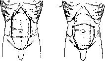
Preparation of the operative field for L. at the present time, thanks to the so-called tanning method, has been greatly simplified: after shaving (if on the day of the operation, then without soap, with alcohol) the skin of the abdomen is smeared with tincture of iodine, a 5% alcoholic solution of tannin or picric acid (the latter solution is especially suitable).-Anesthesia for L. is used predominantly general. It has many advantages over local, causing complete relaxation of the muscles and eliminating involuntary straining of the patient with protrusion of intestinal loops outward. Likewise, suturing of the laparotomy wound under deep general anesthesia is much easier. Nevertheless, in many cases, or on indications from the side of the heart, respiratory organs, kidneys, metabolism (diabetes), general nutrition (cachexia) or on the nature of the operation, local anesthesia is preferred; thus, resection of the stomach, especially in cancer patients, is better tolerated with local anesthesia. With the development of the technique of anesthesia of the p. splanchnici, local anesthesia, one must think, will find still wider application. Spinal anesthesia is used in L. by only a few surgeons. Local anesthesia is very conveniently obtained by the method of spraying the operative field with a 0.5% solution of novocaine (with suprarenin) with additional injection of the same solution along the line of incision. The figures of spraying are seen in figs. 1-3 (according to Haertelio).-The position of the patient for L. depends on the area in which it is planned to operate in the abdominal cavity: the usual horizontal position is changed to a position with the pelvis elevated (Trendelenburg's) for operations in the pelvic cavity and in the lower abdomen or to a position with the lower opening of the thoracic cavity elevated (by placing a roller at the level of the angles of the shoulder blades or by raising the corresponding device of the operating table) for operations on the bile ducts and on the stomach.
The incision of the abdominal wall depends to a large extent on the disease that makes L. necessary; thus, in suppurations in the abdominal cavity making drainage necessary, the incision should be straight and gaping; conversely, in blind suturing of the abdominal wound it is advantageous that the edges of the incision approach each other and that the lines of the tiered sutures do not come one above the other. The location of the incision, and likewise its length, depend even more on the organ to be operated on. The length of the incision should always make it possible to operate in the abdominal cavity, and the incision should be neither shorter nor longer than is necessary for a complete clarification of the relationships in the area of the disease. In this, one must take into account the peculiarities of the anatomical structure of the abdominal wall, keeping in mind that with gross violation of the latter it will not be able after the operation to withstand the intra-abdominal pressure and will give a hernial protrusion. The stability of the abdominal wall is determined by the integrity of its muscular-aponeurotic formations and the preservation of its innervation. It is especially necessary to spare the nerves, since the paralyzed part of the musculature bulges, degenerates, and ceases to retain the contents of the abdominal cavity. The importance of preserving innervation in L. has been elucidated by the works of Kocher, Volkovich, Dyakonov, Sprengel, Valyashko, and others. In view of the anatomical distribution of the nerve trunks in the abdominal wall (see vol. X, art. 229, fig. 33), only an incision along the white line is rational, which can be called the basic laparotomy incision. Usually it involves only part of the white line: for the upper abdomen (fig. 4), for the lower (fig. 4) and for the middle (fig. 5). Its disadvantage is that in the suprapubic part the wound is subjected to strong stretching by the broad abdominal muscles, as a result of which suturing it is often difficult, and after healing of the wound hernial protrusions are often observed; sometimes in the postoperative period complete separation (bursting) of the sutured wound is even observed. The frequently used incision along the lateral edge of the rectus abdominis muscle (fig. 6, a) with displacement of the rectus abdominis muscle toward the middle (the so-called lateral incision of Lennander) was characterized by Kocher as completely inconsistent with the topography of the abdom
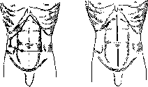
Fig. 3.
Fig. 4.
inal wall (see fig. 6, b). The median incision along the white line (see fig. 5) is the most physiological, since it is made along the line of least resistance and does not cross the aponeurotic fibers of the abdominal muscles. For operations on the upper abdomen, the incision is made from the xiphoid process downward; for operations on the lower abdomen, from the pubis upward; for operations on the middle abdomen, from the navel upward or downward. The transverse incision (see fig. 7) is used for operations on the pelvic organs, for operations on the lower abdomen, and for operations on the upper abdomen when it is necessary to have a wide access. The incision is made parallel to the costal arches. Its advantage is that it does not cross the aponeurotic fibers of the abdominal muscles and that the edges of the wound, when sutured, come together very well. Its disadvantage is that it is difficult to extend it upward or downward. The oblique incision (see fig. 8) is used for operations on the upper abdomen, especially on the gallbladder and liver. It is made from the right or left costal arch downward and outward. Its advantage is that it does not cross the aponeurotic fibers of the abdominal muscles and that it can be easily extended upward or downward. Its disadvantage is that it is difficult to suture the edges of the wound, since they do not come together well. The gridiron incision (see fig. 9) is used for operations on the appendix. It is made in the right iliac region, parallel to the outer edge of the rectus abdominis muscle. Its advantage is that it is small and that it does not cross the aponeurotic fibers of the abdominal muscles. Its disadvantage is that it is difficult to extend it upward or downward. The paramedian incision (see fig. 10) is used for operations on the upper and middle abdomen. It is made parallel to the median line, at a distance of 2-3 cm from it. Its advantage is that it does not cross the aponeurotic fibers of the abdominal muscles and that it can be easily extended upward or downward. Its disadvantage is that it is larger than the median incision. The Pfannenstiel incision (see fig. 11) is used for operations on the pelvic organs. It is made in the pubic region, parallel to the inguinal ligament. Its advantage is that it is hidden by the pubic hair and that it does not cross the aponeurotic fibers of the abdominal muscles. Its disadvantage is that it is difficult to extend it upward or downward.
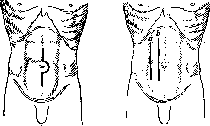
Fig. 5.
Fig. 6.
of the abdominal wall. Instead, the so-called median incision of Lennander (fig. 6, b) with displacement of the rectus muscle to the side should be placed higher than the incision along the linea alba in terms of postoperative strength of the abdominal wall, since the displaced rectus muscle, returning back, covers the sutured deep part of the wound and separates it from the wound of superficial tissues (cul-de-sac incision); but it provides significantly less space. Longitudinal incisions over a small extent with blunt separation to the sides of the fibers of the rectus muscle often allow preservation of the integrity of nerve branches, but with larger incisions it is necessary to cut the nerves, and then part of the muscles to the middle of the longitudinal wound becomes paralyzed and degenerates, causing the corresponding bulging.- Since the time of Sprengel, transverse abdominal incisions have gained general recognition; sometimes they are even called 'physiological,' which is of course incorrect. They well preserve the nerves of the abdominal wall, in combination with bending the body (on a roller) they provide great space, they can be made at any level (fig. 7), deviate to one or
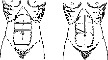
Figure 7.
Figure 8.
the other side, connect them to incisions along the linea alba (thus turning them into an angular incision) (fig. 8). In the subpubic region of the abdomen, a very large space can be obtained by the transverse incision of Bardenheuer (fig. 7); with it, both rectus muscles are cut, and if necessary, it can also be extended to the broad abdominal muscles. Less traumatic (but also provides less space) is the Pfannenstiel incision (fig. 8 (below) and 9), often used by gynecologists: only the tissues from the skin to the rectus sheath are transversely cut, and the separation of the rectus muscles and peritoneum is already carried out in the longitudinal direction. This principle of transverse incision of the fasciae with separation of the medial edges of the rectus muscles, as propagated by Yutsevich, has also been extended to the upper part of the abdomen, above the navel (fig. 10). The Pfannenstiel and Yutsevich incisions thus belong to the group of cul-de-sac incisions along with the longitudinal Lennander incision; however, in the latter, the principle of covering the incision with undamaged tissues is observed more perfectly. Oblique incisions are used for operations in the costal regions and in the iliac fossae (fig. 11). In the costal regions, they are made parallel to the rib edge. In this form, they well open access to organs located in the anterior parts of the subdiaphragmatic space, but they cut across not only muscles but also nerves,
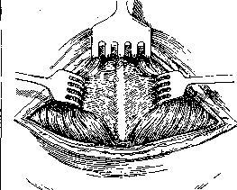
Figure 9.
and therefore cannot be considered rational. On the contrary, oblique incisions in the iliac regions for operations on the cecum on the right and sigmoid colon on the left are well consistent with the anatomical conditions in relation to the nerve branches and at least the external oblique muscle. If the deep broad muscles are not cut with them, but are separated along the course of their fibers, then these incisions should be considered rational.
=="~ its disadvantage lies in the narrowness of the access depth in case of any difficulties. In such cases, it is recommended, stretching the edges of the external oblique muscle, to cut the deep muscles transversely towards the linea alba. If the muscles are not spared, oblique incisions can provide very large space. With the complexity of the topographic relationships of abdominal organs and with the often only clarified during the operation need for greater space, the so-called angular incisions gradually developed, allowing to obtain great space and at the same time preserve the functional capabilities of the abdominal wall after wound healing. A necessary prerequisite for angular incisions was the high development of operative asepsis, which allows one not to be constrained by the breadth of opening the abdominal cavity and to confidently count on the fusion of sutured tissues. Since then, the main incisions in L. have been considered necessary to be made with the possibility of turning them into angular incisions. Of such angular incisions, those shown in fig. 12 (for the stomach), 8 (for the liver) and 13 (for the spleen) should be recognized as quite rational. To the angular incisions belongs the incision of Kehr (fig. 14), in its anatomical characteristics close to longitudinal incisions through the rectus muscle, and of Kausch (fig. 15), close to transverse incisions; both are intended for access to the lower surface of the liver and to the bile ducts. In some cases, the incision of the soft parts of the abdominal wall is insufficient; it may be desirable to extend the incision upward to the lower part of the bony arch of the abdominal cavity,
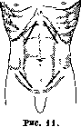
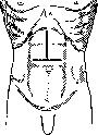
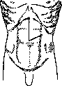
Figure 12.
Figure 13. e.g., for access to the entrance to the stomach and the convex surface of the liver. Of such incisions, one can mention the incision of Auvray with resection of the rib edge (fig. 16) and the incision of Marwedel with temporary turning away of this edge (fig. 17 and 18).- Finally, sometimes simultaneously with L. it is necessary to open the pleural cavity of the corresponding side, even cutting the diaphragm; in such cases, the incision of the abdominal wall is made through the costal cartilages to the corresponding intercostal space, along the latter the pleural cavity is opened, and the wound is widely spread with a retractor (thoracolaparotomy). When opening the peritoneum, it is always desirable to isolate from the skin surface with towels and to grasp the edges of the peritoneal wound with clamps, to prevent unnecessary tearing of it from the intraperitoneal fascia; some even suture the edges of the peritoneal wound, seeing in this a guarantee against infection of the abdominal wall. Spreading the edges of the abdominal wound with hooks, the organ to be operated on is exposed, the intestinal loops are kept away from it with the help of large and long towels, and various retractors are installed. There are retractors of different types: the best ones must still be recognized as wide hooks of the type of spoon mirrors; automatically retaining retractors in the wound often restrict during the operation, especially if the organ to be operated on is poorly extracted outward. If the operation is performed on an organ containing infectious agents, then the free abdominal cavity is carefully isolated from it by
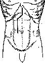

Figure 14.
Figure 15. isolating towels. The towels should be well fastened by their outer ends to the covering sheets, otherwise they can be lost into the abdominal cavity, unnoticed between the intestinal loops, and sutured in the abdomen. Such cases are still observed to this day, although the possibility of such a complication has been sufficiently impressed upon the consciousness of surgeons. Sometimes even instruments are left in the abdominal cavity. Obviously, a certain number of accidents are associated with the need to sometimes during the operation concentrate all attention on one of its moments. At the end of the operation, the abdominal cavity is sutured. For a blind suture of the abdominal wound, two prerequisites are necessary: the absence of surfaces devoid of serous covering and the absence of inflammatory foci that could serve as a source of progressive purulent peritonitis. In the presence of these prerequisites, the peritoneum copes well even with remnants of infected ex4-udate, if such is present in it, and moreover with a blind suture better than with drainage. In the presence of surfaces devoid of serous covering and active infectious foci, it is necessary to resort to drainage. Arguments are made against drainage of the abdominal cavity, that around it adhesions quickly form and the sphere of action of the drainage is thereby extremely narrowed. Nevertheless, everyday experience confirms the benefit and duration
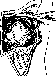
Figure 18.
of action of drainage. The explanation for this is that adhesions can form only when inflammation subsides, when productive processes take precedence over exudative ones; then drainage becomes unnecessary. On the contrary, with the predominance of exudative processes around the drainage, adhesions do not form, and it works well and sufficiently long. In the presence in the abdominal
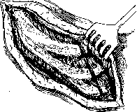
Figure 17.
cavity of surfaces devoid of serous covering, capillary drainage with gauze should be preferred, preferably in the form of a tampon (Mikulicz); in the presence of liquid effusion, tubular drainage is preferable.-The development of the suture of the abdominal cavity required from surgeons a lot of labor and ingenuity (Ott, Dyakonov, Bukhman), but with the development of asepsis it led to the simplest and most understandable forms and methods. At the present time, generally
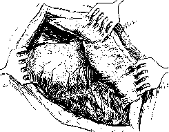
-=**4
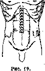
Figure 18. Three-tiered suture of abdominal wound, mostly in 3 tiers: on the peritoneum, on the muscle-aponeurotic formations, and on the skin. Difficulties in suturing arise either from the protrusion of internal organs into the wound or due to the stretching of the wound edges by the force of powerful broad muscles. The first difficulty is eliminated by preparing the intestine for surgery, good anesthesia, lifting the edges of the abdominal wound upward, and holding the internal organs with gauze or special instruments in the form of a plate or splint. In the suprapubic region, where the peritoneum is firmly fused with the posterior lamella of the rectus sheath, it must be included in the suture along with the latter. Closing a gaping abdominal wound is easier to accomplish when done from bottom to top than in the opposite direction. The material for sutures is very varied, but at present it has come down to the usual one in the practice of every surgeon. Subcuticular sutures are better applied as string sutures, as everywhere; the strength and durability of the string are quite sufficient for good healing of the laparotomy wound. The occasional dehiscence of the sutured abdominal wound is in no way connected with the suture material; it indicates a decrease in tissue plasticity, which is connected with the peculiarities of metabolism of a given organism and with various infections, especially of the respiratory tract (influenza). In addition to the tiered suture, sometimes (in very obese subjects, with very great intestinal distension, and when it is necessary to hastily complete the operation) a through suture is used, through the entire thickness of the abdominal wall. Between the main sutures penetrating the entire thickness of the wall, additional sutures are placed on the skin, and sometimes also on the muscle-aponeurotic formations (superficial or subcuticular). This type of suture had great importance previously (Ott, Dyakonov), when subcuticular sutures often suppuration. A bandage for a tightly closed wound is not only unnecessary but rather harmful, as it restricts breathing. A simple gauze roller is sufficient (Fig. 19). For the same reasons, it cannot be recommended to place weight on the abdomen after the operation (a bag of sand). Great caution is required when removing gauze drains inserted into the abdominal cavity. If they were really needed, they can be removed only when sufficiently strong adhesions have formed around them, so as not to rupture upon extraction of the drain; otherwise, a flare-up of peritonitis threatens. The possibility of early removal of drains indicates that there was no need to leave them. Postoperative management after L. requires great attention. It is necessary from the moment of cessation of anesthesia to constantly watch the patient carefully, first of all his breathing and circulation. Postoperative pains begin to trouble the patient as soon as he wakes up from anesthesia; with local anesthesia they arise even sooner. They must be calmed with small doses of morphine or pantopon under the skin, which at the same time improves heart function and makes breathing deeper. If vomiting occurs, the best remedy against it is not to give the patient to drink. Subsequent bronchitis, pneumonia (see Postoperative pneumonia) (Schloffer-15.7%; Mandl-27%; Geinats-26.7%), thrombosis, embolisms are frequent and severe complications. Therefore, as soon as the patient awakens, they are made to take deep breathing movements from time to time, to which he should be accustomed even before the operation; further, they are allowed to turn from side to side as early as possible and to sit and get up as early as possible. Periodic inhalations of CO2 in the nearest postoperative days promote deeper breathing movements and better ventilation of the lungs. If necessary, cardiac drugs and subcutaneous and intravenous infusions are prescribed. It is especially recommended to give warm drop infusions into the rectum (preferably through a Martin's ball). If vomiting persists and there are indications of accumulation of liquid and gas in the stomach, it is necessary to immediately remove them with a tube, if required, repeatedly. It is better to insert a tube unnecessarily than not to insert it when necessary. The same measures help also with hiccough, which can often be stopped by inhaling CO2; persistent hiccough may be considered as an indication of irritation from the peritoneum of the subdiaphragmatic space, usually of an infectious nature. Evacuation of the intestine, if the latter was cleansed before the operation, is not required, and moreover, it cannot be accomplished before the third day, since in the first 48 hours after almost every L. intestinal paresis is observed due to manipulations in the abdominal cavity. On the third day, it is easiest to induce bowel evacuation with a glycerin enema (without water). Until then, only liquid food is given: tea (sugar on the side), broth, water with wine, baked apple; after evacuation, more solid, digestible food is given, which does not give many waste products and gases; milk should be given only with the addition of lime water; buttermilk is well tolerated. Careful observation of the stomach and intestine facilitates the fight against postoperative vomiting, hiccough, and intestinal pains (from the development of gases). Regular evacuation of the intestine is the most reliable means against postoperative adhesions in the abdominal cavity and the development of postoperative intestinal obstruction. As a general rule, it can be accepted: after L. all movements, including that of the intestine, should be limited, dosed, but by no means stopped. This applies to the whole organism and its individual functions. Often patients do not urinate after L., especially after an incision in the lower part of the abdomen. Voluntary urination is facilitated by a sitting position, even better-vertical (standing); if urinary retention still continues, the simplest way is to release it with a catheter. Infusions of glycerin into the bladder and intravenous infusions of urotropin have also been proposed for restoring bladder function.-The most formidable complication of laparotomy is purulent peritonitis. Its frequency has now decreased to such an extent that laparotomy has lost its former character of a dangerous operation; but if nevertheless purulent peritonitis arises from one reason or another, the mortality from it, if it does not reach 100%, is close to this figure (Napalkov) (see Peritonitis).
CANCRORUM
LAPIDES
The most formidable complication of laparotomy is purulent peritonitis. Its frequency has now decreased to such an extent that laparotomy has lost its former character of a dangerous operation; but if nevertheless purulent peritonitis arises from one reason or another, the mortality from it, if it does not reach 100%, is close to this figure (Napalkov) (see Peritonitis).
Related articles
Mentioned in
Cite this page
“Laparotomy.” Soviet Medical Encyclopedia. English translation of Bolshaya Meditsinskaya Entsiklopediya, 1st ed. (Moscow, 1928–1936), ed. N. A. Semashko. https://sovietmedicalencyclopedia.pages.dev/article/laparotomy/