Peptic Ulcer
Historical document, translated for reference. It reflects medical knowledge of the 1920s–30s and is not medical advice.
Summary
Peptic ulcer refers to ulcers that develop secondarily after various operations on the stomach, mainly after gastroenterostomy, at the junction of the stomach and intestine or in the jejunum. This complication became so frequent that it attracted the attention of surgeons worldwide, influencing surgical techniques and indications.
Encyclopedia article (1928–1936)
PEPTIC ULCER, an ulcer that develops secondarily after various operations on the stomach, mainly after gastroenterostomy, at the very junction of the stomach with the intestine or in the jejunum. P. u. has become such a frequent phenomenon that it has attracted the attention of surgeons worldwide. This formidable complication has significantly influenced the attitude toward gastroenterostomy in terms of indications for the operation and has forced a comprehensive review of issues in surgical technique. The first report of secondary P. u. dates back to 1897, but initially it was viewed as an exclusively casuistic curiosity without practical significance; however, in subsequent years the number of such observations steadily increased, and casuistics counted them first in the dozens, and then in the hundreds. At present, in the world literature, more than a thousand described cases can already be counted, and the discussion is about establishing a certain regularity of this complication. Localization of ulcers. All cases of P. u. are divided into two main groups: ulcers developing at the edges of the junction itself (ulcus gastro-jejunale) and ulcers located entirely in the walls of the small intestine (ulcus jejuni). In cases of the first group, the ulcer, having the edge of the junction as its starting point, can spread further from there in the direction of the stomach, as well as in the direction of the small intestine, or simultaneously in both directions, but still more often spreads in the direction of the intestine. Ulcers developing isolated in the small intestine are almost always in the immediate vicinity of the junction, separated from its edges by a strip of several millimeters or receding 1-2 cm. In individual observations, the localization of the ulcer was noted at 7-10 cm, and in the case of Wendel - even at 30 cm from the junction. In one of Mikulicz's cases, an ulcer was found in the edges of a Braun's entero-anastomosis between the afferent and efferent limbs. Ulcers of the jejunum are usually located opposite the opening of the junction, and if they recede to the side, it is more often in the direction of the efferent limb than the afferent. In most cases, the ulcer is single, but many cases of multiple ulcers have also been described, and of different localization. Steinthal described a case of simultaneous finding of four ulcers of almost the same size in the jejunum; this observation is also interesting in that the operation was performed using a Murphy button, which also has 4 holes on each of its halves, which gives reason to establish the development of ulcers in direct connection with the trauma to the intestinal walls during the clamping of the button. The size of ulcers varies widely - from a pea to very extensive erosions, circularly encompassing the jejunum. Isolated ulcers of the jejunum usually have a diameter of 1-2 cm and often have a tendency to spread in depth, causing adhesions with neighboring organs, then extending to them or into the thickness of the abdominal wall. The most characteristic appearance is that of ulcers located isolated in the jejunum; here they retain all the typical properties of a round gastric ulcer: a proper round shape, evenly outlined edges; when such ulcers perforated, a proper round hole, as if punched out with a punch, was found. Often only superficial erosions on the mucous membrane are found, single or scattered, which obviously serve as the starting point for the subsequent development of true ulcers. With prolonged existence and chronic course, the external appearance of the ulcer changes: its periphery becomes infiltrated, the edges thicken, large callus masses form, fusing with the anterior abdominal wall and neighboring organs and involving them in the process; finally, perforations into neighboring organs occur, mainly into the transverse colon; this type of secondary P. u. has a complete analogy with callous penetrating ulcers of the stomach. On the basis of the now very large casuistic material, it is possible to establish the dependence of ulcer development on certain conditions, which sheds light on the cause of their occurrence. First of all, ulcers develop almost exclusively after operations (gastroenterostomy or gastric resection with gastroenterostomy) performed for gastric ulcers, and in only individual observations this occurred after operations for stomach cancer (Braitsyev and others). The fact that in the vast majority of cases it was about ulcers of the pyloric part or duodenum and in only a few cases the ulcer was located remotely from the pylorus in the body of the stomach is also sharply distinguished. The method of gastroenterostomy significantly affects the development of secondary ulcers (see Gastroenterostomy). The true frequency of ulcers of the j. o. has not been established exactly (see Gastroenterostomy). Sharp contradictions and discrepancies in the figures of individual surgeons rather suggest that the evaluation of the material is largely subjective in nature, since the diagnosis of secondary P. u., as will be indicated below, cannot always be established with full certainty not only on clinical symptoms, but also during secondary laparotomies. In addition, too large a percentage of patients escapes later observation or falls into other hands, which naturally makes all calculations inaccurate. The largest percentage falls on the male sex of middle age, the period of greatest working capacity and is clearly connected with the conditions of labor and nutrition; naturally, men of these categories have less opportunity to maintain dietary restrictions for a long period after the operation; it is also possible that the consumption of alcoholic beverages and smoking does not remain without influence on the development of ulcers. Obviously one of the main moments predisposing to the development of ulcers are the socio-domestic conditions in which patients live. However, neither sex, nor age, nor the time elapsed since the operation protects from this complication. Cases of ulcers in infants and in 75-year-old old people have been described. The process of development and course of ulcers is extremely diverse. The development of the ulcer can begin with the very first days after the operation, in others, on the contrary, it passes through many years of an asymptomatic period. In the further course, in some cases, jejunal ulcers complete their entire cycle of development in several days, opening into the abdominal cavity, but in most cases the course takes a chronic character, and gradually neighboring organs and tissues become involved. Extensive adhesions, infiltrates form around, sometimes with delimited purulent accumulations. The method of operation (mainly anterior or posterior gastroenterostomy) has a decisive role in this and determines the path of subsequent complications. In cases of anterior gastroenterostomies, adhesions with the anterior abdominal wall develop first, to which the intestinal loop taken for anastomosis is directly adjacent. Having destroyed the entire thickness of the walls of the stomach or intestine, the ulcer extends to the rectus abdominis muscle, causing extensive infiltrations in it, which later sometimes lead to the formation of an external gastric fistula. After posterior
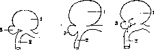
Figure 1.
Figure 2.
Figure 3.
Figure 1. Gastrocolic fistula: 1-stomach; 2-small intestine; 3-transverse colon. Figure 2. Common cloaca between the stomach, transverse colon, and small intestine. Figure 3. Gastrocolic-enteric fistula. Postoperative gastroenterostomy ulcers form adhesions with the posterior abdominal wall, with the mesentery of the transverse colon, with the transverse colon (colon transv.), and in case of perforation of such an ulcer, either diffuse peritonitis develops or a subphrenic abscess forms. The most characteristic feature is the tendency of ulcers following posterior gastroenterostomies to perforate into the transverse colon. Depending on the location of the ulcer, different types of such perforations occur. The perforation opening extends from the jejunum or from the edge of the anastomosis, or finally a common cloaca is formed into which the stomach, jejunum, and transverse colon open (Figs. 1, 2 and 3). The size of such anastomoses or fistulas varies: in some cases it is a narrow channel, in others a wide opening, sometimes with completely even edges resembling an anastomosis made by a surgeon's hand. Fistulas are sometimes so small that they can only be found by straightening the folds of the mucous membrane. The clinical symptoms of P. ulcers are very diverse, depending both on the position of the ulcer and on the nature of subsequent complications. In many cases, the ulcers apparently proceed for a long time without any symptoms, and then suddenly manifest themselves with perforating peritonitis. Such completely unexpected perforations sometimes occur after long periods following the operation, measured in years, during which time nothing can be noted in the patient's condition that would give reason to suspect the formation of a secondary ulcer. A characteristic feature of these cases is that the ulcers were located in the jejunum, there were no adhesions around them, and the serous covering was not involved in the process. However, such cases are still described not often. In most cases, the development of the ulcer does not go unnoticed and gives a series of characteristic symptoms. Usually, symptoms begin to appear after a certain period of completely good general condition; on average, this period is one year, but in individual cases it lasted 8 years or more. It is possible and even very likely that the appearance of painful symptoms does not coincide with the beginning of the development of ulcers, but with one or another complication-penetration of the ulcer deep into the serous covering or into adjacent organs, development of adhesions, narrowing of the anastomosis, etc. The first symptom in most cases is pain in the epigastric region, which in the further course of the disease constitutes the main complaints. The nature of the pain is the same as in ulcers of the stomach and duodenum. Many patients, however, definitely indicate that the new painful sensations are different from those before the operation, that they are more intense and are felt not in the former place, but have moved to the left. The second characteristic symptom is bleeding, which manifests either as bloody vomiting or as tarry stools or occult bleeding. In most cases, there are a number of dyspeptic symptoms, including vomiting. Vomiting usually already serves as a symptom of narrowing of the anastomosis. Due to pain, inability to eat sufficiently, sleepless nights, the general condition and nutrition begin to suffer, and patients often reach an extreme degree of exhaustion. On objective examination, tenderness on pressure in the region of the stomach is noted, sometimes sharply limited to one point, in other cases diffuse throughout the epigastric region, but mainly in its left half or in the region of the navel. Usually, reflex tension of the abdominal muscles is also noted, making palpation difficult. Nevertheless, it is often possible to feel some resistance or a clearly palpable inflammatory tumor is felt. The tumor is usually located along the midline or to the left of it, sometimes at the level of the navel. The size of the tumor varies greatly-from a small nodule to the size of an egg or even a fist. Almost always these tumors are adherent to the anterior abdominal wall and do not move to the side. If the ulcer has already penetrated into the thickness of the anterior abdominal wall, then diffuse infiltration without sharp borders is palpable, sometimes with clearly expressed inflammatory changes on the skin or even with areas of softening. Studies of the chemical composition often show that acidity is increased, but in other cases it remains within normal limits or is even decreased, so the diagnostic value of this sign is small. Cases of ulcer perforation into the transverse colon for a long time may not give any special symptoms different from those just described. But then characteristic signs of a fistula join in, either gradually increasing or suddenly: foul-smelling belching, fecal odor from the mouth, vomiting with admixtures of fecal particles or even consisting almost entirely of liquid or solid fecal masses. On the other hand, persistent diarrhea appears, abundant, watery or in the form of lienteritis; undigested parts of food can be seen with the naked eye in the stools; under the microscope a large amount of fat is revealed, the reaction of the stools is distinctly acidic. General nutrition at this time always sharply decreases. The vomiting itself sometimes causes severe attacks of cyanosis and collapse. The general condition is depressed, but some patients with a gastrocolic fistula for a long time tolerate the presence of feces in the stomach without special disturbances. In cases where the fistulas do not come from the stomach but from the jejunum, the symptoms for a long time may be so little expressed that the fistula itself remains unrecognized, and to prove it, one has to resort to artificial methods-injection of colored fluids into the large intestines followed by pumping out the stomach, inflation of the large intestines with air, which in the presence of a fistula enters the stomach, escaping from it by belching. It is remarkable that the P. ulcer itself after perforation into the transverse colon often heals. Recognition of P. ulcers in certain cases is impossible, as sometimes they occur without symptoms. Unfortunately, it is precisely in such cases that the matter can unexpectedly end with perforating peritonitis. In other cases, recognition can be established with greater or lesser probability, and sometimes with complete certainty. The most characteristic signs are blood in the stomach or in the stool, the presence of a tumor or infiltrate in the anterior abdominal wall; in the presence of painful sensations, vomiting, disturbances of motor function of the stomach, one can only tentatively settle on the diagnosis of postoperative P. ulcer, as the same symptoms can be given by a recurrence of the primary ulcer and narrowing of the anastomosis. However, if the pains reach great intensity, have the peculiarities noted above, if hyperchlorhydria is established, then one should rather lean towards P. ulcer of the anastomosis or jejunum. The presence of symptoms of a gastrocolic fistula naturally immediately clarifies the matter. On X-ray examination, secondary P. ulcers can rarely be proven by direct signs even when using the technique of serial films; this succeeds in no more than 10% of cases. Indirect signs, on which one can rely in the diagnosis of P. ulcers, are the following: 1) tenderness of the anastomosis on pressure, 2) radial convergence of the folds of the gastric mucosa in the direction of the anastomosis, 3) narrowing of the efferent loop by the folds of the mucosa around the presumed ulcer, 4) spasm of the transverse colon in the region of the anastomosis as an expression of the inflammatory process in the mesocolon. The cause of the formation of secondary P. ulcers is quite clearly determined from what has been stated above. There is no doubt that these ulcers are a complete analogy with round ulcers of the stomach, therefore the etiological moments here are of the same order. Of course, this is not all that can be said, since the etiology of round ulcers of the stomach also remains unexplained to the end (see Stomach-ulcer of the stomach, as well as Duodenum-ulcer of the duodenum, and Gastroenterostomy). The main causal factors are irregular nutrition, increased acidity and the digestive action of gastric juice-in relation to postoperative ulcers these factors appear with special obviousness. Physiologically, the mucous membrane of the jejunum is not directly exposed to the action of gastric juice. The conditions change after gastroenterostomy: into the jejunum comes not only the contents of the stomach neutralized by bile and pancreatic secret, and its neutralization here is further hindered by the fact that the play of digestive reflexes is disturbed. Meanwhile, the acidity of gastric juice often turns out to be increased. The significance of insufficient neutralization in the origin of ulcers is confirmed, among other things, by the fact that primary ulcers in the duodenum rapidly decrease in frequency as the distance from the pylorus increases: according to Peri (Regu), out of 149 cases of duodenal ulcers, 123 fell on the first part of the intestine, 16 on the second, and only 1 case on the third. The second argument, however, that the digestive action of gastric juice plays the main role, can be seen in the fact that in cancer of the stomach postoperative ulcers have been observed so far only in individual cases. Of course, the digestive power of gastric juice is determined not only by the figures of acidity, but also by the amount of pepsin.
One can only speak hypothetically about the possibility of a triptic origin of ulcers. In addition to the general causes already considered, other local factors play a role in the development of postoperative ulcers, creating the conditions for peptic action. Such a condition is the violation of the integrity of the mucous membrane at the edges of the anastomosis: on the exposed tissues, the digestive action manifests itself with particular energy. Sometimes the starting point for the development of ulcers was obviously the silk sutures, which for a long time after their removal left deep fistulous tracts. In the case of Ashalm (Ashalme), the suture node lay like in a frame in a separate ulceration of the mucosa. In the immediate vicinity of the anastomosis after gastroenterostomy, as a consequence of trauma, one can see traces of severe circulatory disorders—embolism, ischemia, hemorrhages. The formation of submural hematomas is facilitated by rough kneading of the intestinal walls by hand and especially by clamps. It has already been said that the method of gastroenterostomy is not indifferent in the origin of postoperative ulcers. The above-mentioned etiological factors for the formation of ulcers easily explain this fact. The fact that ulcers develop more often after anterior gastroenterostomies, that the anastomosis according to Braun, and to an even greater extent the operation according to Rou, promotes the development of ulcers, and finally the greatest influence in this regard is the exclusion of the pylorus—all this is easily explained by the fact that the neutralization of gastric contents suffers to an even greater degree under these conditions than with a simple posterior gastroenterostomy. The necessity of preventive measures is obvious, since peptic ulcer is a serious complication, often not yielding to either therapeutic treatment or secondary operative intervention. In addition, it must be emphasized once again that we do not have completely accurate data on the true frequency of secondary ulcers: some of them run in a highly concealed manner and for a long time may not manifest themselves at all. All this makes one think that ulcers develop more often than has been assumed until now. Preventive measures concern two aspects: the technique of the operation and the necessary dietary and medicinal treatment. In the technique of the operation, first place should be given to suturing the entire periphery of the anastomosis with the mucous membrane, either by placing a separate suture on the mucosa or by including the submucosal layer in the suture. The material for sutures (on the mucous and muscular layers) should be catgut, not silk. Intestinal clamps should be applied carefully, avoiding rough trauma to the intestinal walls. In the postoperative period, if the operation was performed for a stomach ulcer, the patient must remain on a suitable diet for a long time (milk-vegetable food, oil); with high acidity, alkaline mineral waters, atropine preparations are prescribed. Treatment. The asymptomatic course of secondary ulcers in some cases forces one to assume that in other cases the ulcer gives symptoms that are not characteristic and weakly expressed. It is possible that sometimes these symptoms are caused by an 'old, unhealed ulcer after the operation'; therefore the measures which are noted as preventive should be applied to every gastroenterostomized patient if even minor and doubtful signs of recurrence appear after the operation. Operation is indicated when therapeutic measures do not lead to the desired result, and in cases complicated by secondary changes that under no conditions yield to conservative therapy, such as the formation of a gastrocolic fistula or perforation into other organs. To proceed to active operative measures immediately at the first suspicion of a secondary ulcer would be incorrect already because the operation represents a difficult task both technically and in essence. It goes without saying that in acute perforations into the abdominal cavity, operation is indicated just as in any other perforations. The complexity of the conditions encountered in secondary ulcers, and the difficulty of establishing these conditions before the operation, in rare cases make it possible to proceed to the operation with a pre-developed and considered plan. Usually this is decided only after the abdominal cavity has already been opened. The methods of operations used by various surgeons for secondary ulcers are very diverse. The main task should be the excision of the ulcer from the anastomosis or intestine within healthy tissues, but this task is often impossible or can be accomplished only at the cost of heroic efforts and great danger. The difficulties increase in cases where the primary stomach ulcer, which served as the reason for the gastroenterostomy, has not yet healed and therefore requires preservation of the anastomosis; with a healed ulcer and a passable pylorus, the task is significantly simplified. Technically, the matter is simpler with anterior gastroenterostomies than with posterior ones. The most common method of operation is the excision of the anastomosis, hermetically suturing the opening in the stomach and restoring the continuity of the small intestine in one way or another (gastroenterostomy). In this form, the operation can be comparatively easily performed with anterior anastomoses. With posterior gastroenterostomy, it is very difficult or even impossible to gain access to the anastomosis due to adhesions and inflammatory infiltrates in the mesentery of the transverse colon. In addition, it is difficult and almost impossible to restore the continuity of the jejunum if the gastroenterostomy was done with a short loop or 'without a loop'. V. Uspensky proposed for such cases an operative method which to a large extent simplifies the matter. According to this method, the path to the posterior anastomosis is laid through the gastrosplenic ligament, the anastomosis with the intestinal loop is drawn into the omental bursa, and the stomach is severed from the anastomosis all around, after which the opening in the stomach ('large') is hermetically sutured. Similarly, the remnants of the gastric walls left as an excess at the edges of the anastomosis ('small stomach') are sutured. Thus the ulcer is not removed, but only excluded, but experience in several cases has shown that such ulcers subsequently heal. In addition to complete excision of the anastomosis, partial excision is applied in suitable cases, i.e., excision of only the ulcer itself, and the resulting defect in the walls of the stomach and intestine is closed with sutures so as not to cause stenosis. This is the simplest method, unfortunately not often feasible. In cases where the anastomosis was successfully excised and the primary stomach ulcer has healed without narrowing of the pylorus, the operation ends there, but some surgeons still add pyloroplasty to it. If there is pyloric stenosis or if the stomach ulcer has not yet healed, pyloroplasty or a new gastroenterostomy is necessary. More active surgeons instead of gastroenterostomy perform stomach resection, i.e., what should have been done already at the first operation. The methods described so far can be called radical. When their execution is impossible or out of fear of too great a risk, some surgeons resort to one or another palliative operations: they applied a second gastroenterostomy or, leaving the stomach alone, performed jejunostomy in order to functionally exclude the stomach, feeding patients through a fistula. Finally, in some cases it was necessary to limit oneself to a trial laparotomy. In cases where the ulcer is located entirely in the jejunum, partial or circular resection is performed. The greatest difficulty is presented by ulcers complicated by the formation of a gastrocolic fistula, and especially—by the formation of a cloaca into which the stomach, jejunum, and transverse colon open. The radical method here is the separation of the adhered organs and the restoration of the integrity and continuity of the stomach, jejunum, and transverse colon. In view of the difficulty and complexity of the operation in this form, some surgeons had to limit themselves to palliative operations—partial resection and exclusion (Fig. 4).

Figure 4. Exclusion of the afferent loop of the transverse colon (1) in gastrocolic fistula; 2—small intestine.
The final outcomes of secondary peptic ulcers are rather sad for both reoperated and non-reoperated patients. Among the non-operated, some died from severe complications of the ulcer or from exhaustion. Reoperated patients show a rather high rate of postoperative mortality and most of them again had recurrences or complications of another kind, requiring further repeated operations. One of Mikulich's patients was operated on five times over a period of 1-2 years, but almost without result. Of 45 cases, operated and non-operated, collected by V. I. Dobrotvorsky, recovery occurred in only 7, 18 died, the others remained in the same condition or only with improvement. S. E. Sokolov, for 125 cases of reoperated patients collected by questionnaire among Russian surgeons in 1926, gives a mortality rate of 30.4%. Cases requiring resection of the transverse colon during the secondary operation gave a mortality rate of 66.6%. In individual surgeons, the postoperative mortality rate in recent years has decreased to 12% (Poshe), but cases of gastrocolic fistulas still gave 30% mortality.
Related articles
Mentioned in
Cite this page
“Peptic Ulcer.” Soviet Medical Encyclopedia. English translation of Bolshaya Meditsinskaya Entsiklopediya, 1st ed. (Moscow, 1928–1936), ed. N. A. Semashko. https://sovietmedicalencyclopedia.pages.dev/article/peptic-ulcer/