False Joint
Historical document, translated for reference. It reflects medical knowledge of the 1920s–30s and is not medical advice.
Summary
False joint (pseudarthrosis) refers to an abnormal mobility in a bone that fails to heal properly. This condition can be congenital or result from pathological processes, trauma, or inadequate treatment of fractures.
Encyclopedia article (1928–1936)
FALSE JOINT, or pseudarthrosis (pseudarthrosis), persistent abnormal mobility along a bone (figs. 1-5). The connection between fragments may be loose-fibrous, dense-scarred, or joint-like. F.J. forms on the basis of developmental anomalies or as a result of pathological or traumatic changes. Congenital F.J. represent a local developmental defect (aplasia). With general defective periosteal bone formation, intrauterine fractures are multiple in nature and not without the ability to spontaneous union. Purely traumatic fractures, if they occur during the intrauterine period, at birth, or in infancy, tend to heal quickly without external assistance. Meanwhile, congenital F.J. are distinguished precisely by 1) localization almost exclusively at the border of the middle and lower thirds of the tibia and 2) inability to form a callus by their own means and local measures. At the moment of birth, the F.J. may not yet exist, but there is only an angular bend or saber-like curvature of the tibia. Under the influence of slight trauma or sharp muscle tension, it forms acutely, and in other cases slowly and without any external provocation
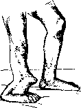
Figure 1.
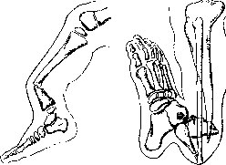
Figure 2.
Figure 3. - fracture and F.J. Hidden forms of such anomalies explain the fact that according to Jungling's collective statistics, out of 100 fractures of tubular bones in childhood (up to 10 years of age), 9 cases occur in the tibia, and out of 100 pseudarthroses in children, 76 occur in the tibial bones. Teratology assigns congenital F.J. of the tibia a medium place in severity in the long series of aplastic changes, ranging from anatomical predisposition of a certain area to fractures to the defect of an entire bone. This area lies at the junction of growth in length from the upper and lower epiphysis. The border strip may remain cartilaginous or fibrous to a greater or lesser extent, may turn into a false joint and into a bone defect. The defect may be of early, embryonic origin and of varying sizes, up to the absence of the entire bone (fig. 6). Of the bones of the tibia, the tibia is much more rarely missing, but the false joint of it is more common, as it is less nourished than the fibula, which is surrounded by muscles on all sides and is sometimes capable of surprising hypertrophy with tibial defects. In congenital pseudarthroses of both bones of the tibia, atrophy of the ends, especially distal, often reaches extreme dimensions. The fragments are thinned, pointed, and amorphously porous. The angular bend at the site of the F.J. becomes increasingly acute, the anterior surface of the tibia abuts against the back of the foot, the entire limb lags in growth and is shorter than the healthy one by 20 or more cm. P. F.J. are formed not without the influence of mechanical factors, but the basis of their origin lies in the pathological process. Diaphyseal osteomyelitis with rapid course quickly disrupts the nutrition of the periosteum or leads to necrosis before the periosteum has time to create a new protective sheath around the dead bone. With chronic course, with the formation of sequestra and fistulas, the reactively abundantly deposited bone may prove insufficiently strong throughout for physiological loading conditions. In both cases, sometimes without traumatic influence, "spontaneously", the bone breaks and due to abnormal nutritional conditions no longer unites. As the infection subsides, in the stage of granulation demarcation and scarring, a fibrous connection forms between the ends of the diaphysis, i.e., a F.J. is formed. The disproportion between pathological passive mobility and loss of active movements will depend on the size of the bone defect, the condition of the soft parts, innervation and blood circulation. - Enchondromas and malignant neoplasms, mainly myelogenic sarcomas, more rarely metastatic carcinomas, by destroying the bone, also create conditions for "spontaneous" fractures with outcome in F.J. It should however be emphasized that in a number of cases not only consolidation occurs, but the formation of a callus even favorably affects the main process. Even more markedly the healing effect of pathological fracture is revealed in fibrous osteitis, if the entire cortical layer was not involved in degeneration and the reactive capacity of the periosteum was preserved. Otherwise, fibrous masses remain between the fragments, and the cavity resembles a F.J. Gummatous and other syphilitic bone lesions are a frequent cause of increased bone fragility and improper fracture union. The danger of F.J. is prevented by timely started specific therapy. - The rarity of pseudarthroses in tuberculosis despite the characteristic osteoporosis of this disease depends on the rarity of diaphyseal involvement by tuberculosis. Destructive displacements of articular ends with some mobility in the new place should be attributed not to F.J., but to pathological dislocations or pseudoarthroses. - Rachitic processes due to the development
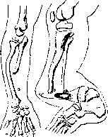
Figure 5.
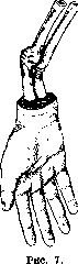
of osteoid tissue and insufficient deposition of lime salts lead to skeletal curvature and delayed union of traumatic fractures, but in childhood do not lead to the formation of F.J. This is prevented by the osteosclerosis that occurs in the reparative stage of the disease. Late rickets and alimentary osteopathies, which received particularly large distribution in Germany at the end and after the imperialist war, more than childhood rickets predispose to insecure, fibrous unions of fragments or to secondary resorption of the callus. Sometimes at the site of a former fracture, even with normal nutritional conditions, without signs of constitutional skeletal disease and without new trauma, several months after consolidation, pain reappears, the callus begins to resorb, and the X-ray film shows instead of the previous solid shadow a transverse crack splitting the bone. Such belt-like rarefactions, or fibrous layers, are called "reconstruction zones" (Umbauzonen) and can turn into F.J. Martin and Muller proved experimentally that after resection of one of two parallel bones, a reconstruction zone or F.J. may appear on the other at the same level. - The role of biochemical, endocrine, mechanical or nervous factors in the pathogenesis of these regressive local changes has not yet been elucidated. Gradual development of erosions with reactive thickening of the periosteum on the tibia and on the metatarsal bones are often found in young soldiers as a consequence of intensified drill exercises. Traumatic F.J. [see separate table (pp. 371-372), figs. 6 and 7] occur most often and do not require for their occurrence either local or general predisposition, although both can play the role of an important conditional factor. The possible time for fracture union varies within very wide limits, and before six months one should not speak of F.J., but only of delayed consolidation. The final failure in a healthy subject may depend on three kinds of causes: lack of bone material between fragments, exhaustion of local osteogenic forces, and finally improper treatment. 1. Too large a diastasis, or divergence of fragments (fig. 7), is the simplest explanation why broken ends do not connect with each other. E.g., so-called "complete" gunshot fractures do not unite, in which large pieces of muscle and bone tissue are torn out. Displacements in which fragments "sit on top", i.e., one fragment abuts against the outer layers of the periosteum of the other, are also an absolute obstacle to the formation of a strong adhesion (fig. 8). Finally, the mechanical role of entrapment of muscles and fasciae between fragments is obvious. Such interpolation of soft parts is probable if crepitation cannot be elicited. It is observed more often in closed fractures than in gunshot fractures. In 120 operations for fractures (among which 92 were closed) Konig (Konig, congress of German surgeons in 1930) found it in 30% of cases. 2. The regenerative capacity of bone depends on the presence of endosteal and periosteal osteoblasts. The death of these cells occurs from the trauma itself or as a result of infection. The more intense the trauma and the heavier the infection, the greater the danger of F.J., since to the mechanical destruction of osteogenic elements are added bacterial influence, purulent decay and necrosis, subsequent circulatory disturbance. If the bone is broken at such a level that the main feeding vessel is simultaneously torn, this circumstance may be sufficient for premature exhaustion
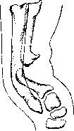
Figure 6.
of osteogenic forces. 3. Improper treatment consists mainly in prolonged immobilization, which leads to atrophy of the bone ends and surrounding soft parts, and in excessive traction, which causes resorption of the fragments. The latter is especially observed in the treatment of fractures of the femoral neck. Incomplete reduction, i.e., when the fragments are not brought into accurate contact, also prevents union. The same applies to insufficient immobilization, which allows movement at the fracture site. Finally, the premature removal of the apparatus before consolidation is complete leads to repeated displacement and non-union. - Among the causes of F.J. in the femur, gunshot injuries take first place, then infections, and finally tumors. In the humerus, gunshot injuries and infections are almost equally common. In the forearm, F.J. is rare, but when it occurs, it is usually in the middle third of the radius. In the bones of the hand, F.J. is observed mainly in the metacarpals and phalanges, often as a result of gunshot injuries. In the foot, F.J. is most common in the metatarsals, especially the second and third. - The clinical picture of F.J. is characterized by abnormal mobility at the fracture site, often accompanied by crepitation. There is no pain on movement, but there may be pain on pressure. The limb is shortened, and there may be deformity. The skin over the F.J. is usually normal, but in cases of infection there may be fistulas and discharge. The muscles are atrophied, and the limb is cold. The prognosis for F.J. is unfavorable, as spontaneous union rarely occurs. Treatment is surgical and consists in removing the fibrous tissue between the fragments, fixing the bone with a plate or nail, and applying a bone graft. In some cases, amputation may be necessary. - Prevention of F.J. consists in proper treatment of fractures, especially in cases of infection or gunshot injuries. Early and adequate immobilization, proper reduction, and avoidance of excessive traction are important. In cases of infection, specific treatment should be started promptly. - The term "false joint" was first used by Ambroise Pare in the 16th century. The condition was described in detail by John Hunter in the 18th century. In the 19th century, the surgical treatment of F.J. was developed by surgeons such as Liston, Syme, and Billroth. In the 20th century, the use of bone grafts and internal fixation devices has improved the results of treatment. - F.J. is a serious complication of fractures that can lead to permanent disability. Early diagnosis and proper treatment are essential to prevent this complication. The prognosis depends on the location of the F.J., the cause of the fracture, and the patient's general condition. With modern surgical techniques, most cases of F.J. can be successfully treated.
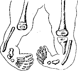
Figure 7.
of osteogenic forces. 3. Improper treatment consists mainly in prolonged immobilization, which leads to atrophy of the bone ends and surrounding soft parts, and in excessive traction, which causes resorption of the fragments. The latter is especially observed in the treatment of fractures of the femoral neck. Incomplete reduction, i.e., when the fragments are not brought into accurate contact, also prevents union. The same applies to insufficient immobilization, which allows movement at the fracture site. Finally, the premature removal of the apparatus before consolidation is complete leads to repeated displacement and non-union. - Among the causes of F.J. in the femur, gunshot injuries take first place, then infections, and finally tumors. In the humerus, gunshot injuries and infections are almost equally common. In the forearm, F.J. is rare, but when it occurs, it is usually in the middle third of the radius. In the bones of the hand, F.J. is observed mainly in the metacarpals and phalanges, often as a result of gunshot injuries. In the foot, F.J. is most common in the metatarsals, especially the second and third. - The clinical picture of F.J. is characterized by abnormal mobility at the fracture site, often accompanied by crepitation. There is no pain on movement, but there may be pain on pressure. The limb is shortened, and there may be deformity. The skin over the F.J. is usually normal, but in cases of infection there may be fistulas and discharge. The muscles are atrophied, and the limb is cold. The prognosis for F.J. is unfavorable, as spontaneous union rarely occurs. Treatment is surgical and consists in removing the fibrous tissue between the fragments, fixing the bone with a plate or nail, and applying a bone graft. In some cases, amputation may be necessary. - Prevention of F.J. consists in proper treatment of fractures, especially in cases of infection or gunshot injuries. Early and adequate immobilization, proper reduction, and avoidance of excessive traction are important. In cases of infection, specific treatment should be started promptly. - The term "false joint" was first used by Ambroise Pare in the 16th century. The condition was described in detail by John Hunter in the 18th century. In the 19th century, the surgical treatment of F.J. was developed by surgeons such as Liston, Syme, and Billroth. In the 20th century, the use of bone grafts and internal fixation devices has improved the results of treatment. - F.J. is a serious complication of fractures that can lead to permanent disability. Early diagnosis and proper treatment are essential to prevent this complication. The prognosis depends on the location of the F.J., the cause of the fracture, and the patient's general condition. With modern surgical techniques, most cases of F.J. can be successfully treated.
of regenerative forces. The interruption of innervation has a harmful effect on the quality of the bone callus, and partial damage to nerve trunks sometimes reflexively delays consolidation. 3. The most frequent cause of the formation of F. j. are errors in treatment. Since the aforementioned harmful factors are removable, the refusal of active intervention for the repositioning of fragments, for combating infection, etc., can contribute to the development of F. j. However, excessive and unskillful radicalism is no less harmful to the cause. During the last war, the antiseptic toilet (or so-called debridement) of gunshot fractures was performed by many surgeons so early and vigorously that due to the removal of fragments still connected with soft tissues, the freshening of fragments, and the excision of muscles, the limb was deprived of the necessary bone material for restoration and the necessary collateral routes for blood supply. The attempt to replace splint immobilization and traction with direct operative fixation of fragments using metal Lenn plates also had the opposite effect in the overwhelming majority of gunshot fractures: purulent osteomyelitis and loosening of screws, a flare-up of infection, prolonged suppuration with the outcome in pseudarthrosis. Another circumstance, which very adversely affects the course of injuries, especially combat ones, is the inevitably associated with evacuation transportation trauma and the non-observance of the principle of continuous immobilization of the limb, even during wound dressings. It should be noted, however, that with extensive comminution of the bone, too vigorous traction, by separating the fragments, delays consolidation. For closed fractures of peacetime, the main therapeutic error leading to the development of F. j. is also insufficient immobilization. In addition, unlike gunshot fractures, the decisive factor is insufficient reduction. It is often impossible to correct the displacement of an obliquely or spirally broken diaphysis of the forearm or tibia, to replace a condyle torn from the joint, without surgery, even if it is only direct traction on the bone. But with proper fixation, it is almost always possible to prevent the development of F. j., provided that the achieved position is maintained for the necessary time without detriment to future function. A considerable number of mysterious cases of fracture recurrence or late F. j. are due to the gradual rupture and obliteration of the bony and vascular bridges between the fragments during the period of outpatient treatment due to oblique loading in removable plaster casts as a result of uncontrolled massage and stereotyped passive gymnastics.-- To reduce the number of F. j., it is necessary to shorten the current periods of general immobilization and extend local ones. In addition, the methods of immobilization in providing first aid and hospital care need to be reviewed and standardized. Therapeutic measures consist of prosthetics or operative interventions. and P a t. a n a t o m y F. j. is very different depending on the type and location of the fracture and on the physiological properties of the affected limb. Loose pseudarthroses with great mobility at the site of F. j. have a connective tissue bridge; they form with large defects in bone material or with significant displacement of fragments. The fragments are sharp, porous, and the bone marrow cavity is closed by a scar. Atrophy is more pronounced the closer the F. j. is located to the epiphyses. The favorite location for such F. j. is the humerus, which generally gives the largest number of traumatic pseudarthroses. Springing pseudarthroses have a dense fibrous connection and a comparatively narrow fissure with a toothed surface and osteoid-cartilaginous deposits. The proximal end is often thicker and embraces the distal one with spurs or a cover. Such F. j. are most often found on the forearm (fig. 9) and tibia, developing gradually and relatively late. "True" F. j. are comparatively rare and require a long time and active movements for their development. They are characterized by the fact that the ends of the fragments are filled and surrounded by smoothly polished fibrous masses or even have a cartilaginous covering. The similarity to a normal joint is increased by the development of a fibrous capsule containing a cavity filled with synovial fluid, and sometimes also iris bodies.
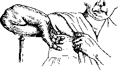
Fig. 9. they form with large defects in bone material or with significant displacement of fragments. The fragments are sharp, porous, and the bone marrow cavity is closed by a scar. Atrophy is more pronounced the closer the F. j. is located to the epiphyses. The favorite location for such F. j. is the humerus, which generally gives the largest number of traumatic pseudarthroses. Springing pseudarthroses have a dense fibrous connection and a comparatively narrow fissure with a toothed surface and osteoid-cartilaginous deposits. The proximal end is often thicker and embraces the distal one with spurs or a cover. Such F. j. are most often found on the forearm (fig. 9) and tibia, developing gradually and relatively late. "True" F. j. are comparatively rare and require a long time and active movements for their development. They are characterized by the fact that the ends of the fragments are filled and surrounded by smoothly polished fibrous masses or even have a cartilaginous covering. The similarity to a normal joint is increased by the development of a fibrous capsule containing a cavity filled with synovial fluid, and sometimes also iris bodies.
E. Osten-Saken. Diseases
Related articles
Mentioned in
Cite this page
“False Joint.” Soviet Medical Encyclopedia. English translation of Bolshaya Meditsinskaya Entsiklopediya, 1st ed. (Moscow, 1928–1936), ed. N. A. Semashko. https://sovietmedicalencyclopedia.pages.dev/article/false-joint/