Fractures
Historical document, translated for reference. It reflects medical knowledge of the 1920s–30s and is not medical advice.
Summary
A comprehensive overview of bone fractures, covering their classification, physical mechanics, and statistical prevalence in the early 20th century. This article details the types of fractures, from simple cracks to complex comminuted injuries, and explains the physics of how external forces cause bone failure.
Encyclopedia article (1928–1936)
FRACTURES, any complete violation of the integrity of a solid object (Wegner), in this case a bone. Fractures, being the result of the most severe injuries, constitute one of the most serious chapters of traumatology. According to Bruns' statistics (London Hospital 300,000 cases of severe injuries, 45,000 fractures), fractures constitute 15% of body injuries. Frequency of fractures of individual bones (in %). Localization of fractures Head . . . Of which: Skull . . . . Facial bones Frequency Localization of fractures Frequency! Bones of the trunk Of which: Spine Pelvis . . . . Ribs . . . . Sternum . . . Scapula . . 3.8 1.42 2.44 0.33 0.31 16.07 0.09 0.86 Upper limbs . . Of which: Clavicle . Shoulder . . . Forearm Hand . . . . 52.60 11.05 Lower limbs
Of which: |Femur .... | Patella ! Shin . . . . I Foot .... 25.88 8.39 1.30 15.53 2.66 Fractures of the limbs constitute more than 3/4 of all fractures, and fractures of the upper limbs occur 2 times more often than fractures of the lower ones. Falling from a height, collapse, being hit by cars, streetcars, carriages, railway catastrophes, airplane accidents, and finally getting caught in the moving parts of industrial and agricultural machines is usually accompanied by fractures. It should be noted that in the statistics of industrial traumatism, fractures constitute from 1% to 2% of all injuries, and in the statistics of agricultural traumatism—from 27% (according to Bolyarsky) to 3.9% (according to Epstein) (the figures are heterogeneous depending on the incompleteness of the registration of minor agricultural traumatism). Statistics in the USA give about 1/2 million fractures annually. According to Bruns' statistics, the greatest number of fractures falls on the age from 20 to 40 years (fractures in men are 4 1/2 times more frequent than in women), i.e., the prime working age. Careful study and elimination of the causes causing fractures is closely related to the study and prevention of traumatism, both industrial, agricultural, and street and domestic. In the USSR, the tasks of combating traumatism, and consequently the prevention of fractures, are the most urgent tasks of the present day. Along with this, an urgent task is the correct organization of the treatment of fractures, the task of achieving the elimination of disability as a consequence of fractures. Classification of fractures. Depending on the force acting on the bone, complete and incomplete fractures are obtained. If the violation of the integrity of the bone is incomplete, i.e., if the connection between the bone particles is not broken throughout the entire extent, then we are dealing with a crack or a fracture. Such cracks on long bones run in a longitudinal direction, and in the form of an incomplete transverse fracture are found on the calcaneus. Their exact recognition is possible only with the help of an X-ray. In a number of cases, the crack heals quickly without special clinical signs (except for pain), in other cases, the healing of the crack is accompanied by a noticeable development of bone callus (periosteal) in the form of a limited periosteal tubercle. On the border between complete fractures and cracks are subperiosteal, i.e., under the periosteum, fractures. In these cases, a crack in the form of a straight or (more often) winding line passes through the entire diameter of the bone. The force acting on the bone here is not great enough to displace the fragments and violate the integrity of the periosteum, which, in the form of a sheath, continues to hold the ends of the bone. Such subperiosteal fractures occur most often on the shin and on the forearm, i.e., on the two-bone sections of the limbs. In these cases the second surviving bone delays the displacement of the fragments and prevents the final rupture of the periosteum. In cases of complete fracture, if under the influence of external violence the bone is separated into a multitude of particles that have lost their mutual connection, we are dealing with fragmentation, and finally, if external violence has caused deformation, i.e., changes in the mutual arrangement of particles, without separating the bone into separate parts, then we are dealing with crushing. The bone possesses certain physical properties (see Bone). These properties change within known limits depending on age, sex, profession, the general physical development of the given subject, and finally on pathological states, among which diseases of metabolism and disorders of the functions of the endocrine system play no small role. The resistance of bone as a material depends on its elasticity. Bone is a body with a low limit of elasticity, as a result of which it is easily subjected to deformation under the influence of external violence. The essence of deformation in the theory of the strength of materials is explained in such a way that under the influence of an external force, the mutual arrangement of molecules in a solid body changes. In this case, the molecules can: 1) either approach each other—compression (Figure 1, A), 2) or move away from each other—tension (B), 3) or move past each other—shear (C).
B
C Figure 1. A-displacement of molecules during compression; B-the same during tension; C-the same during shear. To get a correct idea of the occurrence of fractures, this process should be considered as the result of the interaction of two forces: the living force acting from the outside, causing the rupture of the bone, and the force of cohesion of the molecular particles of the bone, determining its resistance (intermolecular tension). The living force acting from the outside is determined by the formula mv2/2, where m is mass, v is the speed of movement. From this, it is clear that the action of the living force depends on the mass of the object delivering the blow and on the increasing speed of its movement. Thus, for example, a small bullet weighing 14.7 g, flying at a speed of 660 m per second, pierces the bone through. The action of its living force, applied to a very
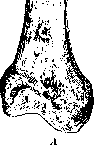
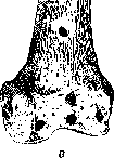
Figure 2. Entrance (A) and exit (B) holes during a through wound of the femoral metaphysis at a distance of 700 m.
small surface, is great enough to overcome the cohesion of particles in a small space. Not having time to spread due to the speed of flight to neighboring particles, the living force overcomes the cohesion of the particles located in its path and carries them away with it (Figure 2). On the other hand, any massive body possessing great weight, but acting slowly, produces destruction of the bone over a large space. Here, the living force, slowly spreading in all directions, will meet the resistance of bone particles over a greater extent. The latter, thanks to the force of cohesion of the particles, will for some time, within the limits of its elasticity (bone), bend, deflect until the living force overcomes the force of cohesion and causes the rupture of particles, clinically—a fracture. Such a picture we observe when the hand gets into a drum. In these cases, we get a bending of the bone, cracks, or fractures from compression. Here, with the slowness of movement, the fracture depends on the mass of the acting body. Thus, an external force acting on the bone can act: 1) in the form of a sharp single push: a) in a transverse direction, causing shear, b) in a longitudinal direction, causing impaction; 2) in the form of prolonged (constant) pressure, gravity, load: a) in a transverse direction, causing bending, b) in a longitudinal direction, causing crushing, c) in the form of a couple of forces, causing torsion. The indicated five main types of deformation determine the basic forms of bone destruction, depending both on the speed and duration of the collision and on the direction of the external force acting on the bone. Fractures from direct impact (single push). The force acting from the outside
can act with a known speed in the form of a push, a direct impact on the bone, and depending on the speed of action it either a) pierces the bone through at the place of its application—the action of a fast-flying bullet on a stationary bone (Figure 2) or produces displacement of particles at the place of its application and a whole bouquet (Figure 3) of cracks (line of bone rupture), spreading in all directions in mutually perpendicular directions—a stellate, comminuted fracture, or b) if the surface of its application, as well as the mass of the acting body, is larger (e.g., a hammer, stone, hoof), it produces a displacement of the entire peripheral segment of the bone in relation to the central one, produces a lateral shear, as if cutting the bone. If at the same time the push, the impact, occurs quickly enough, then we have a direct fracture with even planes (as if a cut surface). If the force of the impact continues to act after the fracture, then further lateral displacement of the fragments occurs (final displacement of fragments along the length of the fracture in tubular bones) (Figure 4). Fractures from bending occur by the same type as the fracture of a stick, the ends of which are trying to be brought closer to each other (Figure 5). When a long bone, fixed at one end, receives a blow or load on the other end, a bending or deflection of the entire limb occurs. For example, with the lower limb fixed while standing, a push from the front, throwing the entire torso back, carries with it the upper part of the femur, which breaks at the place of the bend. The fracture itself occurs in the following way:
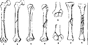
Figure 4. Diagram of fractures of tubular bones: 1-transverse; 2-oblique; 3-longitudinal; 4-spiral; 5-cruciate, or T-shaped; 6-Y-shaped; 7-wedge-shaped; 8-comminuted.
the bone forms an arc, on the concave surface of which compression of molecules occurs, and on the convex surface—tension; since the resistance of solid bodies, including tubular bone, to tension is less than to compression, the rupture of the bone tube during bending occurs first on the convex surface,
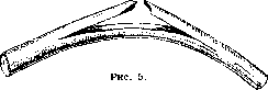
from where it proceeds perpendicularly to the axis of the bone to the middle, the so-called neutral zone, which experiences neither compression nor tension (Fig. 6); upon reaching this point, the line of rupture changes its direction: on a wooden stick, the splitting proceeds along the longitudinal axis (Fig. 5), but on a bone, due to the peculiarities of its structure, the fracture line after passing the neutral zone AB bifurcates and forms a wedge with its base facing the concave side of the bone (Fig. 6). A typical picture of a fracture of a tubular bone from bending is depicted in Fig. 7. Depending on the acting force, the place of fixation, and the length of the free lever, the direction of the rupture line changes,
Figure
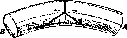
Figure 6.
Figure 6. Diagram of a fracture from bending. Figure 7. Schematic representation of a fracture of the femur from bending. and instead of a wedge, a simple oblique fracture may result (Fig. 8). A combination of a fracture from impact and a fracture from bending occurs with a somewhat slowed impact. Here, the force of the thrust is directly joined by the action of the weight of the entire falling body, due to which the fracture, which began under the influence of the impact, ends under the influence of bending. In cases where the live force acts in the direction of the long axis of the bone, usually from top to bottom, so-called compression fractures (fractures from compression) occur. Such fractures usually occur in connection with the action of a large mass (weight): falling from a height, being buried by earth, etc. Compression fractures most often affect spongy bones—vertebrae, the calcaneus, the spongy part of the tibia. Under the influence of a vertically acting force, the spongy substance of the bone is compressed, the trabeculae are displaced, the porous structure of the bone is disrupted, and the spongy substance turns into a dense, shapeless mass. As for tubular bones, pressure acting along the vertical axis of the bone cylinder causes stretching of its diameter in the middle part, as a result of which the bone cylinder, as it were, bursts and produces a deeply running vertical fissure (Fig. 9); subsequently, transverse cracks from bending join this fissure, as a result of which rhomboid sections of the diaphyseal part of the tubular bone are broken out. Such fractures most often affect the humerus and the tibia. In some cases, longitudinally acting pressure produces an impacted fracture on the epiphyses and metaphyses of long bones. Torsional, or twisted, fractures of tubular bones arise under the influence of the act
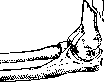
Figure 8. Schematic representation of a fracture of the lower end of the humerus from bending.
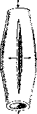
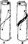
Figure 9. Schematic representation of a longitudinal rupture of a cylinder from longitudinal compression.
Figure 10. Origin of a torsion fracture during twisting around the axis ab. [It is the result] of the action of two forces (so-called "force couple") acting on the bone at different points in parallel but opposite directions. The deformation of the bone resulting from such twisting will be a combination of shear and rupture (Fig. 10). A torsion fracture occurs most often with a fixed lower limb, when, due to inertia, the upper part of the torso continues to move forward and to the side, e.g., the typical skier's fracture (described by Bardenheuer) during a sudden stop while moving, whereby the foot on which the person is standing, trying to maintain balance, is fixed, while the entire limb, during a fall to the side, rotates by inertia outward and forward. As a result, with the leg extended at the knee joint, a torsion fracture of the femur occurs; with the knee bent, [a fracture] of the lower leg (see separate table, Figs. 2-4). The faster the action of the driving forces, the steeper the bending line; the slower the action of the forces, and the greater the distance between the points of their application, the more gently the spiral line runs. Thus, one happens to see that the spiral line of rupture, starting on the lower inner surface of the tibiae, ends near the head of the fibulae. In a whole series of cases, a spiral fracture does not end as such, but having made a half-turn around the bone, it changes its direction, because the action of the gravity of the body falling to the side is added to the torsion action of the "force couple," whereby the second half of the bone cylinder breaks from bending across; thus, a torsion-bending fracture is obtained. If, at the last moment, a blow along the longitudinal axis, which gives a compression fracture, is added to the action of the two aforementioned forces, then a combined torsion-bending-compression fracture may result. It is interesting to note that the endurance of ligaments is many times greater than the endurance of bone. Thus, the lateral ligaments of the knee tear under a load of 390 kg, and the iliofemoral ligament (Bertin's ligament) at 600 kg and more. From this, it is understandable that under the action of a twisting force, we will always encounter a spiral fracture of the femur, and not a rupture of the ligaments. The aforementioned typical fractures are encountered in the most diverse combinations, starting from a direct puncture fracture caused by a pointed bullet, and ending with a comminuted shattered fracture, resulting either from the action of the same bullet at the end of its trajectory or from the action of shell fragments. The same shattered fractures are encountered when caught in a moving machine drum, under the moving wheel of a train, etc. Falls from a height and collapses often give an even more confused picture of a fracture. However, often where even the anamnesis cannot help to establish the mechanism of the fracture, a careful study of an X-ray taken in two projections makes it possible to accurately establish this mechanism. A special type of fracture is represented by so-called avulsion fractures, which are the result of muscular traction during reflex or convulsive contraction of muscles. An example of such avulsion fractures is an avulsion fracture of the transverse processes of the lumbar vertebrae, which is observed in construction workers and miners during excessive reflex bending of the already tense long muscles of the back at the moment of coal, stones, boards, etc., collapsing onto the back. It is necessary to remember that the limit of resistance of a tubular bone to twisting is many times lower than the limit of its resistance to bending. Thus, if the femur breaks during bending under a load of 350-475 kg, then during twisting it breaks already under a load of 140 kg. Displacement of fragments during a bone fracture, which has such great importance in the process of fracture healing and for the further function of the limb, can be primary and secondary; it depends not only on the action and direction of the living force that caused the fracture, but also on the action of muscles contracting immediately following the fracture. A distinction is made between displacement of fragments along the length—ad longitudinem, displacement to the side or lateral displacement—ad latum, and displacement along the axis—ad axin. To the primary displacement is added secondary [displacement], caused by: 1) elastic retraction of muscles, 2) reflex contraction of muscles associated with pain sensations, 3) a change in the position of the limb caused by its weight (e.g., the femur falling outward during a fracture of the femoral neck), and finally 4) unskilled repositioning, insufficient or incorrect fixation of the limb, and unsuccessful transport of the patient. Depending on the shape of the bones, we have fractures of flat, spongy, and tubular bones. Among fractures of flat bones, fractures of the skull, as a spherical hollow body (see Skull), deserve special attention; among fractures of spongy bones—fractures of the spine (see Spine). By anatomical localization along the bone itself, fractures are divided into fractures in the region of the diaphysis, metaphysis, subcapital, supracondylar, and supramalleolar, and finally supra- and intra-articular (see separate table, Figs. 1-11). Each of these types of fractures possesses its own peculiarities, requires special treatment, and gives different degrees of loss of working capacity. Especially great importance is held by intra-articular fractures, often entailing the loss of joint functions. They are often accompanied by so-called fracture-dislocations. On one hand, dislocations are very often accompanied by intra-articular fractures and cracks, chips, partial and complete fractures of epiphyses and articular processes; on the other hand, when the integrity of the epiphysis is violated, the configuration of the joint is destroyed, the articular surfaces change their mutual arrangement; the articular processes lose stability and, under the influence of muscular traction, easily shift to the side—a dislocation results. Among intra-articular fractures, one should single out into a separate group epiphysiolyses—avulsions or detachments of epiphyses on tubular bones. These injuries are encountered up to 22 years of age, i.e., at that age when the period of ossification of the epiphyseal plate has not yet ended. Making an accurate diagnosis of epiphysiolysis is often possible only with the help of an X-ray. Epiphysiolyses are encountered most often at both ends of the femur, at the lower end of the radius and tibia, and at the lower end of the humerus. Along with traumatic fractures, occurring in completely healthy and non-predisposed people under the influence of external violence, there are pathological fractures (fract. spontanea, spontaneous fractures) or, as they are sometimes also called, spontaneous [fractures], occurring from the action of a very insignificant external factor, from barely perceptible violence, sometimes from a simple turn in bed. A predisposing moment is either an intraosseous cyst, which has thinned the walls of the bone to the thickness of cigarette paper, or ostitis fibrosa, or
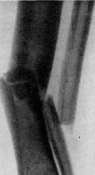
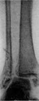
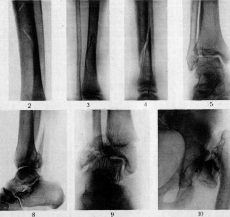
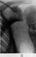
Figure 1. Fracture of both bones of the lower leg with significant displacement along the length and angular curvature. Figure 2. Spiral fracture of the tibia without displacement (in profile). Figure 3 and 4. Spiral fracture of the tibia with displacement in two directions. Figure 5. Fracture of the lateral malleolus. Figure 6. Intermalleolar fracture. Figure 7. Dupuytren's fracture, avulsion of the medial malleolus and fracture of the fibula. Figure 8. Dupuytren's fracture (in profile) with avulsion of the posterior part of the tibia, the so-called posterior marginal fracture. Figure 9. Fracture of the lower leg in the lower third with avulsion of the epiphysis of the tibia. Figure 10. Fracture of the femoral neck with displacement of the femur upward and adduction. Figure 11. Comminuted fracture of the surgical neck of the humerus. To the article Fractures. primary metastatic tumor, or a chronic inflammatory process (gumma, osteomyelitis, etc.). Besides local foci, thanks to pathological thinning of the walls, which purely mechanically prepares for a fracture, general diseases can also serve as a predisposing cause, among which the first place is occupied by osteogenesis imperfecta (see), rickets, which causes bone fragility in childhood, osteomalacia, scurvy, etc. Disorders and diseases of the endocrine system can not only delay or enhance bone growth, but also cause its excessive fragility. Clinically, the division of fractures into closed and open is of great importance. Closed fractures are considered to be fractures without a breach in the integrity of the skin; open—fractures accompanied by a breach in the integrity of the skin. Whether it is a large crushed wound with protruding exposed bone edges, or a small puncture wound inflicted from the inside by a bone fragment, since the entry gates for infection are open, the prognosis and the course of the entire regenerative process in such an open fracture change completely (see below). Gunshot fractures also belong to open fractures, which explains the severity of their course and the difficulty of treatment, associated with the need to fight infection. Open fractures are classified as complicated fractures. Among other complications accompanying fractures, one should mention injuries to vessels, accompanied by severe bleeding with subsequent formation of hematomas, and sometimes the development of aneurysms, then injuries to nerve trunks, ruptures and tears of muscles, fasciae, aponeuroses, etc. Both clinically and pathologically-anatomically, with every fracture, along with damage to the bone, there is also damage to soft tissues—subcutaneous tissue, muscles, fasciae, aponeuroses, large and small vessels and nerves. Depending on the location of the bone, its injuries are accompanied by damage to organs (damage to the brain in fractures of the skull, damage to the bladder and intestines in fractures of the pelvis, damage to the pleura and lung in fractures of the ribs). It is difficult to imagine that the force that caused the fracture left the surrounding tissues and organs undamaged, and it is even more difficult to imagine that the displaced bone fragments did not damage the surrounding tissues and organs. From this point of view, every fracture is essentially a complicated fracture. However, according to the established classification, it is accepted to consider as complicated fractures those accompanied by damage to the skin (infection), damage to large vessels and nerves, and damage to internal organs. Symptomatology and diagnosis of fractures. Clinical symptoms of fractures: 1) deformation, swelling and change in shape in the area of the fracture, 2) bruising in the vicinity of the fracture, 3) pain at the site of the fracture, 4) impairment of limb function, 5) abnormal mobility at the site of the fracture, 6) crepitus at the site of the fracture, 7) shortening of the limb. Comparative diagnostic evaluation of fracture symptoms. Signs of fractures are divided into absolute, occurring in every fracture, and relative, occurring in fractures not always or occurring not only in fractures, but also in other injuries (contusion, distortion, etc.). An absolute sign of a fracture is a striking deformation of a specific section of the organ being examined. In cases where the deformation is sharply pronounced and caused by sharp displacement of fragments, the diagnosis can be made from a distance even by an inexperienced doctor. This is an absolute sign (for example, a bayonet-like fracture of the radius, a fracture of the middle third of the lower leg with a central fragment protruding subcutaneously, a fracture of the humerus in the middle third, sharply protruding when the patient attempts to lift the arm). 1) The deformation caused by a fracture is striking especially in those cases where the bone lies superficially and is not covered by a thick layer of soft tissues. Where the site of the fracture is covered by a muscular sheath, the deformation appears as a diffuse swelling, is not detected upon inspection and is determined with difficulty only with the help of deep palpation (e.g., fracture of the femoral neck or the upper third of the femur, fracture of the head of the humerus, fracture of the pelvic bones, etc.). In cases of impacted fractures and fractures without displacement, this sign is completely absent. 2) Bruising usually appears in characteristic places a few days after the fracture, when extensive hemorrhage, associated with the rupture of vessels of the periosteum, bone marrow (a. nutriciae), rupture of muscles, and sometimes large vessels, penetrates, permeating all soft tissues, from deeply lying areas (surrounding the fracture) into the area of subcutaneous tissue. To the extensive bruising is added the formation of characteristic blisters filled with serous content. Due to the permeation of the thickness of the skin by the effused blood, blood and lymphatic vessels are compressed, serous fluid and lymph ooze to the surface, lifting the horny layer of the skin in the form of blisters. Cases of damage to large vessels during a fracture attract attention due to the increasing severe general condition, a drop in the pulse, general pallor (a picture of general anemia and simultaneously rapidly increasing swelling in the area of the fracture). Bruising, as stated, except in cases of superficially lying fractures, does not appear immediately, but on the 5th-6th day, and therefore in the first days cannot serve as a diagnostic sign. In addition, bruising also occurs with simple contusions, so that by itself it cannot help to make a differential diagnosis. 3) Pain. Constant sharp pain, usually accompanying a fracture, is caused by damage to the periosteum, which is rich in vessels and nerves, and is maintained by the constant irritation of tense, contracted muscles by the fragments of the broken bone. This sign is extremely subjective, depends on the endurance and patience of the victim, and most importantly, it is far from pathognomonic: sharp pain is also experienced with dislocation and sprain. A much more characteristic sign is pain upon pressure at the suspected site of the fracture—sharp pain at a specific point. However, this sign can also be present with contusions, sprains. The most characteristic sign for a fracture is pain along the length during a jolt directed along the axis of the broken bone, e.g., pain experienced when pressing in the longitudinal direction on the heads of the metacarpal and metatarsal bones in fractures of the diaphysis of these bones. 4) Impairment of the function of the given organ relates mainly to fractures of the bones of the limbs and spine. Characteristic is the impairment of the motor function of the given organ: the inability to lift the limb, the inability to step on it. With the integrity of one of two parallel bones, this sign is often masked (e.g., fracture of the fibula with the integrity of the tibia); in impacted fractures, in cracks and fissures, it may be completely absent. The origin of this sign is quite understandable: every movement with an unfixed fracture causes displacement of fragments, which is associated with painful sensations, and therefore is avoided by the patient. Local anesthesia also masks this sign. Thus, after an injection of novocaine into the area of the fracture, the patient can freely lift the limb that is bending at the site of the fracture. In tabetics, deprived of pain sensitivity, this sign is absent—I have had to see tabetics who, despite a fracture (Dupuytren's) of both bones of the lower leg, stepped on the broken leg, after which, having no support, they fell a second time, receiving complex comminuted fractures. 5) Along with the impairment of function, abnormal passive mobility is observed—the limb at the site of the fracture gains the possibility of lateral and rotational movements where this is completely impossible with the integrity of the bone. This sign, characteristic of fractures with displacement of fragments, is convenient for differential diagnosis between dislocation and high periarticular fracture: with dislocation—forced position and limitation of movement, with a fracture—forced position and abnormal passive mobility (e.g., dislocation of the shoulder and fracture of the neck of the humerus). In impacted fractures, this sign is absent. 6) Crepitus—a peculiar crunch when one bone fragment rubs against another—can be obtained artificially for diagnostic purposes. Its presence always indicates a fracture. It is absent in impacted fractures and in cases where there is interposition of muscles or tendons between the fragments. Since eliciting this sign is often very painful for the patient, usually where the diagnosis of a fracture is clear, there is no need to seek it. 7) Shortening is determined by a comparative measurement of the length of the injured and healthy limb. Shortening is detected only in fractures with displacement of fragments along the length of the limb.
It has particular functional significance in fractures of the lower limb, since with large degrees of shortening it leads to disability. Particularly strong shortening is observed in fractures of the femur due to the great contractility of the long two-joint muscles of the thigh. Shortening is measured with a centimeter tape, comparing symmetrically lying distances between bony prominences. For the lower limb, the landmarks are: spina ilii ant. superior, the joint space (or the internal condyle of the femur or the internal edge of the articular surface of the tibiae) and the lower edge of the internal malleolus; for the upper limb—the apex of the acromial process, the external condyle of the humerus, the styloid process of the radius, the styloid process of the ulna. All the above-mentioned signs are characteristic signs of a fracture, and for a correct diagnosis, the presence of several signs simultaneously is necessary. Thorough examination and subsequent careful analysis of all signs are necessary, because while for the provision of first aid it is sufficient only to establish the fact of the presence of a bone fracture, in a hospital or dispensary, where systematic treatment is intended to be conducted, it is necessary, before proceeding to the treatment of the fracture, to clarify the mechanism of its origin, the presence of displacement and the direction of the displaced fragments, the degree of damage to muscles, vessels, and nerves, and finally the general condition of the patient, his age, social, professional, and constitutional characteristics, since all these points must be taken into account when treating a fracture. Methods of examination for fractures: anamnesis—a description as detailed as possible of the mechanism of obtaining the fracture. General and local examination. General examination should be conducted by inspection, without causing unnecessary suffering to the patient with premature manipulations. Clothing must be cut, and the patient must be completely undressed to allow for the comparison of the position of symmetrical parts of the body. Such signs of a fracture as deformation, bruising (if the examination does not take place on the first day), shortening (if it is sharply pronounced) catch the eye during careful inspection and comparison of symmetrical parts. When examining the patient, one should pay attention to the position of the patient and his limbs. There are typical positions, on the basis of which one can immediately make a diagnosis of a fracture, e.g., in fractures of the neck of the femur, the position of the abducted thigh, resting on the bed with its outer surface, is characteristic; in fractures of the pelvis—the so-called "frog position" (according to Volkovich)—legs bent at the knees and evenly spread apart, etc. One can learn about pain from the patient's complaints. To judge the impairment of function, one can immediately ask the patient to move (lift, lower, or turn) the injured limb: if it is a rib—force a deep breath; if it is the spine—ask to sequentially bend and straighten the back, etc. Deformation, swelling, bruising, and impairment of function not only force one to assume a fracture but also indicate the approximate place of its location. After this, one proceeds to palpation, trying by careful feeling along the bone, starting from a known healthy place, to determine the place of greatest tenderness—the painful point. With such systematic feeling, the finger suddenly sinks in, especially often in fractures of the spine, and a definite sensation of a breach in the integrity of the bone is obtained—a sharp pain usually coincides with this, and sometimes crepitus. If all these phenomena are present, then the diagnosis of a fracture is clear. However, it is not always possible to immediately obtain these symptoms—swelling, edema, strongly developed muscles, and abundant subcutaneous tissue make it difficult to determine the place of the fracture. Then one has to try to look for abnormal mobility. For this, the limb is taken with both hands—one above and the other below the suspected place of the fracture—and, moving the limb in two opposite directions, abnormal lateral mobility is obtained; at the same time, crepitus can often be detected. In fractures of the ribs, abnormal mobility and crepitus are determined as follows: at the moment of a deep breath, pressure is applied with the fingers of both hands in different places on the rib in different directions—the abnormal mobility, crepitus, and clicking movement of the rib obtained thereby indicate its fracture. When examining a fracture of the skull or spine, of course, one does not look for crepitus or abnormal mobility. It is sufficient to discover with the help of gentle palpation a deep depression, sinking, or protrusion to make a diagnosis of a skull fracture. Palpation, performed carefully and gently, is a valuable diagnostic tool. For example, when examining a pelvic fracture, palpation of the bony parts corresponding to the most frequent localization of the fracture of the pelvic ring gives a complete idea of the presence of a fracture. During palpatory examination, upon pressure on the head of the fibulae, pain along the area of the external malleolus is pathognomonic for its fracture. Palpation should not be performed roughly and sharply, since in such a case it causes pain and a violent reaction of protest from the patient, which interrupts further examination. To determine shortening in fractures of the limbs, it is necessary to perform measurements, which must be done not on a bed, but on a hard dressing table, strictly starting from symmetrical bony points (especially important in fractures of the lower limbs). Detailed clinical examination—anamnesis, inspection, palpation, and measurement—in the vast majority of cases makes it possible to make a diagnosis of a fracture and to imagine approximately the direction of the fracture line and the position of the fragments. Experience and familiarity with anatomy (muscle attachment) and the function of muscle groups (their influence on the position of fragments in fractures at one level of the bone or another) will help the clinician to sort out typical cases of bone fractures. In addition to all the above-mentioned techniques, we have another excellent diagnostic method—X-ray diagnostics, with the help of which it is desirable to always support and verify the data of clinical examination. If in typical fractures, X-ray images are to some extent a scientific luxury, giving the opportunity to study each fracture in depth, then in a whole series of less typical fractures—impacted, subperiosteal fractures without displacement, avulsion fractures, etc.—X-ray diagnostics is an extremely necessary method. There is a whole series of fractures where it is not possible to make an accurate diagnosis or determine the fracture line even for an experienced clinician-traumatologist. X-rays in the field of study and treatment of fractures have constituted an epoch. Such fractures as avulsion fractures of the transverse processes of the lumbar vertebrae, avulsion fractures of the Schlatter type, fractures of the calcaneus, small bones of the wrist, etc., are now easily diagnosed only because they have been studied with the help of X-rays. For a correct diagnosis of a fracture with the help of X-rays, an image in two projections is necessary, since otherwise, due to the overlapping of shadows, one might not notice the fracture line (see separate table, figures 3 and 4). However, while recognizing the enormous importance of X-ray diagnostics in fractures, one should not neglect general clinical methods of examination and should not lose the experience of old clinicians, acquired by them through long observations and the study of each case. Timely accurate diagnosis, not only of the presence of a fracture but also of its character and direction, is of colossal importance at the present moment. Since fractures affect mainly the working population, persons of heavy physical labor, there is a risk, by missing a difficult-to-diagnose avulsion fracture or an impacted fracture, of sending a worker to work, frivolously considering him a malingerer. The consequences of such an error can be extremely severe for the victim. Hence it is clear with what attention one should treat all kinds of bone injuries, using all available methods of examination. X-rays have played a huge role not only in the matter of diagnosing fractures but also in the study of issues of bone regeneration, and consequently in the development of treatment methods. Healing of fractures. Local reaction. The trauma that caused the bone fracture, the rupture of the periosteum, the abundant hemorrhage associated with the rupture of numerous vessels, the rupture of fasciae, the crushing and maceration of muscles, causes a violent reaction in the surrounding tissues. The local reaction is expressed in the so-called "traumatic inflammation": hyperemia, dilation of vessels, exudation of blood plasma, proliferation of formed elements. In the area of the fracture, processes of an opposite character play out: on the one hand, the dying off of destroyed cells and tissues, and on the other hand, the multiplication and proliferation of young cells and tissues (see Regeneration). With regard to bone tissue, it is definitely proven that the presence of a large number of crushed, necrotizing areas of bone (especially the periosteum) stimulates the regeneration of bone tissue in the zone adjacent to the place of the fracture.
Not to mention the experiments confirming this fact, we have a large clinical experience in this regard: in the first period of the Imperialist War, when many surgeons very carefully removed fragments of crushed bone from the wound along with shreds of periosteum, in a whole series of cases, healing of the fracture did not occur at all (false joints resulted) or occurred at an extremely slow pace, because there were no irritants and not enough material for creation, for the regeneration of bone, because, as the works of some authors (Gaza, Bier, et al.) have proven, the dying elements of crushed tissues are not only irritants to growth, but also material for the construction and restructuring (Umbau) of young regenerating tissues and especially bone tissue. Thus, in the very first days after the trauma, as soon as the bleeding small vessels are thrombosed, with the resorption of the hematoma, proliferation begins, first of connective tissue elements with the formation of connective tissue, the so-called provisional callus, and then of specific elements of osteoid tissue. The provisional callus grows through the blood clot filling the area of the bone fracture and gradually envelops the ends of the bones and nearby bone fragments with a soft, vessel-rich spindle-shaped sheath. By the end of the first week, the development of specific osteoid tissue begins, passing by metaplasia either directly into bone tissue or, in rarer cases, first into cartilage and then into bone. The deposition of calcium salts begins from the inner layers of the periosteal callus. The formation of bone callus proceeds simultaneously from the cambial deep layer of the periosteum (Fig. 11)—periosteal callus, from the vessels and supporting substance of the bone marrow—endosteal callus, from the endosteum of the Haversian canals of the cortical substance—interstitial callus, and finally (according to Bier) from the surrounding muscles—parosteal callus. Various authors attach great importance to one or another source of callus formation. Thus, Lexer attaches exceptional importance to the periosteal callus, which forms in connection with the increased development of periosteal vessels, thanks to the so-called "fracture hyperemia."
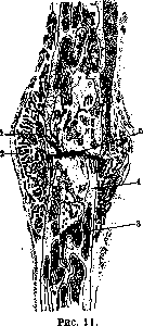
Figure 12. Figure 11. Healing of a rib fracture (magnified through a magnifying glass): 1 and 5—ingrowth of a blood clot; 2—periosteal callus; 3—spongiosa; 4—cortical layer of the diaphysis. Figure 12. Complete restoration of the medullary cavity during the healing of a diaphysis fracture in the correct position.
Lexer and his student Delkeskamp conducted experiments on dogs with artificial fractures of tubular bones, in which they injected vessels during the development of the bone callus (from 1 to 6 weeks). This revealed colossal hyperemia of the vessels of the broken bone. The increase in hyperemia begins from the first week, reaches its apogee at 4–5 weeks, after which the regression of the vessels begins. According to Lexer, periosteal vessels grow even into the medullary cavity of displaced fragments, thus participating in the development of the endosteal callus (Markkallus); he discovered insufficient formation of endosteal callus and a slowing of bone consolidation upon ligation of the nutrient artery. On the other hand, Bier believes that bone regeneration occurs mainly at the expense of the bone marrow (endosteal callus). At the same time, he attributes great importance to the development of parosteal callus from the vessels and elements of the muscle tissue surrounding the fracture. He even asserts that elements of muscle tissue, under favorable conditions, metaplase into bone tissue. Be that as it may, it is clear that for the successful formation of bone callus, it is necessary, on the one hand, to have in the wound elements of resorbing blood, bone, and muscle tissue. These elements, necessary as biochemical irritants not only for connective tissue but also for the bone elements of the callus, are at the same time a nutritional base for the newly building bone tissue; on the other hand, as is evident from the research of Lexer, for the regeneration of bone tissue, active hyperemia of the fracture area is necessary—an increased supply of nutritional materials by blood. Calcium, so necessary for the consolidation of the bone callus, is obtained by it from the blood. The bone callus grows, swells, and increases in size over 4–5 weeks; the final ossification, the so-called "consolidation" of the fracture, occurs between 7 and 10 weeks. The timing of fracture healing, like the timing of ossification and consolidation of the callus, depends on: 1) the age of the patient; in children, consolidation of a fracture occurs within 3–5 weeks (21–35 days), in an adult, consolidation of a diaphyseal fracture requires approximately 60 days; 2) the thickness of the bone. According to Gurlt, the average times for consolidation of a diaphysis fracture with correct positioning of the fragments are as follows: phalangeal bones—2 weeks, metacarpal and metatarsal bones, ribs—3 weeks, clavicle—4 weeks, forearm bones—5 weeks, humeral diaphysis—6 weeks, tibia and neck of the humerus—7 weeks, both bones of the lower leg—8 weeks, femoral diaphysis—10 weeks, femoral neck—12 weeks. * These periods vary greatly both depending on the general condition of the organism, age, sex, etc., and depending on local causes, among which the mutual arrangement of the fragments is of great importance. Where bone fragments are further apart from each other, significantly greater demands are placed on the bone callus, since there occurs not only a simple restoration of integrity, as if soldering the ends of the bone along the plane of the rupture, but during the fusion of strongly displaced fragments located at an angle, a complex process of restructuring the architecture of the bone occurs, a process that can last for years. The moment of consolidation of the callus is far from the end of bone regeneration. By this time, the bone callus is a coarse, loose bone mass of wide-meshed cellular structure, strongly differing from the structure of the surrounding normal bone. Then begins the period of restructuring of the bone callus, which occurs through the resorption of the excess mass of newly formed substance and the strengthening of the bone in certain directions corresponding to the distribution of forces of gravity and pressure according to the general laws of physics and mechanics. The distribution of bone trabeculae, like the structure of the entire normal bone, is conditioned by static and dynamic moments affecting the bone during its development. In the same way, the restructuring of the bone callus * According to recent literature, complete consolidation of a fracture of the femoral neck in the elderly occurs no sooner than in 1/2 year.
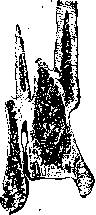
Figure 13. Obliteration of the medullary cavity during the healing of a fracture with large displacement.
occurs by adapting the coarse bone mass to the new conditions of movement and load for it. From this, it is clear that the final remodeling of the bone callus will occur only when the broken limb is placed in normal conditions of movement and load. This is a lengthy process. According to Matti, it takes a year for the bone to acquire its former density, strength, and elasticity, and 2 years for it to acquire its former plasticity. After 2-3 years, in cases where the fragments were positioned satisfactorily, the bone callus is remodeled to such an extent that neither on an X-ray nor on a pathological-anatomical specimen can one find the site of the former fracture. In cases where the fragments were sharply displaced or stood at an angle, a complex system of trabeculae and beams develops through adaptation under the influence of function (constant movements and load), which differs from the usual structure in this area but is obviously fully consistent with the new conditions of statics and dynamics (Fig. 13). In such newly formed bone, the ends of the fragments standing at an angle, being outside the sphere of influence of movement and load during the complete remodeling of the bone, atrophy and disappear over time. The medullary canal in cases with large displacement remains closed for a long time; in cases where the fragments are in contact with the lumens of the medullary canals, the plates closing them are resorbed over time, and the medullary cavity of the entire bone is restored (Fig. 12). Thus, timely and correct repositioning of the fragments accelerates and facilitates the regression of the callus. The function of the limb, by modeling and directing the process of bone reconstruction, shortens the time for the final healing of the fracture. In flat bones, the formation of callus is usually insignificant; it originates from the diploe. In fractures of the vertebrae, callus formation also proceeds through the formation of periosteal and endosteal callus. All the above-mentioned healing times for fractures refer to closed fractures. The healing times for open fractures are significantly lengthened in cases complicated by the introduction of infection and accompanied by signs of inflammation with the development of osteomyelitis and prolonged discharge of sequestra; here the process of fracture healing lasts 8-10 months. It is interesting that the healing process slows down even in open fractures that proceed aseptically. Wier explains the slowing down of bone regeneration in these cases by the fact that the elements necessary as material for building the bone callus are carried away outward by the flowing blood. Mainly, the absence of blood clots, which, according to Wier, stimulate the bone-forming process and serve as material for building new bone, has a harmful effect on regenerative processes. All factors that delay bone regeneration can ultimately lead to the formation of a false joint (see). In open fractures accompanied by the introduction of purulent infection, excessive callus formation is observed. This phenomenon is also observed in closed fractures, especially sharply in cases of incorrect positioning of fragments, in comminuted fractures, and in fractures accompanied by the formation of large hematomas. The excessive development of bone callus in these cases is explained by the increased irritation of osteoblasts by a large amount of destroyed bone tissue and periosteum. The detachment of the periosteum over a large area also contributes to this. In some cases, callus luxurians can have a favorable significance, namely in cases where the excessive periosteal callus in the form of a sheath covers the ununited ends of the broken bone, between which a gap remains. Most often, excessive bone callus is observed in intra-articular fractures of tubular bones in the region of the epiphysis, less often in the region of the diaphysis, and only as an exception in fractures of flat bones. Disordered growths of bone callus, penetrating in the form of osteophytes—bony outgrowths—into the soft tissues, give the impression of myositis ossificans. Such bony growths, especially if they are located in the region of a joint, severely hinder and limit the function of the joint. In some cases, in young subjects, when the fracture line passes through the metaphysis of a tubular bone (lower leg), excessive (gigantic) growth of the damaged limb is observed. This growth is obviously caused by irritation of the zone of growing osteoid tissue, caused by trauma, hemorrhage, compression, and partial necrosis of damaged cells and tissues.
Speaking about the healing processes of fractures, one cannot limit oneself to questions of the regeneration of bone tissue as such. As already stated, in a fracture, one is usually also dealing with a more or less serious trauma to the tissues surrounding the fracture. Muscle tissue suffers particularly in this regard, which is of great importance, since for the restoration of the functions of the affected organ (limb), timely healing of the muscle tissue is necessary. Muscle tissue, as a highly differentiated tissue, usually does not regenerate but is replaced by a connective tissue scar. However, it has been possible to experimentally achieve the regeneration of specific muscle tissue. In cases of muscle tears arising in connection with a fracture of a nearby bone, muscles regenerate as specific tissue on the condition of early initiated movements. Muscle elements develop from the preserved cells of the perimysium under the influence of functional load. Movement in this case plays the role of a specific stimulus (Bildungsreiz). Conversely, prolonged rest during the healing period leads to the filling of defects with connective tissue, which turns into a dense scar, subsequently fixing the retracted muscles in the state in which they were in the first moments after the fracture. Muscles that have contracted and are fixed by a scar subsequently undergo atrophy and finally lose both their physical elasticity and physiological contractility. With the timely application of movement, the innervation of the traumatized muscles is also restored. Regeneration of fasciae.
and tendon stretches in the area of muscle fiber attachment can also occur in two ways. With complete rest, the fragments of aponeuroses and fasciae heal with a coarse connective tissue scar, which after some time densifies, shrinks, and subsequently, by fixing the surrounding tissues, limits and hinders movement. According to the research of Roux, constant tension is also a formative stimulus here, contributing to the development of elastic elements of the connective tissue scar. "Tension, thanks to its dynamic action, gives direction to the connective tissue (elastic) fibers." Where complete rest of the entire limb was maintained during healing, and consequently of the muscles and fasciae (for example, a circular plaster cast), all free spaces are filled with coarse scar connective tissue, which turns into a dense, shrunken scar, devoid of specific muscle and elastic elements, and consequently incapable of restoring the function of the damaged organ. From analyses of the regeneration processes of various tissues damaged during fractures (of the limbs), it is evident what an important role the active state—movement, functional load—of the injured organ plays. These data should serve as a guiding thread when applying various methods of treating fractures, mainly of the limbs (the locomotor apparatus), where the restoration of functions plays a paramount role. The treatment of limb fractures pursues not only the restoration of the anatomical integrity of the bone but also the complete restoration of the organ's function. The latter is especially important at the present moment, when all treatment methods are being reviewed from the point of view of the speed and completeness of restoring the patient's ability to work. When evaluating the numerous proposed methods of treating fractures, one should proceed: 1) from the biological foundations of tissue regeneration (bone, muscle, connective, fascial tissue); 2) from the foundations of the biomechanics of the locomotor apparatus; 3) from the social mandate that our socialist state presents to us—to return the lost ability to work to the worker in the shortest possible time. Development of the doctrine on the treatment of fractures. Throughout the history of medicine, two methods of treating fractures have been constantly struggling with each other: the method of immobilization, rest of the limb, which at one time was considered the only way to restore the anatomical integrity of the bone, and the method of treatment with movement and massage, a method that provides rapid restoration of function, often at the expense of anatomical relationships. The development of medical knowledge, especially the development of technical methods for treating fractures, proceeded empirically. Since the time of Hippocrates, the treatment of fractures by traction and counter-traction has been used, with the simultaneous use of guiding splints. In the Middle Ages, hardening circular bandages (made of proteins) were most widely used. In the 18th century (1713–1788), Percival Pott, based on a careful study of the mechanism of fractures, having clarified that the displacement of fragments is the result of muscle contraction, came to the decision to treat limb fractures by placing the muscles in a relaxed state. When treating fractures of the lower limb, he recommended a semi-bent position on the side, which, of course, did not yet yield brilliant results. Subsequently, as a further stage in the development of his method, the treatment of lower limb fractures on a double inclined plane—the so-called "Planum inclinatum duplex"—was used in England. This method was justly reproached for the fact that the main role in it was played by the comfortable "position" of the limb, and the further retention of fragments in the correct position was not achieved. In any case, Pott's method already contained a grain of truth: namely, that muscle relaxation is a necessary prerequisite for reposition and the correct position of bone fragments. In 1812, Sauter first added treatment by traction to the Pott method—treatment in a semi-bent position. However, he did not have the technical capabilities for the correct application of traction (rubber adhesive plaster, which can withstand heavy weights, was invented in America much later, in 1839). Therefore, even under Sauter, the extension bandage in a semi-bent position did not receive further development and widespread application. Although the ideas underlying it were correct, there were not yet technical possibilities for further development. The method of Pott and Sauter was forgotten for a long time when plaster began to be used in surgery in the middle of the 19th century. The military doctor Mathijsen in 1852 first introduced the circular plaster cast into use. Having great advantages as a quickly hardening material possessing great strength, plaster received wide distribution. The method of treating fractures with circular plaster casts has since been firmly established among surgeons and especially among orthopedists as the only correct method of treating fractures. The research of Pott and all the benefits of the semi-bent position of the limb during fractures were forgotten. It is not for nothing that many surgeons (Steinmann) believe that the introduction of the circular plaster cast long delayed the further development of the correct doctrine on the treatment of fractures. Meanwhile, the disadvantages of prolonged immobilization with plaster casts soon made themselves felt. To weaken the negative aspects of the plaster cast, the so-called "staged" treatment with plaster casts was proposed, which were changed every 14–20 days; when changing the bandage, the incorrect position of the fragments was corrected, and massage was performed, after which the limb was again encased in plaster. To combat complications of a general nature (pneumonia, embolism, severe muscle atrophy), patients were encouraged to get up and move around in the cast; outpatient treatment in a plaster cast was even used. However, these palliatives could not eliminate the harmful influence of plaster. Dissatisfied with the results of treatment with a circular plaster cast, surgeons, in pursuit of the correct anatomical position of the fragments, turned to the method of open reduction and to the fixation of fragments with metal plates, ivory, staples, nails, wire, etc. (see Bone suture). This method became possible after the triumph of asepsis in surgery. Even greater disappointment in the plaster cast occurred when X-rays revealed that the circular plaster cast, previously used for the sake of restoring the anatomical integrity of the bone, does not actually achieve its goal, being unable to hold the fragments in the correct position for a long time. The healing of a fracture, the regeneration of bone and muscle, is a dynamic process during which both the pathological-anatomical and physiological state of the damaged limb is constantly changing. Concluding the entire limb for this complex period in a "plaster robe" (Wegner) is an antiphysiological method of treatment. Not to mention pressure sores, ischemic paralysis, and similar complications, which can be considered as shortcomings of the technique of applying plaster, but also such shortcomings of treatment as muscle atrophy, joint stiffness, and prolonged circulatory disorders (edema, congestive phenomena) made one doubt the usefulness of this method. A limb removed from a plaster cast after 7 weeks of treatment was a barely mobile, unbending stick (log), which only with difficulty, after long and persistent follow-up treatment with massage and mechanotherapy, adapted to its function. The subsequent "follow-up treatment" in these cases required more time than the entire period of callus consolidation. The method of open treatment of fractures at one time received wide distribution in France, Belgium, England, and America. However, disappointment soon followed here too, when it became clear that surgical intervention delays bone regeneration and, despite an anatomically good result, is far from brilliant from the point of view of restoring functions. In contrast to the anatomical direction in the treatment of fractures, a direction that, in pursuit of an anatomically correct position of the fragments, forgot about the function of the limb, the method of "free functional treatment" arose. The leader of this method, Lucas-Championniere (1895), proclaimed the correct slogan "Le mouvement c'est la vie" ("movement is life"). However, in the pursuit of restoring functions, he completely neglected (incorrectly, of course) the anatomical position of the fragments and the subsequent shape of the limb. His method—early active gymnastics and massage, applied from the very first days—sometimes yielded good results, but since it was carried out without simultaneous traction and without any fixation of the limb, large deformities, shortenings, displacements, and subluxations sometimes occurred. Both these methods—the method of complete immobilization and the method of movement and gymnastics (functional)—struggled with each other, each having its ardent supporters. Both were one-sided, and neither one nor the other yielded entirely good
FRACTURES
The question could only be solved dialectically, i.e., not "either-or," according to the laws of formal logic, but "and," "and." One cannot pose the question this way: either an anatomically good result or a functionally good result; or prolonged fixation of fragments and complete rest or no fixation and only movement. The result must be good both anatomically and functionally. The question was solved only when it became possible to find a way to apply simultaneously constant fixation of fragments in a semi-flexed position and early free movements, performed without disturbing the mutual fixation of the fragments. It was not possible to find and develop this method in detail immediately. The use of adhesive plaster in America by Swift in 1862, holding large weights, contributed to the development of the extension dressing method. The possibility of widespread use of adhesive plaster traction appeared. Bardenheuer in the 80s developed in detail the technique of adhesive plaster traction for fractures and thereby made a great contribution to the doctrine of fracture treatment. However, he failed to implement the basic principle put forward by Pott, Sauter, and later Lorinser and Middeldorpf—traction in a semi-flexed position. Bardenheuer applied traction to the limb in an extended, straightened position, due to which he had to use extremely large weights, and yet he did not always manage to achieve good results. Only starting with Zuppinger, who in 1905 constructed his apparatus, movable splints for treatment by traction in a semi-flexed position, does the doctrine of fracture treatment finally enter the correct path—functional treatment of limbs with simultaneous traction, fixing the fragments in an anatomically correct position. Ziegler, Matti, Böhler, and other authors developed the methodology of this traction and invented a number of valuable apparatuses. An excellent theoretical justification for the treatment of fractures by traction in a semi-flexed "physiological" position was given by Wegner, who created a specific methodology in this direction at the Kharkov Medico-Mechanical Institute, the essence of which consists in the use of free adhesive plaster traction in a position of absolute physiological rest of the limb without any splints or apparatuses, with systematic exercises of active movements, starting from the first days. The use of skeletal traction according to Steinmann gave a further impetus to the development of the modern method of functional treatment of fractures (see Traction, Vol. VI, 117-118 and 121-122, Table II and III). Such was the long path of searching for correct methods of treating fractures, a path not yet finished, but the further direction of which is clear to us at the present time.
Basic tasks of fracture treatment. The first task in the treatment of fractures is the reposition of fragments. As already stated, the displacement of fragments depends on the direction and force of the impact that caused the fracture, i.e., on the mechanism of the injury and on the action of the contracted muscles. Physiologically, each group of muscles is in a state of some tension. At the moment of the fracture, this tension instantly ceases, the attachment points of the muscles approach each other, and the muscle mechanically shortens. This contraction is the contraction of the muscle as a physical body, determined by its elasticity. If one cuts a muscle on a corpse, it will also contract by virtue of its elasticity (like a rubber tube). However, this is not enough: in a fracture, the free bone fragments, shifted from their usual place, irritate the surrounding muscles, cause painful sensations, and induce further reflex contraction; this contraction is caused by the physiological contractility of the muscles. Under normal conditions, physiological contraction is carried out as a normal voluntary contraction of muscles, while the elastic contractility of muscles does not manifest itself under these conditions, because it is hindered by the constant state of tension in which the muscle is found when the bone is intact. In a fracture, these two properties of muscles—their physical and their physiological contractility—acting simultaneously, cause the displacement of fragments and hold them in an incorrect position. Thus, in order to put the displaced fragments back into place, it is necessary to overcome both the elastic and the reflex (physiological) contraction. The latter, under the influence of trauma, soon passes into a state of "reflex traumatic hypertonia," which leads to persistent histological changes in the muscle tissue, hindering the "reposition of fragments." This manipulation, which usually does not present particularly great difficulties in the first hours, subsequently becomes more and more complex with every hour, and even more so with every day. A prolonged state of muscle retraction leads to persistent histological changes in the tissue of the contracted muscles, which after 5-6 days can only be stretched to normal with great difficulty. If 10-14 days have passed since the moment of the fracture, we are already dealing with persistent muscle retraction acting on an entire limb segment, the so-called myogenic contracture, against which it is often impossible to fight. Considering this, it is strongly recommended to perform the reduction of fragments in a fracture immediately upon the patient's admission to the hospital. To eliminate the effect of physiological reflex contraction on the separated fragments, Böhler recommends injecting a 2% solution of novocaine into the fracture site. Novocaine, mixing with the extravasated blood, evenly bathes the bone fragments and the torn periosteum; deep local anesthesia ensues, pain and muscle irritation cease, and 3-5 minutes after the injection, the reflex muscle contraction disappears, which makes possible not only a detailed painless examination of the fracture area but also a relatively easy, completely painless reposition of the fragments. However, it must be remembered that anesthesia, by destroying the pain reflex, destroys only the reflex physiological contraction; it does not act on the elastic retraction of muscles. Even more so, it will not act on the myogenic contracture that occurs several days after the injury. Besides muscle contractions, an obstacle to the reposition of fragments can be the interposition of muscles and fascia, the presence of an impacted fracture, and the interlocking and interposition of displaced bone fragments. However, in the vast majority of cases, in the first hours after a fracture, with the help of gradual traction and counter-traction (sometimes forced flexion and extension under local anesthesia), it is possible to put the fragments back into place. In old cases, it is necessary to use great force under anesthesia to destroy the elastic retraction of muscles and to break down newly formed adhesions that are turning into persistent, dense scars. If an X-ray shows that nothing could be achieved even with these rough manipulations, then it is better to abandon the futile attempts, which cause trauma to the entire limb, and proceed to a bloody operation. Thus
Since the greatest displacement usually occurs (in cases of two-joint muscles) along the length, the fragments can be positioned correctly only by extending the limb along its length, i.e., by applying a pulling force in the direction of the long axis of the limb. By such simple traction in the longitudinal direction, we stretch the contracted muscle. However, it is known that a force acting in the direction opposite to muscle contraction causes tension in the muscle, which is expressed as resistance to passive stretching. The greater the force applied, the greater the resistance this will cause. According to Weber's law, muscle tension increases in proportion to the square of the stretch. However, if traction of the limb is applied while the muscles are relaxed, then a many times smaller force will be needed for the repositioning of the fragments. Therefore, traction according to the method of Zuppinger, Ziegler, Matti, and Wegner is applied, directed along the long axis of the broken limb with the entire limb in a semi-flexed position. Such a semi-flexed position, 'semiflexio', which Wegner calls the 'mean physiological position', gives the limb muscles a state of 'absolute physiological rest'. This is a position of complete physiological equilibrium between groups of antagonists: flexors and extensors, abductors and adductors, pronators and supinators. Since flexors are always and everywhere stronger than extensors, it is clear that physiological equilibrium of the muscles will always be obtained in the semi-flexed position of the limb. It has been possible to calculate precisely the angles of flexion and abduction that must be observed in order to give the limbs a mean physiological position. These angles are as follows (according to Wegner). For the lower limb: angle of flexion of the hip joint—45° (60–30°); angle of flexion of the knee joint—140° (from 130° to 150°); angle of plantar flexion of the foot—10°. For the upper limb: in the shoulder joint, the angle of abduction of the arm from the trunk outward—60–70°; the angle formed by the arm with the frontal plane of the entire trunk—35°; rotation of the arm inward—45°; angle of flexion of the forearm to the arm—110°; the forearm should be in a state midway between pronation and supination in relation to the arm, at an angle of 75°; angle of palmar flexion of the hand—10°; angle of abduction of the hand—15%. In order to achieve such a mean physiological position of the limb, it is necessary, firstly, to flex the limb so that the midpoints of the articular surfaces facing each other lie opposite one another, and secondly, to counterbalance the force of gravity of all segments of the limb. According to Fischer, the weight of individual segments of the limb of an adult male is expressed in the following figures: weight of the thigh—6.96 kg, weight of the lower leg—3.06 kg, weight of the foot—1.01 kg (entire lower limb—11.03 kg); weight of the arm—2.04 kg, weight of the forearm—1.42 kg, weight of the hand—0.04 kg (entire upper limb—3.50 kg). The weight of the limb segments can be counterbalanced 1) either on slings with corresponding weights on pulleys; 2) or by placing hard bolsters and pillows under the flexed limb according to Wegner (see Traction); 3) or by placing the limb on corresponding improvised or specially manufactured devices and apparatuses (made of wood or metal or bent pipes) (according to Zuppinger, Ziegler, Bell, Braun, Matti, and others). Having given the limb such a mean physiological position, traction of the broken bone is applied along its longitudinal axis. In such a position, due to the complete relaxation of all muscle groups, much less traction force is required than in the straightened position of the limb. In a whole series of cases, such a physiological position given to the limb immediately after the fracture, thanks to the relaxation of the muscles, automatically gives the fragments the correct position; for example, a posterior supracondylar fracture with displacement of the posterior fragment together with the calcaneus and the entire foot upward and backward (due to the strongest contraction of the calf muscles) easily falls into place and is held in place as soon as the entire limb is given a physiological position on slings. Usually, however, especially in cases where one is dealing with powerful muscles, it is necessary to apply greater or lesser traction along the length.
Traction for the repositioning of fragments is applied 1) either at one time, and is carried out directly by the surgeon's hands while the trunk and the central part of the limb are fixed by an assistant [it is possible to fix by tying to a stationary object (table) with a sheet or towel], 2) or repositioning is achieved by prolonged traction of the limb in a semi-flexed position, by an arrangement on the bed, hanging a corresponding weight that carries out traction along the long axis of the limb. Manual reduction under local anesthesia is usually carried out on the forearm and lower leg, where, in addition to displacement along the length, lateral displacement and rotation are observed. After such manual repositioning, it is necessary to perform fixation of the fragments in the repositioned position (see below). In fractures of the arm and especially the thigh, prolonged traction is more often used, in which the repositioning of the fragments is achieved gradually. Usually, for repositioning in these cases, traction along the long axis of the limb is sufficient, since lateral displacement with sufficient traction in length (in the physiological position of the limb) is achieved by the fact that the muscles, brought into a normal state, cover the ends of the bones with a powerful sheath, directing the fragments to their former place. This does not always happen, since much depends on the level and direction of the fracture line. Where this has not occurred, traction and counter-traction in the lateral direction are added to the traction along the longitudinal axis. The fact is that displacement along the length is usually carried out by long muscles stretched across two joints (two-joint muscles), which, at the moment of the fracture, contracting, strive to bring the points of attachment closer; lateral displacement, however, is carried out firstly by the force of gravity of the entire unsupported limb (e.g., the outward falling of the thigh in a fracture of the femoral neck), and secondly by the action of short one-joint muscles, the force of action of which depends on the length of the lever remaining free (after the fracture) and the proximity of the fracture site to the joint. In a periarticular fracture, a small peripheral fragment forms a single whole with the entire peripheral segment, taking the position into which the force of gravity of this segment and its contracting muscles draw it. In cases of periarticular fractures, when the short periarticular fragment lies centrally and when the fracture line passes above the site of muscle attachment (fracture of the neck of the humerus and neck of the femur), the short fragment remains outside the influence of the periarticular muscles and takes a position determined by the shape (spherical) of the joint and the direction of the fracture line. Thus, the first task (the first stage) in the treatment of fractures is the repositioning of the fragments.
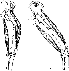
In this, the basic rule must be observed: 'the peripheral fragment must be placed against the central one' (this is insisted upon, among others, by Kulenkampff). And indeed, in a fracture, we are deprived of any possibility of influencing the central fragment, which takes the position given to it by the contracted muscles attached to it, fixed at the other end to the bones of the upper or lower girdle or to centrally located, relatively stationary points. The surgeon only has to, knowing the direction of the central fragment, place the peripheral one against it so that the axes of both fragments coincide and the fracture surfaces touch. To achieve this, the entire peripheral segment of the limb (together with the peripheral fragment) is given the position required by the position of the central fragment (Figs. 14 and 15). In the vast majority, the correct position of the fragments is achieved (by simple traction) in the semi-flexed mean physiological position. However, in epiphyseal fractures, where the contraction of one-joint muscles acts on the central fragment, traction in the physiological position is insufficient for complete repositioning. In these cases, starting from the physiological position, one has to deviate from it in one direction or another in order to place the entire peripheral segment either in a state of extreme abduction, or in a state of sharp flexion, or even extension. For example, in a high subtrochanteric fracture of the femur, the central fragment is sharply abducted outward by the action of the powerful gluteal muscles (Fig. 16). To place the peripheral fragment against it, the usual mean physiological position with abduction of 45° is not enough, but it is necessary to perform a more sharp abduction (Fig. 17). Conversely, in a fracture of the femur in the lower third (Fig. 18), where the central fragment is brought to the midline by the action of the adductor muscles, it is necessary to perform more sharp adduction (Fig. 17).
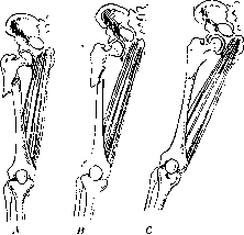
Figure 14. Fracture of the upper third of the femur with strong protrusion of the upper fragment. Figure 15. Coaptation of fragments by traction in a position flexed at the hip joint. Figure 16. Schematic representation of the action of muscles in a fracture of the upper third of the femur. Figure 17. A—abduction of the upper fragment and displacement with adduction of the lower one in a subtrochanteric fracture; B—incomplete coaptation with longitudinal traction alone; C—complete coaptation by traction with abduction.
and to place the peripheral segment together with the knee and lower leg as close as possible to the midline, sometimes completely eliminating the abduction angle. If correct and timely repositioning constitutes the first task (stage) of the treatment of fractures, then the second, no less important task is fixation, holding the fragments in the correct position; for many centuries, orthopedic surgeons and traumatologists have struggled with this task. The difficulty in resolving this task, as mentioned, consisted in the fact that they sought to achieve fixation of the fragments by means of complete immobilization of the entire limb. A heavy circular cast, as X-ray images have proven, not only failed to achieve the goal but also exerted an extremely harmful influence, causing atrophy of muscles and joints. Immobilization of the entire limb deprived it of the possibility of movement necessary for the proper course of regeneration processes and for the subsequent function of the limb. By using constant traction in the mid-physiological position, the second task was also solved in an extremely ingenious and simple way: gradual fixation of the fragments with the simultaneous possibility of constant movements in all joints. This method perfectly combines two opposite principles—rest (relative) and movement. While preserving the correct mutual arrangement of the fragments, it allows for movement of the entire limb. This creates the physiologically necessary conditions for the regeneration not only of the bone but also of the muscles, fasciae, and aponeuroses. At the same time, healthy muscles are not subjected to atrophy, which is so dangerous for an injured limb, and the joints, in turn, thanks to constant movements and the proper distribution of synovial fluid, do not become desolate (the capsule does not shrink) and retain the entire necessary range of motion.

Constant exercises of the limb, carried out during treatment by this method, contribute to the proper circulation of blood both throughout the entire limb and at the site of the fracture, in the newly forming bone tissue. The movement of muscles enhances the suction action of the venous system and the pumping action of the arterial system. The resorption of the hematoma and the desorption of the decay products of crushed tissues are accelerated, and at the same time, the supply of nutritional elements of the blood (including Ca) is increased. The arterial hyperemia of the fracture, necessary according to Lexer, is achieved in this case in the most complete manner. This method of treatment has received the quite correct name of the functional method, as it is carried out the entire time by developing the function of the injured organ. Functional treatment in this case is carried out from the first days and does not cease during the entire period of treatment. With this method, the patient does not need subsequent after-treatment and re-treatment. The function of the organ develops and is restored in parallel and simultaneously with the regeneration of all tissues. Movement of the limb accelerates the consolidation of the callus and subsequently shapes it, and at the same time facilitates its reverse development, compaction, and architectural restructuring of the entire fracture area. Functional treatment in its modern development by no means neglects, as was the case before, the anatomically correct standing of the fragments, since it is clear that the most rapid restoration of the integrity of the bone will occur with correct coaptation of the bone fragments; an anatomically correct position will simultaneously ensure the best functional conditions for the muscles and joints. The functional method, ensuring both correct anatomical-topographical relations and the function of the entire limb as a whole, is at the present time the method of choice. If early movements of the injured limb play a large role in fractures of the diaphyses of bones, then they acquire even greater significance in intra-articular fractures. There is nothing more dangerous for an intra-articular fracture than complete, prolonged immobilization: with such treatment, ankylosis is guaranteed. Therefore, in a whole series of cases with intra-articular fractures, treatment by early movement is used; particularly good results are obtained when treating intra-articular fractures of the elbow and shoulder joints with such a method (for more details, see Elbow joint). Without contrasting function with anatomy, as Lucas-Championnière and others did, while striving to achieve perfection in both respects, the modern surgeon, however, in cases that are particularly complex and do not yield to correct repositioning (neglected, incorrectly treated, complicated cases), will not always strive to restore ideally correct anatomical relations, but in a whole series of cases, sometimes even neglecting unpleasant deformation, will develop first and foremost the function of the injured organ. This method, the development of which traumatologists and orthopedists are currently occupied with, is not as simple as it seems: it requires from the doctor not only knowledge of the anatomy and physiology of the motor apparatus but also a thorough knowledge of physics and biomechanics. At the same time, it requires the availability of instruments and apparatus for applying traction in the corresponding position. Apparatus is necessary, which can be significantly simplified and, with some ingenuity on the part of the doctor and medical personnel, replaced by simple devices that do not require special expenses. Repositioning of the fragments, setting up and arranging the traction, and constant maintenance of the position advantageous for the patient require constant attention, care, and control from the doctor and all the attending staff. However, the efforts spent on this are fully repaid by the results that a correctly applied method of functional treatment provides. According to literary data summarizing the materials of various authors, functional treatment costs the state 2-3 times less than treatment with a circular plaster cast (Steinmann), not to mention treatment by the bloody method, which, when applied to tubular bones, is the most expensive method of treatment. The high cost of the latter two methods is explained by the duration of subsequent treatment: slowed bone regeneration in the case of bloody intervention and the need for subsequent after-treatment (prolonged massage and mechanotherapy) in the case of circular plaster casts. The Austrian traumatologist-orthopedist Böhler, who created a school for the treatment of fractures, uses, along with traction, the so-called Ungepolsterter Gipsverband in cases of fractures of the lower leg and forearm, i.e., a circular plaster cast applied without any padding (without cotton wool) and covering only one adjacent joint. Such a plaster cast, applied after repositioning of the fragments in the first hours after the fracture (before the formation of edema), in the hands of a specialist who perfectly masters plaster technique, gives good results on the condition that the patient, as is done in Böhler's clinic, uses all possibilities of early movement and early standing. The disadvantages of such a cast are as follows: 1) with the slightest errors in technique, it causes pressure sores and circulatory disturbance; 2) this cast is heavy, hinders movements, and, like any circular plaster cast, disrupts blood circulation, restricts joints, and leads to muscle atrophy. In the treatment of fractures of the lower leg and forearm, the use of a cardboard-plaster cast according to the Volkovich method gives good results. A Volkovich splint, prepared from plaster-impregnated cardboard, is applied to the lower leg in the form of a stirrup splint, which fixes the fracture site well (without any padding), with the knee joint remaining free and the possibility of movement in the ankle joint also being preserved. The advantages of cardboard-plaster splints and plaster splints over a circular cast are as follows: comparative lightness, absence of circular compression that hinders blood circulation; with sufficient plasticity of the splints and splints made ex tempore, the possibility of modeling them according to the requirements of each given case. In those cases where there is no need for traction and where, after repositioning of the fragments, only their fixation is needed (e.g., on the lower leg and forearm), preference is given to the Volkovich splint or plaster splints over circular plaster casts. With the development of surgical technique and asepsis, the bloody method of treating fractures with the use of bone suture has become widespread. However, not to mention the particular sensitivity of bone tissue to infection, one must remember that any bloody intervention delays the healing of a fracture. Nevertheless, with appropriate indications, i.e., in those cases where it is impossible to achieve repositioning by bloodless means or, having achieved repositioning, it is impossible to hold the repositioned fragments in the correct position, one can immediately obtain good results by means of bloody intervention. In a whole series of cases (e.g., interposition of muscles, overriding of fragments, etc.), a bloody repositioning alone is often sufficient, which in more complex cases must be accompanied by fixation of the fragments with the help of a bone suture, wire, a metal or bone plate, a metal screw, or a bone nail.
All the above-mentioned methods—the method of traction and functional treatment, the circular plaster cast method, the circular plaster cast without padding (Ungepolsterter Gipsverband), cardboard-plaster splints, open reduction, and bone suturing—have their indications and contraindications in the treatment of fractures. However, evaluating them from the point of view of the requirements set forth above, one must still recognize at the present moment that the basic, leading method is the method of functional treatment of fractures. This method, firstly, creates the best biological conditions for the tissues for regeneration; secondly, based on the laws of biomechanics, it contributes to the restoration of the motor apparatus; and finally, thirdly, it makes it possible to fully restore the lost working capacity of the worker in the shortest possible time. To be convinced of the latter position not only theoretically, but also practically, it is necessary to check the results of the treatment of fractures of the extremities using various methods, doing this not at the moment of discharge, but over the course of several months, sometimes even years, observing the patient even when he has started work (or has become permanently disabled). Then it will become clear that for judging the results, a satisfactory position of the fragments on an X-ray at the moment of discharge is insufficient. As is known, the bone callus undergoes a long reverse cycle of development. The periods for the restoration of bone integrity in fractures of the diaphyses (without displacement) are as follows: the beginning of callus formation—after 2-3 weeks, increase in callus—after 4-5 weeks, ossification of the callus—after 7-10 weeks, demarcation of the callus—after 3-4 months, disappearance of the fracture line (on X-ray)—after 6-8 months, density of normal bone—after 8-10 months, plasticity of normal bone—up to 2 years. From these figures, it is evident that even according to schematized data, bone reconstruction after a fracture is a long and complex process. During the subsequent period, when not only bone remodeling occurs, but also the adaptation of muscles and joints, load and work can still have a strong effect on the not-yet-fully strengthened callus. Moreover, a small bone deformity over time can either smooth out or, conversely, turn into a deformity that hinders the main function of the extremity. Muscle atrophy can turn into their complete degeneration, joint stiffness into persistent ankylosis, and edema and congestive phenomena into persistent circulatory disorders. Local deviations from the norm over the years affect the statics and dynamics of the entire body. A patient discharged with "satisfactory" results in the first period of bone healing may ultimately turn out to be a disabled person incapable of work.
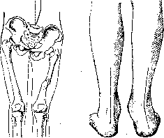
What requirements, from the point of view of restoring function and, consequently, working capacity, are imposed on an extremity after a healed fracture? First of all, a dense, well-consolidated plastic callus that holds the bone fragments in the correct position, preservation of the correct direction of the axis of the extremity, normal mobility in all joints, preservation of muscle strength, absence of circulatory and lymphatic disorders, and absence of trophic disorders or any deviations associated with damage to the nervous system. If we dwell separately on the function of the upper and lower extremities, it is evident that the requirements imposed on a healed fracture have their own specific features for both the upper and lower extremities. For the upper extremity, where it is especially important to preserve the full range of motion in all joints, where there is a risk of obtaining a limitation of movement, it is necessary to pay special attention to the position of the fingers and hand. A semi-flexed position of the fingers with dorsal flexion of the hand is the most advantageous in terms of working function; for the forearm, the most advantageous position is the middle one between pronation and supination; for the shoulder—a position of abduction and flexion at 45°. A vicious state of the upper extremity, leading to disability, is not so much the curvature of one or another bone (shoulder, forearm, etc.) as its fixation in an incorrect position; for example, the position of the shoulder in a state of adduction, the forearm in a state of pronation, the hand in a state of flexion, and the fingers in a sharply extended position, with the thumb abducted, is a vicious position that impairs working capacity and causes disability in a worker.
The lower extremity is subject to requirements of stability, endurance, and the preservation of a normal gait; therefore, the following are of great importance here: 1) shortening, whereby a shortening of up to 2-3 cm is fully corrected by the lowering of the pelvis (Figure 19), without causing any disorders in gait, although even a small curvature causes changes in the statics of the entire body; large shortening is caused by fractures of the femur due to the power of the pelvic girdle muscles; therefore, in fractures of the femur, treatment by traction is the method of choice; 2) change in the axis of the extremity due to adduction of the femur (in fractures of the femoral neck), valgus deformity of the foot (Figure 20) (in ankle fractures), and recurvation (in fractures of the lower leg in the middle third); these changes in the axis of the lower extremity disrupt the statics of the body, make walking difficult, and, if they cannot be corrected by subsequent orthopedic manipulations and prosthetics, lead to disability; 3) stiffness of the joints of the lower extremity (although it is a lesser evil than joint instability). Ankylosis of the joints of the lower extremity leads to disability, especially in those cases where they fix the joint in an unfavorable position (knee in a flexed position, foot in a state of adduction, etc.). Circulatory disorders, edema, congestive phenomena, and subsequent varicose veins develop especially often in connection with the incorrect position of the fragments and prolonged inactivity of the lower extremity. These disorders, reaching severe degrees, entail disability.
Figure 19. Compensation of shortening of the femur by tilting the pelvis and scoliosis of the spine.
Figure 20. Pes valgus in a malunited ankle fracture.
fracture of the femur on average after 17 weeks. Steinman and Wegner, who applied the functional method of treatment, speak of similarly good results. The immediate task of Soviet surgeons is to summarize the vast material of existing clinics, institutes, and hospitals, to produce a comparative assessment based on this material, to determine indications for various methods of treating fractures of the extremities, and to work out, based on scientific data, the periods for the restoration of working capacity in fractures of the bones of the extremities. The current state of our scientific knowledge, illuminating the biochemical essence of bone and muscle regeneration, familiarity with the laws of biomechanics of the extremities, the development of mechanics and technology, which makes it possible to apply traction in any position (adhesive plaster, skeletal), traction using all kinds of nails, staples, wires, etc., the high development of operative technique, allowing for bloody intervention in particularly complex cases, and finally, the development of X-ray technology, which makes it possible to study and check not only the position of fragments in various types of fractures but also all stages of callus development—all this is a guarantee of the approach of the moment when disability after a fracture will pass into the realm of legends, and we will learn to return all cases of extremity fractures to work.
The treatment of fractures should not be limited to the use of splints and traction. Recently, along with massage, gymnastics, and mechanotherapy, methods of physiotherapeutic treatment, which influence both the regenerative processes in the bone and the improvement of blood and lymph circulation in the entire damaged extremity, have been gaining great importance.
Related articles
Mentioned in
Cite this page
“Fractures.” Soviet Medical Encyclopedia. English translation of Bolshaya Meditsinskaya Entsiklopediya, 1st ed. (Moscow, 1928–1936), ed. N. A. Semashko. https://sovietmedicalencyclopedia.pages.dev/article/fractures/