Femur
Historical document, translated for reference. It reflects medical knowledge of the 1920s–30s and is not medical advice.
Summary
The article provides a detailed anatomical description of the femur, including its embryological development, structure, musculature, vascular and nervous supply. It explains the bone's characteristics, its differences between genders and ages, and its relationship with surrounding muscles and vessels.
Encyclopedia article (1928–1936)
FEMUR (femur). Embryology of the femur. In the beginning of the 5th week of intrauterine life, in the central part of the tubercle from which the lower extremity is to develop, a condensation of mesenchyme is observed. This condensed area represents the rudiment of the skeleton of the lower extremity. By the 8th week, its division into the femur, tibia, and foot is already noticeable. Ossification begins from the 3rd month. For the diaphysis of the femur, an ossification center appears on the 42nd day. Four additional ossification centers are intended for the head, greater and lesser trochanters, and the lower end of the bone. At the same time, the musculature begins to differentiate, first in the proximal part of the femur. The vessels, initially forming a vascular network, then differentiate, with the powerful a. ischiadica existing as the main trunk until it is replaced in the embryo at 15.5 mm in length (in the 2nd month) by a. femoralis. - The foundation of the femur is the femur bone, the longest (74 of the body's height) and strongest bone of the skeleton, withstanding pressure up to 10 atmospheres (Lesgaft). The diaphysis of the femur bone appears three-sided; its anterior surface is slightly convex forward, the lateral surfaces behind are separated by a rough line (linea aspera femoris), which below diverges into two lips (labia). The upper end is curved, compared to Lesgaft, in the form of a "crane" and forms a neck (collum), somewhat compressed from front to back, which transitions into the head (caput), representing 2/3 of a spheroid. The size of the neck and the angle it forms with the body vary: in the newborn it is barely noticeable, in the male the angle is more obtuse than in the female. On the upper end of the body is located, usually easily palpable, the greater trochanter (trochanter major), corresponding to the middle of the Roser-Nelatone line (between the anterior superior iliac spine and the ischial tuberosity). Below and behind it is the lesser trochanter (troch. minor) - the site of attachment of m. ilio-psoas. Below, the femur bone widens and ends with two condyles (condyli medialis and lateralis) for articulation with the tibia and patella. The diaphysis of the femur forms an angle with the head, Figure 1. Anterior muscles of the right thigh: 1-m. psoas major; 2-m. iliacus internus; 3-m. pectineus; 4-m. tensor fasciae latae; 5-m. adductor longus; 6-m. rectus femoris; 7-m. gracilis; 8-m. sartorius; 9-m. adductor magnus; 10-m. vastus lateralis; 11-m. vastus medialis. which varies in people of different sexes and ages, due to which the femurs converge downward. The convergence of the femur is more pronounced in women than in men.
Muscles of the femur (see Figure 1-4) are divided into three groups: 1) On the anterior side of the thigh are located most superficially - m. tensor fasciae latae (tensor of the fascia lata), beginning near the spina iliaca ant. sup. and ending in the tractus ilio-tibialis (see below); it fixes the point of termination here of part of the fibers of musculus glutaei max., slightly abducts and flexes the thigh, and m. sartorius (tailor's muscle), also beginning from the crest and ending near the tuberositas tibiae; it flexes the knee and slightly rotates the tibia inward; along its medial edge are located the femoral artery and nerve. In the depression between the diverging -
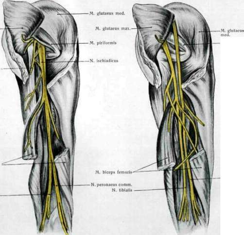
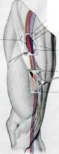
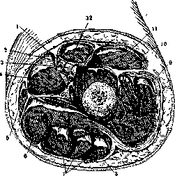
Figure 1. Topography of n. and v. femoralis (according to Kleiweg). Figure 2. Cross-section of the middle third of the left thigh: 1-n. saphenus; 2-a. femoralis; 3-v. femoralis; 4-m. adductor longus; 5-m. gracilis; 6-m. adductor magnus; 7-group of flexors; 8-n. ischiadicus; 9-m. vastus lateralis; 10-m. rectus femoris; 11-m. vastus medialis; 12-m. sartorius (according to Zuckerkandl).
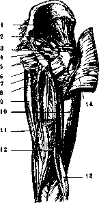
muscles lies the rectus muscle (m. rectus femoris), the superficial head of the quadriceps muscle (m. quadriceps cruris), beginning from the inferior crest; 3 deep mm. vasti lateralis, medialis and intermedialis, surround the femur bone, beginning from the base of the greater trochanter, the medial lip of the rough line and its smooth anterior surface; the common terminal tendon, containing (as a sesamoid bone) the patella under the name lig. patellae, attaches to the roughness of the tibia, the muscle as a whole is a powerful extensor of the knee; the rectus, in addition, is also a flexor of the thigh. 2) On the medial side are located muscles beginning from the pubic and ischial bones and, with the exception of m. gracilis, which ends near the roughness of the tibia, find their terminal attachment on the rough line of the femur and bear the name mm. pectineus, adductores longus, brevis, magnus (and minimus), they adduct the femur and partly weakly rotate it inward. In the tendon of the great adductor muscle there is an opening for the passage of vasa femoralia - hiatus tendineus adductorius. 3) On the posterior side lie 3 flexors of the knee, extensors in the hip joint and at the same time rotators of the tibia, taking origin from the ischial tuberosity: m. biceps femoris, the short head of which begins from the rough line of the femur, ends on the head of the fibula; mm. semitendinosus and semimembranosus end near the roughness of the tibia. Numerous bursae are located at the beginning and end of the muscles.
Vessels and nerves of the femur. The main artery of the femur, a. femoralis, serves as a continuation of a. iliacae ext.; it is located on the anteromedial side of the thigh from the Poupart's ligament to the hiatus tendineus adductorius. Its proximal part lies in the femoral triangle (see), then it passes under the sartorius muscle and finally lies lateral to it. To the artery, the femoral vein first adjoins from the medial side, then from behind and laterally. From the branches of the femoral art., a. epigastrica superficialis, branching under the Poupart's ligament, ramifies in the anterior abdominal wall, a. circumflexa ilium superficialis runs parallel to the ligament, aa. pudendae ext.- in the external genital organs; the largest branch, almost equal in caliber to the main trunk, is the deep artery--a. profunda femoris, departing 3-4 cm below the Poupart's ligament; from it go aa. circumflexae femor. medialis and lateralis and aa. perforantes (3-5), anastomosing with each other and with neighboring branches and supplying, mainly, the adductor and flexor muscles; further-6-8 muscular branches and the superior articular artery of the knee. From the slit in the tendon of the great adductor muscle until it divides into the anterior and posterior tibial arteries, it changes its name to the popliteal artery, giving branches to the knee joint. In addition, branches of the inferior gluteal and obturator arteries go to the thigh. Collateral circulation when ligating a. femoralis is easily restored in case of ligation below the departure of a. profunda femoris, i.e., approximately 5 cm below the Poupart's ligament, due to the well-developed collaterals of the latter. A. prof. fem. supplies all muscles of the thigh while the main trunk, a. femoralis, carries blood, mainly, for the underlying parts of the extremity. With concentrated branching of the femoral artery and its high division, the anatomomechanical conditions of blood flow are worse than with gradual branching of the branches (the so-called main type of architecture of a. femoralis).
Vena femoralis in the upper part of the femur receives v. saphena magna, collecting blood from the subcutaneous veins of the leg. Valves in v. femoralis above the place of influx of v. saphenae mag. are sometimes absent or insufficient; below this place v. femoralis usually has two or three pairs of valves. Superficial and deep lymphatic vessels of the femur collect lymph into the glands located in Scarpa's triangle and in gl. inguinalis. Lymphatic vessels of the gluteal region collect lymph into gl. hypogastricae. The lateral part of the skin of the femur is innervated by n. cutaneus fem. lat.-N. femoralis (from LI, LII, LIII and L IV) gives motor and sensitive
branches for the anterior surface of the Femur. In some extreme cases, its main trunk gives off secondary branches gradually, while in others, a concentrated departure of branches in the form of a bundle is observed. The continuation of the n. femoralis is the n. saphenus, which gives branches to the knee joint. The adductor muscles of the Femur are innervated by the n. obturatorius, descending from the pelvic cavity through the canalis obturatorius. The n. ischiadicus, supplying the posterior surface of the Femur, emerges from the lower edge of the m. glutaei max. and descends vertically downward. In some cases, at the level of the m. piriformis, it divides into two trunks (n. tibialis and n. peronaeus), while in other cases, its trunk divides at the border between the middle and lower thirds of the Femur (see table). The n. cutaneus femoris post, runs more superficially than the n. ischiadici, vertically under the aponeurosis, supplying sensory endings to the skin of the gluteal region and the posterior surface of the Femur. All muscles of the Femur are covered by fascia lata, connected with the fasciae of the mm. glutaei, perineum, and the Poupart ligament; it reaches particular power on the anterolateral side of the Femur, where it received the name tractus ilio-tibialis Maissiati. From the inner surface of the fascia lata to the bone, the septa intermusculare mediale and laterale extend, separating the flexor muscles from the extensor muscles; in addition, each muscle has its own fascial covering, in some cases connected with the fascia lata. The deep lamina is formed due to the fascia ilio-pectinea. Under the medial part of the Poupart ligament in the fascia lata, corresponding to the place of entry into the femoral vein of the v. saphenae, there is a crescent-shaped notch—the lower part of the anulus femoralis externus, and the fossa delimited by it, on the bottom of which the large femoral vessels are visible, is called the oval fossa (fossa ovalis). The skin of the Femur is loosely connected to the fascia, easily movable, its contractility is about 5 cm. The subcutaneous fatty layer of the thigh can be very well developed, especially in women in the upper part of the thigh.
Related articles
Mentioned in
Cite this page
“Femur.” Soviet Medical Encyclopedia. English translation of Bolshaya Meditsinskaya Entsiklopediya, 1st ed. (Moscow, 1928–1936), ed. N. A. Semashko. https://sovietmedicalencyclopedia.pages.dev/article/femur/